
Updated prevalence of latent prostate cancer in Chinese population and comparison of biopsy results: An autopsy-based study
-
GRAPHICAL ABSTRACT
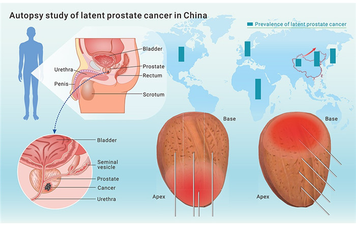
 DownLoad:
Full size image
DownLoad:
Full size image
-
PUBLIC SUMMARY
- ■ We conducted prostate autopsies to investigate latent cancer epidemiology and measure the accuracy of prostate biopsy.
- ■ In recent decades, the prevalence of latent prostate cancer has increased in China.
- ■ Transperineal and transrectal biopsies exhibited similar sensitivities but different preferential areas.
- ■ Both methods of biopsy missed one-third of cases with large lesions, most of which were in the anterior prostate.
-
ABSTRACT
Prostate cancer detected by autopsy is named latent prostate cancer. As the repertoire of clinical prostate cancer, latent cancer may better reflect the disease burden. Unlike clinical prostate specimens, which are obtained exclusively from biopsy-positive cases, prostate specimens obtained through autopsy provide information on biopsy-negative cases, helping calculate the true sensitivity of prostate biopsy. From 2014 to 2021, we collected autopsy specimens of the prostate from body donors in China and performed transperineal and transrectal biopsies on specimens before step-sectioning and pathological measurements. We found that the crude prevalence of latent prostate cancer in middle-aged and elderly men was 35.1% (81/231), which was higher than previous estimates for Chinese populations. The overall per-patient sensitivities of transperineal and transrectal biopsies were not significantly different (33.3% vs. 32.1%, p = 0.82), but the two approaches differed in preferential sampling area along the proximal-distal axis of the prostate. Transperineal biopsy had a higher sensitivity for detecting clinically significant lesions in the distal third (34.7% vs. 16.3%, p = 0.02) and distal half (30.6% vs. 18.1%, p = 0.04), while transrectal biopsy had a higher sensitivity for lesions in the proximal half (25.0% vs. 13.9%, p = 0.046). Both transperineal and transrectal methods of biopsy missed most small lesions (< 0.1 mL) and 35.3% (6/17) of large lesions (> 0.5 mL). In conclusion, the prevalence of latent prostate cancer in China has increased over the past 2 decades. Systematic transperineal and transrectal methods of biopsy had comparable sensitivities but had different preferential sampling areas. Both approaches miss one-third of large lesions. -

-
REFERENCES
[1] Sung, H., Ferlay, J., Siegel, R.L., et al. (2021). Global Cancer Statistics 2020: GLOBOCAN estimates of incidence and mortality worldwide for 36 cancers in 185 countries. CA. Cancer J. Clin. 71, 209–249. [2] Breslow, N., Chan, C.W., Dhom, G., et al. (1977). Latent carcinoma of prostate at autopsy in seven areas. The International Agency for Research on Cancer, Lyons, France. Int. J. Cancer 20, 680–688. [3] Jahn, J.L., Giovannucci, E.L., and Stampfer, M.J. (2015). The high prevalence of undiagnosed prostate cancer at autopsy: implications for epidemiology and treatment of prostate cancer in the Prostate-specific Antigen-era. Int. J. Cancer 137, 2795–2802. [4] Rebbeck, T.R., and Haas, G.P. (2014). Temporal trends and racial disparities in global prostate cancer prevalence. Can. J. Urol. 21, 7496–7506. [5] Gu, F.L., Xia, T.L., and Kong, X.T. (1994). Preliminary study of the frequency of benign prostatic hyperplasia and prostatic cancer in China. Urology 44, 688–691. [6] Zhang, Y.H., Zhang, J.S., Yang, L., et al. (2004). Occurence of prostatic adenocarcinoma in senile autopsy cases. Chinese Journal of Urology 25, 118–120. (in Chinese). [7] Zhou, Y., Mai, Z., Yan, W., et al. (2021). The characteristics and spatial distributions of prostate cancer in autopsy specimens. Prostate 81, 135–141. [8] Baco, E., Ukimura, O., Rud, E., et al. (2015). Magnetic resonance imaging-transectal ultrasound image-fusion biopsies accurately characterize the index tumor: correlation with step-sectioned radical prostatectomy specimens in 135 patients. Eur. Urol. 67, 787–794. [9] Camtosun, A., and Gökçe, H. (2019). Comparison of prostate biopsy pathology and radical prostatectomy pathologies. Dicle Tıp Dergisi, 133–139. [10] Le, J.D., Tan, N., Shkolyar, E., et al. (2015). Multifocality and prostate cancer detection by multiparametric magnetic resonance imaging: correlation with whole-mount histopathology. Eur. Urol. 67, 569–576. [11] Mai, Z., Xiao, Y., Yan, W., et al. (2018). Comparison of lesions detected and undetected by template-guided transperineal saturation prostate biopsy. BJU Int. 121, 415–420. [12] Mottet, N., van den Bergh, R.C.N., Briers, E., et al. (2021). EAU-EANM-ESTRO-ESUR-SIOG guidelines on prostate cancer-2020 update. Part 1: Screening, diagnosis, and local treatment with curative intent. Eur. Urol. 79, 243–262. [13] Emiliozzi, P., Corsetti, A., Tassi, B., et al. (2003). Best approach for prostate cancer detection: a prospective study on transperineal versus transrectal six-core prostate biopsy. Urology 61, 961–966. [14] Jiang, C.-Y., Shen, P.-F., Wang, C., et al. (2019). Comparison of diagnostic efficacy between transrectal and transperineal prostate biopsy: a propensity score-matched study. Asian J. Androl. 21, 612–617. [15] Hara, R., Jo, Y., Fujii, T., et al. (2008). Optimal approach for prostate cancer detection as initial biopsy: prospective randomized study comparing transperineal versus transrectal systematic 12-core biopsy. Urology 71, 191–195. [16] Takenaka, A., Hara, R., Ishimura, T., et al. (2008). A prospective randomized comparison of diagnostic efficacy between transperineal and transrectal 12-core prostate biopsy. Prostate Cancer Prostatic Dis. 11, 134–138. [17] Cerruto, M.A., Vianello, F., D'Elia, C., et al. (2014). Transrectal versus transperineal 14-core prostate biopsy in detection of prostate cancer: a comparative evaluation at the same institution. Arch. Ital. Urol. Androl. 86, 284–287. [18] Cowan, T., Baker, E., McCray, G., et al. (2020). Detection of clinically significant cancer in the anterior prostate by transperineal biopsy. BJU Int. 126, 33–37. [19] Chiu, P.K.F., Lo, K.L., Teoh, J.Y.C., et al. (2021). Sectoral cancer detection and tolerability of freehand transperineal prostate biopsy under local anaesthesia. Prostate Cancer Prostatic Dis. 24, 431–438. [20] Ong, W.L., Weerakoon, M., Huang, S., et al. (2015). Transperineal biopsy prostate cancer detection in first biopsy and repeat biopsy after negative transrectal ultrasound-guided biopsy: the Victorian Transperineal Biopsy Collaboration experience. BJU Int. 116, 568–576. [21] Lee, C.U., Sung, S.H., Jang, C.T., et al. (2019). Cancer location in upgrading and detection after transperineal template-guided mapping biopsy for patients in active surveillance and negative transrectal ultrasonography-guided prostate biopsy. Urol. Int. 103, 262–269. [22] Mai, Z., Zhou, Z., Yan, W., et al. (2018). The transverse and vertical distribution of prostate cancer in biopsy and radical prostatectomy specimens. BMC Cancer 18, 1205. [23] Meyer, A.R., Mamawala, M., Winoker, J.S., et al. (2021). Transperineal prostate biopsy improves the detection of clinically sgnificant prostate cancer among men on active surveillance. J. Urol. 205, 1069–1074. [24] Delongchamps, N.B., de la Roza, G., Jones, R., et al. (2009). Saturation biopsies on autopsied prostates for detecting and characterizing prostate cancer. BJU Int. 103, 49–54. [25] Haas, G.P., Delongchamps, N.B., Jones, R.F., et al. (2007). Needlebiopsies on autopsy prostates: sensitivity of cancer detection based on true prevalence. J. Natl. Cancer Inst. 99, 1484–1489. [26] Crawford, E.D., Wilson, S.S., Torkko, K.C., et al. (2005). Clinical staging of prostate cancer: a computer-simulated study of transperineal prostate biopsy. BJU Int. 96, 999–1004. [27] Rocco, B., de Cobelli, O., Leon, M.E., et al. (2006). Sensitivity and detection rate of a 12-core trans-perineal prostate biopsy: preliminary report. Eur. Urol. 49, 827–833. [28] Stamatiou, K., Alevizos, A., Agapitos, E., et al. (2006). Incidence of impalpable carcinoma of the prostate and of non-malignant and precarcinomatous lesions in Greek male population: an autopsy study. Prostate 66, 1319–1328. [29] Epstein, J.I., Egevad, L., Amin, M.B., et al. (2016). The 2014 International Society of Urological Pathology (ISUP) consensus conference on Gleason grading of prostatic carcinoma: definition of grading patterns and proposal for a new grading system. Am. J. Surg. Pathol. 40, 244–252. [30] Nevoux, P., Ouzzane, A., Ahmed, H.U., et al. (2012). Quantitative tissue analyses of prostate cancer foci in an unselected cystoprostatectomy series. BJU Int. 110, 517–523. [31] Schned, A.R., Wheeler, K.J., Hodorowski, C.A., et al. (1996). Tissue-shrinkage correction factor in the calculation of prostate cancer volume. Am. J. Surg. Pathol. 20, 1501–1506. [32] Van der Kwast, T.H., Amin, M.B., Billis, A., et al. (2011). International Society of Urological Pathology (ISUP) consensus conference on handling and staging of radical prostatectomy specimens. Working group 2: T2 substaging and prostate cancer volume. Mod. Pathol. 24, 16–25. [33] Eklund, M., Jäderling, F., Discacciati, A., et al. (2021). MRI-targeted or standard siopsy in € prostate cancer screening. N. Engl. J. Med. 385, 908–920. [34] Greene, D.R., Fitzpatrick, J.M., and Scardino, P.T. (1995). Anatomy of the prostate and distribution of early prostate cancer. Semin. Surg. Oncol. 11, 9–22. [35] Armitage, P. (1955). Tests for linear trends in proportions and frequencies. Biometrics 11, 375–386. [36] Cochran, W.G. (1954). Some methods for strengthening the common χ2 tests. Biometrics 10, 417–451. [37] Koletsi, D., and Pandis, N. (2017). Matched analysis for paired binary data (McNemar test). Am. J. Orthod. Dentofacial Orthop. 151, 222–223. [38] McHugh, M.L. (2012). Interrater reliability: the kappa statistic. Biochem. Med. 22, 276–282. [39] Inaba, H., Kimura, T., Onuma, H., et al. (2020). Tumor location and pathological features of latent and incidental prostate cancer in contemporary Japanese men. J. Urol. 204, 267–272. [40] Hitosugi, M., Mukaisho, K., Kido, M., et al. (2017). No change in theprevalenceof latent prostate cancer over the last 10 years: a forensic autopsy study in Japan. Biomed. Res. 38, 307–312. [41] Kido, M., Hitosugi, M., Ishii, K., et al. (2015). Latent prostate cancer in Japanese men who die unnatural deaths: a forensic autopsy study. Prostate 75, 917–922. [42] Zlotta, A.R., Egawa, S., Pushkar, D., et al. (2013). Prevalence of prostate cancer on autopsy: cross-sectional study on unscreened Caucasian and Asian men. J. Natl. Cancer Inst. 105, 1050–1058. [43] Powell, I.J., Bock, C.H., Ruterbusch, J.J., et al. (2010). Evidence supports afaster growthrate and/or earlier transformation to clinically significant prostate cancer in black than in white American men, and influences racial progression and mortality disparity. J. Urol. 183, 1792–1796. [44] Soos, G., Tsakiris, I., Szanto, J., et al. (2005). The prevalence of prostate carcinoma and its precursor in Hungary: an autopsy study. Eur. Urol. 48, 739–744. [45] Kimura, T., Takahashi, H., Okayasu, M., et al. (2016). Time trends in histological features of latent prostate cancer in Japan. J. Urol. 195, 1415–1420. [46] Hamdy, F.C., Donovan, J.L., Lane, J.A., et al. (2023). Fifteen-year outcomes after monitoring, surgery, or radiotherapy for prostate cancer. N. Engl. J. Med. 388, 1547–1558. [47] Bergengren, O., Pekala, K.R., Matsoukas, K., et al. (2023). 2022 update on prostate cancer epidemiology and risk factors-a systematic review. Eur. Urol. 84, 191–206. [48] Matoso, A., and Epstein, J.I. (2019). Defining clinically significant prostate cancer on the basis of pathological findings. Histopathology 74, 135–145. [49] Ahdoot, M., Wilbur, A.R., Reese, S.E., et al. (2020). MRI-Targeted, systematic, and combined biopsy for prostate cancer diagnosis. N. Engl. J. Med. 382, 917–928. [50] Kasivisvanathan, V., Rannikko, A.S., Borghi, M., et al. (2018). MRI-targeted or standard biopsy for prostate-cancer diagnosis. N. Engl. J. Med. 378, 1767–1777. [51] Stranne, J., Mottet, N., Rouvière, O., et al. (2023). Systematic biopsies as a complement to magnetic resonance imaging-targeted biopsies: "To be or not to be. Eur. Urol. 83, 381–384. [52] Johnson, D.C., Raman, S.S., Mirak, S.A., et al. (2019). Detection of individual prostate cancer foci via multiparametric magnetic resonance imaging. Eur. Urol. 75, 712–720. -
ABOUT THIS ARTICLE
Cite this article:
Yuliang Chen, Zhien Zhou, Yi Zhou, Zhipeng Mai, Shijie Jin, Zhen Liang, Zhiyuan Shang, Yuzhi Zuo, Yu Xiao, Wenze Wang, Haibo Wang, Weigang Yan. Updated prevalence of latent prostate cancer in Chinese population and comparison of biopsy results: An autopsy-based study[J]. The Innovation, 2024, 5(1). https://doi.org/10.1016/j.xinn.2023.100558
Welcome!
To request copyright permission to republish or share portions of our works, please visit Copyright Clearance Center's (CCC) Marketplace website at marketplace.copyright.com.
Export File
Citation
Yuliang Chen, Zhien Zhou, Yi Zhou, Zhipeng Mai, Shijie Jin, Zhen Liang, Zhiyuan Shang, Yuzhi Zuo, Yu Xiao, Wenze Wang, Haibo Wang, Weigang Yan. Updated prevalence of latent prostate cancer in Chinese population and comparison of biopsy results: An autopsy-based study[J]. The Innovation, 2024, 5(1). https://doi.org/10.1016/j.xinn.2023.100558

Format
Content

-
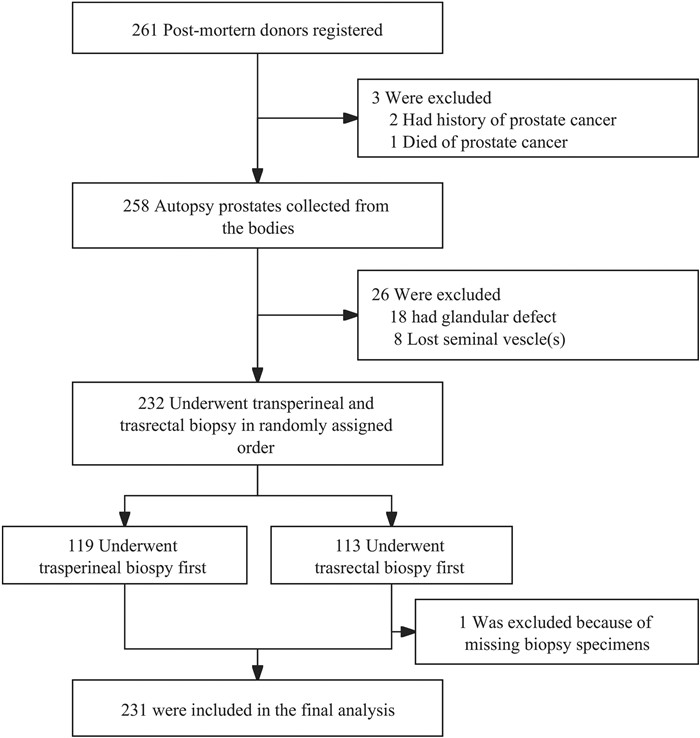
Figure 1.
Enrollment of the specimens
-
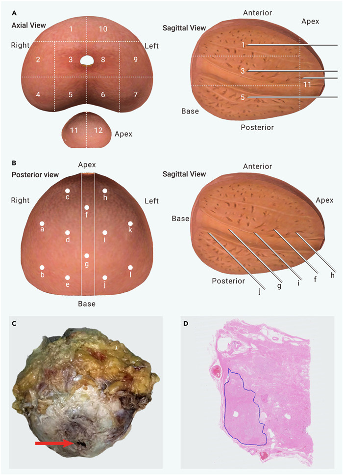
Figure 2.
Biopsy plan, autopsy specimen, and digital slice
-
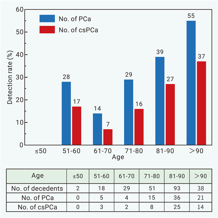
Figure 3.
Prevalence of all prostate cancer and csPCa in different age groups
-
Figure 4.
Rates of lesions missed by both TP and TR methods of biopsy, stratified by tumor volume (TV), grade group (GrG), and tumor location
