
M2 macrophage extracellular vesicles in bone regeneration: Applications and challenges
-
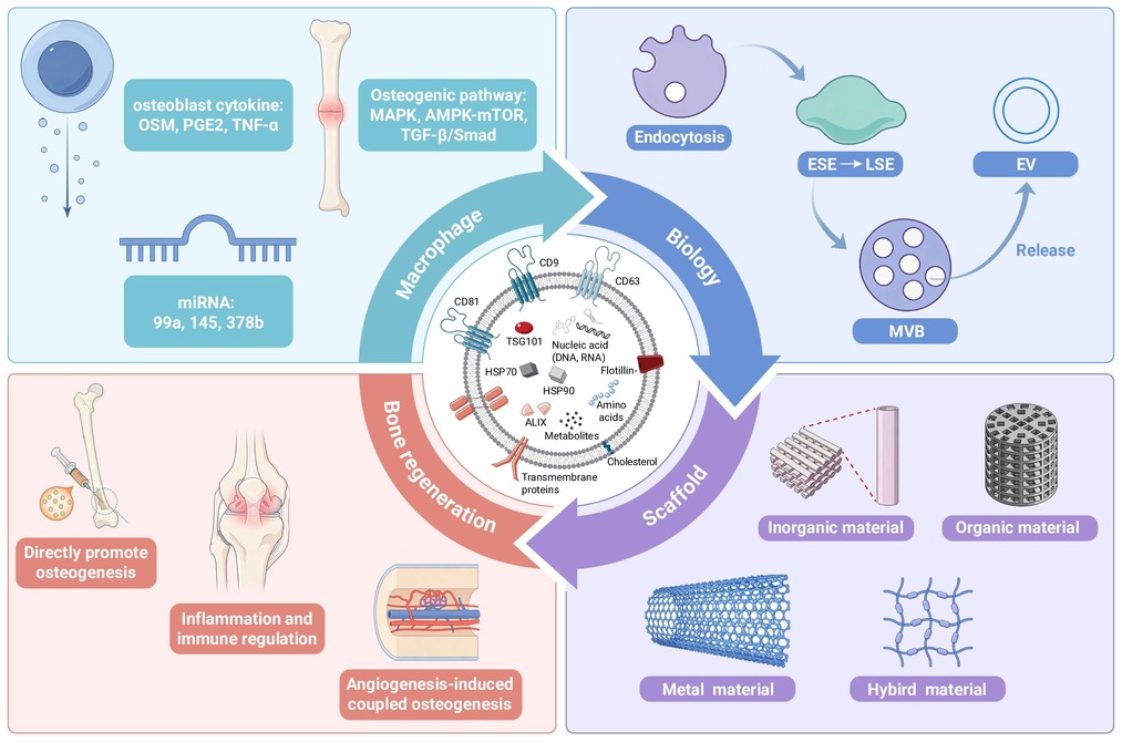
GRAPHICAL ABSTRACT
 DownLoad:
Full size image
-
PUBLIC SUMMARY
-
M2 macrophage extracellular vesicles promote bone repair by reducing inflammation and stimulating blood vessel growth.
When combined with functionalized scaffolds, these vesicles significantly improve implant integration and defect healing.
Challenges remain in targeting, scalable production, and quality control for clinical use.
-
-
ABSTRACT
Extracellular vesicles (EVs) can carry bioactive molecules and mediateintercellular communication. Among EVs from various sources, those derivedfrom M2 macrophages (M2-EVs) have attracted widespread attention in thefield of bone regeneration due to their inherent anti-inflammatory andosteogenic properties. M2-EVs promote osteogenic differentiation ofmesenchymal stem cells by delivering specific microRNAs and proteins thatactivate key signaling pathways, such as MAPK, TGF-β/Smad, AMPK-mTOR,and PI3K/Akt. They also modulate the osteoimmune microenvironment bysuppressing inflammation and inducing M2 polarization, while enhancingangiogenesis through VEGF and miR-21 transfer. In bone tissue engineering,M2-EVs can be integrated with various scaffolds—such as β-TCP, PCLfibrous scaffolds, and hydrogels—to improve osseointegration and bonedefect repair, as confirmed in preclinical models. However, clinical translationfaces challenges, including insufficient targeting, heterogeneity, isolationstandardization, and quality control. Despite these limitations, M2-EVsrepre-sent a promising cell-free therapeutic strategy for bone repair. Futureresearch may focus on engineering approaches to enhance their specificity,stability, and large-scale production, ultimately unlocking their full potential inregenerative medicine.-
Keywords:
- M2 macrophage /
- extracellular vesicles /
- bone regeneration /
- osteogenesis /
- immunomodulation /
- scaffolds
-

-
REFERENCES
[1] González E. and Falcón-Pérez J. M. (2025). Expanding horizons: Next-generation and interdisciplinary advances in the applications of extracellular vesicles. J Extracell Biol 4:e70101. DOI:10.1002/jex2.70101 [2] Couch Y., Buzàs E. I., Di Vizio D., et al. (2021). A brief history of nearly EV-erything - The rise and rise of extracellular vesicles. J Extracell Vesicles 10:e12144. DOI:10.1002/jev2.12144 [3] Biswas S., Gangadaran P., Dhara C., et al. (2025). Extracellular vesicles in osteogenesis: A comprehensive review of mechanisms and therapeutic potential for bone regeneration. Curr Issues Mol Biol 47:675. DOI:10.3390/cimb47080675 [4] Epelman S., Lavine K. J. and Randolph G. J. (2014). Origin and functions of tissue macrophages. Immunity 41:21−35. DOI:10.1016/j.immuni.2014.06.013 [5] Tong L., van Wijnen A. J., Wang H., et al. (2024). Advancing bone biology: The mutual promotion of biology and pioneering technologies. Innov Life 2:100078. DOI:10.59717/j.xinn-life.2024.100078 [6] Pajarinen J., Lin T., Gibon E., et al. (2019). Mesenchymal stem cell-macrophage crosstalk and bone healing. Biomaterials 196:80−89. DOI:10.1016/j.biomaterials.2017.12.025 [7] Gu Q. L., Yang H. L. and Shi Q. (2017). Macrophages and bone inflammation. J. Orthop. Transl. 10:86−93. DOI:10.1016/j.jot.2017.05.002 [8] Peng M., Li N., Wang H., et al. (2025). Macrophages: Subtypes, distribution, polarization, immunomodulatory functions, and therapeutics. MedComm 6:e70304. DOI:10.1002/mco2.70304 [9] Zhou S., Zhu Y., Wu Y., et al. (2025). New insights on metabolic reprogramming in macrophage plasticity. Int Immunopharmacol 157:114797. DOI:10.1016/j.intimp.2025.114797 [10] Daneshvar A., Nemati P., Azadi A., et al. (2024). M2 macrophage-derived exosomes for bone regeneration: A systematic review. Arch Oral Biol 166:106034. DOI:10.1016/j.archoralbio.2024.106034 [11] Zhang B.B., Zha Xi D.W., Li C., et al. (2022). M2 macrophagy-derived exosomal miRNA-26a-5p induces osteogenic differentiation of bone mesenchymal stem cells. J Orthop Surg Res 17:137. DOI:10.1186/s13018-022-03029-0 [12] Ho C. C., Hsu T. T., Lin Y. H., et al. (2026). 3D-printed dictamni-calcium silicate scaffolds modulate the osteoimmune microenvironment and enhance macrophage-derived exosomal miR-21 signaling in vascularized bone regeneration. J Nanobiotechnology 24:596. DOI:10.1186/s12951-026-04507-x [13] Yin L., Zhang Z., Lin P., et al. (2025). Three-dimensional printed PCL/nHA/SA/COL structure-mediated M2 macrophage polarization enhances angiogenesis and osteogenic differentiation of bone marrow mesenchymal stem cells. Int Immunopharmacol 167:115679. DOI:10.1016/j.intimp.2025.115679 [14] Kalluri R. and LeBleu V. S. (2020). The biology, function, and biomedical applications of exosomes. Science 367:eaau6977. DOI:10.1126/science.aau6977 [15] Kahlert C. and Kalluri R. (2013). Exosomes in tumor microenvironment influence cancer progression and metastasis. J. Mol. Med. 91:431−437. DOI:10.1007/s00109-013-1020-6 [16] Kalluri R. (2016). The biology and function of exosomes in cancer. J. Clin. Invest. 126:1208−1215. DOI:10.1172/jci81135 [17] Corso G., Mäger I., Lee Y., et al. (2017). Reproducible and scalable purification of extracellular vesicles using combined bind-elute and size exclusion chromatography. Sci Rep 7:11561. DOI:10.1038/s41598-017-10646-x [18] Li B., Wu W., Xu W., et al. (2025). Advances of extracellular vesicles isolation and detection frontier technology: from heterogeneity analysis to clinical application. J Nanobiotechnology 23:678. DOI:10.1186/s12951-025-03768-2 [19] Kim T., Hong J. W. and Lee L. P. (2025). Efficient methods of isolation and purification of extracellular vesicles. Nano Converg 12:45. DOI:10.1186/s40580-025-00509-x [20] Oksvold M. P., Neurauter A. and Pedersen K. W. (2015). Magnetic bead-based isolation of exosomes. Methods Mol Biol 1218:465−481. DOI:10.1007/978-1-4939-1538-5_27 [21] CNCB-NGDC Members and Partners. (2022). Database resources of the national genomics data center, china national center for bioinformation in 2022. Nucleic Acids Res 50:D27−D38. DOI:10.1093/nar/gkab951 [22] Sidhom K., Obi P. O. and Saleem A. (2020). A review of exosomal isolation methods: Is size exclusion chromatography the best option. Int J Mol Sci 21:6466. DOI:10.3390/ijms21186466 [23] Liu W. Z., Ma Z. J. and Kang X. W. (2022). Current status and outlook of advances in exosome isolation. Anal Bioanal Chem 414:7123−7141. DOI:10.1007/s00216-022-04253-7 [24] Mazahir F. and Yadav A. K. (2024). Recent progress in engineered extracellular vesicles and their biomedical applications. Life Sci 350:122747. DOI:10.1016/j.lfs.2024.122747 [25] Malvicini R., De Lazzari G., Tolomeo A. M., et al. (2024). Influence of the isolation method on characteristics and functional activity of mesenchymal stromal cell-derived extracellular vesicles. Cytotherapy 26:157−170. DOI:10.1016/j.jcyt.2023.11.001 [26] Pascucci L. and Scattini G. (2021). Imaging extracelluar vesicles by transmission electron microscopy: Coping with technical hurdles and morphological interpretation. Biochim Biophys Acta Gen Subj 1865:129648. DOI:10.1016/j.bbagen.2020.129648 [27] Comfort N., Cai K., Bloomquist T. R., et al. (2021). Nanoparticle tracking analysis for the quantification and size determination of extracellular vesicles. J Vis Exp 2021:62447. DOI:10.3791/62447 [28] Ekström K., Omar O., Granéli C., et al. (2013). Monocyte exosomes stimulate the osteogenic gene expression of mesenchymal stem cells. PLoS One 8:e75227. DOI:10.1371/journal.pone.0075227 [29] Arab T., Mallick E. R., Huang Y., et al. (2021). Characterization of extracellular vesicles and synthetic nanoparticles with four orthogonal single-particle analysis platforms. J Extracell Vesicles 10:e12079. DOI:10.1002/jev2.12079 [30] Volkman A. and Gowans J. L. (1965). Origin of macrophages from bone marrow in rat. Brit. J. Exp. Pathol. 46:62−70. [31] Mosser D. M. and Edwards J. P. (2008). Exploring the full spectrum of macrophage activation. Nat. Rev. Immunol. 8:958−969. DOI:10.1038/nri2448 [32] He P., Dai M., Li Z., et al. (2024). Effect of connexin 43 in LPS/IL-4-induced macrophage M1/M2 polarization: An observational study. Medicine (Baltimore) 103:e37811. DOI:10.1097/md.0000000000037811 [33] Messex J. K. and Liou G. Y. (2021). Targeting btk signaling in the microenvironment of solid tumors as a feasible cancer therapy option. Cancers (Basel) 13:2198. DOI:10.3390/cancers13092198 [34] Luo Q. T., Li X. Y., Zhong W. C., et al. (2022). Dicalcium silicate-induced mitochondrial dysfunction and autophagy-mediated macrophagic inflammation promotes osteogenic differentiation of BMSCs. Regen. Biomater. 9:rbab075. DOI:10.1093/rb/rbab075 [35] Hwang S. M., Chung G., Kim Y. H., et al. (2019). The role of maresins in inflammatory pain: Function of macrophages in wound regeneration. Int. J. Mol. Sci. 20:5849. DOI:10.3390/ijms20235849 [36] Tedesco S., Bolego C., Toniolo A., et al. (2015). Phenotypic activation and pharmacological outcomes of spontaneously differentiated human monocyte-derived macrophages. Immunobiology 220:545−554. DOI:10.1016/j.imbio.2014.12.008 [37] Shabo I. and Svanvik J. (2011). Expression of macrophage antigens by tumor cells. Dittmar T. and Zanker K. S. (ed). Cell fusion in health and disease II: cell fusion in disease. (Springer-verlag Berlin), pp: 141-150. DOI: 10.1007/978-94-007-0782-5_7 [38] Raes G., De Baetselier P., Noël W., et al. (2002). Differential expression of FIZZ1 and Ym1 in alternatively versus classically activated macrophages. J. Leukoc. Biol. 71:597−602. [39] Salomon B. and Bluestone J. A. (2001). Complexities of CD28/B7: CTLA-4 costimulatory pathways in autoimmunity and transplantation. Annu Rev Immunol 19:225−252. DOI:10.1146/annurev.immunol.19.1.225 [40] Holtrop T., Budding K., Brandsma A. M., et al. (2022). Targeting the high affinity receptor, FcγRI, in autoimmune disease, neuropathy, and cancer. Immunother Adv 2:ltac011. DOI:10.1093/immadv/ltac011 [41] Chen M. T., Lin H. S., Shen C., et al. (2015). Pu.1-regulated long noncoding rna lnc-mc controls human monocyte/macrophage differentiation through interaction with microrna 199a-5p. Mol. Cell. Biol. 35:3212-3224. DOI:10.1128/mcb.00429-15 [42] Cao J., Dong R., Jiang L., et al. (2019). Lncrna-mm2p identified as a modulator of macrophage m2 polarization. Cancer Immunol. Res. 7:292−305. DOI:10.1158/2326-6066.Cir-18-0145 [43] Michalski M. N. and McCauley L. K. (2017). Macrophages and skeletal health. Pharmacol. Ther. 174:43−54. DOI:10.1016/j.pharmthera.2017.02.017 [44] Wynn T. A. (2008). Cellular and molecular mechanisms of fibrosis. J. Pathol. 214:199−210. DOI:10.1002/path.2277 [45] Wynn T. A. and Ramalingam T. R. (2012). Mechanisms of fibrosis: therapeutic translation for fibrotic disease. Nat. Med. 18:1028−1040. DOI:10.1038/nm.2807 [46] Wynn T. A. and Barron L. (2010). Macrophages: Master regulators of inflammation and fibrosis. Semin. Liver Dis. 30:245−257. DOI:10.1055/s-0030-1255354 [47] Weiskopf K. and Weissman I. L. (2015). Macrophages are critical effectors of antibody therapies for cancer. MAbs 7:303−310. DOI:10.1080/19420862.2015.1011450 [48] Mills C. D. (2012). M1 and m2 macrophages: Oracles of health and disease. Crit. Rev. Immunol. 32:463−488. DOI:10.1615/CritRevImmunol.v32.i6.10 [49] Boniakowski A. E., Kimball A. S., Jacobs B. N., et al. (2017). Macrophage-mediated inflammation in normal and diabetic wound healing. J. Immunol. 199:17−24. DOI:10.4049/jimmunol.1700223 [50] Sica A. and Mantovani A. (2012). Macrophage plasticity and polarization: in vivo veritas. J. Clin. Invest. 122:787−795. DOI:10.1172/jci59643 [51] Sawa-Wejksza K. and Kandefer-Szerszen M. (2018). Tumor-associated macrophages as target for antitumor therapy. Arch. Immunol. Ther. Exp. 66:97−111. DOI:10.1007/s00005-017-0480-8 [52] Le G., Wen R., Fang H., et al. (2025). Exosomal miR-122 derived from M2 macrophages induces osteogenic differentiation of bone marrow mesenchymal stem cells in the treatment of alcoholic osteonecrosis of the femoral head. J Orthop Surg Res 20:107. DOI:10.1186/s13018-025-05515-7 [53] Zhang B., Li Y. L., Yu Y., et al. (2018). Microrna-378 promotes osteogenesis-angiogenesis coupling in bmmscs for potential bone regeneration. Anal. Cell. Pathol. 2018:1−9. DOI:10.1155/2018/8402390 [54] Kang M. Y., Huang C. C., Lu Y., et al. (2020). Bone regeneration is mediated by macrophage extracellular vesicles. Bone 141:115627. DOI:10.1016/j.bone.2020.115627 [55] Lee D. Y., Deng Z. Q., Wang C. H., et al. (2007). MicroRNA-378 promotes cell survival, tumor growth, and angiogenesis by targeting SuFu and Fus-1 expression. Proc. Natl. Acad. Sci. U. S. A. 104:20350−20355. DOI:10.1073/pnas.0706901104 [56] You L., Gu W. S., Chen L., et al. (2014). MiR-378 overexpression attenuates high glucose-suppressed osteogenic differentiation through targeting CASP3 and activating PI3K/Akt signaling pathway. Int. J. Clin. Exp. Pathol. 7:7249−7261. [57] Jia J., Tian Q., Ling S., et al. (2013). MiR-145 suppresses osteogenic differentiation by targeting Sp7. FEBS Lett 587:3027−3031. DOI:10.1016/j.febslet.2013.07.030 [58] Lin Z., Xiong Y., Sun Y., et al. (2023). Circulating miRNA-21-enriched extracellular vesicles promote bone remodeling in traumatic brain injury patients. Experimental and Molecular Medicine 55:587−596. DOI:10.1038/s12276-023-00956-8 [59] Liu G. Z., Cao R. M., Liu Q. M., et al. (2025). M2 macrophages-derived exosomes for osteonecrosis of femoral head treatment: modulating neutrophil extracellular traps formation and endothelial phenotype transition. Bone Res. 13:1. DOI:10.1038/s41413-025-00412-5 [60] Sun F., Luo J. H., Yue T. T., et al. (2021). The role of hydrogen sulphide signalling in macrophage activation. Immunology 162:3−10. DOI:10.1111/imm.13253 [61] Nomachi A., Yoshinaga M., Liu J., et al. (2013). Moesin controls clathrin-mediated s1pr1 internalization in t cells. PLoS One 8:e82590. DOI:10.1371/journal.pone.0082590 [62] Zhou Y. K., Han C. S., Zhu Z. L., et al. (2024). M2 exosomes modified by hydrogen sulfide promoted bone regeneration by moesin mediated endocytosis. Bioact. Mater. 31:192−205. DOI:10.1016/j.bioactmat.2023.08.006 [63] Wu X. L., Jin S. S., Wang Q. B., et al. (2026). Calcium phosphate nanoparticle-immobilized macrophage-derived extracellular vesicle nanohybrid facilitates diabetic bone regeneration. Adv. Mater. 38:19. DOI:10.1002/adma.202509410 [64] Rafieezadeh D. (2024). Extracellular vesicles and their therapeutic applications: a review article (part 2). Int J Physiol Pathophysiol Pharmacol 16:81−88. DOI:10.62347/aupq6330 [65] Yan W. H., Li T. H., Yin T. Y., et al. (2020). M2 macrophage-derived exosomes promote the c-KIT phenotype of vascular smooth muscle cells during vascular tissue repair after intravascular stent implantation. Theranostics 10:10712−10728. DOI:10.7150/thno.46143 [66] Mata R., Yao Y. J., Cao W. B., et al. (2021). The dynamic inflammatory tissue microenvironment: Signality and disease therapy by biomaterials. Research 2021:4189516. DOI:10.34133/2021/4189516 [67] Champagne C. M., Takebe J., Offenbacher S., et al. (2002). Macrophage cell lines produce osteoinductive signals that include bone morphogenetic protein-2. Bone 30:26−31. DOI:10.1016/s8756-3282(01)00638-x [68] Pirraco R. P., Reis R. L. and Marques A. P. (2013). Effect of monocytes/macrophages on the early osteogenic differentiation of hBMSCs. J. Tissue Eng. Regen. Med. 7:392−400. DOI:10.1002/term.535 [69] Liu H., Zhang H., Han Y. F., et al. (2022). Bacterial extracellular vesicles-based therapeutic strategies for bone and soft tissue tumors therapy. Theranostics 12:6576−6594. DOI:10.7150/thno.78034 [70] Hoff P., Gaber T., Strehl C., et al. (2016). Immunological characterization of the early human fracture hematoma. Immunol Res 64:1195−1206. DOI:10.1007/s12026-016-8868-9 [71] Pajarinen J., Lin T., Gibon E., et al. (2019). Mesenchymal stem cell-macrophage crosstalk and bone healing. Biomaterials 196:80−89. DOI:10.1016/j.biomaterials.2017.12.025 [72] Zhang J., Pan J. and Jing W. (2020). Motivating role of type H vessels in bone regeneration. Cell Prolif 53:e12874. DOI:10.1111/cpr.12874 [73] Wang L., Zhou S., Liu B., et al. (2006). Locally applied nerve growth factor enhances bone consolidation in a rabbit model of mandibular distraction osteogenesis. J Orthop Res 24:2238−2245. DOI:10.1002/jor.20269 [74] Park B. W., Kim J. R., Lee J. H., et al. (2006). Expression of nerve growth factor and vascular endothelial growth factor in the inferior alveolar nerve after distraction osteogenesis. Int J Oral Maxillofac Surg 35:624−630. DOI:10.1016/j.ijom.2006.02.019 [75] Guihard P., Danger Y., Brounais B., et al. (2012). Induction of osteogenesis in mesenchymal stem cells by activated monocytes/macrophages depends on oncostatin m signaling. Stem Cells 30:762−772. DOI:10.1002/stem.1040 [76] Guihard P., Boutet M. A., Brounais-Le Royer B., et al. (2015). Oncostatin m, an inflammatory cytokine produced by macrophages, supports intramembranous bone healing in a mouse model of tibia injury. Am. J. Pathol. 185:765−775. DOI:10.1016/j.ajpath.2014.11.008 [77] Ninomiya T., Hosoya A., Hiraga T., et al. (2011). Prostaglandin E2 receptor EP4-selective agonist (ONO-4819) increases bone formation by modulating mesenchymal cell differentiation. Eur. J. Pharmacol. 650:396−402. DOI:10.1016/j.ejphar.2010.10.021 [78] Lu L. Y., Loi F., Nathan K., et al. (2017). Pro-inflammatory M1 macrophages promote Osteogenesis by mesenchymal stem cells via the COX-2-prostaglandin E2 pathway. J. Orthop. Res. 35:2378−2385. DOI:10.1002/jor.23553 [79] Ryoo H. M., Lee M. H. and Kim Y. J. (2006). Critical molecular switches involved in BMP-2-induced osteogenic differentiation of mesenchymal cells. Gene 366:51−57. DOI:10.1016/j.gene.2005.10.011 [80] Cho H. H., Shin K. K., Kim Y. J., et al. (2010). Nf-κb activation stimulates osteogenic differentiation of mesenchymal stem cells derived from human adipose tissue by increasing taz expression. J. Cell. Physiol. 223:168−177. DOI:10.1002/jcp.22024 [81] Briolay A., Lencel P., Bessueille L., et al. (2013). Autocrine stimulation of osteoblast activity by Wnt5a in response to TNF-α in human mesenchymal stem cells. Biochem. Biophys. Res. Commun. 430:1072−1077. DOI:10.1016/j.bbrc.2012.12.036 [82] Chang M. K., Raggatt L. J., Alexander K. A., et al. (2008). Osteal tissue macrophages are intercalated throughout human and mouse bone lining tissues and regulate osteoblast function in vitro and in vivo. J. Immunol. 181:1232−1244. DOI:10.4049/jimmunol.181.2.1232 [83] Alexander K. A., Chang M. K., Maylin E. R., et al. (2011). Osteal macrophages promote in vivo intramembranous bone healing in a mouse tibial injury model. J. Bone Miner. Res. 26:1517−1532. DOI:10.1002/jbmr.354 [84] Kholodenko B. N. and Birtwistle M. R. (2009). Four-dimensional dynamics of MAPK information-processing systems. Wiley Interdiscip. Rev. Syst. Biol 1:28−44. DOI:10.1002/wsbm.16 [85] Hao J., Hu Y. X., Li Y. M., et al. (2017). Involvement of JNK signaling in IL4-induced M2 macrophage polarization. Exp. Cell Res. 357:155−162. DOI:10.1016/j.yexcr.2017.05.010 [86] Ge C. X., Xiao G. Z., Jiang D., et al. (2007). Critical role of the extracellular signal-regulated kinase-MAPK pathway in osteoblast differentiation and skeletal development. J. Cell Biol. 176:709−718. DOI:10.1083/jcb.200610046 [87] Niu H. Y., Ma Y. F., Wu G. Y., et al. (2019). Multicellularity-interweaved bone regeneration of BMP-2-loaded scaffold with orchestrated kinetics of resorption and osteogenesis. Biomaterials 216:119216. DOI:10.1016/j.biomaterials.2019.05.027 [88] Liu X. Z., Li X., Tao Y. C., et al. (2020). TCDD inhibited the osteogenic differentiation of human fetal palatal mesenchymal cells through AhR and BMP-2/TGF-β/Smad signaling. Toxicology 431:152353. DOI:10.1016/j.tox.2019.152353 [89] Byles V., Covarrubias A. J., Ben-Sahra I., et al. (2013). The TSC-mTOR pathway regulates macrophage polarization. Nat. Commun. 4:2834. DOI:10.1038/ncomms3834 [90] Han R., Gao J., Zhai H., et al. (2016). RAD001 (everolimus) attenuates experimental autoimmune neuritis by inhibiting the mTOR pathway, elevating Akt activity and polarizing M2 macrophages. Exp Neurol 280:106−114. DOI:10.1016/j.expneurol.2016.04.005 [91] Koh Y. C., Yang G. L., Lai C. S., et al. (2018). Chemopreventive effects of phytochemicals and medicines on m1/m2 polarized macrophage role in inflammation-related diseases. Int. J. Mol. Sci. 19:2208. DOI:10.3390/ijms19082208 [92] He Y., Gao Y., Zhang Q., et al. (2020). Il-4 switches microglia/macrophage m1/m2 polarization and alleviates neurological damage by modulating the jak1/stat6 pathway following ich. Neuroscience 437:161−171. DOI:10.1016/j.neuroscience.2020.03.008 [93] Vergadi E., Ieronymaki E., Lyroni K., et al. (2017). Akt signaling pathway in macrophage activation and m1/m2 polarization. J. Immunol. 198:1006−1014. DOI:10.4049/jimmunol.1601515 [94] Vlashi R., Zhang X., Wu M., et al. (2023). Wnt signaling: Essential roles in osteoblast differentiation, bone metabolism and therapeutic implications for bone and skeletal disorders. Genes Dis 10:1291−1317. DOI:10.1016/j.gendis.2022.07.011 [95] Drager J., Harvey E. J. and Barralet J. (2015). Hypoxia signalling manipulation for bone regeneration. Expert Rev. Mol. Med. 17:e6. DOI:10.1017/erm.2015.4 [96] Hall A. (1998). Rho GTPases and the actin cytoskeleton. Science 279:509−514. DOI:10.1126/science.279.5350.509 [97] MacNeil S. (2007). Progress and opportunities for tissue-engineered skin. Nature 445:874−880. DOI:10.1038/nature05664 [98] Sacks M. S., Schoen F. J. and Mayer J. E. (2009). Bioengineering challenges for heart valve tissue engineering. Annu. Rev. Biomed. Eng. 11:289−313. DOI:10.1146/annurev-bioeng-061008-124903 [99] Ouyang J., Xie A., Zhou J., et al. (2022). Minimally invasive nanomedicine: nanotechnology in photo-/ultrasound-/radiation-/magnetism-mediated therapy and imaging. Chem. Soc. Rev. 51:4996−5041. DOI:10.1039/d1cs01148k [100] Hosseini V., Maroufi N. F., Saghati S., et al. (2019). Current progress in hepatic tissue regeneration by tissue engineering. J. Transl. Med. 17:383. DOI:10.1186/s12967-019-02137-6 [101] Okuda T., Ioku K., Yonezawa I., et al. (2007). The effect of the microstructure of β-tricalcium phosphate on the metabolism of subsequently formed bone tissue. Biomaterials 28:2612−2621. DOI:10.1016/j.biomaterials.2007.01.040 [102] Zhang J. Y., Liu X. L., Li H. Y., et al. (2016). Exosomes/tricalcium phosphate combination scaffolds can enhance bone regeneration by activating the PI3K/Akt signaling pathway. Stem Cell Res. Ther. 7:136. DOI:10.1186/s13287-016-0391-3 [103] Fu X. K., Li Y. Y., Huang T. L., et al. (2018). Runx2/osterix and zinc uptake synergize to orchestrate osteogenic differentiation and citrate containing bone apatite formation. Adv. Sci. 5:1700755. DOI:10.1002/advs.201700755 [104] Liao J. G., Li Y. Q., Li H. Y., et al. (2018). Preparation, bioactivity and mechanism of nano-hydroxyapatite/sodium alginate/chitosan bone repair material. J. Appl. Biomater. Funct. Mater. 16:28−35. DOI:10.5301/jabfm.5000372 [105] Liang B., Liang J. M., Ding J. N., et al. (2019). Dimethyloxaloylglycine-stimulated human bone marrow mesenchymal stem cell-derived exosomes enhance bone regeneration through angiogenesis by targeting the AKT/mTOR pathway. Stem Cell Res. Ther. 10:335. DOI:10.1186/s13287-019-1410-y [106] Wen Z. H., Li S. Y., Liu Y., et al. (2025). An engineered M2 macrophage-derived exosomes-loaded electrospun biomimetic periosteum promotes cell recruitment, immunoregulation, and angiogenesis in bone regeneration. Bioact. Mater. 50:95−115. DOI:10.1016/j.bioactmat.2025.03.027 [107] Xue R. Y., Qian Y. N., Li L. H., et al. (2017). Polycaprolactone nanofiber scaffold enhances the osteogenic differentiation potency of various human tissue-derived mesenchymal stem cells. Stem Cell Res. Ther. 8:148. DOI:10.1186/s13287-017-0588-0 [108] Zha Y., Li Y. W., Lin T. Y., et al. (2021). Progenitor cell-derived exosomes endowed with VEGF plasmids enhance osteogenic induction and vascular remodeling in large segmental bone defects. Theranostics 11:397−409. DOI:10.7150/thno.50741 [109] Jin S., Wen J., Zhang Y., et al. (2024). M2 macrophage-derived exosome-functionalized topological scaffolds regulate the foreign body response and the coupling of angio/osteoclasto/osteogenesis. Acta Biomater 177:91−106. DOI:10.1016/j.actbio.2024.01.043 [110] Jin S., Li J., Zhou Z., et al. (2027). PTGFRN- and IL-4-overexpressing exosome-functionalized Janus fibrous scaffolds for enhanced heterogeneous tissue regeneration. Biomaterials 336:124440. DOI:10.1016/j.biomaterials.2026.124440 [111] Kapanen A., Ryhänen J., Danilov A., et al. (2001). Effect of nickel-titanium shape memory metal alloy on bone formation. Biomaterials 22:2475−2480. DOI:10.1016/s0142-9612(00)00435-x [112] Zhang Z. C., Xu R. G., Yang Y., et al. (2021). Micro/nano-textured hierarchical titanium topography promotes exosome biogenesis and secretion to improve osseointegration. J. Nanobiotechnol. 19:78. DOI:10.1186/s12951-021-00826-3 [113] Zhai M. M., Zhu Y., Yang M. Y., et al. (2020). Human mesenchymal stem cell derived exosomes enhance cell-free bone regeneration by altering their mirnas profiles. Adv. Sci. 7:2001334. DOI:10.1002/advs.202001334 [114] Wei F., Li M. T., Crawford R., et al. (2019). Exosome-integrated titanium oxide nanotubes for targeted bone regeneration. Acta Biomater. 86:480−492. DOI:10.1016/j.actbio.2019.01.006 [115] Guan P. F., Liu C., Xie D. H., et al. (2022). Exosome-loaded extracellular matrix-mimic hydrogel with anti-inflammatory property Facilitates/promotes growth plate injury repair. Bioact. Mater. 10:145−158. DOI:10.1016/j.bioactmat.2021.09.010 [116] Zhao X., Chen X., Deng Y., et al. (2023). A novel adhesive dual-sensitive hydrogel for sustained release of exosomes derived from M2 macrophages promotes repair of bone defects. Mater Today Bio 23:100840. DOI:10.1016/j.mtbio.2023.100840 [117] Mondschein R. J., Kanitkar A., Williams C. B., et al. (2017). Polymer structure-property requirements for stereolithographic 3D printing of soft tissue engineering scaffolds. Biomaterials 140:170−188. DOI:10.1016/j.biomaterials.2017.06.005 [118] Li S., Li W. Q., Wu X. G., et al. (2024). Immune cell-derived extracellular vesicles for precision therapy of inflammatory-related diseases. J. Control. Release 368:533−547. DOI:10.1016/j.jconrel.2024.03.007 [119] Li H., Feng Y., Zheng X., et al. (2022). M2-type exosomes nanoparticles for rheumatoid arthritis therapy via macrophage re-polarization. J. Control. Release 341:16−30. DOI:10.1016/j.jconrel.2021.11.019 [120] Cui Y., Hong S. B., Xia Y. H., et al. (2023). Melatonin engineering m2 macrophage-derived exosomes mediate endoplasmic reticulum stress and immune reprogramming for periodontitis therapy. Adv. Sci. 10:2302029. DOI:10.1002/advs.202302029 [121] Ma T. L., Chen S. J., Wang J. H., et al. (2024). Enhanced osteolysis targeted therapy through fusion of exosomes derived from m2 macrophages and bone marrow mesenchymal stem cells: Modulating macrophage polarization. Small 20:14. DOI:10.1002/smll.202303506 [122] Yang J., Tang X. L., Chen L., et al. (2025). Liver-derived exosomal mirna in nafld: Mechanisms of action, biomarkers, and therapeutic applications. Curr. Med. Chem. 32:3606−3619. DOI:10.2174/0109298673276581231210170332 [123] Hou J., Chen K. X., He C., et al. (2024). Aged bone marrow macrophages drive systemic aging and age-related dysfunction via extracellular vesicle-mediated induction of paracrine senescence. Nature Aging 4:1562−1581. DOI:10.1038/s43587-024-00694-0 [124] Xie L., Chen J. Y., Hu H. C., et al. (2024). Engineered M2 macrophage-derived extracellular vesicles with platelet membrane fusion for targeted therapy of atherosclerosis. Bioact. Mater. 35:447−460. DOI:10.1016/j.bioactmat.2024.02.015 [125] Zuo T. C., Shi C. G., Li J. Y., et al. (2026). M2 macrophage-derived exosome nanoplatform for targeted H2S delivery to alleviate hepatic ischemia-reperfusion injury via synergistic anti-oxidative, anti-inflammatory, and anti-ferroptotic effects. Acta Biomater. 212:593−612. DOI:10.1016/j.actbio.2025.12.041 [126] Wiklander O. P. B., Mamand D. R., Mohammad D. K., et al. (2024). Antibody-displaying extracellular vesicles for targeted cancer therapy. Nat. Biomed. Eng 8:1453−1468. DOI:10.1038/s41551-024-01214-6 [127] He C., Hu C., He W. Z., et al. (2024). Macrophage-derived extracellular vesicles regulate skeletal stem/ progenitor Cell lineage fate and bone deterioration in obesity. Bioact. Mater. 36:508−523. DOI:10.1016/j.bioactmat.2024.06.035 [128] Guan X., Li C. Y., Wang X. Y., et al. (2025). Engineered M2 macrophage-derived exosomes: mechanisms and therapeutic potential in inflammation regulation and regenerative medicine. Acta Biomater. 203:38−58. DOI:10.1016/j.actbio.2025.07.039 [129] Manno M., Bongiovanni A., Margolis L., et al. (2025). The physico-chemical landscape of extracellular vesicles. Nat. Rev. Bioeng. 3:68−82. DOI:10.1038/s44222-024-00255-5 [130] Limongi T., Milla P., Stella B., et al. (2026). Regulatory challenges and opportunities for cell-derived extracellular vesicles in pharmaceutical development: A european and global perspective. J Extracell Vesicles 15:e70332. DOI:10.1002/jev2.70332 -
ABOUT THIS ARTICLE
Cite this article:
Lu P., Luo F., Zhou Z., et al. (2026). M2 macrophage extracellular vesicles in bone regeneration: Applications and challenges. The Innovation Reviews 1:100003. https://doi.org/10.59717/j.tirv.2026.100003
Welcome!
To request copyright permission to republish or share portions of our works, please visit Copyright Clearance Center's (CCC) Marketplace website at marketplace.copyright.com.
Export File
Citation
Lu P., Luo F., Zhou Z., et al. (2026). M2 macrophage extracellular vesicles in bone regeneration: Applications and challenges. The Innovation Reviews 1:100003. https://doi.org/10.59717/j.tirv.2026.100003

Format
Content
-
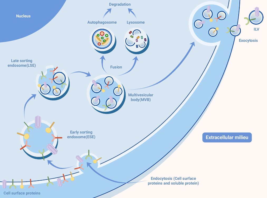
Figure 1.
Extracellular vesicle biogenesis: Plasma membrane endocytosis internalizes cell surface and soluble proteins → early sorting endosomes (ESEs) → late sorting endosomes (LSEs) → multivesicular bodies (MVBs) containing intraluminal vesicles (ILVs)
-
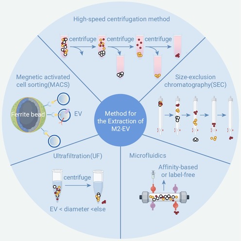
Figure 2.
Classic extracellular vesicle isolation methods: high-speed centrifugation method; magnetic-activated cell sorting (MACS); size-exclusion chromatography (SEC); ultrafiltration (UF); microfluidics.
-
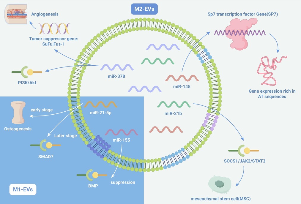
Figure 3.
Osteogenic role of miRNAs: miR-378a positively regulates osteogenesis, promotes MSC osteogenic differentiation, and is highly enriched in M2-EVs
-
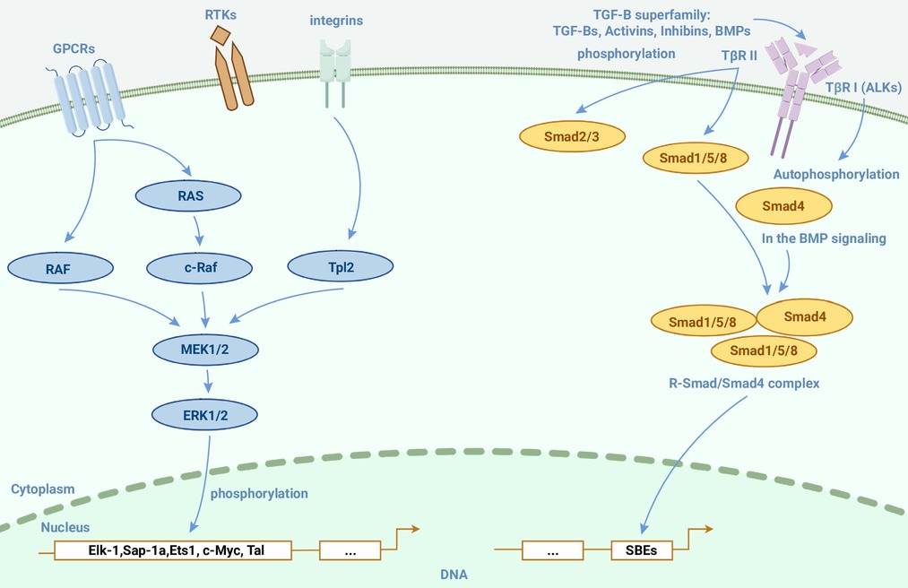
Figure 4.
MAPK signaling pathway and TGF-β/Smad signaling pathway involved in osteogenesis
-
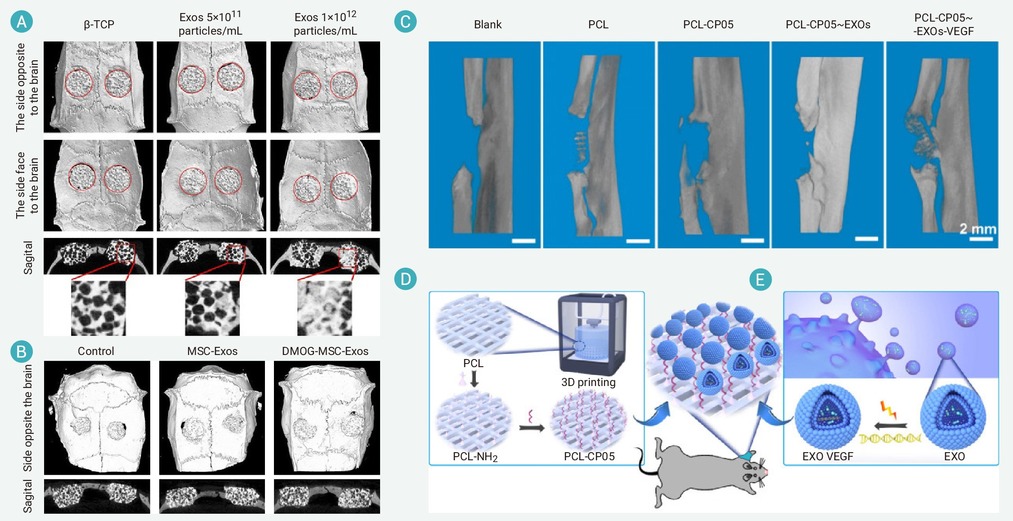
Figure 5.
Comparative evaluation of bone defect repair for different biomaterial scaffolds loaded with various engineered extracellular vesicles
-
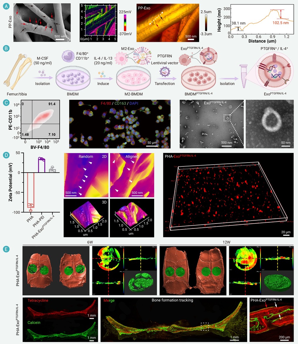
Figure 6.
Preparation, characterization and bone regeneration of PTGFRN/IL-4-EV-modified Janus electrospun nanofibrous scaffolds
-
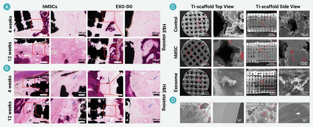
Figure 7.
Histological and SEM characterization of EV-modified metal scaffolds and hydrogel biomaterials
