
Expression dynamics of IL-1 family genes in the healthy human brain suggest non-immune functional roles
-
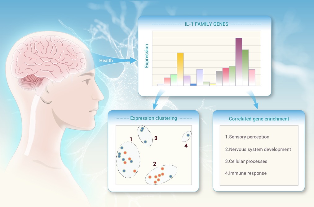
GRAPHICAL ABSTRACT
 DownLoad:
Full size image
DownLoad:
Full size image
-
PUBLIC SUMMARY
-
Cytokines and receptors of the IL-1 family are mediators of inflammation and defense immune functions in the human body.
However, their substantial expression in the healthy brain suggests that they may have other functions.
Analysis of gene expression and co-expression suggests brain-specific roles.
These molecules may have physiological functions in neurogenesis, neurometabolism and neurotransmission.
-
-
ABSTRACT
Interleukin-1 (IL-1) family genes include 22 cytokines and receptors with different evolutionary histories. While generally associated with inflammation-related immune defensive reactions, several IL-1 family genes are expressed at measurable levels in the healthy brain, as assessed in animal models, although their functions are still obscure. In this study, we investigated the expression and possible functions of these genes in the healthy human brain, using RNA-seq samples retrieved from three different databases. Some of them, such as SIGIRR, IL33 and IL1RAPL1, are highly expressed across multiple regions, while others, e.g., IL1F10 and IL36B, are undetectable. Co-expression analysis identified genes with positive and negative expression correlation with IL-1 family genes. Upon enrichment analysis of the correlated genes, it was found that, at variance with their systemic expression and immune-related function, expression of several IL-1 family genes shows a brain-specific pattern that correlates with neurological functions. For instance, IL1RAPL2 expression positively correlates with genes involved in brain development and synaptic functions and negatively correlates with genes involved in fatty acid oxidation, suggesting promotion of neurogenesis. IL1RL1, IL1RAP and IL1RAPL1 positively correlate with genes involved in sensory smell perception and transmission. SIGIRR positively correlates with genes involved in translation and protein synthesis in the brain. IL33 positively correlates with genes involved in fatty acid oxidation, suggesting regulation of neurogenesis. Thus, this study suggests that several IL-1 family genes, which are constitutively expressed in the healthy human brain, may have a non-immune non-inflammatory role in sustaining and regulating brain-specific functions. -

-
REFERENCES
[1] The IL-1 cytokine and receptor family (2018). Immunol. Rev. 281: 1–247. DOI: 10.1111/imr.12588 [2] Boraschi D., Italiani P., Weil S., et al. (2018). The family of the interleukin-1 receptors. Immunol. Rev. 281:197−232. DOI:10.1111/imr.12606 [3] Garlanda C., Dinarello C.A. and Mantovani A. (2013). The Interleukin-1 family: Back to the future. Immunity 39:1003−1018. DOI:10.1016/j.immuni.2013.11.010 [4] Gaballa J.M., Falkesgaard Hojen J., De Graaf, D.M., et al. (2024). International nomenclature guidelines for the IL-1 family of cytokines and receptors. Nat. Immunol. 25:581−582. DOI:10.1038/s41590-024-01777-1 [5] Rivers-Auty J., Daniels M.J.D., Colliver I., et al. (2018). Redefining the ancestral origins of the interleukin-1 superfamily. Nat. Commun. 9:1156. DOI:10.1038/s41467-018-03362-1 [6] Boraschi D. (2022). What Is IL-1 for? The functions of Interleukin-1 across evolution. Front. Immunol. 13: 872155. DOI: 10.3389/fimmu.2022.872155 [7] Rothwell N.J., Luheshi G.N. (2000). Interleukin 1 in the brain: Biology, pathology and therapeutic target. Trends Neurosci. 23:618−625. DOI:10.1016/S0166-2236(00)01661-1 [8] Boraschi D., Italiani P., Migliorini P., et al. (2023). Cause or consequence? The role of IL-1 family cytokines and receptors in neuroinflammatory and neurodegenerative diseases. Front. Immunol. 14: 1128190. DOI: 10.3389/fimmu. 2023.1128190 [9] Aguet F., Barbeira A.N., Bonazzola R., et al. (2020). The GTEx Consortium atlas of genetic regulatory effects across human tissues. Science 369:1318−1330. DOI:10.1126/science.aaz1776 [10] Sjöstedt E., Zhong W., Fagerberg L., et al. (2020). An atlas of the protein-coding genes in the human, pig, and mouse brain. Science 367:eaay5947. DOI:10.1126/science.aay5947 [11] Uhlén M., Fagerberg L., Hallström B.M., et al. (2015). Tissue-based map of the human proteome. Science 347:1260419. DOI:10.1126/science.1260419 [12] Zhao S., Ye Z. and Stanton R. (2020). Misuse of RPKM or TPM normalization when comparing across samples and sequencing protocols. RNA 26:903−909. DOI:10.1261/RNA.074922.120 [13] Robinson M.D. and Oshlack A. (2010). A scaling normalization method for differential expression analysis of RNA-seq data. Genome Biol. 11:R25. DOI:10.1186/gb-2010-11-3-r25 [14] Kuleshov M.V., Jones M.R., Rouillard A.D., et al. (2016). Enrichr: A comprehensive gene set enrichment analysis web server 2016 update. Nucleic Acids Res. 44:W90−W97. DOI:10.1093/nar/gkw377 [15] Heinz S., Benner C., Spann N., et al. (2010). Simple combinations of lineage-determining transcription factors prime cis-regulatory elements required for macrophage and B cell Identities. Mol. Cell 38:576−589. DOI:10.1016/j.molcel.2010.05.004 [16] Lee J.K., Chung J., Druey K.M., et al. (2012). RGS10 exerts a neuroprotective role through the PKA/c-AMP response-element (CREB) pathway in dopaminergic neuron-like cells. J. Neurochem. 122:333−342. DOI:10.1111/J.1471-4159.2012.07780.x [17] van Dam S., Võsa U., van der Graaf A., et al. (2018). Gene co-expression analysis for functional classification and gene-disease predictions. Brief. Bioinform. 19:575−592. DOI:10.1093/bib/bbw139 [18] Mosley B., De Imus C., Friend D., et al. (1996). Dual oncostatin M (OSM) receptors. Cloning and characterization of an alternative signaling subunit conferring OSM-specific receptor activation. J. Biol. Chem. 271:32635−32643. DOI:10.1074/jbc.271.51.32635 [19] Shi S., Gu S., Han T., et al. (2020). Inhibition of MAN2A1 enhances the immune response to anti-PD-L1 in human tumors. Clin. Cancer Res. 26:5990−6002. DOI:10.1158/1078-0432.CCR-20-0778 [20] Tang W., Lai Y.H., Han X.D., et al. (1997). Murine Hn1 on chromosome 11 is expressed in hemopoietic and brain tissues. Mamm. Genome 8:695−696. DOI:10.1007/S003359900540 [21] Vallet A.-E., Verscheren A., Petiot P., et al. (2012). Neurological features in adult Triple-A (Allgrove) syndrome. J. Neurol. 259:39−46. DOI:10.1007/s00415-011-6115-9 [22] Knobloch M., Pilz G.A., Ghesquière B., et al. (2017). A fatty acid oxidation-dependent metabolic shift regulates adult neural stem cell activity. Cell Rep. 20:2144−2155. DOI:10.1016/j.celrep.2017.08.029 [23] Roncaglia P., Martone M.E., Hill D.P., et al. (2013). The gene ontology (GO) cellular component ontology: Integration with SAO (subcellular anatomy ontology) and other recent developments. J. Biomed. Semantics 4:20. DOI:10.1186/2041-1480-4-20 [24] Salem S., Salem D. and Gros P. (2020). Role of IRF8 in immune cells functions, protection against infections, and susceptibility to inflammatory diseases. Hum. Genet. 139:707−721. DOI:10.1007/s00439-020-02154-2 [25] Thie G., Ekici, M. and Rössler O.G. (2015). RE-1 silencing transcription factor (REST): A regulator of neuronal development and neuronal/endocrine function. Cell Tissue Res. 359:99−109. DOI:10.1007/s00441-014-1963-0 [26] Crombie E.M., Cleverley K., Timmers H.T.M., et al. (2024). The roles of TAF1 in neuroscience and beyond. R. Soc. Open Sci. 11:240790. DOI:10.1098/RSOS.240790 [27] Mariani J.N., Mansky B., Madsen P.M., et al. (2024). Repression of developmental transcription factor networks triggers aging-associated gene expression in human glial progenitor cells. Nat. Commun. 15:1−19. DOI:10.1038/s41467-024-48118-2 [28] Hutter S., Bolin S., Weishaupt H., et al. (2017). Modeling and targeting MYC genes in childhood brain tumors. Genes 8:107. DOI:10.3390/genes8040107 [29] Min X.-L., Liu H.-J., Dou X.-K., et al. (2024). Extracellular vesicles from neural stem cells carry microRNA-16-5p to reduce corticosterone-induced neuronal injury in depression rats. Neuroscience 538:95−109. DOI:10.1016/j.neuroscience.2023.09.016 [30] Hoss A.G., Kartha V.K., Dong X., et al. (2014). MicroRNAs located in the Hox gene clusters are implicated in Huntington’s disease pathogenesis. PLoS Genet. 10:e1004188. DOI:10.1371/journal.pgen.1004188 [31] Hausser J., Mayo A., Keren L., et al. (2019). Central dogma rates and the trade-off between precision and economy in gene expression. Nat. Commun. 10:68. DOI:10.1038/s41467-018-07391-8 [32] Jin H., Gardner R.J., Viswesvaraiah R., et al. (2000). Two novel members of the interleukin-1 receptor gene family, one deleted in Xp22.1-Xp21.3 mental retardation. Eur. J. Hum. Genet. 8: 87–94. DOI: 10.1038/sj.ejhg.5200415 [33] Irwin L.N., Byers D.M. (2012). Novel odors affect gene expression for cytokines and proteinases in the rat amygdala and hippocampus. Brain Res. 1489:1−7. DOI:10.1016/j.brainres.2012.10.034 [34] Lin C.C. and Huang T.L. (2021). SIGIRR and TNFAIP3 are differentially expressed in both PBMC and TNF-α secreting cells of patients with major depressive disorder. Front. Psychiatry 12:698257. DOI:10.3389/fpsyt.2021.698257 -
ABOUT THIS ARTICLE
Cite this article:
Babarinde I. A. and Boraschi D. (2026). Expression dynamics of IL-1 family genes in the healthy human brain suggest non-immune functional roles. The Innovation Life 4:100183. https://doi.org/10.59717/j.xinn-life.2026.100183
Welcome!
To request copyright permission to republish or share portions of our works, please visit Copyright Clearance Center's (CCC) Marketplace website at marketplace.copyright.com.
Supplementary Information
-
download_supplementary
Tables
Article Metrics
Relative Articles
Cited by
Export File
Citation
Babarinde I. A. and Boraschi D. (2026). Expression dynamics of IL-1 family genes in the healthy human brain suggest non-immune functional roles. The Innovation Life 4:100183. https://doi.org/10.59717/j.xinn-life.2026.100183

Format
Content
-
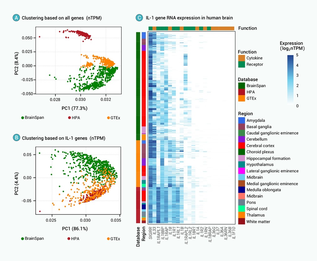
Figure 1.
Dynamics of IL-1 family gene expression across different human brain regions
-
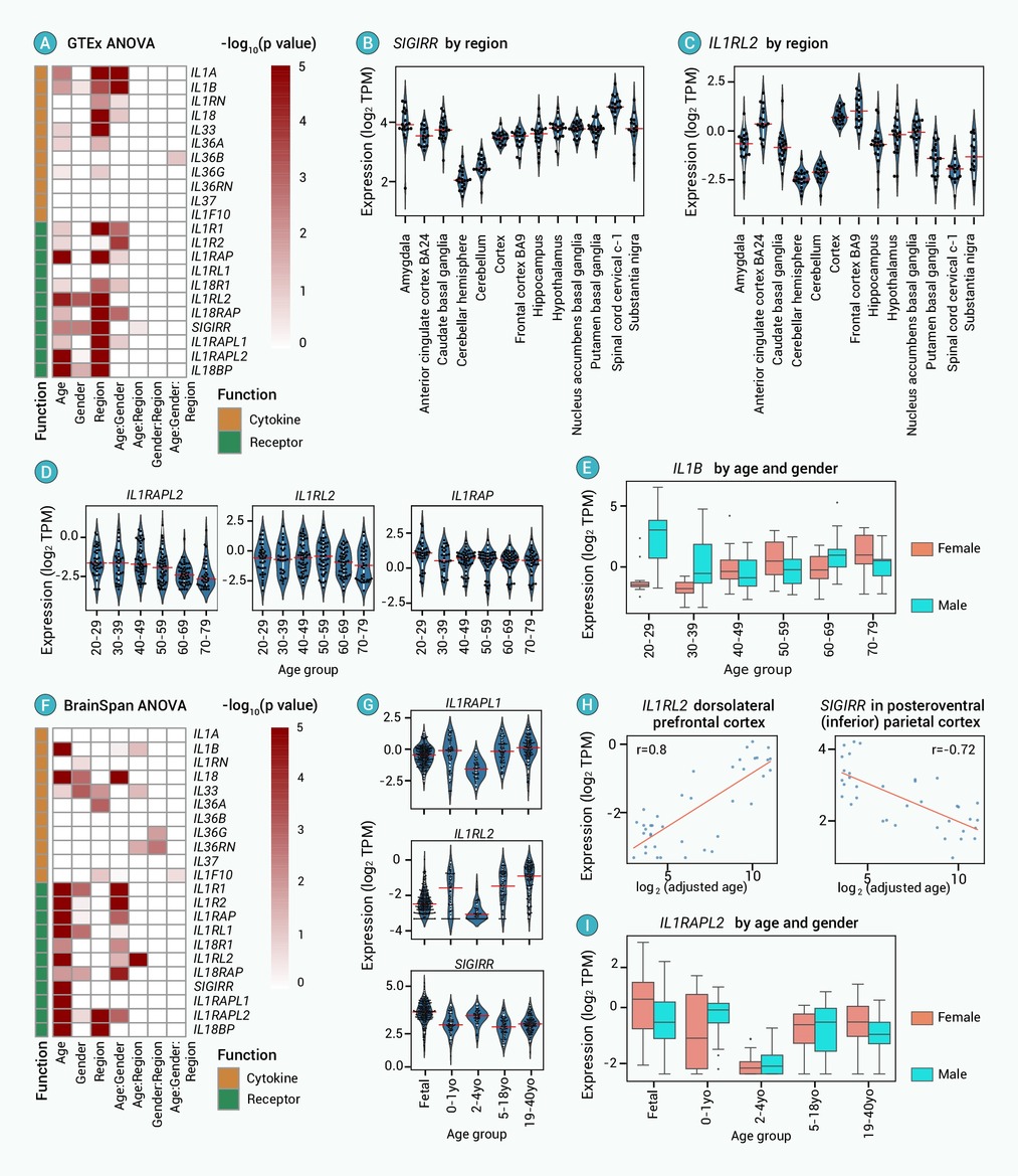
Figure 2.
Assessment of factors affecting the expression levels of IL-1 family genes in the human brain
-
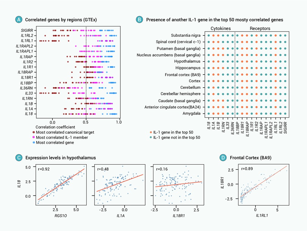
Figure 3.
Correlated expression of IL-1 family genes across different brain regions
-
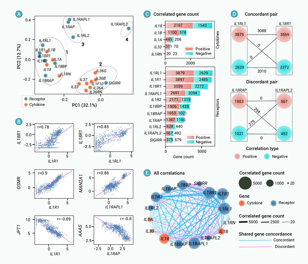
Figure 4.
Co-expression analysis of IL-1 genes in the human brain
-
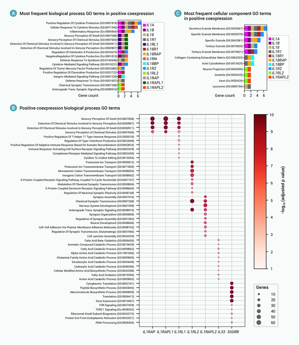
Figure 5.
Gene ontology enrichment analysis for potential functions of positively correlated genes
-
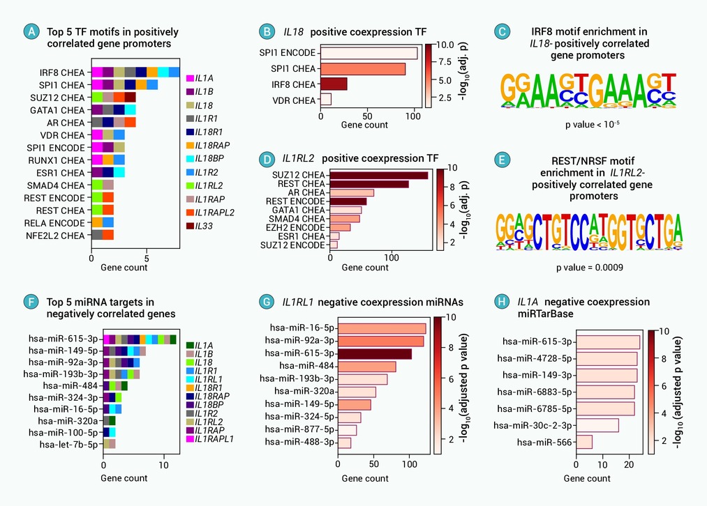
Figure 6.
Transcription factor and miRNA enrichment analysis highlights the potential expression regulation of correlated genes
