
-
GRAPHICAL ABSTRACT
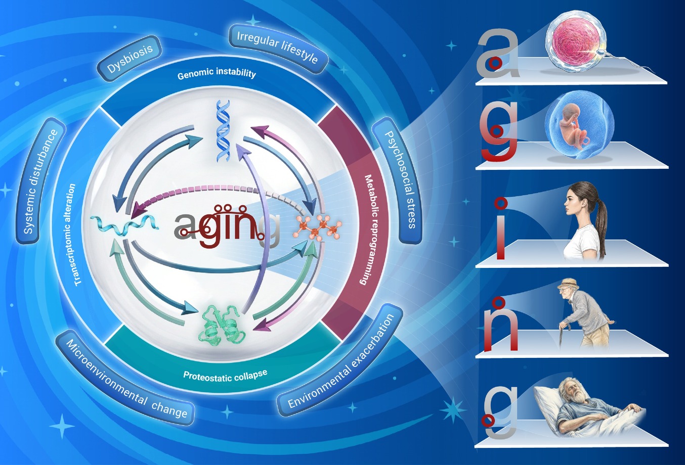
 DownLoad:
Full size image
-
PUBLIC SUMMARY
-
The Central Dogma of Aging extends the canonical DNA→RNA→protein information flow to metabolites.
Layer-specific imbalances in anabolism, modification and catabolism propagate across levels to drive aging.
The exposome acts hierarchically on the four layers and thereby sets the aging trajectory.
This framework offers a rationale for aging interventions at any layer, and the exposome.
-
-
ABSTRACT
Aging is driven by interconnected molecular and cellular changes that lead to functional decline and increased disease susceptibility. Here, we propose a novel "Central Dogma of Aging" hypothesis that extends the classical central dogma of molecular biology (DNA > RNA > protein) to encompass DNA, RNA, protein, and metabolite. The four layers are interconnected and mutually regulated by a 4C (Coding, Catalysis, Constitution and Catabolism) rule, referred to as the Central Dogma; each is further modulated by anabolism (synthesis), modification (post-synthesis alterations), and catabolism (degradation) process. Aging arises from imbalances in anabolism, modification, and catabolism across DNA, RNA, proteins, and metabolites, and is dynamically challenged by the exposome—cumulative environmental stimuli. The Central Dogma of Aging offers a comprehensive model for aging mechanisms, with implications for targeted therapies to promote healthiness and longevity. -

-
REFERENCES
[1] Hou Y., Dan X., Babbar M., et al. (2019). Ageing as a risk factor for neurodegenerative disease. Nat. Rev. Neurol. 15:565−581. DOI:10.1038/s41582-019-0244-7 [2] Guo J., Huang X., Dou L., et al. (2022). Aging and aging-related diseases: From molecular mechanisms to interventions and treatments. Signal Transduct. Target Ther. 7:391. DOI:10.1038/s41392-022-01251-0 [3] Pu F., Chen W., Li C., et al. (2024). Heterogeneous associations of multiplexed environmental factors and multidimensional aging metrics. Nat. Commun. 15:4921. DOI:10.1038/s41467-024-49283-0 [4] Argentieri M. A., Amin N., Nevado-Holgado A. J., et al. (2025). Integrating the environmental and genetic architectures of aging and mortality. Nat. Med. 31:1016−1025. DOI:10.1038/s41591-024-03483-9 [5] Lopez-Otin C., Blasco M. A., Partridge L., et al. (2023). Hallmarks of aging: An expanding universe. Cell 186:243−278. DOI:10.1016/j.cell.2022.11.001 [6] Kroemer G., Maier A. B., Cuervo A. M., et al. (2025). From geroscience to precision geromedicine: Understanding and managing aging. Cell 188:2043−2062. DOI:10.1016/j.cell.2025.03.011 [7] Crick F. (1970). Central dogma of molecular biology. Nature 227:561−563. DOI:10.1038/227561a0 [8] Cheng F. and Wang L. (2024). Slowing down the central dogma rate for alleviating aging. Innov. Life 2:100049. DOI:10.59717/j.xinn-life.2024.100049 [9] Shen X., Wang C., Zhou X., et al. (2024). Nonlinear dynamics of multi-omics profiles during human aging. Nat. Aging 4:1619−1634. DOI:10.1038/s43587-024-00692-2 [10] Ahadi S., Zhou W., Schussler-Fiorenza Rose S. M., et al. (2020). Personal aging markers and ageotypes revealed by deep longitudinal profiling. Nat. Med. 26:83−90. DOI:10.1038/s41591-019-0719-5 [11] Rutledge J., Oh H. and Wyss-Coray T. (2022). Measuring biological age using omics data. Nat. Rev. Genet. 23:715−727. DOI:10.1038/s41576-022-00511-7 [12] Niedernhofer L. J., Garinis G. A., Raams A., et al. (2006). A new progeroid syndrome reveals that genotoxic stress suppresses the somatotroph axis. Nature 444:1038−1043. DOI:10.1038/nature05456 [13] Schumacher B., Pothof J., Vijg J., et al. (2021). The central role of DNA damage in the ageing process. Nature 592:695−703. DOI:10.1038/s41586-021-03307-7 [14] Bujarrabal-Dueso A., Garinis G. A., Robbins P. D., et al. (2025). Targeting DNA damage in ageing: Towards supercharging DNA repair. Nat. Rev. Drug Discovery 24:785−807. DOI:10.1038/s41573-025-01212-6 [15] Kunkel T. A. (2004). DNA replication fidelity. J. Biol. Chem. 279:16895−16898. DOI:10.1074/jbc.R400006200 [16] Loeb L. A. and Monnat R. J., Jr. (2008). DNA polymerases and human disease. Nat. Rev. Genet. 9:594−604. DOI:10.1038/nrg2345 [17] Harley C. B., Futcher A. B. and Greider C. W. (1990). Telomeres shorten during ageing of human fibroblasts. Nature 345:458−460. DOI:10.1038/345458a0 [18] Chang E. and Harley C. B. (1995). Telomere length and replicative aging in human vascular tissues. Proc. Natl. Acad. Sci. USA 92:11190−11194. DOI:10.1073/pnas.92.24.11190 [19] Herbig U., Jobling W. A., Chen B. P., et al. (2004). Telomere shortening triggers senescence of human cells through a pathway involving ATM, p53, and p21(CIP1), but not p16(INK4a). Mol. Cell 14:501−513. DOI:10.1016/s1097-2765(04)00256-4 [20] Crabbe L., Jauch A., Naeger C. M., et al. (2007). Telomere dysfunction as a cause of genomic instability in Werner syndrome. Proc. Natl. Acad. Sci. USA 104:2205−2210. DOI:10.1073/pnas.0609410104 [21] Rossiello F., Jurk D., Passos J. F., et al. (2022). Telomere dysfunction in ageing and age-related diseases. Nat. Cell Biol. 24:135−147. DOI:10.1038/s41556-022-00842-x [22] Benayoun B. A., Pollina E. A. and Brunet A. (2015). Epigenetic regulation of ageing: Linking environmental inputs to genomic stability. Nat. Rev. Mol. Cell Biol. 16:593−610. DOI:10.1038/nrm4048 [23] Pal S. and Tyler J. K. (2016). Epigenetics and aging. Sci. Adv. 2:e1600584. DOI:10.1126/sciadv.1600584 [24] Yang J. H., Hayano M., Griffin P. T., et al. (2023). Loss of epigenetic information as a cause of mammalian aging. Cell 186:305-326 e327. DOI:10.1016/j.cell.2022.12.027. [25] Wu Z., Qu J. and Liu G. H. (2024). Roles of chromatin and genome instability in cellular senescence and their relevance to ageing and related diseases. Nat. Rev. Mol. Cell Biol. 25:979−1000. DOI:10.1038/s41580-024-00775-3 [26] Debes C., Papadakis A., Gronke S., et al. (2023). Ageing-associated changes in transcriptional elongation influence longevity. Nature 616:814−821. DOI:10.1038/s41586-023-05922-y [27] Papantonis A., Antebi A., Partridge L., et al. (2025). Age-associated changes in transcriptional elongation and their effects on homeostasis. Trends Cell Biol. 35:645−650. DOI:10.1016/j.tcb.2024.11.005 [28] Hao X., Shiromoto Y., Sakurai M., et al. (2022). ADAR1 downregulation by autophagy drives senescence independently of RNA editing by enhancing p16(INK4a) levels. Nat. Cell Biol. 24:1202−1210. DOI:10.1038/s41556-022-00959-z [29] Zhang J., Ao Y., Zhang Z., et al. (2020). Lamin A safeguards the m(6) A methylase METTL14 nuclear speckle reservoir to prevent cellular senescence. Aging Cell 19:e13215. DOI:10.1111/acel.13215 [30] Wu Z., Lu M., Liu D., et al. (2023). M(6)A epitranscriptomic regulation of tissue homeostasis during primate aging. Nat. Aging 3:705−721. DOI:10.1038/s43587-023-00393-2 [31] Fu Y., Jiang F., Zhang X., et al. (2024). Perturbation of METTL1-mediated tRNA N(7)- methylguanosine modification induces senescence and aging. Nat. Commun. 15:5713. DOI:10.1038/s41467-024-49796-8 [32] van Sluis M., Gonzalo-Hansen C., Li Q., et al. (2026). Mechanisms of transcription-coupled repair and DNA damage surveillance in health and disease. Nat. Rev. Mol. Cell Biol. 27:234−251. DOI:10.1038/s41580-025-00915-3 [33] Bahar R., Hartmann C. H., Rodriguez K. A., et al. (2006). Increased cell-to-cell variation in gene expression in ageing mouse heart. Nature 441:1011−1014. DOI:10.1038/nature04844 [34] Wei Y. N., Hu H. Y., Xie G. C., et al. (2015). Transcript and protein expression decoupling reveals RNA binding proteins and miRNAs as potential modulators of human aging. Genome Biol. 16:41. DOI:10.1186/s13059-015-0608-2 [35] Zheng H., Seit-Nebi A., Han X., et al. (2013). A posttranslational modification cascade involving p38, Tip60, and PRAK mediates oncogene-induced senescence. Mol. Cell 50:699−710. DOI:10.1016/j.molcel.2013.04.013 [36] Zhang C. and Cuervo A. M. (2008). Restoration of chaperone-mediated autophagy in aging liver improves cellular maintenance and hepatic function. Nat. Med. 14:959−965. DOI:10.1038/nm.1851 [37] Bobkova N. V., Evgen'ev M., Garbuz D. G., et al. (2015). Exogenous Hsp70 delays senescence and improves cognitive function in aging mice. Proc. Natl. Acad. Sci. USA 112:16006−16011. DOI:10.1073/pnas.1516131112 [38] Edifizi D., Nolte H., Babu V., et al. (2017). Multilayered reprogramming in response to persistent DNA damage in C. elegans. Cell Rep. 20:2026−2043. DOI:10.1016/j.celrep.2017.08.028 [39] Stein K. C., Morales-Polanco F., van der Lienden J., et al. (2022). Ageing exacerbates ribosome pausing to disrupt cotranslational proteostasis. Nature 601:637−642. DOI:10.1038/s41586-021-04295-4 [40] Jankowski C. S. R., Samarah L. Z., MacArthur M. R., et al. (2025). Aged mice exhibit widespread metabolic changes but preserved major fluxes. Cell Metab. 37:2280-2294 e2284. DOI:10.1016/j.cmet.2025.09.009. [41] Alonso-Lavin A. J., Bajic D. and Poyatos J. F. (2021). Tolerance to NADH/NAD(+) imbalance anticipates aging and anti-aging interventions. iScience 24:102697. DOI:10.1016/j.isci.2021.102697 [42] Menendez J. A., Alarcon T. and Joven J. (2014). Gerometabolites: The pseudohypoxic aging side of cancer oncometabolites. Cell Cycle 13:699−709. DOI:10.4161/cc.28079 [43] Grimbs S., Selbig J., Bulik S., et al. (2007). The stability and robustness of metabolic states: Identifying stabilizing sites in metabolic networks. Mol. Syst. Biol. 3:146. DOI:10.1038/msb4100186 [44] Lopez-Otin C., Blasco M. A., Partridge L., et al. (2013). The hallmarks of aging. Cell 153:1194−1217. DOI:10.1016/j.cell.2013.05.039 [45] Li S., Agudelo Garcia P. A., Aliferis C., et al. (2025). Advancing biological understanding of cellular senescence with computational multiomics. Nat. Genet. 57:2381−2394. DOI:10.1038/s41588-025-02314-y [46] Hoffman J. M., Lyu Y., Pletcher S. D., et al. (2017). Proteomics and metabolomics in ageing research: From biomarkers to systems biology. Essays Biochem. 61:379−388. DOI:10.1042/EBC20160083 [47] Imai S. and Guarente L. (2014). NAD+ and sirtuins in aging and disease. Trends Cell Biol. 24:464−471. DOI:10.1016/j.tcb.2014.04.002 [48] Villa E., Sahu U., O'Hara B. P., et al. (2021). mTORC1 stimulates cell growth through SAM synthesis and m(6)A mRNA-dependent control of protein synthesis. Mol. Cell 81:2076-2093 e2079. DOI:10.1016/j.molcel.2021.03.009. [49] Hu W. L., Jin L., Xu A., et al. (2018). GUARDIN is a p53-responsive long non-coding RNA that is essential for genomic stability. Nat. Cell Biol. 20:492−502. DOI:10.1038/s41556-018-0066-7 [50] de Lorenzo V. (2014). From the selfish gene to selfish metabolism: Revisiting the central dogma. Bioessays 36:226−235. DOI:10.1002/bies.201300153 [51] Hernandez H., Santamaria-Garcia H., Moguilner S., et al. (2025). The exposome of healthy and accelerated aging across 40 countries. Nat. Med. 31:3089−3100. DOI:10.1038/s41591-025-03808-2 [52] Johnson F. B., Sinclair D. A. and Guarente L. (1999). Molecular biology of aging. Cell 96:291−302. DOI:10.1016/s0092-8674(00)80567-x [53] Jacobs P. A., Court Brown W. M. and Doll R. (1961). Distribution of human chromosome counts in relation to age. Nature 191:1178−1180. DOI:10.1038/1911178a0 [54] Vijg J. and Dolle M. E. (2002). Large genome rearrangements as a primary cause of aging. Mech. Ageing Dev. 123:907−915. DOI:10.1016/s0047-6374(02)00028-3 [55] Dolle M. E. and Vijg J. (2002). Genome dynamics in aging mice. Genome Res. 12:1732−1738. DOI:10.1101/gr.125502 [56] Chandra T., Ewels P. A., Schoenfelder S., et al. (2015). Global reorganization of the nuclear landscape in senescent cells. Cell Rep. 10:471−483. DOI:10.1016/j.celrep.2014.12.055 [57] Lodato M. A., Rodin R. E., Bohrson C. L., et al. (2018). Aging and neurodegeneration are associated with increased mutations in single human neurons. Science 359:555−559. DOI:10.1126/science.aao4426 [58] Choudhury S., Huang A. Y., Kim J., et al. (2022). Somatic mutations in single human cardiomyocytes reveal age-associated DNA damage and widespread oxidative genotoxicity. Nat. Aging 2:714−725. DOI:10.1038/s43587-022-00261-5 [59] Vijg J., Schumacher B., Abakir A., et al. (2023). Mitigating age-related somatic mutation burden. Trends Mol. Med. 29:530−540. DOI:10.1016/j.molmed.2023.04.002 [60] Shaban H. A. and Gasser S. M. (2025). Dynamic 3D genome reorganization during senescence: Defining cell states through chromatin. Cell Death Differ. 32:9−15. DOI:10.1038/s41418-023-01197-y [61] Bacolla A., Pradhan S., Roberts R. J., et al. (1999). Recombinant human DNA (cytosine-5) methyltransferase. II. Steady-state kinetics reveal allosteric activation by methylated DNA. J. Biol. Chem. 274:33011−33019. DOI:10.1074/jbc.274.46.33011 [62] Suetake I., Shinozaki F., Miyagawa J., et al. (2004). DNMT3L stimulates the DNA methylation activity of Dnmt3a and Dnmt3b through a direct interaction. J. Biol. Chem. 279:27816−27823. DOI:10.1074/jbc.M400181200 [63] Oberdoerffer P., Michan S., McVay M., et al. (2008). SIRT1 redistribution on chromatin promotes genomic stability but alters gene expression during aging. Cell 135:907−918. DOI:10.1016/j.cell.2008.10.025 [64] Pazolli E., Alspach E., Milczarek A., et al. (2012). Chromatin remodeling underlies the senescence-associated secretory phenotype of tumor stromal fibroblasts that supports cancer progression. Cancer Res. 72:2251−2261. DOI:10.1158/0008-5472.CAN-11-3386 [65] Marttila S., Kananen L., Hayrynen S., et al. (2015). Ageing-associated changes in the human DNA methylome: Genomic locations and effects on gene expression. BMC Genomics 16:179. DOI:10.1186/s12864-015-1381-z [66] Sen P., Lan Y., Li C. Y., et al. (2019). Histone acetyltransferase p300 induces de novo super-enhancers to drive cellular senescence. Mol. Cell 73:684-698 e688. DOI:10.1016/j.molcel.2019.01.021 [67] Xu C., Wang L., Fozouni P., et al. (2020). SIRT1 is downregulated by autophagy in senescence and ageing. Nat. Cell Biol. 22:1170−1179. DOI:10.1038/s41556-020-00579-5 [68] Wang K., Liu H., Hu Q., et al. (2022). Epigenetic regulation of aging: Implications for interventions of aging and diseases. Signal Transduct. Target Ther. 7:374. DOI:10.1038/s41392-022-01211-8 [69] Weaver K. J., Holt R. A., Henry E., et al. (2023). Effects of hunger on neuronal histone modifications slow aging in Drosophila. Science 380:625−632. DOI:10.1126/science.ade1662 [70] Makarov V. L., Hirose Y. and Langmore J. P. (1997). Long G tails at both ends of human chromosomes suggest a C strand degradation mechanism for telomere shortening. Cell 88:657−666. DOI:10.1016/s0092-8674(00)81908-x [71] Wright W. E., Tesmer V. M., Huffman K. E., et al. (1997). Normal human chromosomes have long G-rich telomeric overhangs at one end. Genes Dev. 11:2801−2809. DOI:10.1101/gad.11.21.2801 [72] Huffman K. E., Levene S. D., Tesmer V. M., et al. (2000). Telomere shortening is proportional to the size of the G-rich telomeric 3'-overhang. J. Biol. Chem. 275:19719−19722. DOI:10.1074/jbc.M002843200 [73] Shammas M. A. (2011). Telomeres, lifestyle, cancer, and aging. Curr. Opin. Clin. Nutr. Metab. Care 14:28−34. DOI:10.1097/MCO.0b013e32834121b1 [74] Hewitt G., Jurk D., Marques F. D., et al. (2012). Telomeres are favoured targets of a persistent DNA damage response in ageing and stress-induced senescence. Nat. Commun. 3:708. DOI:10.1038/ncomms1708 [75] Wang Y., Wang X., Flores E. R., et al. (2016). Dysfunctional telomeres induce p53-dependent and independent apoptosis to compromise cellular proliferation and inhibit tumor formation. Aging Cell 15:646−660. DOI:10.1111/acel.12476 [76] Lan Y. Y., Heather J. M., Eisenhaure T., et al. (2019). Extranuclear DNA accumulates in aged cells and contributes to senescence and inflammation. Aging Cell 18:e12901. DOI:10.1111/acel.12901 [77] Li Y., Tian X., Luo J., et al. (2024). Molecular mechanisms of aging and anti-aging strategies. Cell Commun. Signal 22:285. DOI:10.1186/s12964-024-01663-1 [78] Goncalves T., Cunniffe S., Ma T. S., et al. (2025). Elevated reactive oxygen species can drive the alternative lengthening of telomeres pathway in ATRX-null cancers. Nucleic Acids Res. 53:gkaf061. DOI:10.1093/nar/gkaf061 [79] Wang C., Martens D. S., Bustamante M., et al. (2025). The multi-omics signatures of telomere length in childhood. BMC Genomics 26:75. DOI:10.1186/s12864-025-11209-5 [80] Sedelnikova O. A., Horikawa I., Zimonjic D. B., et al. (2004). Senescing human cells and ageing mice accumulate DNA lesions with unrepairable double-strand breaks. Nat. Cell Biol. 6:168−170. DOI:10.1038/ncb1095 [81] Morandini F., Lu J. Y., Rechsteiner C., et al. (2025). Transposable element 5mC methylation state of blood cells predicts age and disease. Nat. Aging 5:193−204. DOI:10.1038/s43587-024-00757-2 [82] Wang F., Pan X., Kalmbach K., et al. (2013). Robust measurement of telomere length in single cells. Proc. Natl. Acad. Sci. USA 110:E1906−1912. DOI:10.1073/pnas.1306639110 [83] Rowland T. J., Dumbovic G., Hass E. P., et al. (2019). Single-cell imaging reveals unexpected heterogeneity of telomerase reverse transcriptase expression across human cancer cell lines. Proc. Natl. Acad. Sci. USA 116:18488−18497. DOI:10.1073/pnas.1908275116 [84] Narita M., Nunez S., Heard E., et al. (2003). Rb-mediated heterochromatin formation and silencing of E2F target genes during cellular senescence. Cell 113:703−716. DOI:10.1016/s0092-8674(03)00401-x [85] Nikolov D. B. and Burley S. K. (1997). RNA polymerase II transcription initiation: A structural view. Proc. Natl. Acad. Sci. USA 94:15−22. DOI:10.1073/pnas.94.1.15 [86] Yamaguchi Y., Takagi T., Wada T., et al. (1999). NELF, a multisubunit complex containing RD, cooperates with DSIF to repress RNA polymerase II elongation. Cell 97:41−51. DOI:10.1016/s0092-8674(00)80713-8 [87] Oakes C. C., Smiraglia D. J., Plass C., et al. (2003). Aging results in hypermethylation of ribosomal DNA in sperm and liver of male rats. Proc. Natl. Acad. Sci. USA 100:1775−1780. DOI:10.1073/pnas.0437971100 [88] Cheung A. C., Sainsbury S. and Cramer P. (2011). Structural basis of initial RNA polymerase II transcription. EMBO J. 30:4755−4763. DOI:10.1038/emboj.2011.396 [89] D'Aquila P., Montesanto A., Mandala M., et al. (2017). Methylation of the ribosomal RNA gene promoter is associated with aging and age-related decline. Aging Cell 16:966−975. DOI:10.1111/acel.12603 [90] Sharifi S. and Bierhoff H. (2018). Regulation of RNA polymerase I transcription in development, disease, and aging. Annu. Rev. Biochem. 87:51−73. DOI:10.1146/annurev-biochem-062917-012612 [91] Vos S. M., Farnung L., Urlaub H., et al. (2018). Structure of paused transcription complex Pol II-DSIF-NELF. Nature 560:601−606. DOI:10.1038/s41586-018-0442-2 [92] Dai D. P., Gan W., Hayakawa H., et al. (2018). Transcriptional mutagenesis mediated by 8-oxoG induces translational errors in mammalian cells. Proc. Natl. Acad. Sci. USA 115:4218−4222. DOI:10.1073/pnas.1718363115 [93] Kang D., Baek Y. and Lee J. S. (2022). Mechanisms of RNA and protein quality control and their roles in cellular senescence and age-related diseases. Cells 11:4062. DOI:10.3390/cells11244062 [94] Sharifi S., Chaudhari P., Martirosyan A., et al. (2024). Reducing the metabolic burden of rRNA synthesis promotes healthy longevity in Caenorhabditis elegans. Nat. Commun. 15:1702. DOI:10.1038/s41467-024-46037-w [95] Schiksnis E. C., Nicastro I. A. and Pasquinelli A. E. (2024). Full-length direct RNA sequencing reveals extensive remodeling of RNA expression, processing and modification in aging Caenorhabditis elegans. Nucleic Acids Res. 52:13896−13913. DOI:10.1093/nar/gkae1064 [96] Sun P., Miller B. A., Sakai A. P., et al. (2026). RNA imbalance as a hallmark of cellular ageing. Nat. Cell Biol. 28:1102−1112. DOI:10.1038/s41556-026-01946-4 [97] Lee Y. and Rio D. C. (2015). Mechanisms and regulation of alternative pre-mRNA splicing. Annu. Rev. Biochem. 84:291−323. DOI:10.1146/annurev-biochem-060614-034316 [98] Han X., Xing L., Hong Y., et al. (2024). Nuclear RNA homeostasis promotes systems-level coordination of cell fate and senescence. Cell Stem Cell 31:694-716 e611. DOI:10.1016/j.stem.2024.03.015 [99] van der Werf I., Sneifer J. and Jamieson C. (2024). RNA modifications shape hematopoietic stem cell aging: Beyond the code. FEBS Lett. 598:2774−2775. DOI:10.1002/1873-3468.15014 [100] Wang Y., Wang C., Dai X., et al. (2025). Deciphering the multifaceted role of circular RNA in aging: From molecular mechanisms to therapeutic potentials. Noncoding RNA Res. 14:129−144. DOI:10.1016/j.ncrna.2025.05.015 [101] Lykke-Andersen J., Shu M. D. and Steitz J. A. (2000). Human Upf proteins target an mRNA for nonsense-mediated decay when bound downstream of a termination codon. Cell 103:1121−1131. DOI:10.1016/s0092-8674(00)00214-2 [102] Brewer G. (2002). Messenger RNA decay during aging and development. Ageing Res. Rev. 1:607−625. DOI:10.1016/s1568-1637(02)00023-5 [103] Wang W., Martindale J. L., Yang X., et al. (2005). Increased stability of the p16 mRNA with replicative senescence. EMBO Rep. 6:158−164. DOI:10.1038/sj.embor.7400346 [104] Son H. G., Seo M., Ham S., et al. (2017). RNA surveillance via nonsense-mediated mRNA decay is crucial for longevity in daf-2/insulin/IGF-1 mutant C. elegans. Nat. Commun. 8:14749. DOI:10.1038/ncomms14749 [105] Mullani N., Porozhan Y., Mangelinck A., et al. (2021). Reduced RNA turnover as a driver of cellular senescence. Life Sci. Alliance 4:e202000809. DOI:10.26508/lsa.202000809 [106] Wu Z., Zhang W., Qu J., et al. (2024). Emerging epigenetic insights into aging mechanisms and interventions. Trends Pharmacol. Sci. 45:157−172. DOI:10.1016/j.tips.2023.12.002 [107] Guillet C., Prod'homme M., Balage M., et al. (2004). Impaired anabolic response of muscle protein synthesis is associated with S6K1 dysregulation in elderly humans. FASEB J. 18:1586−1587. DOI:10.1096/fj.03-1341fje [108] Syntichaki P., Troulinaki K. and Tavernarakis N. (2007). eIF4E function in somatic cells modulates ageing in Caenorhabditis elegans. Nature 445:922−926. DOI:10.1038/nature05603 [109] Fry C. S., Drummond M. J., Glynn E. L., et al. (2011). Aging impairs contraction-induced human skeletal muscle mTORC1 signaling and protein synthesis. Skelet. Muscle 1:11. DOI:10.1186/2044-5040-1-11 [110] Rogers A. N., Chen D., McColl G., et al. (2011). Life span extension via eIF4G inhibition is mediated by posttranscriptional remodeling of stress response gene expression in C. elegans. Cell Metab. 14:55−66. DOI:10.1016/j.cmet.2011.05.010 [111] You J. S., Anderson G. B., Dooley M. S., et al. (2015). The role of mTOR signaling in the regulation of protein synthesis and muscle mass during immobilization in mice. Dis. Model Mech. 8:1059−1069. DOI:10.1242/dmm.019414 [112] Plubell D. L., Wilmarth P. A., Zhao Y., et al. (2017). Extended multiplexing of Tandem Mass Tags (TMT) labeling reveals age and high fat diet specific proteome changes in mouse epididymal adipose tissue. Mol. Cell. Proteomics 16:873−890. DOI:10.1074/mcp.M116.065524 [113] Anisimova A. S., Meerson M. B., Gerashchenko M. V., et al. (2020). Multifaceted deregulation of gene expression and protein synthesis with age. Proc. Natl. Acad. Sci. USA 117:15581−15590. DOI:10.1073/pnas.2001788117 [114] Kim H. S. and Pickering A. M. (2023). Protein translation paradox: Implications in translational regulation of aging. Front. Cell Dev. Biol. 11:1129281. DOI:10.3389/fcell.2023.1129281 [115] Choksi K. B., Boylston W. H., Rabek J. P., et al. (2004). Oxidatively damaged proteins of heart mitochondrial electron transport complexes. Biochim. Biophys. Acta 1688:95−101. DOI:10.1016/j.bbadis.2003.11.007 [116] Hains P. G. and Truscott R. J. (2010). Age-dependent deamidation of lifelong proteins in the human lens. Invest. Ophthalmol. Vis. Sci. 51:3107−3114. DOI:10.1167/iovs.09-4308 [117] Beltrao P., Bork P., Krogan N. J., et al. (2013). Evolution and functional cross-talk of protein post-translational modifications. Mol. Syst. Biol. 9:714. DOI:10.1002/msb.201304521 [118] Hollingsworth T. J., Radic M. Z., Beranova-Giorgianni S., et al. (2018). Murine retinal citrullination declines with age and is mainly dependent on peptidyl arginine deiminase 4 (PAD4). Invest. Ophthalmol. Vis. Sci. 59:3808−3815. DOI:10.1167/iovs.18-24118 [119] Leutert M., Entwisle S. W. and Villen J. (2021). Decoding post-translational modification crosstalk with proteomics. Mol. Cell. Proteomics 20:100129. DOI:10.1016/j.mcpro.2021.100129 [120] Murphy M. P., Bayir H., Belousov V., et al. (2022). Guidelines for measuring reactive oxygen species and oxidative damage in cells and in vivo. Nat. Metab. 4:651−662. DOI:10.1038/s42255-022-00591-z [121] Lee J. M., Hammaren H. M., Savitski M. M., et al. (2023). Control of protein stability by post-translational modifications. Nat. Commun. 14:201. DOI:10.1038/s41467-023-35795-8 [122] Johnston E. and Buckley M. (2023). Age-related changes in post-translational modifications of proteins from whole male and female skeletal elements. Molecules 28:4899. DOI:10.3390/molecules28134899 [123] Poudel S., Chuang C.-L., Shrestha H. K., et al. (2025). Pan-PTM profiling identifies post-translational modifications associated with exceptional longevity and preservation of skeletal muscle function in Drosophila. npj Aging 11:23. DOI:10.1038/s41514-025-00215-2 [124] Klionsky D. J. (2007). Autophagy: From phenomenology to molecular understanding in less than a decade. Nat. Rev. Mol. Cell Biol. 8:931−937. DOI:10.1038/nrm2245 [125] Cuanalo-Contreras K., Mukherjee A. and Soto C. (2013). Role of protein misfolding and proteostasis deficiency in protein misfolding diseases and aging. Int. J. Cell Biol. 2013:638083. DOI:10.1155/2013/638083 [126] Aman Y., Schmauck-Medina T., Hansen M., et al. (2021). Autophagy in healthy aging and disease. Nat. Aging 1:634−650. DOI:10.1038/s43587-021-00098-4 [127] Hamazaki J. and Murata S. (2024). Relationships between protein degradation, cellular senescence, and organismal aging. J. Biochem. 175:473−480. DOI:10.1093/jb/mvae016 [128] Yang X., Wei L., Zhong S., et al. (2025). Liquid-liquid phase separation of RBM33 facilitates hippocampus aging by inducing microglial senescence by activating CDKN1A. Int. J. Biol. Macromol. 310:142986. DOI:10.1016/j.ijbiomac.2025.142986 [129] Li Y., Jin X., Yu C., et al. (2025). Phase separation of MRG15 delays cellular senescence. Commun. Biol. 8:688. DOI:10.1038/s42003-025-08071-2 [130] Lian K. and Nan F. (2026). Small molecules modulate the biomolecular condensates and liquid‒liquid phase separation. Acta Pharm. Sin. B. 16:2756−2772. DOI:10.1016/j.apsb.2026.01.032 [131] Nyström T. (2005). Role of oxidative carbonylation in protein quality control and senescence. EMBO J. 24:1311−1317. DOI:10.1038/sj.emboj.7600599 [132] Olinger B. and Basisty N. (2026). A compendium of circulating biomarkers of senescence in humans: Insights on mechanistic impact across health domains and modulation by therapeutic interventions. Ageing Res. Rev. 121:103254. DOI:10.1016/j.arr.2026.103254 [133] de Lima Camillo L. P., Asif M. H., Horvath S., et al. (2025). Histone mark age of human tissues and cell types. Sci. Adv. 11:eadk9373. DOI:10.1126/sciadv.adk9373 [134] Breen L. and Phillips S. M. (2011). Skeletal muscle protein metabolism in the elderly: Interventions to counteract the 'anabolic resistance' of ageing. Nutr. Metab. (Lond.) 8:68. DOI:10.1186/1743-7075-8-68 [135] Zhu J. and Thompson C. B. (2019). Metabolic regulation of cell growth and proliferation. Nat. Rev. Mol. Cell Biol. 20:436−450. DOI:10.1038/s41580-019-0123-5 [136] Han Y., Hu Z., Cui A., et al. (2019). Post-translational regulation of lipogenesis via AMPK-dependent phosphorylation of insulin-induced gene. Nat. Commun. 10:623. DOI:10.1038/s41467-019-08585-4 [137] Le Couteur D. G., Solon-Biet S. M., Cogger V. C., et al. (2020). Branched chain amino acids, aging and age-related health. Ageing Res. Rev. 64:101198. DOI:10.1016/j.arr.2020.101198 [138] Lee G., Kim Y. Y., Jang H., et al. (2022). SREBP1c-PARP1 axis tunes anti-senescence activity of adipocytes and ameliorates metabolic imbalance in obesity. Cell Metab. 34:702-718 e705. DOI:10.1016/j.cmet.2022.03.010. [139] Bonnet L., Alexandersson I., Baboota R. K., et al. (2022). Cellular senescence in hepatocytes contributes to metabolic disturbances in NASH. Front. Endocrinol. (Lausanne) 13:957616. DOI:10.3389/fendo.2022.957616 [140] Vanweert F., Schrauwen P. and Phielix E. (2022). Role of branched-chain amino acid metabolism in the pathogenesis of obesity and type 2 diabetes-related metabolic disturbances BCAA metabolism in type 2 diabetes. Nutr. Diabetes 12:35. DOI:10.1038/s41387-022-00213-3 [141] Moldakozhayev A. and Gladyshev V. N. (2023). Metabolism, homeostasis, and aging. Trends Endocrinol. Metab. 34:158−169. DOI:10.1016/j.tem.2023.01.003 [142] Ayala A., Munoz M. F. and Arguelles S. (2014). Lipid peroxidation: Production, metabolism, and signaling mechanisms of malondialdehyde and 4-hydroxy-2-nonenal. Oxid. Med. Cell. Longev. 2014:360438. DOI:10.1155/2014/360438 [143] Furman D., Chang J., Lartigue L., et al. (2017). Expression of specific inflammasome gene modules stratifies older individuals into two extreme clinical and immunological states. Nat. Med. 23:174−184. DOI:10.1038/nm.4267 [144] Adav S. S. and Wang Y. L. (2021). Metabolomics signatures of aging: Recent advances. Aging Dis. 12:646−661. DOI:10.14336/Ad.2020.0909 [145] Balashova E. E., Maslov D. L., Trifonova O. P., et al. (2022). Metabolome profiling in aging studies. Biology (Basel) 11:1570. DOI:10.3390/biology11111570 [146] Trivedi D., Hollywood K. A., Xu Y., et al. (2024). Metabolomic heterogeneity of ageing with ethnic diversity: A step closer to healthy ageing. Metabolomics 21:9. DOI:10.1007/s11306-024-02199-8 [147] Roubenoff R. (2003). Catabolism of aging: Is it an inflammatory process. Curr. Opin. Clin. Nutr. 6:295−299. DOI:10.1097/01.mco.0000068965.34812.62 [148] Newgard C. B., An J., Bain J. R., et al. (2009). A branched-chain amino acid-related metabolic signature that differentiates obese and lean humans and contributes to insulin resistance. Cell Metab. 9:311−326. DOI:10.1016/j.cmet.2009.02.002 [149] Mills E. L., Kelly B., Logan A., et al. (2016). Succinate dehydrogenase supports metabolic repurposing of mitochondria to drive inflammatory macrophages. Cell 167:457-470 e413. DOI:10.1016/j.cell.2016.08.064 [150] Ravera S., Podesta M., Sabatini F., et al. (2019). Discrete changes in glucose metabolism define aging. Sci. Rep. 9:10347. DOI:10.1038/s41598-019-46749-w [151] Biswas D., Dao K. T., Mercer A., et al. (2020). Branched-chain ketoacid overload inhibits insulin action in the muscle. J. Biol. Chem. 295:15597−15621. DOI:10.1074/jbc.RA120.013121 [152] Parkhitko A. A., Filine E., Mohr S. E., et al. (2020). Targeting metabolic pathways for extension of lifespan and healthspan across multiple species. Ageing Res. Rev. 64:101188. DOI:10.1016/j.arr.2020.101188 [153] Lu Y., Xu K., Lin D., et al. (2022). Multi-omics analysis reveals neuroinflammation, activated glial signaling, and dysregulated synaptic signaling and metabolism in the hippocampus of aged mice. Front. Aging Neurosci. 14:964429. DOI:10.3389/fnagi.2022.964429 [154] Sun L., Li Z., Hu C., et al. (2023). Age-dependent changes in the gut microbiota and serum metabolome correlate with renal function and human aging. Aging Cell 22:e14028. DOI:10.1111/acel.14028 [155] Sebastiani P., Monti S., Lustgarten M. S., et al. (2024). Metabolite signatures of chronological age, aging, survival, and longevity. Cell Rep. 43:114913. DOI:10.1016/j.celrep.2024.114913 [156] Perez R. F., Tezanos P., Penarroya A., et al. (2024). A multiomic atlas of the aging hippocampus reveals molecular changes in response to environmental enrichment. Nat. Commun. 15:5829. DOI:10.1038/s41467-024-49608-z [157] Xu F., Guo Y., Thomas S. C., et al. (2025). Succinate modulates oral dysbiosis and inflammation through a succinate receptor 1 dependent mechanism in aged mice. Int. J. Oral Sci. 17:47. DOI:10.1038/s41368-025-00376-6 [158] Panyard D. J., Yu B. and Snyder M. P. (2022). The metabolomics of human aging: Advances, challenges, and opportunities. Sci. Adv. 8:eadd6155. DOI:10.1126/sciadv.add6155 [159] Goncalves S., Yin K., Ito Y., et al. (2021). COX2 regulates senescence secretome composition and senescence surveillance through PGE(2). Cell Rep. 34:108860. DOI:10.1016/j.celrep.2021.108860 [160] Balakrishnan R., Mori M., Segota I., et al. (2022). Principles of gene regulation quantitatively connect DNA to RNA and proteins in bacteria. Science 378:eabk2066. DOI:10.1126/science.abk2066 [161] Cortez D. (2019). Replication-Coupled DNA Repair. Mol. Cell 74:866−876. DOI:10.1016/j.molcel.2019.04.027 [162] Finkel T. and Holbrook N. J. (2000). Oxidants, oxidative stress and the biology of ageing. Nature 408:239−247. DOI:10.1038/35041687 [163] Poetsch A. R., Boulton S. J. and Luscombe N. M. (2018). Genomic landscape of oxidative DNA damage and repair reveals regioselective protection from mutagenesis. Genome Biol. 19:215. DOI:10.1186/s13059-018-1582-2 [164] Kiseleva O. I., Arzumanian V. A., Ikhalaynen Y. A., et al. (2024). Multiomics of aging and aging-related diseases. Int. J. Mol. Sci. 25:13671. DOI:10.3390/ijms252413671 [165] Ren P., Zheng C., Pang Y., et al. (2025). Single-cell analysis of the somatic mutational landscape in human chondrocytes during aging and in osteoarthritis. Nat. Aging 5:2417−2431. DOI:10.1038/s43587-025-01011-z [166] Bahat A., Milenkovic D., Cors E., et al. (2025). Ribonucleotide incorporation into mitochondrial DNA drives inflammation. Nature 647:726−734. DOI:10.1038/s41586-025-09541-7 [167] McCulloch S. D. and Kunkel T. A. (2008). The fidelity of DNA synthesis by eukaryotic replicative and translesion synthesis polymerases. Cell Res. 18:148−161. DOI:10.1038/cr.2008.4 [168] Gao Z., Moorjani P., Sasani T. A., et al. (2019). Overlooked roles of DNA damage and maternal age in generating human germline mutations. Proc. Natl. Acad. Sci. USA 116:9491−9500. DOI:10.1073/pnas.1901259116 [169] Burhans W. C. and Weinberger M. (2007). DNA replication stress, genome instability and aging. Nucleic Acids Res. 35:7545−7556. DOI:10.1093/nar/gkm1059 [170] Alexander J. L., Barrasa M. I. and Orr-Weaver T. L. (2015). Replication fork progression during re-replication requires the DNA damage checkpoint and double-strand break repair. Curr. Biol. 25:1654−1660. DOI:10.1016/j.cub.2015.04.058 [171] Graf M., Bonetti D., Lockhart A., et al. (2017). Telomere length determines TERRA and R-loop regulation through the cell cycle. Cell 170:72−85.e14. DOI:10.1016/j.cell.2017.06.006 [172] Banin S., Moyal L., Shieh S., et al. (1998). Enhanced phosphorylation of p53 by ATM in response to DNA damage. Science 281:1674−1677. DOI:10.1126/science.281.5383.1674 [173] Matsuoka S., Ballif B. A., Smogorzewska A., et al. (2007). ATM and ATR substrate analysis reveals extensive protein networks responsive to DNA damage. Science 316:1160−1166. DOI:10.1126/science.1140321 [174] Liu B., Wang J., Chan K. M., et al. (2005). Genomic instability in laminopathy-based premature aging. Nat. Med. 11:780−785. DOI:10.1038/nm1266 [175] Herr L. M., Schaffer E. D., Fuchs K. F., et al. (2024). Replication stress as a driver of cellular senescence and aging. Commun. Biol. 7:616. DOI:10.1038/s42003-024-06263-w [176] Serrano A. L. and Andres V. (2004). Telomeres and cardiovascular disease: Does size matter. Circ. Res. 94:575−584. DOI:10.1161/01.RES.0000122141.18795.9C [177] Victorelli S. and Passos J. F. (2020). Telomeres: Beacons of autocrine and paracrine DNA damage during skin aging. Cell Cycle 19:532−540. DOI:10.1080/15384101.2020.1728016 [178] Di Micco R., Krizhanovsky V., Baker D., et al. (2021). Cellular senescence in ageing: From mechanisms to therapeutic opportunities. Nat. Rev. Mol. Cell Biol. 22:75−95. DOI:10.1038/s41580-020-00314-w [179] Yamaguchi H., Calado R. T., Ly H., et al. (2005). Mutations in TERT, the gene for telomerase reverse transcriptase, in aplastic anemia. N. Engl. J. Med. 352:1413−1424. DOI:10.1056/NEJMoa042980 [180] Jaskelioff M., Muller F. L., Paik J. H., et al. (2011). Telomerase reactivation reverses tissue degeneration in aged telomerase-deficient mice. Nature 469:102−106. DOI:10.1038/nature09603 [181] Jiricny J. (2006). The multifaceted mismatch-repair system. Nat. Rev. Mol. Cell Biol. 7:335−346. DOI:10.1038/nrm1907 [182] Hegde M. L., Hazra T. K. and Mitra S. (2008). Early steps in the DNA base excision/single-strand interruption repair pathway in mammalian cells. Cell Res. 18:27−47. DOI:10.1038/cr.2008.8 [183] Xu Z., Zhang L., Zhang W., et al. (2015). SIRT6 rescues the age related decline in base excision repair in a PARP1-dependent manner. Cell Cycle 14:269−276. DOI:10.4161/15384101.2014.980641 [184] Durik M., Kavousi M., van der Pluijm I., et al. (2012). Nucleotide excision DNA repair is associated with age-related vascular dysfunction. Circulation 126:468−478. DOI:10.1161/CIRCULATIONAHA.112.104380 [185] Kunkel T. A. and Erie D. A. (2015). Eukaryotic mismatch repair in relation to DNA replication. Annu. Rev. Genet. 49:291−313. DOI:10.1146/annurev-genet-112414-054722 [186] Mao Z., Tian X., Van Meter M., et al. (2012). Sirtuin 6 (SIRT6) rescues the decline of homologous recombination repair during replicative senescence. Proc. Natl. Acad. Sci. USA 109:11800−11805. DOI:10.1073/pnas.1200583109 [187] White R. R. and Vijg J. (2016). Do DNA double-strand breaks drive aging. Mol. Cell 63:729−738. DOI:10.1016/j.molcel.2016.08.004 [188] Pal S., Postnikoff S. D., Chavez M., et al. (2018). Impaired cohesion and homologous recombination during replicative aging in budding yeast. Sci. Adv. 4:eaaq0236. DOI:10.1126/sciadv.aaq0236 [189] Seluanov A., Mittelman D., Pereira-Smith O. M., et al. (2004). DNA end joining becomes less efficient and more error-prone during cellular senescence. Proc. Natl. Acad. Sci. USA 101:7624−7629. DOI:10.1073/pnas.0400726101 [190] Lombard D. B., Chua K. F., Mostoslavsky R., et al. (2005). DNA repair, genome stability, and aging. Cell 120:497−512. DOI:10.1016/j.cell.2005.01.028 [191] Li M., Yang X., Lu X., et al. (2018). APE1 deficiency promotes cellular senescence and premature aging features. Nucleic Acids Res. 46:5664−5677. DOI:10.1093/nar/gky326 [192] Lin Y., Raj J., Li J., et al. (2020). APE1 senses DNA single-strand breaks for repair and signaling. Nucleic Acids Res. 48:1925−1940. DOI:10.1093/nar/gkz1175 [193] Liu L., Trimarchi J. R., Smith P. J., et al. (2002). Mitochondrial dysfunction leads to telomere attrition and genomic instability. Aging Cell 1:40−46. DOI:10.1046/j.1474-9728.2002.00004.x [194] Yousefzadeh M., Henpita C., Vyas R., et al. (2021). DNA damage-how and why we age. eLife 10:e62852. DOI:10.7554/eLife.62852 [195] Titus S., Li F., Stobezki R., et al. (2013). Impairment of BRCA1-related DNA double-strand break repair leads to ovarian aging in mice and humans. Sci. Transl. Med. 5:172ra121. DOI:10.1126/scitranslmed.3004925 [196] Sedic M., Skibinski A., Brown N., et al. (2015). Haploinsufficiency for BRCA1 leads to cell-type-specific genomic instability and premature senescence. Nat. Commun. 6:7505. DOI:10.1038/ncomms8505 [197] Panier S., Wang S. and Schumacher B. (2024). Genome instability and DNA repair in somatic and reproductive aging. Annu. Rev. Pathol. 19:261−290. DOI:10.1146/annurev-pathmechdis-051122-093128 [198] Robles S. J. and Adami G. R. (1998). Agents that cause DNA double strand breaks lead to p16INK4a enrichment and the premature senescence of normal fibroblasts. Oncogene 16:1113−1123. DOI:10.1038/sj.onc.1201862 [199] Clarke T. L. and Mostoslavsky R. (2022). DNA repair as a shared hallmark in cancer and ageing. Mol. Oncol. 16:3352−3379. DOI:10.1002/1878-0261.13285 [200] Gluck S., Guey B., Gulen M. F., et al. (2017). Innate immune sensing of cytosolic chromatin fragments through cGAS promotes senescence. Nat. Cell Biol. 19:1061−1070. DOI:10.1038/ncb3586 [201] Liu H., Zhang H., Wu X., et al. (2018). Nuclear cGAS suppresses DNA repair and promotes tumorigenesis. Nature 563:131−136. DOI:10.1038/s41586-018-0629-6 [202] Horvath S. (2013). DNA methylation age of human tissues and cell types. Genome Biol. 14:R115. DOI:10.1186/gb-2013-14-10-r115 [203] Lu A. T., Fei Z., Haghani A., et al. (2023). Universal DNA methylation age across mammalian tissues. Nat. Aging 3:1144−1166. DOI:10.1038/s43587-023-00462-6 [204] Agger K., Cloos P. A., Rudkjaer L., et al. (2009). The H3K27me3 demethylase JMJD3 contributes to the activation of the INK4A-ARF locus in response to oncogene- and stress-induced senescence. Genes Dev. 23:1171−1176. DOI:10.1101/gad.510809 [205] Ito T., Teo Y. V., Evans S. A., et al. (2018). Regulation of cellular senescence by polycomb chromatin modifiers through distinct DNA damage- and histone methylation-dependent pathways. Cell Rep. 22:3480−3492. DOI:10.1016/j.celrep.2018.03.002 [206] Li R., Teng Y., Guo Y., et al. (2023). Aging-related decrease of histone methyltransferase SUV39H1 in adipose-derived stem cells enhanced SASP. Mech. Ageing Dev. 215:111868. DOI:10.1016/j.mad.2023.111868 [207] Bracken A. P., Dietrich N., Pasini D., et al. (2006). Genome-wide mapping of Polycomb target genes unravels their roles in cell fate transitions. Genes Dev. 20:1123−1136. DOI:10.1101/gad.381706 [208] Tordella L., Khan S., Hohmeyer A., et al. (2016). SWI/SNF regulates a transcriptional program that induces senescence to prevent liver cancer. Genes Dev. 30:2187−2198. DOI:10.1101/gad.286112.116 [209] Wolf B. K., Zhao Y., McCray A., et al. (2023). Cooperation of chromatin remodeling SWI/SNF complex and pioneer factor AP-1 shapes 3D enhancer landscapes. Nat. Struct. Mol. Biol. 30:10−21. DOI:10.1038/s41594-022-00880-x [210] Wu Y. L., Lin Z. J., Li C. C., et al. (2023). Epigenetic regulation in metabolic diseases: Mechanisms and advances in clinical study. Signal Transduct. Target Ther. 8:98. DOI:10.1038/s41392-023-01333-7 [211] Mortusewicz O., Schermelleh L., Walter J., et al. (2005). Recruitment of DNA methyltransferase I to DNA repair sites. Proc. Natl. Acad. Sci. USA 102:8905−8909. DOI:10.1073/pnas.0501034102 [212] Dabin J., Fortuny A. and Polo S. E. (2016). Epigenome maintenance in response to DNA damage. Mol. Cell 62:712−727. DOI:10.1016/j.molcel.2016.04.006 [213] Soto-Palma C., Niedernhofer L. J., Faulk C. D., et al. (2022). Epigenetics, DNA damage, and aging. J. Clin. Invest. 132:e158446. DOI:10.1172/JCI158446 [214] Qiu L., Xu W., Lu X., et al. (2023). The HDAC6-RNF168 axis regulates H2A/H2A. X ubiquitination to enable double-strand break repair. Nucleic Acids Res. 51:9166−9182. DOI:10.1093/nar/gkad631 [215] Liu X., Liu Z., Wu Z., et al. (2023). Resurrection of endogenous retroviruses during aging reinforces senescence. Cell 186:287-304 e226. DOI:10.1016/j.cell.2022.12.017 [216] Yang H., Wang H., Ren J., et al. (2017). cGAS is essential for cellular senescence. Proc. Natl. Acad. Sci. USA 114:E4612−E4620. DOI:10.1073/pnas.1705499114 [217] Mao J., Zhang Q., Zhuang Y., et al. (2024). Reactivation of senescence-associated endogenous retroviruses by ATF3 drives interferon signaling in aging. Nat. Aging 4:1794−1812. DOI:10.1038/s43587-024-00745-6 [218] Kerepesi C., Zhang B., Lee S. G., et al. (2021). Epigenetic clocks reveal a rejuvenation event during embryogenesis followed by aging. Sci. Adv. 7:eabg6082. DOI:10.1126/sciadv.abg6082 [219] Kabacik S., Lowe D., Fransen L., et al. (2022). The relationship between epigenetic age and the hallmarks of aging in human cells. Nat. Aging 2:484−493. DOI:10.1038/s43587-022-00220-0 [220] Levine M. E., Lu A. T., Quach A., et al. (2018). An epigenetic biomarker of aging for lifespan and healthspan. Aging (Albany N.Y.) 10:573−591. DOI:10.18632/aging.101414 [221] Lu A. T., Quach A., Wilson J. G., et al. (2019). DNA methylation GrimAge strongly predicts lifespan and healthspan. Aging (Albany N.Y.) 11:303−327. DOI:10.18632/aging.101684 [222] Belsky D. W., Caspi A., Arseneault L., et al. (2020). Quantification of the pace of biological aging in humans through a blood test, the DunedinPoAm DNA methylation algorithm. eLife 9:e54870. DOI:10.7554/eLife.54870 [223] Dou Z., Ghosh K., Vizioli M. G., et al. (2017). Cytoplasmic chromatin triggers inflammation in senescence and cancer. Nature 550:402−406. DOI:10.1038/nature24050 [224] Martinez P. and Blasco M. A. (2017). Telomere-driven diseases and telomere-targeting therapies. J. Cell Biol. 216:875−887. DOI:10.1083/jcb.201610111 [225] Serifoglu N., Allavena G., Lopes-Bastos B., et al. (2025). cGAS-STING are responsible for premature aging of telomerase-deficient zebrafish. EMBO J. 44:4666−4680. DOI:10.1038/s44318-025-00482-5 [226] Cheng H., Xuan H., Green C. D., et al. (2018). Repression of human and mouse brain inflammaging transcriptome by broad gene-body histone hyperacetylation. Proc. Natl. Acad. Sci. USA 115:7611−7616. DOI:10.1073/pnas.1800656115 [227] Boisvert M. M., Erikson G. A., Shokhirev M. N., et al. (2018). The aging astrocyte transcriptome from multiple regions of the mouse brain. Cell Rep. 22:269−285. DOI:10.1016/j.celrep.2017.12.039 [228] Tabula Muris C., Overall c., Logistical c., et al. (2018). Single-cell transcriptomics of 20 mouse organs creates a Tabula Muris. Nature 562:367−372. DOI:10.1038/s41586-018-0590-4 [229] Ham S. and Lee S. V. (2020). Advances in transcriptome analysis of human brain aging. Exp. Mol. Med. 52:1787−1797. DOI:10.1038/s12276-020-00522-6 [230] Tabula Muris C. (2020). A single-cell transcriptomic atlas characterizes ageing tissues in the mouse. Nature 583:590−595. DOI:10.1038/s41586-020-2496-1 [231] Perez-Gomez A., Buxbaum J. N. and Petrascheck M. (2020). The aging transcriptome: Read between the lines. Curr. Opin. Neurobiol. 63:170−175. DOI:10.1016/j.conb.2020.05.001 [232] Soheili-Nezhad S., Ibanez-Sole O., Izeta A., et al. (2024). Time is ticking faster for long genes in aging. Trends Genet. 40:299−312. DOI:10.1016/j.tig.2024.01.009 [233] Arif M., Lehoczki A., Hasko G., et al. (2025). Global and tissue-specific transcriptomic dysregulation in human aging: Pathways and predictive biomarkers. Geroscience 47:5917−5936. DOI:10.1007/s11357-025-01672-z [234] Gyenis A., Chang J., Demmers J., et al. (2023). Genome-wide RNA polymerase stalling shapes the transcriptome during aging. Nat. Genet. 55:268−279. DOI:10.1038/s41588-022-01279-6 [235] Varga B. and Kerepesi C. (2025). CpGeneAge: Multi-omics aging clocks associated with Nf-κB signaling pathway in aging. bioRxiv:2025.2006.2026.661785. DOI:10.1101/2025.06.26.661785 [236] Liu J. Y., Souroullas G. P., Diekman B. O., et al. (2019). Cells exhibiting strong p16(INK4a) promoter activation in vivo display features of senescence. Proc. Natl. Acad. Sci. USA 116:2603−2611. DOI:10.1073/pnas.1818313116 [237] Zhao H., Liu Z., Chen H., et al. (2024). Identifying specific functional roles for senescence across cell types. Cell 187:7314-7334 e7321. DOI:10.1016/j.cell.2024.09.021 [238] Wu Z., Shi Y., Lu M., et al. (2020). METTL3 counteracts premature aging via m6A-dependent stabilization of MIS12 mRNA. Nucleic Acids Res. 48:11083−11096. DOI:10.1093/nar/gkaa816 [239] Cookson M. R. (2012). Aging--RNA in development and disease. Wiley Interdiscip. Rev. RNA 3:133-143. DOI:10.1002/wrna.109 [240] Li Q., Li X., Tang H., et al. (2017). NSUN2-mediated m5C methylation and METTL3/METTL14-mediated m6A methylation cooperatively enhance p21 translation. J. Cell Biochem. 118:2587−2598. DOI:10.1002/jcb.25957 [241] Casella G., Tsitsipatis D., Abdelmohsen K., et al. (2019). mRNA methylation in cell senescence. Wiley Interdiscip. Rev. RNA 10:e1547. DOI:10.1002/wrna.1547 [242] Cheng Y., Wang S., Zhang H., et al. (2024). A non-canonical role for a small nucleolar RNA in ribosome biogenesis and senescence. Cell 187:4770-4789 e4723. DOI:10.1016/j.cell.2024.06.019 [243] Cortes-Lopez M., Gruner M. R., Cooper D. A., et al. (2018). Global accumulation of circRNAs during aging in Caenorhabditis elegans. BMC Genomics 19:8. DOI:10.1186/s12864-017-4386-y [244] Anver S., Sumit A. F., Sun X. M., et al. (2024). Ageing-associated long non-coding RNA extends lifespan and reduces translation in non-dividing cells. EMBO Rep. 25:4921−4949. DOI:10.1038/s44319-024-00265-9 [245] Seo M., Seo K., Hwang W., et al. (2015). RNA helicase HEL-1 promotes longevity by specifically activating DAF-16/FOXO transcription factor signaling in Caenorhabditis elegans. Proc. Natl. Acad. Sci. USA 112:E4246−4255. DOI:10.1073/pnas.1505451112 [246] Park S., Park H.-E. H., Son H. G., et al. (2017). The role of RNA helicases in aging and lifespan regulation. Transl. Med. Aging 1:24−31. DOI:10.1016/j.tma.2017.08.001 [247] Anisimova A. S., Alexandrov A. I., Makarova N. E., et al. (2018). Protein synthesis and quality control in aging. Aging (Albany N.Y.) 10:4269−4288. DOI:10.18632/aging.101721 [248] Huang Y., Zhu S., Yao S., et al. (2025). Unraveling aging from transcriptomics. Trends Genet. 41:218−235. DOI:10.1016/j.tig.2024.09.006 [249] Luo Y., Na Z. and Slavoff S. A. (2018). P-bodies: Composition, properties, and functions. Biochemistry 57:2424−2431. DOI:10.1021/acs.biochem.7b01162 [250] Li J., Chen K., Dong X., et al. (2022). YTHDF1 promotes mRNA degradation via YTHDF1-AGO2 interaction and phase separation. Cell Prolif. 55:e13157. DOI:10.1111/cpr.13157 [251] Rajappa A., Patel T., Raju S. S., et al. (2026). Aging associated RNA loss impairs the dynamics of RNA and protein condensates. bioRxiv:2026.2002.2016.705223. DOI:10.64898/2026.02.16.705223 [252] Moaddel R., Ubaida-Mohien C., Tanaka T., et al. (2021). Proteomics in aging research: A roadmap to clinical, translational research. Aging Cell 20:e13325. DOI:10.1111/acel.13325 [253] Rhine K., Li R., Kopalle H. M., et al. (2025). Neuronal aging causes mislocalization of splicing proteins and unchecked cellular stress. Nat. Neurosci. 28:1174−1184. DOI:10.1038/s41593-025-01952-z [254] Johnson A. A., Shokhirev M. N., Wyss-Coray T., et al. (2020). Systematic review and analysis of human proteomics aging studies unveils a novel proteomic aging clock and identifies key processes that change with age. Ageing Res. Rev. 60:101070. DOI:10.1016/j.arr.2020.101070 [255] Jackson R. J., Hellen C. U. and Pestova T. V. (2010). The mechanism of eukaryotic translation initiation and principles of its regulation. Nat. Rev. Mol. Cell Biol. 11:113−127. DOI:10.1038/nrm2838 [256] Kim H. S., Parker D. J., Hardiman M. M., et al. (2023). Early-adulthood spike in protein translation drives aging via juvenile hormone/germline signaling. Nat. Commun. 14:5021. DOI:10.1038/s41467-023-40618-x [257] Ding Y., Zuo Y., Zhang B., et al. (2025). Comprehensive human proteome profiles across a 50-year lifespan reveal aging trajectories and signatures. Cell 188:5763−5784.e5726. DOI:10.1016/j.cell.2025.06.047 [258] Spoel S. H. (2018). Orchestrating the proteome with post-translational modifications. J. Exp. Bot. 69:4499−4503. DOI:10.1093/jxb/ery295 [259] Kaushik S. and Cuervo A. M. (2018). The coming of age of chaperone-mediated autophagy. Nat. Rev. Mol. Cell Biol. 19:365−381. DOI:10.1038/s41580-018-0001-6 [260] Hipp M. S., Kasturi P. and Hartl F. U. (2019). The proteostasis network and its decline in ageing. Nat. Rev. Mol. Cell Biol. 20:421−435. DOI:10.1038/s41580-019-0101-y [261] Strang K. H., Croft C. L., Sorrentino Z. A., et al. (2018). Distinct differences in prion-like seeding and aggregation between Tau protein variants provide mechanistic insights into tauopathies. J. Biol. Chem. 293:2408−2421. DOI:10.1074/jbc.M117.815357 [262] Ottens F., Franz A. and Hoppe T. (2021). Build-UPS and break-downs: Metabolism impacts on proteostasis and aging. Cell Death Differ. 28:505−521. DOI:10.1038/s41418-020-00682-y [263] Mauthe M., Kampinga H. and Reggiori F. (2025). Aggregate fragmentation: The ticket to aggrephagy. Autophagy 21:3422−3424. DOI:10.1080/15548627.2025.2562893 [264] Brestoff J. R. and Artis D. (2015). Immune regulation of metabolic homeostasis in health and disease. Cell 161:146−160. DOI:10.1016/j.cell.2015.02.022 [265] Marschallinger J., Iram T., Zardeneta M., et al. (2020). Lipid-droplet-accumulating microglia represent a dysfunctional and proinflammatory state in the aging brain. Nat. Neurosci. 23:194−208. DOI:10.1038/s41593-019-0566-1 [266] McReynolds M. R., Chellappa K., Chiles E., et al. (2021). NAD(+) flux is maintained in aged mice despite lower tissue concentrations. Cell Syst. 12:1160-1172 e1164. DOI:10.1016/j.cels.2021.09.001 [267] Hornburg D., Wu S., Moqri M., et al. (2023). Dynamic lipidome alterations associated with human health, disease and ageing. Nat. Metab. 5:1578−1594. DOI:10.1038/s42255-023-00880-1 [268] Halade G. V., Upadhyay G., Marimuthu M., et al. (2024). Exercise reduces pro-inflammatory lipids and preserves resolution mediators that calibrate macrophage-centric immune metabolism in spleen and heart following obesogenic diet in aging mice. J. Mol. Cell. Cardiol. 188:79−89. DOI:10.1016/j.yjmcc.2024.02.004 [269] Wang Z., Zhu H. and Xiong W. (2025). Metabolism and metabolomics in senescence, aging, and age-related diseases: A multiscale perspective. Front. Med. 19:200−225. DOI:10.1007/s11684-024-1116-0 [270] Madeo F., Eisenberg T., Pietrocola F., et al. (2018). Spermidine in health and disease. Science 359:eaan2788. DOI:10.1126/science.aan2788 [271] Lin S. J., Defossez P. A. and Guarente L. (2000). Requirement of NAD and SIR2 for life-span extension by calorie restriction in Saccharomyces cerevisiae. Science 289:2126−2128. DOI:10.1126/science.289.5487.2126 [272] Ogawa T., Tsubakiyama R., Kanai M., et al. (2016). Stimulating S-adenosyl-l-methionine synthesis extends lifespan via activation of AMPK. Proc. Natl. Acad. Sci. USA 113:11913−11918. DOI:10.1073/pnas.1604047113 [273] Selberg S., Blokhina D., Aatonen M., et al. (2019). Discovery of small molecules that activate RNA methylation through cooperative binding to the METTL3-14-WTAP complex active site. Cell Rep. 26:3762-3771 e3765. DOI:10.1016/j.celrep.2019.02.100 [274] Zhang H., Alsaleh G., Feltham J., et al. (2019). Polyamines control eIF5A hypusination, TFEB translation, and autophagy to reverse B cell senescence. Mol. Cell 76:110-125 e119. DOI:10.1016/j.molcel.2019.08.005 [275] Ao Y., Zhang J., Liu Z., et al. (2019). Lamin A buffers CK2 kinase activity to modulate aging in a progeria mouse model. Sci. Adv. 5:eaav5078. DOI:10.1126/sciadv.aav5078 [276] Xie N., Zhang L., Gao W., et al. (2020). NAD(+) metabolism: Pathophysiologic mechanisms and therapeutic potential. Signal Transduct. Target Ther. 5:227. DOI:10.1038/s41392-020-00311-7 [277] Hwang I., Uchida H., Dai Z., et al. (2021). Cellular stress signaling activates type-I IFN response through FOXO3-regulated lamin posttranslational modification. Nat. Commun. 12:640. DOI:10.1038/s41467-020-20839-0 [278] Covarrubias A. J., Perrone R., Grozio A., et al. (2021). NAD(+) metabolism and its roles in cellular processes during ageing. Nat. Rev. Mol. Cell Biol. 22:119−141. DOI:10.1038/s41580-020-00313-x [279] Fang W., Chen S., Jin X., et al. (2023). Metabolomics in aging research: Aging markers from organs. Front. Cell Dev. Biol. 11:1198794. DOI:10.3389/fcell.2023.1198794 [280] Huang H., Chen Y., Xu W., et al. (2025). Decoding aging clocks: New insights from metabolomics. Cell Metab. 37:34−58. DOI:10.1016/j.cmet.2024.11.007 [281] Aragon A. A., Tipton K. D. and Schoenfeld B. J. (2023). Age-related muscle anabolic resistance: Inevitable or preventable. Nutr. Rev. 81:441−454. DOI:10.1093/nutrit/nuac062 [282] Khutornenko A. A., Roudko V. V., Chernyak B. V., et al. (2010). Pyrimidine biosynthesis links mitochondrial respiration to the p53 pathway. Proc. Natl. Acad. Sci. USA 107:12828−12833. DOI:10.1073/pnas.0910885107 [283] Avanesov A. S., Ma S., Pierce K. A., et al. (2014). Age- and diet-associated metabolome remodeling characterizes the aging process driven by damage accumulation. eLife 3:e02077. DOI:10.7554/eLife.02077 [284] Yang L., You J., Yang X., et al. (2025). ACSS2 drives senescence-associated secretory phenotype by limiting purine biosynthesis through PAICS acetylation. Nat. Commun. 16:2071. DOI:10.1038/s41467-025-57334-3 [285] Wiley C. D., Sharma R., Davis S. S., et al. (2021). Oxylipin biosynthesis reinforces cellular senescence and allows detection of senolysis. Cell Metab. 33:1124-1136 e1125. DOI:10.1016/j.cmet.2021.03.008 [286] Qian C., Wang Q., Qiao Y., et al. (2025). Arachidonic acid in aging: New roles for old players. J. Adv. Res. 70:79−101. DOI:10.1016/j.jare.2024.05.003 [287] Chen J., Deng J. C., Zemans R. L., et al. (2022). Age-induced prostaglandin E(2) impairs mitochondrial fitness and increases mortality to influenza infection. Nat. Commun. 13:6759. DOI:10.1038/s41467-022-34593-y [288] Nagano T., Awai Y., Kuwaba S., et al. (2021). Riboflavin transporter SLC52A1, a target of p53, suppresses cellular senescence by activating mitochondrial complex II. Mol. Biol. Cell 32:br10. DOI:10.1091/mbc.E21-05-0262 [289] Yerevanian A., Murphy L. M., Emans S., et al. (2022). Riboflavin depletion promotes longevity and metabolic hormesis in Caenorhabditis elegans. Aging Cell 21:e13718. DOI:10.1111/acel.13718 [290] Zhu D., Wu J. Z., Griffin P. T., et al. (2025). Metabolomics biomarkers of frailty: A longitudinal study of aging female and male mice. NPJ Aging 11:40. DOI:10.1038/s41514-025-00237-w [291] Brookes P. S. and Jimenez A. G. (2021). Metabolomics of aging in primary fibroblasts from small and large breed dogs. Geroscience 43:1683−1696. DOI:10.1007/s11357-021-00388-0 [292] Xu P., Kong Y. M., Palmer N. D., et al. (2024). Integrated multi-omic analyses uncover the effects of aging on cell-type regulation in glucose-responsive tissues. Aging Cell 23:e14199. DOI:10.1111/acel.14199 [293] Basu R., Dalla Man C., Campioni M., et al. (2006). Effects of age and sex on postprandial glucose metabolism: Differences in glucose turnover, insulin secretion, insulin action, and hepatic insulin extraction. Diabetes 55:2001−2014. DOI:10.2337/db05-1692 [294] Hasin Y., Seldin M. and Lusis A. (2017). Multi-omics approaches to disease. Genome Biol. 18:83. DOI:10.1186/s13059-017-1215-1 [295] Aging Atlas C. (2021). Aging Atlas: A multi-omics database for aging biology. Nucleic Acids Res. 49:D825−D830. DOI:10.1093/nar/gkaa894 [296] Ji Q., Jiang X., Wang M., et al. (2024). Multimodal omics approaches to aging and age-related diseases. Phenomics 4:56−71. DOI:10.1007/s43657-023-00125-x [297] Solovev I., Shaposhnikov M. and Moskalev A. (2020). Multi-omics approaches to human biological age estimation. Mech. Ageing Dev. 185:111192. DOI:10.1016/j.mad.2019.111192 [298] Singh P. P. and Benayoun B. A. (2023). Considerations for reproducible omics in aging research. Nat. Aging 3:921−930. DOI:10.1038/s43587-023-00448-4 [299] Jeffries A. M., Yu T., Ziegenfuss J. S., et al. (2025). Single-cell transcriptomic and genomic changes in the ageing human brain. Nature 646:657−666. DOI:10.1038/s41586-025-09435-8 [300] Rivero-Segura N. A., Bello-Chavolla O. Y., Barrera-Vazquez O. S., et al. (2020). Promising biomarkers of human aging: In search of a multi-omics panel to understand the aging process from a multidimensional perspective. Ageing Res. Rev. 64:101164. DOI:10.1016/j.arr.2020.101164 [301] Mattick J. S., Amaral P. P., Carninci P., et al. (2023). Long non-coding RNAs: Definitions, functions, challenges and recommendations. Nat. Rev. Mol. Cell Biol. 24:430−447. DOI:10.1038/s41580-022-00566-8 [302] Cruickshanks H. A., McBryan T., Nelson D. M., et al. (2013). Senescent cells harbour features of the cancer epigenome. Nat. Cell Biol. 15:1495−1506. DOI:10.1038/ncb2879 [303] Xie W., Kagiampakis I., Pan L., et al. (2018). DNA methylation patterns separate senescence from transformation potential and indicate cancer risk. Cancer Cell 33:309-321 e305. DOI:10.1016/j.ccell.2018.01.008 [304] Vaidya H., Jelinek J. and Issa J. J. (2025). DNA methylation, aging, and cancer. Epigenomes 9:18. DOI:10.3390/epigenomes9020018 [305] Fleming A. M., Ding Y. and Burrows C. J. (2017). Oxidative DNA damage is epigenetic by regulating gene transcription via base excision repair. Proc. Natl. Acad. Sci. USA 114:2604−2609. DOI:10.1073/pnas.1619809114 [306] Stoeger T., Grant R. A., McQuattie-Pimentel A. C., et al. (2022). Aging is associated with a systemic length-associated transcriptome imbalance. Nat. Aging 2:1191−1206. DOI:10.1038/s43587-022-00317-6 [307] Donnio L. M. and Giglia-Mari G. (2025). Keep calm and reboot - how cells restart transcription after DNA damage and DNA repair. FEBS Lett. 599:275−294. DOI:10.1002/1873-3468.14964 [308] Walther D. M., Kasturi P., Zheng M., et al. (2017). Widespread proteome remodeling and aggregation in aging C. elegans. Cell 168:944. DOI:10.1016/j.cell.2016.12.041 [309] Chien Y., Scuoppo C., Wang X., et al. (2011). Control of the senescence-associated secretory phenotype by NF-κB promotes senescence and enhances chemosensitivity. Genes Dev. 25:2125−2136. DOI:10.1101/gad.17276711 [310] Sabath N., Levy-Adam F., Younis A., et al. (2020). Cellular proteostasis decline in human senescence. Proc. Natl. Acad. Sci. USA 117:31902−31913. DOI:10.1073/pnas.2018138117 [311] Maan K., Baghel R., Dhariwal S., et al. (2023). Metabolomics and transcriptomics based multi-omics integration reveals radiation-induced altered pathway networking and underlying mechanism. npj Syst. Biol. Appl. 9:42. DOI:10.1038/s41540-023-00305-5 [312] Xiong J., Zhu X., Guo Y., et al. Multi-omic underpinnings of heterogeneous aging across multiple organ systems. Cell Genomics 5:101032. DOI:10.1016/j.xgen.2025.101032 [313] Mori M. P., Lozoya O. A., Brooks A. M., et al. (2025). Mitochondrial membrane hyperpolarization modulates nuclear DNA methylation and gene expression through phospholipid remodeling. Nat. Commun. 16:4029. DOI:10.1038/s41467-025-59427-5 [314] Bonkowski M. S. and Sinclair D. A. (2016). Slowing ageing by design: The rise of NAD(+) and sirtuin-activating compounds. Nat. Rev. Mol. Cell Biol. 17:679−690. DOI:10.1038/nrm.2016.93 [315] Etchegaray J. P. and Mostoslavsky R. (2016). Interplay between metabolism and epigenetics: A nuclear adaptation to environmental changes. Mol. Cell 62:695−711. DOI:10.1016/j.molcel.2016.05.029 [316] Best L., Dost T., Esser D., et al. (2025). Metabolic modelling reveals the aging-associated decline of host-microbiome metabolic interactions in mice. Nat. Microbiol. 10:973−991. DOI:10.1038/s41564-025-01959-z [317] Wen J. (2025). Refining the generation, interpretation and application of multi-organ, multi-omics biological aging clocks. Nat. Aging 5:1897−1913. DOI:10.1038/s43587-025-00928-9 [318] Zhang H., Ryu D., Wu Y., et al. (2016). NAD+ repletion improves mitochondrial and stem cell function and enhances life span in mice. Science 352:1436−1443. DOI:10.1126/science.aaf2693 [319] Kang J., Benjamin D. I., Kim S., et al. (2024). Depletion of SAM leading to loss of heterochromatin drives muscle stem cell ageing. Nat. Metab. 6:153−168. DOI:10.1038/s42255-023-00955-z [320] Perez J M. J., Lam A., Weissleder C., et al. (2026). The mitochondrial unfolded protein response in human microglia disrupts neuronal–glial communication and promotes senescence. Nat. Neurosci. 29:1858−1872. DOI:10.1038/s41593-026-02320-1 [321] Han Y.-M., Ramprasath T. and Zou M.-H. (2020). β-hydroxybutyrate and its metabolic effects on age-associated pathology. Exp. Mol. Med. 52:548−555. DOI:10.1038/s12276-020-0415-z [322] Newman J. C., Covarrubias A. J., Zhao M., et al. (2017). Ketogenic diet reduces midlife mortality and improves memory in aging mice. Cell Metab. 26:547−557.e548. DOI:10.1016/j.cmet.2017.08.004 [323] Eisenberg T., Abdellatif M., Schroeder S., et al. (2016). Cardioprotection and lifespan extension by the natural polyamine spermidine. Nat. Med. 22:1428−1438. DOI:10.1038/nm.4222 [324] Hofer S. J., Daskalaki I., Bergmann M., et al. (2024). Spermidine is essential for fasting-mediated autophagy and longevity. Nat. Cell Biol. 26:1571−1584. DOI:10.1038/s41556-024-01468-x [325] Asadi Shahmirzadi A., Edgar D., Liao C. Y., et al. (2020). Alpha-ketoglutarate, an endogenous metabolite, extends lifespan and compresses morbidity in aging mice. Cell Metab. 32:447-456 e446. DOI:10.1016/j.cmet.2020.08.004 [326] Yamauchi S., Sugiura Y., Yamaguchi J., et al. (2024). Mitochondrial fatty acid oxidation drives senescence. Sci. Adv. 10:eado5887. DOI:10.1126/sciadv.ado5887 [327] Du J., Sarkar R., Wang L., et al. (2026). Gut microbiota-derived acetate safeguards the colonic epithelial acetyl-CoA reserve to avert colonic senescence. bioRxiv. DOI:10.64898/2026.05.15.725523 [328] Wang Y., Shen Y., Liu J., et al. (2026). Flavin adenine dinucleotide is an endogenous suppressor for cytosolic DNA and RNA sensors to modulate innate immunity. Immunity 59:2413−2428.e2419. DOI:10.1016/j.immuni.2026.06.010 [329] Denzel M. S., Storm N. J., Gutschmidt A., et al. (2014). Hexosamine pathway metabolites enhance protein quality control and prolong life. Cell 156:1167−1178. DOI:10.1016/j.cell.2014.01.061 [330] Saeedi Saravi S. S., Pugin B., Constancias F., et al. (2025). Gut microbiota-dependent increase in phenylacetic acid induces endothelial cell senescence during aging. Nat. Aging 5:1025−1045. DOI:10.1038/s43587-025-00864-8 [331] Zhou Y., Hu G. and Wang M. C. (2021). Host and microbiota metabolic signals in aging and longevity. Nat. Chem. Biol. 17:1027−1036. DOI:10.1038/s41589-021-00837-z [332] Akbar M., Yadav S., Naseer A., et al. (2026). Gut microbiota-derived succinate links proteostasis collapse to α-synuclein pathology and aging. iScience 29:115941. DOI:10.1016/j.isci.2026.115941 [333] Sun C., Li J., Dong L., et al. (2025). Lactylation: A novel epigenetic regulator of cellular senescence. Aging Dis. 17:890−906. DOI:10.14336/ad.2025.0277 [334] Park A. Y., Leney-Greene M., Lynberg M., et al. (2024). GIMAP5 deficiency reveals a mammalian ceramide-driven longevity assurance pathway. Nat. Immunol. 25:282−293. DOI:10.1038/s41590-023-01691-y [335] Kolora S. R. R., Owens G. L., Vazquez J. M., et al. (2021). Origins and evolution of extreme life span in Pacific Ocean rockfishes. Science 374:842−847. DOI:10.1126/science.abg5332 [336] Tao M., Chen J., Cui C., et al. (2024). Identification of a longevity gene through evolutionary rate covariation of insect mito-nuclear genomes. Nat. Aging 4:1076−1088. DOI:10.1038/s43587-024-00641-z [337] Bou Sleiman M., Roy S., Gao A. W., et al. (2022). Sex- and age-dependent genetics of longevity in a heterogeneous mouse population. Science 377:eabo3191. DOI:10.1126/science.abo3191 [338] Zhang Z., Tian X., Lu J. Y., et al. (2023). Increased hyaluronan by naked mole-rat Has2 improves healthspan in mice. Nature 621:196−205. DOI:10.1038/s41586-023-06463-0 [339] Farfariello V., Gordienko D. V., Mesilmany L., et al. (2022). TRPC3 shapes the ER-mitochondria Ca(2+) transfer characterizing tumour-promoting senescence. Nat. Commun. 13:956. DOI:10.1038/s41467-022-28597-x [340] Morgan M. J. and Liu Z. G. (2011). Crosstalk of reactive oxygen species and NF-kappaB signaling. Cell Res. 21:103−115. DOI:10.1038/cr.2010.178 [341] Jiapaer Z., Su D., Hua L., et al. (2022). Regulation and roles of RNA modifications in aging-related diseases. Aging Cell 21:e13657. DOI:10.1111/acel.13657 [342] Freund A., Laberge R. M., Demaria M., et al. (2012). Lamin B1 loss is a senescence-associated biomarker. Mol. Biol. Cell 23:2066−2075. DOI:10.1091/mbc.E11-10-0884 [343] Ren X., Li B., Xu C., et al. (2022). High expression of Piezo1 induces senescence in chondrocytes through calcium ions accumulation. Biochem. Biophys. Res. Commun. 607:138−145. DOI:10.1016/j.bbrc.2022.03.119 [344] Ding J., Chen Y., Zhao Y. J., et al. (2022). Acid-sensitive ion channel 1a mediates osteoarthritis chondrocyte senescence by promoting Lamin B1 degradation. Biochem. Pharmacol. 202:115107. DOI:10.1016/j.bcp.2022.115107 [345] L'Hote V., Courbeyrette R., Pinna G., et al. (2021). Ouabain and chloroquine trigger senolysis of BRAF-V600E-induced senescent cells by targeting autophagy. Aging Cell 20:e13447. DOI:10.1111/acel.13447 [346] Sattler F. R. (2013). Growth hormone in the aging male. Best Pract. Res. Clin. Endocrinol. Metab. 27:541−555. DOI:10.1016/j.beem.2013.05.003 [347] Junnila R. K., List E. O., Berryman D. E., et al. (2013). The GH/IGF-1 axis in ageing and longevity. Nat. Rev. Endocrinol. 9:366−376. DOI:10.1038/nrendo.2013.67 [348] Camon C., Garratt M. and Correa S. M. (2024). Exploring the effects of estrogen deficiency and aging on organismal homeostasis during menopause. Nat. Aging 4:1731−1744. DOI:10.1038/s43587-024-00767-0 [349] Monteiro R., Teixeira D. and Calhau C. (2014). Estrogen signaling in metabolic inflammation. Mediators Inflamm. 2014:615917. DOI:10.1155/2014/615917 [350] Coppe J. P., Kauser K., Campisi J., et al. (2006). Secretion of vascular endothelial growth factor by primary human fibroblasts at senescence. J. Biol. Chem. 281:29568−29574. DOI:10.1074/jbc.M603307200 [351] Grunewald M., Kumar S., Sharife H., et al. (2021). Counteracting age-related VEGF signaling insufficiency promotes healthy aging and extends life span. Science 373:eabc8479. DOI:10.1126/science.abc8479 [352] Matsuda S., Revandkar A., Dubash T. D., et al. (2023). TGF-beta in the microenvironment induces a physiologically occurring immune-suppressive senescent state. Cell Rep. 42:112129. DOI:10.1016/j.celrep.2023.112129 [353] Kiourtis C., Terradas-Terradas M., Gee L. M., et al. (2024). Hepatocellular senescence induces multi-organ senescence and dysfunction via TGFbeta. Nat. Cell Biol. 26:2075−2083. DOI:10.1038/s41556-024-01543-3 [354] Lu H., Jia C., Wu D., et al. (2021). Fibroblast growth factor 21 (FGF21) alleviates senescence, apoptosis, and extracellular matrix degradation in osteoarthritis via the SIRT1-mTOR signaling pathway. Cell Death Dis. 12:865. DOI:10.1038/s41419-021-04157-x [355] Ma S., Ji Z., Zhang B., et al. (2024). Spatial transcriptomic landscape unveils immunoglobin-associated senescence as a hallmark of aging. Cell 187:7025-7044 e7034. DOI:10.1016/j.cell.2024.10.019 [356] Franceschi C., Garagnani P., Parini P., et al. (2018). Inflammaging: A new immune-metabolic viewpoint for age-related diseases. Nat. Rev. Endocrinol. 14:576−590. DOI:10.1038/s41574-018-0059-4 [357] Wang B., Han J., Elisseeff J. H., et al. (2024). The senescence-associated secretory phenotype and its physiological and pathological implications. Nat. Rev. Mol. Cell Biol. 25:958−978. DOI:10.1038/s41580-024-00727-x [358] Sladitschek-Martens H. L., Guarnieri A., Brumana G., et al. (2022). YAP/TAZ activity in stromal cells prevents ageing by controlling cGAS-STING. Nature 607:790−798. DOI:10.1038/s41586-022-04924-6 [359] Iijima H., Gilmer G., Wang K., et al. (2023). Age-related matrix stiffening epigenetically regulates alpha-Klotho expression and compromises chondrocyte integrity. Nat. Commun. 14:18. DOI:10.1038/s41467-022-35359-2 [360] Fu B., Shen J., Zou X., et al. (2024). Matrix stiffening promotes chondrocyte senescence and the osteoarthritis development through downregulating HDAC3. Bone Res. 12:32. DOI:10.1038/s41413-024-00333-9 [361] Gilmer G., Iijima H., Hettinger Z. R., et al. (2025). Menopause-induced 17beta-estradiol and progesterone loss increases senescence markers, matrix disassembly and degeneration in mouse cartilage. Nat. Aging 5:65−86. DOI:10.1038/s43587-024-00773-2 [362] Zhang X., Cao D., Xu L., et al. (2023). Harnessing matrix stiffness to engineer a bone marrow niche for hematopoietic stem cell rejuvenation. Cell Stem Cell 30:378-395 e378. DOI:10.1016/j.stem.2023.03.005 [363] Sephel G. C. and Davidson J. M. (1986). Elastin production in human skin fibroblast cultures and its decline with age. J. Invest. Dermatol. 86:279−285. DOI:10.1111/1523-1747.ep12285424 [364] Skora B. and Szychowski K. A. (2025). Proteostasis and autophagy disruption by the aging-related VGVAPG hexapeptide - preliminary insights into a potential novel elastin-induced neurodegeneration pathway in an in vitro human cellular neuron model. Neurochem. Int. 187:105992. DOI:10.1016/j.neuint.2025.105992 [365] Alfonzo M. C., Al Saedi A., Fulzele S., et al. (2022). Extracellular vesicles as communicators of senescence in musculoskeletal aging. JBMR Plus 6:e10686. DOI:10.1002/jbm4.10686 [366] Sanz-Ros J., Romero-Garcia N., Mas-Bargues C., et al. (2022). Small extracellular vesicles from young adipose-derived stem cells prevent frailty, improve health span, and decrease epigenetic age in old mice. Sci. Adv. 8:eabq2226. DOI:10.1126/sciadv.abq2226 [367] Chen X., Luo Y., Zhu Q., et al. (2024). Small extracellular vesicles from young plasma reverse age-related functional declines by improving mitochondrial energy metabolism. Nat. Aging 4:814−838. DOI:10.1038/s43587-024-00612-4 [368] Fafian-Labora J. A., Rodriguez-Navarro J. A. and O'Loghlen A. (2020). Small extracellular vesicles have GST activity and ameliorate senescence-related tissue damage. Cell Metab. 32:71-86 e75. DOI:10.1016/j.cmet.2020.06.004 [369] Hu S., Li Z., Zhu D., et al. (2026). Microneedle delivery of small extracellular vesicles from young blood to treat endothelial senescence. Adv. Mater. 38:e2418352. DOI:10.1002/adma.202418352 [370] Hou J., Chen K. X., He C., et al. (2024). Aged bone marrow macrophages drive systemic aging and age-related dysfunction via extracellular vesicle-mediated induction of paracrine senescence. Nat. Aging 4:1562−1581. DOI:10.1038/s43587-024-00694-0 [371] Weise M., De-Levi S., Barnes K. M., et al. (2001). Effects of estrogen on growth plate senescence and epiphyseal fusion. Proc. Natl. Acad. Sci. USA 98:6871−6876. DOI:10.1073/pnas.121180498 [372] Chesnokova V., Zhou C., Ben-Shlomo A., et al. (2013). Growth hormone is a cellular senescence target in pituitary and nonpituitary cells. Proc. Natl. Acad. Sci. USA 110:E3331−3339. DOI:10.1073/pnas.1310589110 [373] Chesnokova V., Zonis S., Apostolou A., et al. (2021). Local non-pituitary growth hormone is induced with aging and facilitates epithelial damage. Cell Rep. 37:110068. DOI:10.1016/j.celrep.2021.110068 [374] Phillips P. A., Rolls B. J., Ledingham J. G., et al. (1984). Reduced thirst after water deprivation in healthy elderly men. N. Engl. J. Med. 311:753−759. DOI:10.1056/NEJM198409203111202 [375] Luckey A. E. and Parsa C. J. (2003). Fluid and electrolytes in the aged. Arch. Surg. 138:1055−1060. DOI:10.1001/archsurg.138.10.1055 [376] Farrell M. J., Zamarripa F., Shade R., et al. (2008). Effect of aging on regional cerebral blood flow responses associated with osmotic thirst and its satiation by water drinking: A PET study. Proc. Natl. Acad. Sci. USA 105:382−387. DOI:10.1073/pnas.0710572105 [377] Schlanger L. E., Bailey J. L. and Sands J. M. (2010). Electrolytes in the aging. Adv. Chronic Kidney Dis. 17:308−319. DOI:10.1053/j.ackd.2010.03.008 [378] Bencivenga L., De Souto Barreto P., Rolland Y., et al. (2022). Blood pressure variability: A potential marker of aging. Ageing Res. Rev. 80:101677. DOI:10.1016/j.arr.2022.101677 [379] Higashi Y., Kihara Y. and Noma K. (2012). Endothelial dysfunction and hypertension in aging. Hypertens. Res. 35:1039−1047. DOI:10.1038/hr.2012.138 [380] Benetos A., Petrovic M. and Strandberg T. (2019). Hypertension management in older and frail older patients. Circ. Res. 124:1045−1060. DOI:10.1161/CIRCRESAHA.118.313236 [381] Majewska J. and Krizhanovsky V. (2025). Immune surveillance of senescent cells in aging and disease. Nat. Aging 5:1415−1424. DOI:10.1038/s43587-025-00910-5 [382] Wang T. W., Johmura Y., Suzuki N., et al. (2022). Blocking PD-L1-PD-1 improves senescence surveillance and ageing phenotypes. Nature 611:358−364. DOI:10.1038/s41586-022-05388-4 [383] Iltis C., Moskalevska I., Debiesse A., et al. (2025). A ganglioside-based immune checkpoint enables senescent cells to evade immunosurveillance during aging. Nat. Aging 5:219−236. DOI:10.1038/s43587-024-00776-z [384] Cai Y., Wang S., Qu J., et al. (2022). Rejuvenation of tissue stem cells by intrinsic and extrinsic factors. Stem Cells Transl. Med. 11:231−238. DOI:10.1093/stcltm/szab012 [385] Brunet A., Goodell M. A. and Rando T. A. (2023). Ageing and rejuvenation of tissue stem cells and their niches. Nat. Rev. Mol. Cell Biol. 24:45−62. DOI:10.1038/s41580-022-00510-w [386] Matteini F., Montserrat-Vazquez S. and Florian M. C. (2024). Rejuvenating aged stem cells: Therapeutic strategies to extend health and lifespan. FEBS Lett. 598:2776−2787. DOI:10.1002/1873-3468.14865 [387] Ocampo A., Reddy P., Martinez-Redondo P., et al. (2016). In vivo amelioration of age-associated hallmarks by partial reprogramming. Cell 167:1719-1733 e1712. DOI:10.1016/j.cell.2016.11.052 [388] Sun G., Li K., Ping J., et al. (2025). A single-cell transcriptomic atlas reveals senescence and inflammation in the post-tuberculosis human lung. Nat. Microbiol. 10:2073−2091. DOI:10.1038/s41564-025-02050-3 [389] Barcena C., Valdes-Mas R., Mayoral P., et al. (2019). Healthspan and lifespan extension by fecal microbiota transplantation into progeroid mice. Nat. Med. 25:1234−1242. DOI:10.1038/s41591-019-0504-5 [390] Yan R., Zhang Y., Zhang J., et al. (2025). Soluble N-terminal region of prion protein causes rapid neurodegeneration in prion disease. Sci. Adv. 11:eadw6867. DOI:10.1126/sciadv.adw6867 [391] Mathiasen S. L., Gall-Mas L., Pateras I. S., et al. (2021). Bacterial genotoxins induce T cell senescence. Cell Rep. 35:109220. DOI:10.1016/j.celrep.2021.109220 [392] Hormaechea-Agulla D., Matatall K. A., Le D. T., et al. (2021). Chronic infection drives Dnmt3a-loss-of-function clonal hematopoiesis via IFNgamma signaling. Cell Stem Cell 28:1428-1442 e1426. DOI:10.1016/j.stem.2021.03.002 [393] Bodogai M., O'Connell J., Kim K., et al. (2018). Commensal bacteria contribute to insulin resistance in aging by activating innate B1a cells. Sci. Transl. Med. 10:eaat4271. DOI:10.1126/scitranslmed.aat4271 [394] Zheng Y., Liu X., Le W., et al. (2020). A human circulating immune cell landscape in aging and COVID-19. Protein Cell 11:740−770. DOI:10.1007/s13238-020-00762-2 [395] Wang S., Yao X., Ma S., et al. (2021). A single-cell transcriptomic landscape of the lungs of patients with COVID-19. Nat. Cell Biol. 23:1314−1328. DOI:10.1038/s41556-021-00796-6 [396] Peng H., Pfeiffer S., Varynskyi B., et al. (2025). Prion-induced ferroptosis is facilitated by RAC3. Nat. Commun. 16:5385. DOI:10.1038/s41467-025-60793-3 [397] Hernandez-Arciga U., Stamenkovic C., Yadav S., et al. (2025). Dietary methionine restriction started late in life promotes healthy aging in a sex-specific manner. Sci. Adv. 11:eads1532. DOI:10.1126/sciadv.ads1532 [398] Richardson N. E., Konon E. N., Schuster H. S., et al. (2021). Lifelong restriction of dietary branched-chain amino acids has sex-specific benefits for frailty and life span in mice. Nat. Aging 1:73−86. DOI:10.1038/s43587-020-00006-2 [399] Han H. S., Ahn E., Park E. S., et al. (2023). Impaired BCAA catabolism in adipose tissues promotes age-associated metabolic derangement. Nat. Aging 3:982−1000. DOI:10.1038/s43587-023-00460-8 [400] Luo T., Zhao L., Feng C., et al. (2025). Asparagine prevents intestinal stem cell aging via the autophagy-lysosomal pathway. Aging Cell 24:e14423. DOI:10.1111/acel.14423 [401] Sol J., Fernandez-Bernal A., Mota-Martorell N., et al. (2025). Ether lipids and sphingolipids drive sex-specific human aging dynamics. Redox Biol. 85:103779. DOI:10.1016/j.redox.2025.103779 [402] Janssens G. E., Molenaars M., Herzog K., et al. (2024). A conserved complex lipid signature marks human muscle aging and responds to short-term exercise. Nat. Aging 4:681−693. DOI:10.1038/s43587-024-00595-2 [403] Li X., Wang J., Wang L., et al. (2022). Lipid metabolism dysfunction induced by age-dependent DNA methylation accelerates aging. Signal Transduct. Target Ther. 7:162. DOI:10.1038/s41392-022-00964-6 [404] Roh K., Noh J., Kim Y., et al. (2023). Lysosomal control of senescence and inflammation through cholesterol partitioning. Nat. Metab. 5:398−413. DOI:10.1038/s42255-023-00747-5 [405] Pini M., Czibik G., Sawaki D., et al. (2021). Adipose tissue senescence is mediated by increased ATP content after a short-term high-fat diet exposure. Aging Cell 20:e13421. DOI:10.1111/acel.13421 [406] Suda M., Shimizu I., Katsuumi G., et al. (2021). Senolytic vaccination improves normal and pathological age-related phenotypes and increases lifespan in progeroid mice. Nat. Aging 1:1117−1126. DOI:10.1038/s43587-021-00151-2 [407] Gliniak C. M., Gordillo R., Youm Y. H., et al. (2025). FGF21 promotes longevity in diet-induced obesity through metabolic benefits independent of growth suppression. Cell Metab. 37:1547-1567 e1546. DOI:10.1016/j.cmet.2025.05.011. [408] Qu Q., Chen Y., Wang Y., et al. (2025). Lithocholic acid binds TULP3 to activate sirtuins and AMPK to slow down ageing. Nature 643:201−209. DOI:10.1038/s41586-024-08348-2 [409] Ma S., Sun S., Geng L., et al. (2020). Caloric restriction reprograms the single-cell transcriptional landscape of Rattus norvegicus aging. Cell 180:984−1001.e1022. DOI:10.1016/j.cell.2020.02.008 [410] Acosta-Rodriguez V., Rijo-Ferreira F., Izumo M., et al. (2022). Circadian alignment of early onset caloric restriction promotes longevity in male C57BL/6J mice. Science 376:1192−1202. DOI:10.1126/science.abk0297 [411] Hofer S. J. and Madeo F. (2023). Sex- and timing-specific effects of fasting and caloric restriction. Cell Metab. 35:1091−1093. DOI:10.1016/j.cmet.2023.06.001 [412] Hu F., Yu L., Tu X., et al. (2025). A high-fiber diet mimics aging-related signatures of caloric restriction in mammals. Nat. Commun. 16:5917. DOI:10.1038/s41467-025-61046-z [413] Voisin S., Seale K., Jacques M., et al. (2024). Exercise is associated with younger methylome and transcriptome profiles in human skeletal muscle. Aging Cell 23:e13859. DOI:10.1111/acel.13859 [414] Kang J. S., Kim J. H., Kim M. J., et al. (2025). Exercise-induced CLCF1 attenuates age-related muscle and bone decline in mice. Nat. Commun. 16:4743. DOI:10.1038/s41467-025-59959-w [415] Geng L., Ping J., Wu R., et al. (2025). Systematic profiling reveals betaine as an exercise mimetic for geroprotection. Cell 188:5426−5428. DOI:10.1016/j.cell.2025.07.030 [416] Liu L., Kim S., Buckley M. T., et al. (2023). Exercise reprograms the inflammatory landscape of multiple stem cell compartments during mammalian aging. Cell Stem Cell 30:689-705 e684. DOI:10.1016/j.stem.2023.03.016. [417] Feng M., Li M., Lou J., et al. (2025). Early-life exercise extends healthspan but not lifespan in mice. Nat. Commun. 16:6328. DOI:10.1038/s41467-025-61443-4 [418] Lyons C. E., Pallais J. P., McGonigle S., et al. (2025). Chronic social stress induces p16-mediated senescent cell accumulation in mice. Nat. Aging 5:48−64. DOI:10.1038/s43587-024-00743-8 [419] Klopack E. T., Crimmins E. M., Cole S. W., et al. (2022). Social stressors associated with age-related T lymphocyte percentages in older US adults: Evidence from the US Health and Retirement Study. Proc. Natl. Acad. Sci. USA 119:e2202780119. DOI:10.1073/pnas.2202780119 [420] Harvanek Z. M., Fogelman N., Xu K., et al. (2021). Psychological and biological resilience modulates the effects of stress on epigenetic aging. Transl. Psychiatry 11:601. DOI:10.1038/s41398-021-01735-7 [421] Zhang G., Lian Y., Li Q., et al. (2025). Vagal pathway activation links chronic stress to decline in intestinal stem cell function. Cell Stem Cell 32:778−794.e710. DOI:10.1016/j.stem.2025.02.016 [422] Patke A., Young M. W. and Axelrod S. (2020). Molecular mechanisms and physiological importance of circadian rhythms. Nat. Rev. Mol. Cell Biol. 21:67−84. DOI:10.1038/s41580-019-0179-2 [423] Mohawk J. A., Green C. B. and Takahashi J. S. (2012). Central and peripheral circadian clocks in mammals. Annu. Rev. Neurosci. 35:445−462. DOI:10.1146/annurev-neuro-060909-153128 [424] Takahashi J. S. (2017). Transcriptional architecture of the mammalian circadian clock. Nat. Rev. Genet. 18:164−179. DOI:10.1038/nrg.2016.150 [425] Gamble K. L., Berry R., Frank S. J., et al. (2014). Circadian clock control of endocrine factors. Nat. Rev. Endocrinol. 10:466−475. DOI:10.1038/nrendo.2014.78 [426] Challet E. (2019). The circadian regulation of food intake. Nat. Rev. Endocrinol. 15:393−405. DOI:10.1038/s41574-019-0210-x [427] Katewa S. D., Akagi K., Bose N., et al. (2016). Peripheral circadian clocks mediate dietary restriction-dependent changes in lifespan and fat metabolism in Drosophila. Cell Metab. 23:143−154. DOI:10.1016/j.cmet.2015.10.014 [428] Yamazaki S., Straume M., Tei H., et al. (2002). Effects of aging on central and peripheral mammalian clocks. Proc. Natl. Acad. Sci. USA 99:10801−10806. DOI:10.1073/pnas.152318499 [429] Chen C. Y., Logan R. W., Ma T., et al. (2016). Effects of aging on circadian patterns of gene expression in the human prefrontal cortex. Proc. Natl. Acad. Sci. USA 113:206−211. DOI:10.1073/pnas.1508249112 [430] Kondratov R. V., Kondratova A. A., Gorbacheva V. Y., et al. (2006). Early aging and age-related pathologies in mice deficient in BMAL1, the core component of the circadian clock. Genes Dev. 20:1868−1873. DOI:10.1101/gad.1432206 [431] Kunieda T., Minamino T., Katsuno T., et al. (2006). Cellular senescence impairs circadian expression of clock genes in vitro and in vivo. Circ. Res. 98:532−539. DOI:10.1161/01.RES.0000204504.25798.a8 [432] Liang C., Liu Z., Song M., et al. (2021). Stabilization of heterochromatin by CLOCK promotes stem cell rejuvenation and cartilage regeneration. Cell Res. 31:187−205. DOI:10.1038/s41422-020-0385-7 [433] Liang C., Ke Q., Liu Z., et al. (2022). BMAL1 moonlighting as a gatekeeper for LINE1 repression and cellular senescence in primates. Nucleic Acids Res. 50:3323−3347. DOI:10.1093/nar/gkac146 [434] Jomova K., Raptova R., Alomar S. Y., et al. (2023). Reactive oxygen species, toxicity, oxidative stress, and antioxidants: Chronic diseases and aging. Arch. Toxicol. 97:2499−2574. DOI:10.1007/s00204-023-03562-9 [435] Ryan S., Taylor C. T. and McNicholas W. T. (2005). Selective activation of inflammatory pathways by intermittent hypoxia in obstructive sleep apnea syndrome. Circulation 112:2660−2667. DOI:10.1161/CIRCULATIONAHA.105.556746 [436] Rogers R. S., Wang H., Durham T. J., et al. (2023). Hypoxia extends lifespan and neurological function in a mouse model of aging. PLoS Biol. 21:e3002117. DOI:10.1371/journal.pbio.3002117 [437] Ding E., Wang Y., Liu J., et al. (2022). A review on the application of the exposome paradigm to unveil the environmental determinants of age-related diseases. Hum. Genomics 16:54. DOI:10.1186/s40246-022-00428-6 [438] Chen S., Liu Y., Yi Y., et al. (2025). Long-term impacts of heatwaves on accelerated ageing. Nat. Clim. Change 15:1000−1007. DOI:10.1038/s41558-025-02407-w [439] Lee H. J., Alirzayeva H., Koyuncu S., et al. (2023). Cold temperature extends longevity and prevents disease-related protein aggregation through PA28γ-induced proteasomes. Nat. Aging 3:546−566. DOI:10.1038/s43587-023-00383-4 [440] King K. E., McCormick J. J. and Kenny G. P. (2025). The effect of 7-day cold water acclimation on autophagic and apoptotic responses in young males. Adv. Biol. (Weinh.) 9:e2400111. DOI:10.1002/adbi.202400111 [441] Zhao Z., Cao J., Niu C., et al. (2022). Body temperature is a more important modulator of lifespan than metabolic rate in two small mammals. Nat. Metab. 4:320−326. DOI:10.1038/s42255-022-00545-5 [442] Tan C. L. and Knight Z. A. (2018). Regulation of body temperature by the nervous system. Neuron 98:31−48. DOI:10.1016/j.neuron.2018.02.022 [443] Cannon B. and Nedergaard J. (2004). Brown adipose tissue: Function and physiological significance. Physiol. Rev. 84:277−359. DOI:10.1152/physrev.00015.2003 [444] Berry D. C., Jiang Y., Arpke R. W., et al. (2017). Cellular aging contributes to failure of cold-induced beige adipocyte formation in old mice and humans. Cell Metab. 25:481. DOI:10.1016/j.cmet.2017.01.011 [445] Liu Z., Hu W., Tan X., et al. (2026). White adipose atrophy exacerbates cold stress and accelerates aging in male mice. Nat. Commun. 17:5390. DOI:10.1038/s41467-026-71857-3 [446] Conti B., Sanchez-Alavez M., Winsky-Sommerer R., et al. (2006). Transgenic mice with a reduced core body temperature have an increased life span. Science 314:825−828. DOI:10.1126/science.1132191 [447] Guijas C., Montenegro-Burke J. R., Cintron-Colon R., et al. (2020). Metabolic adaptation to calorie restriction. Sci. Signal. 13:eabb2490. DOI:10.1126/scisignal.abb2490 [448] Gao Y., Sun Y., Frank K. M., et al. (1998). A critical role for DNA end-joining proteins in both lymphogenesis and neurogenesis. Cell 95:891−902. DOI:10.1016/s0092-8674(00)81714-6 [449] de Feraudy S., Revet I., Bezrookove V., et al. (2010). A minority of foci or pan-nuclear apoptotic staining of gammaH2AX in the S phase after UV damage contain DNA double-strand breaks. Proc. Natl. Acad. Sci. USA 107:6870−6875. DOI:10.1073/pnas.1002175107 [450] Ohira Y., Yoshinaga T., Nomura T., et al. (2002). Gravitational unloading effects on muscle fiber size, phenotype and myonuclear number. Adv. Space Res. 30:777−781. DOI:10.1016/s0273-1177(02)00395-2 [451] Scott J. M., Stoudemire J., Dolan L., et al. (2022). Leveraging spaceflight to advance cardiovascular research on earth. Circ. Res. 130:942−957. DOI:10.1161/CIRCRESAHA.121.319843 [452] Wu F., Du H., Overbey E., et al. (2024). Single-cell analysis identifies conserved features of immune dysfunction in simulated microgravity and spaceflight. Nat. Commun. 15:4795. DOI:10.1038/s41467-023-42013-y [453] Mao W., Huai Y., An L., et al. (2024). Microgravity inhibits cell proliferation and promotes senescence and apoptosis in embryonic stem cells. Space Sci. Technol. 4:0104. DOI:10.34133/space.0104 [454] Huang J., Sun L., Yin Y., et al. (2025). High matrix stiffness triggers testosterone decline in aging males by disrupting stem Leydig cell pool homeostasis. Cell Rep. 44:116207. DOI:10.1016/j.celrep.2025.116207 [455] Dutta S., Goodrich J. M., Dolinoy D. C., et al. (2023). Biological aging acceleration due to environmental exposures: An exciting new direction in toxicogenomics research. Genes (Basel) 15:16. DOI:10.3390/genes15010016 [456] Khodasevich D., Gladish N., Daredia S., et al. (2025). Exposome-wide association study of environmental chemical exposures and epigenetic aging in the national health and nutrition examination survey. Aging (Albany N.Y.) 17:408−430. DOI:10.18632/aging.206201 [457] Maitre L., Bustamante M., Hernandez-Ferrer C., et al. (2022). Multi-omics signatures of the human early life exposome. Nat. Commun. 13:7024. DOI:10.1038/s41467-022-34422-2 [458] Peters A., Nawrot T. S. and Baccarelli A. A. (2021). Hallmarks of environmental insults. Cell 184:1455−1468. DOI:10.1016/j.cell.2021.01.043 [459] Qian M., Liu Z., Peng L., et al. (2018). Boosting ATM activity alleviates aging and extends lifespan in a mouse model of progeria. eLife 7:e34836. DOI:10.7554/eLife.34836 [460] Yu J., Li T. and Zhu J. (2023). Gene therapy strategies targeting aging-related diseases. Aging Dis. 14:398−417. DOI:10.14336/AD.2022.00725 [461] Bujarrabal-Dueso A., Sendtner G., Meyer D. H., et al. (2023). The DREAM complex functions as conserved master regulator of somatic DNA-repair capacities. Nat. Struct. Mol. Biol. 30:475−488. DOI:10.1038/s41594-023-00942-8 [462] Koblan L. W., Erdos M. R., Wilson C., et al. (2021). In vivo base editing rescues Hutchinson-Gilford progeria syndrome in mice. Nature 589:608−614. DOI:10.1038/s41586-020-03086-7 [463] Plesa A. M., Shadpour M., Boyden E., et al. (2023). Transcriptomic reprogramming for neuronal age reversal. Hum. Genet. 142:1293−1302. DOI:10.1007/s00439-023-02529-1 [464] Dang W., Steffen K. K., Perry R., et al. (2009). Histone H4 lysine 16 acetylation regulates cellular lifespan. Nature 459:802−807. DOI:10.1038/nature08085 [465] Liu B., Ghosh S., Yang X., et al. (2012). Resveratrol rescues SIRT1-dependent adult stem cell decline and alleviates progeroid features in laminopathy-based progeria. Cell Metab. 16:738−750. DOI:10.1016/j.cmet.2012.11.007 [466] Liu B., Wang Z., Zhang L., et al. (2013). Depleting the methyltransferase Suv39h1 improves DNA repair and extends lifespan in a progeria mouse model. Nat. Commun. 4:1868. DOI:10.1038/ncomms2885 [467] Bernardes de Jesus B., Schneeberger K., Vera E., et al. (2011). The telomerase activator TA-65 elongates short telomeres and increases health span of adult/old mice without increasing cancer incidence. Aging Cell 10:604−621. DOI:10.1111/j.1474-9726.2011.00700.x [468] Bugai A., Quaresma A. J. C., Friedel C. C., et al. (2019). P-TEFb activation by RBM7 shapes a pro-survival transcriptional response to genotoxic stress. Mol. Cell 74:254-267 e210. DOI:10.1016/j.molcel.2019.01.033. [469] Gulen M. F., Samson N., Keller A., et al. (2023). cGAS-STING drives ageing-related inflammation and neurodegeneration. Nature 620:374−380. DOI:10.1038/s41586-023-06373-1 [470] Yang J. H., Petty C. A., Dixon-McDougall T., et al. (2023). Chemically induced reprogramming to reverse cellular aging. Aging (Albany N.Y.) 15:5966−5989. DOI:10.18632/aging.204896 [471] Labbadia J. and Morimoto R. I. (2015). The biology of proteostasis in aging and disease. Annu. Rev. Biochem. 84:435−464. DOI:10.1146/annurev-biochem-060614-033955 [472] Palmer J. E., Wilson N., Son S. M., et al. (2025). Autophagy, aging, and age-related neurodegeneration. Neuron 113:29−48. DOI:10.1016/j.neuron.2024.09.015 [473] Jia X., Fan J., Wu X., et al. (2024). A novel metabolomic aging clock predicting health outcomes and its genetic and modifiable factors. Adv. Sci. (Weinh.) 11:e2406670. DOI:10.1002/advs.202406670 [474] Upadhyay A. (2021). Natural compounds in the regulation of proteostatic pathways: An invincible artillery against stress, ageing, and diseases. Acta Pharm. Sin. B. 11:2995−3014. DOI:10.1016/j.apsb.2021.01.006 [475] Nadeem J., Sultana R., Parveen A., et al. (2025). Recent advances in anti-aging therapeutic strategies targeting DNA damage response and senescence-associated secretory phenotype-linked signaling cascade. Cell Biochem. Funct. 43:e70046. DOI:10.1002/cbf.70046 [476] Kain V., Ingle K. A., Kachman M., et al. (2018). Excess ω-6 fatty acids influx in aging drives metabolic dysregulation, electrocardiographic alterations, and low-grade chronic inflammation. Am. J. Physiol. Heart Circ. Physiol. 314:H160−H169. DOI:10.1152/ajpheart.00297.2017 [477] Xiong Y., Li X., Liu J., et al. (2024). Omega-3 PUFAs slow organ aging through promoting energy metabolism. Pharmacol. Res. 208:107384. DOI:10.1016/j.phrs.2024.107384 [478] Loffredo F. S., Steinhauser M. L., Jay S. M., et al. (2013). Growth differentiation factor 11 is a circulating factor that reverses age-related cardiac hypertrophy. Cell 153:828−839. DOI:10.1016/j.cell.2013.04.015 [479] Widjaja A. A., Lim W. W., Viswanathan S., et al. (2024). Inhibition of IL-11 signalling extends mammalian healthspan and lifespan. Nature 632:157−165. DOI:10.1038/s41586-024-07701-9 [480] Sola P., Mereu E., Bonjoch J., et al. (2023). Targeting lymphoid-derived IL-17 signaling to delay skin aging. Nat. Aging 3:688−704. DOI:10.1038/s43587-023-00431-z [481] Baker D. J., Wijshake T., Tchkonia T., et al. (2011). Clearance of p16Ink4a-positive senescent cells delays ageing-associated disorders. Nature 479:232−236. DOI:10.1038/nature10600 [482] Baker D. J., Childs B. G., Durik M., et al. (2016). Naturally occurring p16(Ink4a)-positive cells shorten healthy lifespan. Nature 530:184−189. DOI:10.1038/nature16932 [483] Zhu Y., Tchkonia T., Pirtskhalava T., et al. (2015). The Achilles' heel of senescent cells: From transcriptome to senolytic drugs. Aging Cell 14:644−658. DOI:10.1111/acel.12344 [484] Yosef R., Pilpel N., Tokarsky-Amiel R., et al. (2016). Directed elimination of senescent cells by inhibition of BCL-W and BCL-XL. Nat. Commun. 7:11190. DOI:10.1038/ncomms11190 [485] Xu M., Pirtskhalava T., Farr J. N., et al. (2018). Senolytics improve physical function and increase lifespan in old age. Nat. Med. 24:1246−1256. DOI:10.1038/s41591-018-0092-9 [486] Carver C. M., Rodriguez S. L., Atkinson E. J., et al. (2025). IL-23R is a senescence-linked circulating and tissue biomarker of aging. Nat. Aging 5:291−305. DOI:10.1038/s43587-024-00752-7 [487] Zhang L., Pitcher L. E., Prahalad V., et al. (2023). Targeting cellular senescence with senotherapeutics: Senolytics and senomorphics. FEBS J. 290:1362−1383. DOI:10.1111/febs.16350 [488] Yoshida S., Nakagami H., Hayashi H., et al. (2020). The CD153 vaccine is a senotherapeutic option for preventing the accumulation of senescent T cells in mice. Nat. Commun. 11:2482. DOI:10.1038/s41467-020-16347-w [489] Amor C., Feucht J., Leibold J., et al. (2020). Senolytic CAR T cells reverse senescence-associated pathologies. Nature 583:127−132. DOI:10.1038/s41586-020-2403-9 [490] Yang D., Sun B., Li S., et al. (2023). NKG2D-CAR T cells eliminate senescent cells in aged mice and nonhuman primates. Sci. Transl. Med. 15:eadd1951. DOI:10.1126/scitranslmed.add1951 [491] Eskiocak O., Gewolb J., Shah V., et al. (2025). Anti-uPAR CAR T cells reverse and prevent aging-associated defects in intestinal regeneration and fitness. Nat. Aging 6:108−126. DOI:10.1038/s43587-025-01022-w [492] Sun S., Meng Y., Li M., et al. (2023). CD133(+) endothelial-like stem cells restore neovascularization and promote longevity in progeroid and naturally aged mice. Nat. Aging 3:1401−1414. DOI:10.1038/s43587-023-00512-z [493] Lei J., Xin Z., Liu N., et al. (2025). Senescence-resistant human mesenchymal progenitor cells counter aging in primates. Cell 188:5039-5061 e5035. DOI:10.1016/j.cell.2025.05.021. [494] Jeon O. H., Mehdipour M., Gil T. H., et al. (2022). Systemic induction of senescence in young mice after single heterochronic blood exchange. Nat. Metab. 4:995−1006. DOI:10.1038/s42255-022-00609-6 [495] Shiels P. G., Painer J., Natterson-Horowitz B., et al. (2021). Manipulating the exposome to enable better ageing. Biochem. J. 478:2889−2898. DOI:10.1042/BCJ20200958 [496] Kalia V., Belsky D. W., Baccarelli A. A., et al. (2022). An exposomic framework to uncover environmental drivers of aging. Exposome 2:osac002. DOI:10.1093/exposome/osac002 -
ABOUT THIS ARTICLE
Cite this article:
Zhang J., Su W., Duan J., et al. (2026). The central dogma of aging. The Innovation Life 4:100262. https://doi.org/10.59717/j.xinn-life.2026.100262
Welcome!
To request copyright permission to republish or share portions of our works, please visit Copyright Clearance Center's (CCC) Marketplace website at marketplace.copyright.com.
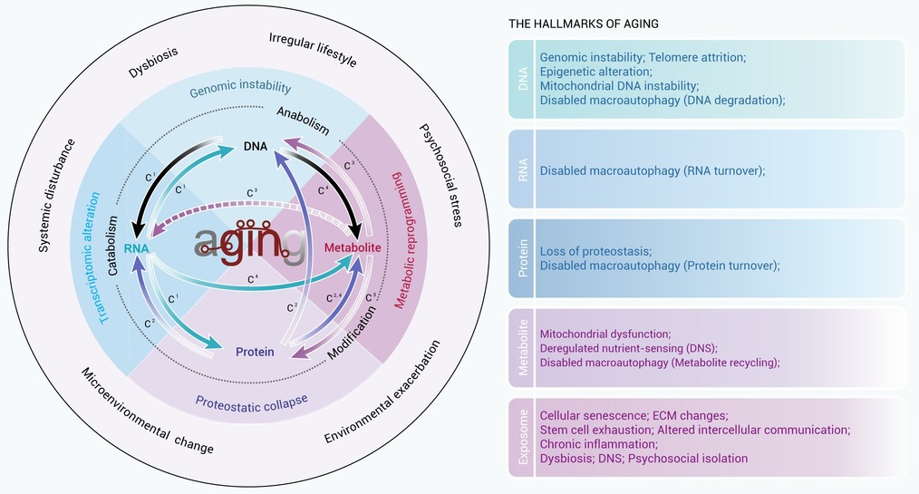
Export File
Citation
Zhang J., Su W., Duan J., et al. (2026). The central dogma of aging. The Innovation Life 4:100262. https://doi.org/10.59717/j.xinn-life.2026.100262

Format
Content
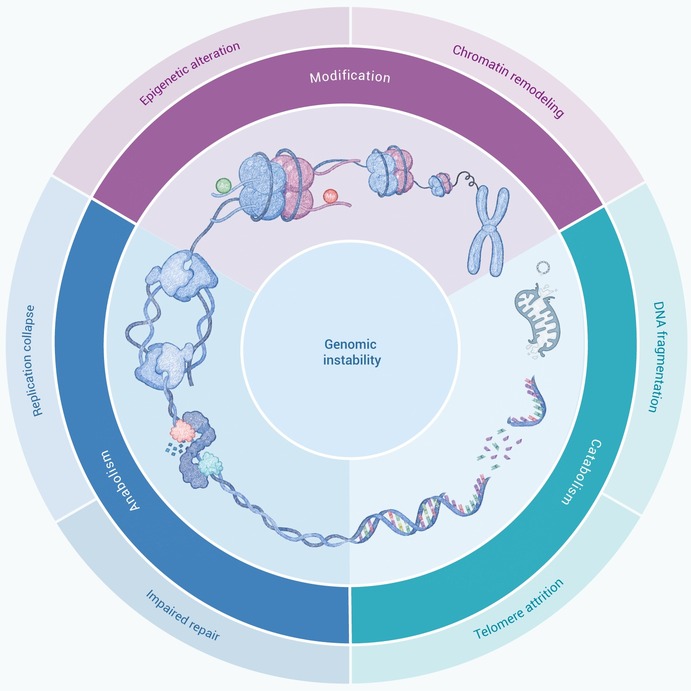
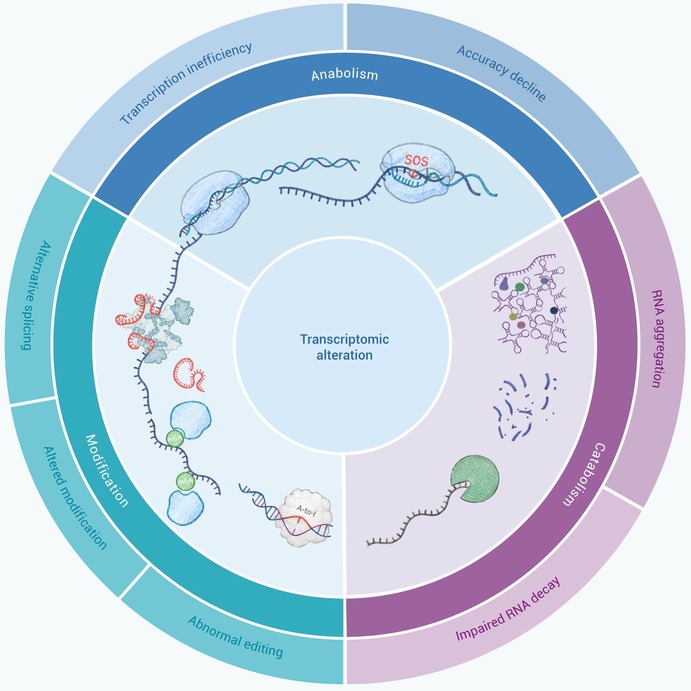
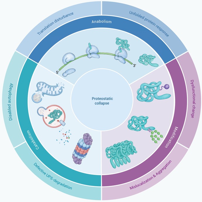
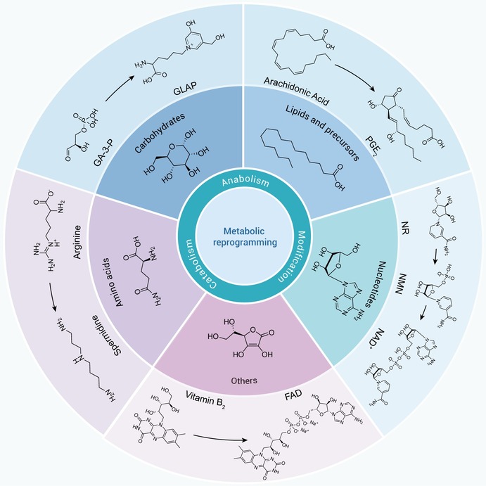
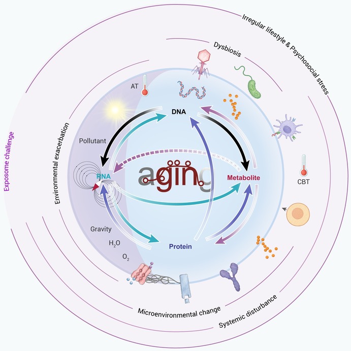
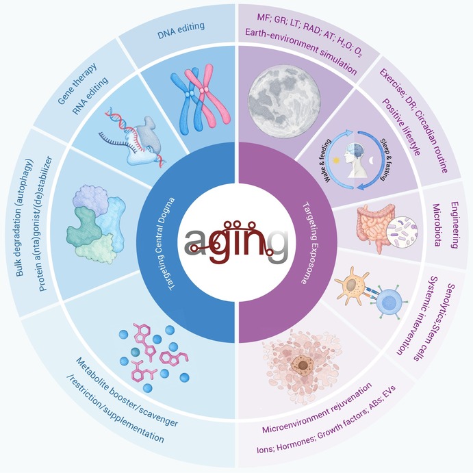
-
Figure 1.
The central dogma of aging
-
Figure 2.
Genomic instability
-
Figure 3.
Transcriptomic alteration
-
Figure 4.
Proteostasis collapse
-
Figure 5.
Metabolic reprogramming
-
Figure 6.
The Exposome: A hierarchical modulator of aging
-
Figure 7.
Targeting the central dogma to mitigate aging
