
Engineering traps in Tm-doped calcium titanate for visible and near-infrared double band persistent luminescence bioimaging
-
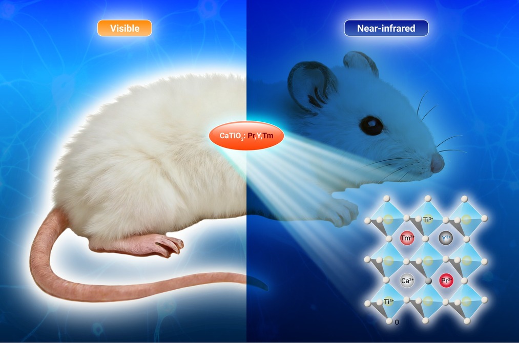
GRAPHICAL ABSTRACT
 DownLoad:
Full size image
DownLoad:
Full size image
-
PUBLIC SUMMARY
-
Y surpassed Sc, La, Gd, and Lu in enhancing the persistent luminescence (PersL) of CaTiO3:Tm.
Al, Ga, and In decreased the near-infrared (NIR) PersL of CaTiO3:Tm.
Three basic laws were derived for optimizing the NIR PersL.
CaTiO3:Tm, Y, Pr showed visible and NIR PersL at 613 and 803 nm, respectively.
CaTiO3:Tm, Pr, Y was applied to multi-modal imaging both in vitro and in vivo.
-
-
ABSTRACT
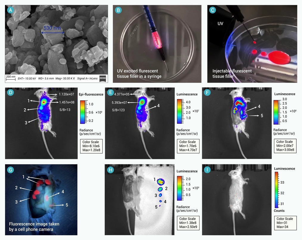
CaTiO3:Tm (CTT) has unusual red-light charged near-infrared (NIR) persistent luminescence (PersL) property at ~ 800 nm. In this study, trap engineering was realized by co-doping CTT with 8 non-luminescent trivalent ions. Some basic laws in optimizing the NIR PersL of CTT were established based on the observation that rare earth dopants, including Sc/Y/La/Gd/Lu, at the A site of the ABO3 perovskite structure of CaTiO3 increased the PersL while the boron group dopants, including Al/Ga/In, at the B site led to decreased PersL. Y was found as an efficient co-dopant to realize enhanced NIR PersL in CaTiO3:Tm,Y (CTT-Y) with a ~60% enhancement vs. CTT. Thermal-stimulated luminescence (TSL) study indicate that Y is the most efficient element in generating abundant energy traps while slightly increased the trap depth from 320 to 326 K. Further, by choosing Pr as the 3rd dopant, visible-NIR double band fluorescence and PersL was realized in CaTiO3:Tm,Y,Pr (CTT-Y-Pr) at 613 and ~800 nm, which were ~5 and 2 times of those of CaTiO3:Tm,Pr (CTT-Pr), respectively. CTT-Y-Pr was applied to realize four-modal optical anti-counterfeiting imaging in vitro including visible fluorescence, visible PersL, invisible NIR fluorescence, and invisible NIR PersL. Moreover, visible fluorescence and NIR PersL imaging in vivo was applied to observe implanted tissue filler and its post-surgical tissue cleaning by using CTT-Y-Pr as an optical imaging contrast reagent, which was hard to retrieve once it was injected within body and formed unwanted tiny debris.-
Keywords:
- Calcium titanate /
- afterglow /
- near-infrared /
- tissue filler imaging /
- rare earth
-

-
REFERENCES
[1] Jin L., Mo W., Wang Z., et al. (2025). Day-long organic persistent luminescence in flexible polymeric materials. Angew. Chem. Int. Edit. 64:e202506261. DOI:10.1002/anie.202506261 [2] Lin P., Shi J., Liu L., et al. (2025). Spatial confinement growth of high-performance persistent luminescence nanoparticles for image-guided sonodynamic therapy. Acta Biomater. 192:279−289. DOI:10.1016/j.actbio.2024.12.011 [3] Wang R., Shi J., Luo X., et al. (2025). Magnetic persistent luminescence nanoparticles repolarize tumor-associated macrophages for enhanced cancer immunotherapy. ACS Materials Lett. 7:2094−2104. DOI:10.1021/acsmaterialslett.5c00170 [4] Yao B., Xu F., Tian Z., et al. (2025). Strong persistent luminescence NaYF4-based nanoparticles combined with manipulated hyperfractionated irradiation for X-ray-excited photodynamic therapy enhancement. ACS Appl. Mater. Inter. 17:16561−16575. DOI:10.1021/acsami.4c20049 [5] Zhou L., Chen R., Li X., et al. (2025). Multicolor persistent luminescence for high-sensitivity optical temperature sensing, human motion detection, and multimodal anticounterfeiting materials. ACS Appl. Mater. Inter. 17:25451−25466. DOI:10.1021/acsami.5c01483 [6] Abulipizi G., Yu Z., Lin S., et al. (2023). Trap engineering in LiInSi2O6:Cr,Pr by excess cristobalite silica for X-ray activated near-infrared persistent luminescence imaging in vivo. Ceram. Int. 49:39664−39670. DOI:10.1016/j.ceramint.2023.09.321 [7] Chen K., Xiong Y., Wang D., et al. (2024). A facile strategy for achieving polymeric afterglow materials with wide color-tunability and persistent near-infrared luminescence. Adv. Funct. Mater. 34:202312883. DOI:10.1002/adfm.202312883 [8] Feng Y., Yang X., Rao Q., et al. (2024). Persistent luminescence lifetime-based near-infrared nanoplatform via deep learning for high-fidelity biosensing of hypochlorite. Anal. Chem. 96:7240−7247. DOI:10.1021/acs.analchem.4c00899 [9] Luo X., Shi J., Wang R., et al. (2024). Near-infrared persistent luminescence nanoprobe for early detection of atherosclerotic plaque. ACS Nano 18:6500−6512. DOI:10.1021/acsnano.3c12136 [10] Wang Y., Zhang P., Tian X., et al. (2025). Persistent scintillators for X-ray repetitive imaging with stable energy traps. J. Rare Earth. 43:1161−1168. DOI:10.1016/j.jre.2025.01.002 [11] Yan L.-X., Yan Z.-Y., Zhao X., et al. (2024). Size-independent boosting of near-infrared persistent luminescence in nano-phosphors via a magnesium doping strategy. J. Colloid. Interf. Sci. 662:11−18. DOI:10.1016/j.jcis.2024.02.008 [12] Zhu Y., Wen Y., Xie Y., et al. (2025). Intelligent hierarchical targeting near-infrared persistent luminescence nanosystem for improved nuclear delivery and simultaneous visualization/therapy of EBV-associated cancer. ACS Appl. Mater. Inter. 17:3072−3083. DOI:10.1021/acsami.4c20023 [13] Zhu Q., Xiahou J., Guo Y., et al. (2019). Zn3Ga2Ge2O10:Cr3+ uniform microspheres: Template-free synthesis, tunable bandgap/trap depth, and in vivo rechargeable near-infrared-persistent luminescence. ACS App. Bio. Mater. 2:577−587. DOI:10.1021/acsabm.8b00734 [14] Maldiney T., Bessiere A., Seguin J., et al. (2014). The in vivo activation of persistent nanophosphors for optical imaging of vascularization, tumours and grafted cells. Nat. Mater. 13:418−426. DOI:10.1038/Nmat3908 [15] Miao S. H., Lv X. L., Shan X. H., et al. (2024). Ultraviolet-B and near-infrared dual-band luminescence in Bi2+/Bi3+ codoped persistent phosphor for optical storage application. ACS Appl. Mater. Interfaces 16:23585−23595. DOI:10.1021/acsami.4c02522 [16] Ding Y. C., So B., Cao J. K., et al. (2023). Light delivery, acoustic read-out, and optical thermometry using ultrasound-induced mechanoluminescence and the near-infrared persistent luminescence of CaZnOS:Nd. Adv. Opt. Mater. 11:202300331. DOI:10.1002/adom.202300331 [17] Su J. Y., Pang R., Tan T., et al. (2024). A novel near-infrared emitting Sr2LuSbO6:Fe3+ phosphor with persistent luminescence performance. Adv. Opt. Mater. 12:202303187. DOI:10.1002/adom.202303187 [18] Zhou Z. H., He F. Q., Song E. H., et al. (2023). Broadband and multimode near-infrared emitter based on Cr3+-activated stannate for multifunctional applications. Adv. Opt. Mater. 11:202202466. DOI:10.1002/adom.202202466 [19] Zhao Y. L., Wang X. D., Wang Q. H., et al. (2024). Abnormal spectral broadening of ordered-structure near-infrared phosphor La2CaHfO6:Cr3+. J. Mater. Chem. C 12:10532−10539. DOI:10.1039/d4tc01036a [20] Pan L., Delaey M., Wang Y., et al. (2024). Structural and optical properties of Cr ion-doped near-infrared long persistent luminescence silicogermanate phosphors with broad emission bands. J. Alloy. Compd. 983:173853. DOI:10.1016/j.jallcom.2024.173853 [21] Huang L., Lin L., Xie W., et al. (2020). Near-infrared persistent luminescence in a Cr3+-doped perovskite for low-irradiance imaging. Chem. Mater. 32:5579−5588. DOI:10.1021/acs.chemmater.0c00807 [22] Abulipizi G., Wang Y. X., Fang Y. L., et al. (2025). Persistent luminescent NaYTi2O6:Cr activated in the near-infrared bio-imaging window as a self-luminescent ceramic coating for visible Ti-implant in vivo. Ceram. Int. 51:6959−6967. DOI:10.1016/j.ceramint.2024.12.130 [23] Yu Z., Abulipizi G., Xu Y., et al. (2024). Stabilization of Ti3+ in CaTiO3 by doping Y3+ to generate visible light-activated near-infrared persistent luminescence. J. Alloy. Compd. 983:173879. DOI:10.1016/j.jallcom.2024.173879 [24] Xu Y., Abulipizi G., Wang Y., et al. (2024). Near-infrared persistent luminescence of CaTiO3:Cr,Y for imaging of bone implants using red-light illumination instead of X-ray. ACS Appl. Mater. Interfaces 16:55823−55831. DOI:10.1021/acsami.4c13865 [25] Abulipizi G., Zhou J., Qu C., et al. (2025). Unusual single band 803 nm persistent luminescence of Tm-doped calcium titanate charged in the near-infrared bio-imaging window. ACS Appl. Mater. Interfaces 17:36912−36921. DOI:10.1021/acsami.5c07800 [26] Kamioka H., Igarashi A., Watabe K., et al. (2025). Pr and Al doping effects on the red emission and long lasting afterglow in CaTiO3 single crystals. Opt. Mater. 159:116663. DOI:10.1016/j.optmat.2025.116663 [27] Yin S., Chen D. and Tang W. (2007). Combustion synthesis and luminescent properties of CaTiO3:Pr, Al persistent phosphors. J. Alloy. Compd. 441:327−331. DOI:10.1016/j.jallcom.2006.09.120 [28] Huang K., Li Z. J., Li Y., et al. (2021). Three-dimensional colloidal controlled growth of core-shell heterostructured persistent luminescence nanocrystals. Nano Lett. 21:4903−4910. DOI:10.1021/acs.nanolett.0c04940 [29] Qiao J. W., Zhang S., Zhou X. Q., et al. (2022). Near-infrared light-emitting diodes utilizing a Europium-activated calcium oxide phosphor with external quantum efficiency of up to 54.7%. Adv. Mater. 34:202201887. DOI:10.1002/adma.202201887. [30] Liu L., Shi J. P., Wang J. Y., et al. (2024). Biodegradable persistent luminescence nanoparticles as pyroptosis inducer for high-efficiency tumor immunotherapy. Adv. Sci. 11:202406340. DOI:10.1002/advs.202406340 [31] Rakov N., Matias F. and Xiao M. F. (2024). An efficient red light-emissive process from Eu3+ doped amorphous-derived CaAl2Si2O8 ceramic powders: Down-shifted luminescence with high thermal stability under 532 nm excitation. Ceram. Int. 50:16064−16075. DOI:10.1016/j.ceramint.2024.02.086 [32] V. Nikiforov I., V. Iliina E., Lazoryak B. I., et al. (2024). Photoluminescence, structural and antibacterial properties of co-doped β-Ca3(PO4)2-type phosphates Ca8CuRE(PO4)7 (RE = Eu-Er). J. Rare. Earth. 42:1658−1668. DOI:10.1016/j.jre.2023.07.023 [33] Van H. N., Cuong L. T., Nguyen D.-H., et al. (2025). Effect of Sr substituted on multifunction pure green emission of rare-earth-element doped HA/β-TCP nanocomposite for optical thermometer. Ceram. Int. 51:16894−16903. DOI:10.1016/j.ceramint.2024.07.119 [34] Piotrowski W. M., Ristic Z., Dramicanin M. D., et al. (2022). Modification of the thermometric performance of the lifetime-based luminescent thermometer exploiting Ti3+ emission in SrTiO3 and CaTiO3 by doping with lanthanide ions. J. Alloy. Compd. 906:164398. DOI:10.1016/j.jallcom.2022.164398 [35] Chen Y. F. and Pan Z. W. (2023). Red/NIR/SWIR multi-band persistent luminescent nanoparticles as ultrasensitive multi-channel tracers in water and crude oil/water emulsions. Nano Res. 16:12706−12712. DOI:10.1007/s12274-023-6116-x [36] Yin X., Zhong H., Liu L., et al. (2024). X-ray-activated Bi3+/Pr3+ co-doped LiYGeO4 phosphor with UV and NIR dual-emissive persistent luminescence. J. Rare Earth. 42:955−961. DOI:10.1016/j.jre.2023.03.008 [37] Zhao T., Sun W., Wang S., et al. (2025). Low-dose X-ray induced long afterglow NIR luminescence from Cr3+ doped Zn1_xCdxGa2O4 spinel solid solutions. J. Rare Earth. 43:246−252. DOI:10.1016/j.jre.2023.12.006 [38] Wang Y., Abulipizi G., Zhou J., et al. (2025). Persistent luminescence of Dy-doped strontium pyrophosphate with double traps for room temperature qualitative and heated quantitative X-ray dosimetry by naked eyes. J. Alloy. Compd. 1022:179900. DOI:10.1016/j.jallcom.2025.179900 [39] Gao L., Liu Y., Su J., et al. (2025). Modulation of near-infrared afterglow luminescence in inorganic nanomaterials for biological applications. Adv. Mater. 37:202419349. DOI:10.1002/adma.202419349 [40] Shang R., Yang F., Gao G., et al. (2024). Bioimaging and prospects of night pearls-based persistence phosphors in cancer diagnostics. Exploration 4:20230124−20230124. DOI:10.1002/exp.20230124 [41] Wei Y. and Wang J. (2024). X-ray/γ-ray/ultrasound-activated persistent luminescence phosphors for deep tissue bioimaging and therapy. ACS Appl. Mater. Interfaces 16:56519−56544. DOI:10.1021/acsami.4c11585 [42] Yang X., Waterhouse G. I. N., Lu S., et al. (2023). Recent advances in the design of afterglow materials: mechanisms, structural regulation strategies and applications. Chem. Soc. Rev. 52:8005−8058. DOI:10.1039/d2cs00993e [43] Cai G. Y., Delgado T., Richard C., et al. (2023). ZGSO Spinel nanoparticles with dual emission of NIR persistent luminescence for anti-counterfeiting applications. Materials 16:1132. DOI:10.3390/ma16031132 [44] Cai G. Y., Naillon T., Seguin J., et al. (2024). ZGSO:Cr3+, Ni2+ persistent phosphors with dual emission in NIR-I and SWIR ranges for bio-imaging applications. Small 20:202406507. DOI:10.1002/smll.202406507 -
ABOUT THIS ARTICLE
Cite this article:
Ding L., Zhou J., Abulipizi G., et al. (2026). Engineering traps in Tm-doped calcium titanate for visible and near-infrared double band persistent luminescence bioimaging. The Innovation Materials 4:100200. https://doi.org/10.59717/j.xinn-mater.2026.100200
Welcome!
To request copyright permission to republish or share portions of our works, please visit Copyright Clearance Center's (CCC) Marketplace website at marketplace.copyright.com.
Export File
Citation
Ding L., Zhou J., Abulipizi G., et al. (2026). Engineering traps in Tm-doped calcium titanate for visible and near-infrared double band persistent luminescence bioimaging. The Innovation Materials 4:100200. https://doi.org/10.59717/j.xinn-mater.2026.100200

Format
Content
-
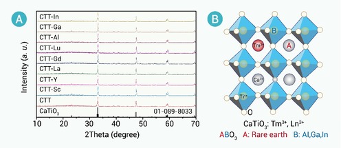
Figure 1.
Crystal structure of the as-synthesized CTT-Ln phosphors
-
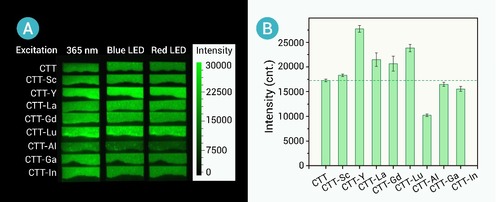
Figure 2.
Comparison of the PersL intensities of CTT-Ln with CTT
-
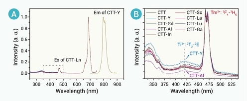
Figure 3.
Photoluminescence spectra of CTT-Ln
-
Figure 4.
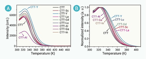
Thermal-stimulated luminescence of CTT-Ln phosphors charged by a 365 nm UV lamp
-
Figure 5.
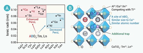
Proposed mechanism of the influence of Ln3+ co-doping in PersL CTT-Ln
-
Figure 6.
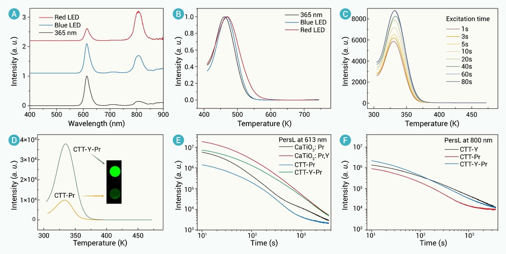
Visible-NIR double band PersL of CTT-Y-Pr
-
Figure 7.
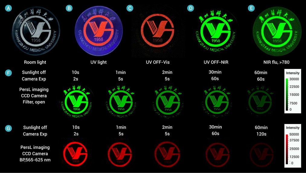
Multi-modal optical anti-counterfeiting based on CTT-Y and CTT-Y-Pr. Digital images taken by a cellphone camera
-
Figure 8.
PersL imaging of CTT-Y-Pr labeled silicone tissue filler
