
Interpretable and robust multimodal data integration for precise treatment response and survival prediction in gastric cancer
-
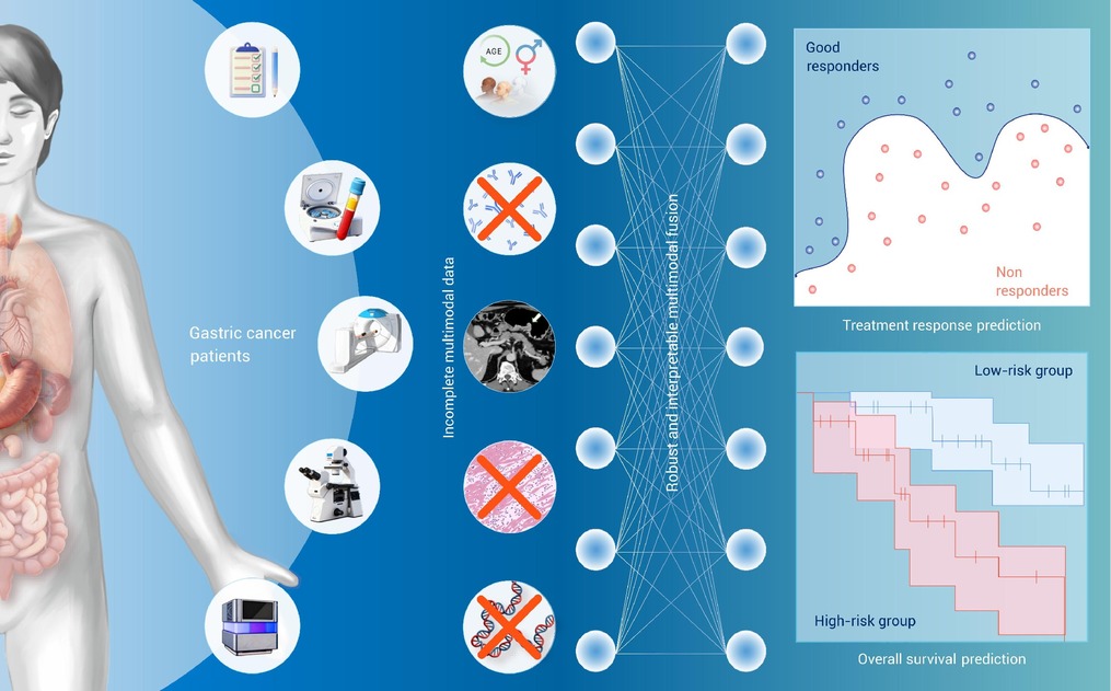
GRAPHICAL ABSTRACT
 DownLoad:
Full size image
DownLoad:
Full size image
-
PUBLIC SUMMARY
-
This study proposes an interpretable and robust multimodal fusion framework for gastric cancer analysis.
This model robustly integrates multiple data sources to predict treatment response and patient survival.
This model shows reliable performance across multiple datasets and can handle incomplete data scenarios.
Its interpretability identifies key factors, providing trustworthy insights for treatment personalization.
-
-
ABSTRACT
Gastric cancer (GC) was the fifth most prevalent cancer in 2022. Integrating multimodal data enables significant improvement in precise GC analysis. However, the heterogeneity and incompleteness of multimodal data pose challenges for effective integration. The lack of interpretability in models hinders their trustworthiness in clinical practice. Therefore, this study presented an interpretable and robust multimodal data integration framework for GC analysis (iMD4GC), facilitating effective integration of diverse data sources to predict treatment response and overall survival. Three multimodal datasets were collected for model evaluation: GastricRes (n=698) for response prediction, GastricSur (n=801) for survival analysis, and TCGA-STAD (n=400) for further survival analysis. The iMD4GC achieved an area under the receiver operating characteristic curve (AUC) of 0.803 (95% CI 0.680-0.925) on GastricRes, a concordance index (c-index) of 0.715 (95% CI 0.646-0.784) and a time-dependent AUC of 0.739 (95% CI 0.644-0.833) on GastricSur, and a c-index of 0.661 (95% CI 0.504-0.818) and a time-dependent AUC of 0.690 (95% CI 0.500-0.881) on TCGA-STAD. The comparative analysis underscores the reliability of iMD4GC in integrating diverse multimodal data and addressing missing modalities. Further interpretability analysis identified pivotal factors across various data modalities, validating the alignment with clinical expertise and providing essential insights for informed decision-making in clinical contexts. The promising performance of iMD4GC highlights its potential in advancing clinical decision-making, personalizing treatment strategies, and improving patient prognosis in GC. Inherent interpretability fosters transparent decision analysis and provides valuable insights for clinicians, promoting its trustworthiness and acceptance in clinical settings. -

-
REFERENCES
[1] Bray F., Laversanne M., Sung H., et al. (2024). Global cancer statistics 2022: GLOBOCAN estimates of incidence and mortality worldwide for 36 cancers in 185 countries. CA-Cancer J. Clin. 74:229−263. DOI:10.3322/caac.21834 [2] Zurlo I.V., Schino M., Strippoli A., et al. (2022). Predictive value of NLR, TILs (CD4+/CD8+) and PD-L1 expression for prognosis and response to preoperative chemotherapy in gastric cancer. Cancer Immunol. Immunother. 71:45−55. DOI:10.1007/s00262-021-02960-1 [3] Liu Z., Wang Y., Shan F., et al. (2021). Treatment switch in poor responders with locally advanced gastric cancer after neoadjuvant chemotherapy. Ann. Surg. Oncol. 28:8892−8907. DOI:10.1245/s10434-021-10087-x [4] Yoshikawa T., Sasako M., Yamamoto S., et al. (2009). Phase II study of neoadjuvant chemotherapy and extended surgery for locally advanced gastric cancer. Br. J. Surg. 96:1015−1022. DOI:10.1002/bjs.6665 [5] Lowy A.M., Mansfield P.F., Leach S.D., et al. (1999). Response to neoadjuvant chemotherapy best predicts survival after curative resection of gastric cancer. Ann. Surg. 229:303−308. DOI:10.1097/00000658-199903000-00001 [6] Fu J., Tang L., Li Z.Y., et al. (2020). Diffusion kurtosis imaging in the prediction of poor responses of locally advanced gastric cancer to neoadjuvant chemotherapy. Eur. J. Radiol. 128:108974. DOI:10.1016/j.ejrad.2020.108974 [7] Zhang J., Cui Y., Wei K., et al. (2022). Deep learning predicts resistance to neoadjuvant chemotherapy for locally advanced gastric cancer: A multicenter study. Gastric Cancer 25:1050−1059. DOI:10.1007/s10120-022-01328-3 [8] Wang S., Zhu Y., Yu L., et al. (2019). RMDL: Recalibrated multi-instance deep learning for whole slide gastric image classification. Med. Image Anal. 58:101549. DOI:10.1016/j.media.2019.101549 [9] Yu H., Zhang X., Song L., et al. (2021). Large-scale gastric cancer screening and localization using multi-task deep neural network. Neurocomputing 448:290−300. DOI:10.1016/j.neucom.2021.03.006 [10] Meng L., Dong D., Chen X., et al. (2020). 2D and 3D CT radiomic features performance comparison in characterization of gastric cancer: A multi-center study. IEEE J. Biomed. Health Inform. 25:755−763. DOI:10.1109/jbhi.2020.3002805 [11] Yuan M., Xia Y., Chen X., et al. (2023). Cluster-induced mask transformers for effective opportunistic gastric cancer screening on non-contrast CT scans. Med. Image Comput. Comput.-Assist. Interv. 14223:146-156. DOI:10.1007/978-3-031-439049_15 [12] Du G., Zeng Y., Chen D., et al. (2023). Application of radiomics in precision prediction of diagnosis and treatment of gastric cancer. Jpn. J. Radiol. 41:245−257. DOI:10.1007/s11604-022-01352-4 [13] Jiang Y., Zhou K., Sun Z., et al. (2023). Non-invasive tumor microenvironment evaluation and treatment response prediction in gastric cancer using deep learning radiomics. Cell Rep. Med. 4:101146. DOI:10.1016/j.xcrm.2023.101146 [14] Chen T., Li X., Mao Q., et al. (2022). An artificial intelligence method to assess the tumor microenvironment with treatment outcomes for gastric cancer patients after gastrectomy. J. Transl. Med. 20:100. DOI:10.1186/s12967-022-03298-7 [15] Li X., Zhang Y., Zhang Y., et al. (2010). Survival prediction of gastric cancer by a seven-microRNA signature. Gut 59:579−585. DOI:10.1136/gut.2008.175497 [16] Oh S.E., Seo S.W., Choi M.G., et al. (2018). Prediction of overall survival and novel classification of patients with gastric cancer using the survival recurrent network. Ann. Surg. Oncol. 25:1153−1159. DOI:10.1016/j.ejso.2018.10.287 [17] Chen D., Fu M., Chi L., et al. (2022) Prognostic and predictive value of a pathomics signature in gastric cancer. Nat. Commun. 13:6903. DOI:10.1038/s41467-022-34703-w [18] Xu Q., Sun Z., Li X., et al. (2021). Advanced gastric cancer: CT radiomics prediction and early detection of downstaging with neoadjuvant chemotherapy. Eur. Radiol. 31:8765−8774. DOI:10.1007/s00330-021-07962-2 [19] Sun J., Wang X., Zhang Z., et al. (2021). The sensitivity prediction of neoadjuvant chemotherapy for gastric cancer. Front. Oncol. 11:641304. DOI:10.3389/fonc.2021.641304 [20] Cui Y., Zhang J., Li Z., et al. (2022). A CT-based deep learning radiomics nomogram for predicting the response to neoadjuvant chemotherapy in patients with locally advanced gastric cancer: A multicenter cohort study. EClinicalMedicine 46:101348. DOI:10.1016/j.eclinm.2022.101348 [21] Gu J., Tong T., He C., et al. (2022). Deep learning radiomics of ultrasonography can predict response to neoadjuvant chemotherapy in breast cancer at an early stage of treatment: a prospective study. Eur. Radiol. 32:2099−2109. DOI:10.1007/s00330-021-08293-y [22] Wang X., Zhang Y., Yang M., et al. (2024). Dynamic ultrasound-based modeling predictive of response to neoadjuvant chemotherapy in patients with early breast cancer. Sci. Rep. 14:31644. DOI:10.1038/s41598-024-80409-y [23] Hottat N.A., Badr D.A., Lecomte S., et al. (2023). Assessment of diffusion-weighted MRI in predicting response to neoadjuvant chemotherapy in breast cancer patients. Sci. Rep. 13:614. DOI:10.1038/s41598-023-27787-x [24] Ogier du Terrail J., Leopold A., Joly C., et al. (2023). Federated learning for predicting histological response to neoadjuvant chemotherapy in triple-negative breast cancer. Nat. Med. 29:135−146. DOI:10.1038/s41591-022-02155-w [25] Zhang L., Dong D., Zhang W., et al. (2020). A deep learning risk prediction model for overall survival in patients with gastric cancer: A multicenter study. Radiother. Oncol. 150:73−80. DOI:10.2139/ssrn.3514655 [26] Zhong H., Wang T., Hou M., et al. (2024). Deep learning radiomics nomogram based on enhanced CT to predict the response of metastatic lymph nodes to neoadjuvant chemotherapy in locally advanced gastric cancer. Ann. Surg. Oncol. 31:421−432. DOI:10.1245/s10434-023-14508-x [27] Li J., Yin H., Wang Y., et al. (2023). Multiparametric MRI-based radiomics nomogram for early prediction of pathological response to neoadjuvant chemotherapy in locally advanced gastric cancer. Eur. Radiol. 33:2746−2756. DOI:10.1007/s00330-022-09219-y [28] Zhang Y., Chen Q., Yang Z., et al. (2019). BioWordVec, improving biomedical word embeddings with subword information and MeSH. Sci. Data 6:52. DOI:10.1038/s41597-019-0055-0 [29] Wasserthal J., Breit H.C., Meyer M.T., et al. (2023). TotalSegmentator: Robust segmentation of 104 anatomic structures in CT images. Radiology: Artif. Intell. 5:e230024. DOI:10.1148/ryai.230024 [30] He K., Zhang X., Ren S., et al. (2016). Deep residual learning for image recognition. CVPR 2016:770-778. DOI:10.1109/cvpr.2016.90 [31] Vaswani A., Shazeer N., Parmar N., et al. (2017). Attention is all you need. Adv. Neural Inf. Process. Syst. arXiv preprint. DOI:10.48550/arXiv.1706.03762. [32] Wang X., Yang S., Zhang J., et al. (2022). Transformer-based unsupervised contrastive learning for histopathological image classification. Med. Image Anal. 81:102559. DOI:10.1016/j.media.2022.102559 [33] Du J., Jia P., Dai Y., et al. (2019). Gene2vec: Distributed representation of genes based on co-expression. BMC Genomics 20:7−15. DOI:10.1186/s12864-018-5370-x [34] Tarvainen A. andValpola H. (2017). Mean teachers are better role models: Weight-averaged consistency targets improve semi-supervised deep learning results. Proc. 31st Int. Conf. Neural Inf. Process. Syst. pp:1195-1204 [35] Zadeh A., Liang P.P., Mazumder N., et al. (2018). Memory fusion network for multi-view sequential learning. Proc. AAAI Conf. Artif. Intell 2018:5634-5641. DOI:10.1609/aaai.v32i1.12021 [36] Tsai Y.H.H., Bai S., Liang P.P., et al. (2019). Multimodal transformer for unaligned multimodal language sequences. Proc. 57th Annu. Meet. Assoc. Comput. Linguist 2019:6558-6569. DOI:10.18653/v1/p19-1656 [37] Cui C., Liu H., Liu Q., et al. (2022) Survival prediction of brain cancer with incomplete radiology, pathology, genomic, and demographic data. Med. Image Comput. Comput. Assist. Interv 2022:626-635. DOI:10.1007/978-3-031-16443-9_60 [38] Choromanski K.M., Likhosherstov V., Dohan D., et al. (2020). Rethinking attention with performers. International Conference on Learning Representations. arXiv. 2009.14794 DOI:10.48550/arXiv.2009.14794 [39] Xiong Y., Zeng Z., Chakraborty R., et al. (2021). Nyströmformer: A nyström-based algorithm for approximating self-attention. Proc. AAAI Conf. Artif. Intell 2021:14138-14148. DOI:10.1609/aaai.v35i16.17664 [40] Hung H. and Chiang C.T. (2010). Estimation methods for time‐dependent AUC models with survival data. Can. J. Stat. 38:8−26. DOI:10.1002/cjs.10046 [41] Lambert J. and Chevret S. (2016). Summary measure of discrimination in survival models based on cumulative/dynamic time-dependent ROC curves. Stat. Methods Med. Res. 25:2088−2102. DOI:10.1177/0962280213515571 [42] Sundararajan M., Taly A., and Yan Q. (2017). Axiomatic attribution for deep networks. Int. Conf. Mach. Learn 70:3319-3328. DOI:10.48550/arXiv.1703.01365 [43] Gadermayr M. and Tschuchnig M. (2024). Multiple instance learning for digital pathology: A review of the state-of-the-art, limitations & future potential. Comput. Med. Imaging Graph. 112:102337−102337. DOI:10.1016/j.compmedimag.2024.102337 [44] Tang W., Huang S., Zhang X., et al. (2023). Multiple instance learning framework with masked hard instance mining for whole slide image classification. Proc. IEEE/CVF Int. Conf. Comput. Vis 2023:4078-4087. DOI:10.1109/ICCV51070.2023.00377 [45] Graham S., Vu Q.D., Raza S.E.A., et al. (2019). Hover-net: Simultaneous segmentation and classification of nuclei in multi-tissue histology images. Med. Image Anal. 58:101563. DOI:10.1016/j.media.2019.101563 [46] Selvaraju R.R., Cogswell M., Das A., et al. (2017). Grad-cam: Visual explanations from deep networks via gradient-based localization. Proc. IEEE Int. Conf. Comput. Vis. 2017:618-626. DOI:10.1109/iccv.2017.74 [47] Yang Y.C., Wang S.W., Wu I.C., et al. (2009). A tumorigenic homeobox (HOX) gene expressing human gastric cell line derived from putative gastric stem cell. Eur. J. Gastroenterol. Hepatol. 21:1016−1023. DOI:10.1097/MEG.0b013e32831d7b9c [48] Ma Y.Y., Zhang Y., Mou X.Z., et al. (2017). High level of homeobox A9 and PBX homeobox 3 expression in gastric cancer correlates with poor prognosis. Oncol. Lett. 14:5883−5889. DOI:10.3892/ol.2017.6937 [49] Shenoy U.S., Adiga D., Alhedyan F., et al. (2023). HOXA9 transcription factor is a double-edged sword: From development to cancer progression. Cancer Metastasis Rev. 43:709−728. DOI:10.1007/s10555-023-10159-2 [50] Ge S., Xia X., Ding C., et al. (2018). A proteomic landscape of diffuse-type gastric cancer. Nat. Commun. 9:1012. DOI:10.1007/s10120-021-01226-0 [51] Yan Q., Zeng P., Zhou X., et al. (2021). RBMX suppresses tumorigenicity and progression of bladder cancer by interacting with the hnRNP A1 protein to regulate PKM alternative splicing. Oncogene 40:2635−2650. DOI:10.1038/s41388-021-01666-z [52] Wang X., Xu K., Liao X., et al. (2022). Construction of a survival nomogram for gastric cancer based on the cancer genome atlas of m6A-related genes. Front. Genet. 13:936658. DOI:10.3389/fgene.2022.936658 [53] Mu G., Zhu Y., Dong Z., et al. (2021). Calmodulin 2 facilitates angiogenesis and metastasis of gastric cancer via STAT3/HIF-1A/VEGF-A mediated macrophage polarization. Front. Oncol. 11:727306. DOI:10.3389/fonc.2021.727306 [54] Sun J., Cao Q., Lin S., et al. (2023). Knockdown of CALM2 increases the sensitivity to afatinib in HER2-amplified gastric cancer cells by regulating the Akt/FoxO3a/Puma axis. Toxicol. Vitro 87:105531. DOI:10.1016/j.tiv.2022.105531 [55] Hu Y., Chen C., Tong X., et al. (2021). NSUN2 modified by SUMO-2/3 promotes gastric cancer progression and regulates mRNA m5C methylation. Cell Death Dis. 12:842. DOI:10.1038/s41419-021-04127-3 [56] Chen R.J., Ding T., Lu M.Y., et al. (2024). Towards a general-purpose foundation model for computational pathology. Nat. Med. 30:850−862. DOI:10.1038/s41591-024-02857-3 -
ABOUT THIS ARTICLE
Cite this article:
Zhou F., Xu Y., Cui Y., et al. (2025). Interpretable and robust multimodal data integration for precise treatment response and survival prediction in gastric cancer. The Innovation Informatics 1:100011. https://doi.org/10.59717/j.xinn-inform.2025.100011
Welcome!
To request copyright permission to republish or share portions of our works, please visit Copyright Clearance Center's (CCC) Marketplace website at marketplace.copyright.com.
Export File
Citation
Zhou F., Xu Y., Cui Y., et al. (2025). Interpretable and robust multimodal data integration for precise treatment response and survival prediction in gastric cancer. The Innovation Informatics 1:100011. https://doi.org/10.59717/j.xinn-inform.2025.100011

Format
Content
-
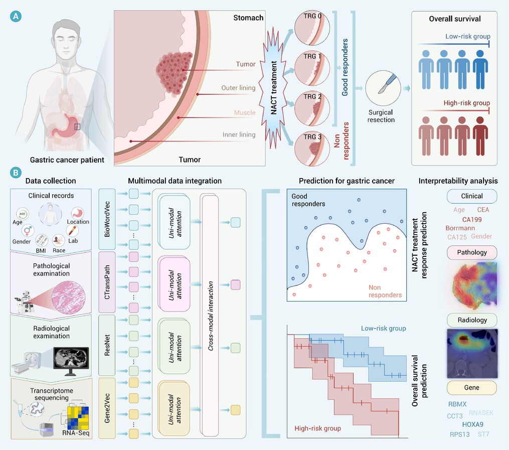
Figure 1.
Overview of integrating multimodal data to predict treatment response and overall survival for GC patients
-
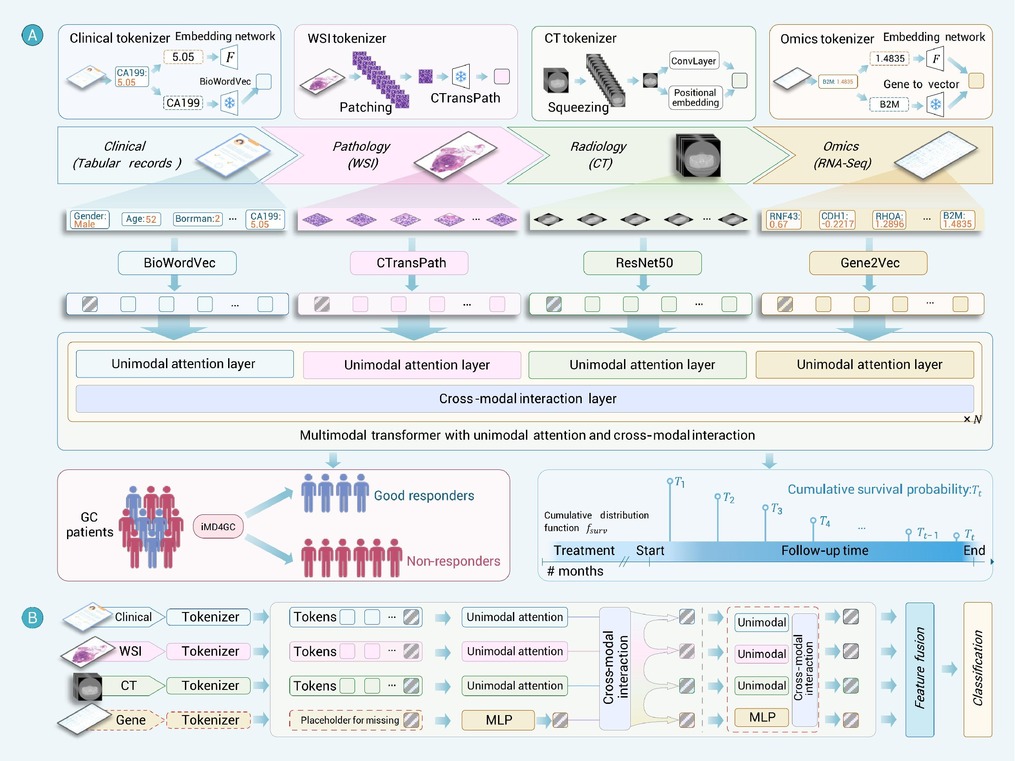
Figure 2.
Overall framework
-
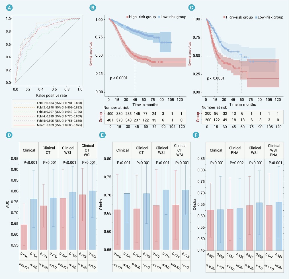
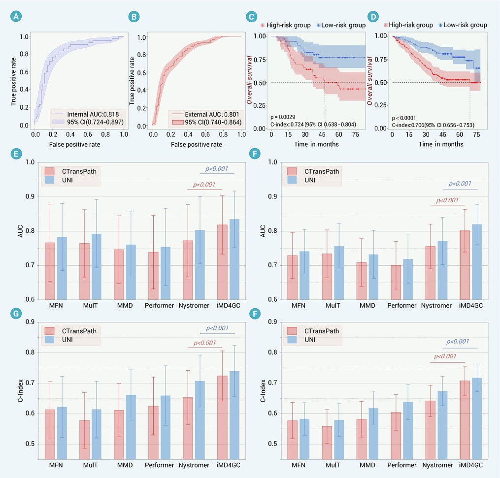
Figure 3.
ROC curves for treatment prediction, Kaplan-Meier curves for survival analysis, and comparative analysis for knowledge distillation
-
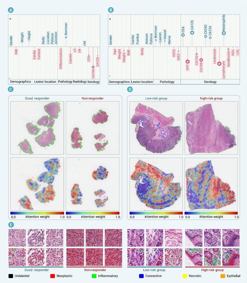
Figure 4.
Interpretability analysis of clinical records and pathological images
-
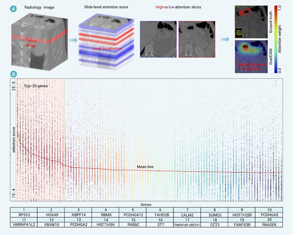
Figure 5.
Interpretability analysis of radiology images and transcriptomics profiles for GC analysis
-
Figure 6.
Cross-center validation results
