
A brain-computer interface roadmap for diagnosing and treating neurological disorders
-
GRAPHICAL ABSTRACT
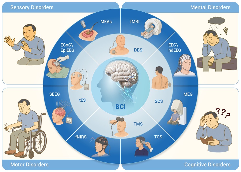
 DownLoad:
Full size image
DownLoad:
Full size image
-
PUBLIC SUMMARY
-
Brain-computer interfaces (BCI) achieve the treatment of neurological diseases through decoding and modulation.
BCI can provide personalized treatment for neurological diseases through closed-loop neuromodulation.
Key challenges include decoding accuracy, long-term biocompatibility, and real-world clinical translation.
-
-
ABSTRACT
Currently, the global incidence of neurological disorders is on a continuous upward trend, posing severe challenges to the medical field. However, traditional diagnosis and treatment methods for such diseases are associated with risks arising from invasive procedures, generally low diagnostic and therapeutic efficiency, and more critically, they struggle to achieve precise and personalized treatment, failing to fully meet the individual needs of patients. Against this backdrop, exploring rapid, efficient, and safe diagnosis and treatment protocols for brain diseases has become a core research direction. The emergence of Brain-computer interface (BCI) technology provides a highly promising solution to break through this dilemma and is expected to fundamentally revolutionize the diagnostic and therapeutic models for neurological diseases. By accurately capturing and analyzing brain signals, BCI offers a brand-new pathway for restoring lost physiological functions in patients, as well as regulating and enhancing brain activity, bringing new hope to numerous patients afflicted by neurological disorders. Here we systematically review the latest research progress of BCI technology in recent years and focus on analyzing its potential clinical application value in the fields of sensory disorders, motor disorders, cognitive disorders, and mental disorders. The research insights and technical directions summarized in this review aim to provide inspiration for subsequent research in this field, promote the development of BCI technology towards a more mature and practical direction, and ultimately provide more effective diagnostic and therapeutic means for patients with neurological disorders, helping them improve their quality of life and regain hope for health. -

-
REFERENCES
[1] Wang Z., Wang S.-N., Xu T.-Y., et al. (2017). Organoid technology for brain and therapeutics research. CNS Neurosci. Ther. 23:771−778. DOI:10.1111/cns.12754 [2] Issack P.S, Cunningham M.E, Pumberger M, et al. (2012). Degenerative lumbar spinal stenosis: Evaluation and management. JAAOS 20:527−535. DOI:10.5435/JAAOS-20-08-527 [3] Cruz M.V., Jamal S. and Sethuraman S.C. (2025). A comprehensive survey of brain-computer interface technology in health care: Research perspectives. J. Med. Signals Sens. 15:6−16. DOI:10.4103/jmss.jmss_49_24 [4] Mohanty R., Sinha A.M., Remsik A.B., et al. (2018). Machine learning classification to identify the stage of brain-computer interface therapy for stroke rehabilitation using functional connectivity. Front. Neurosci. 12:353. DOI:10.3389/fnins.2018.00353 [5] Ma J., Wang Y., Yang J., et al. (2024). In vivo bioelectronic nose based on a bioengineered rat for the explosive odors with Benzene ring detection and identification. ICBBE 24:110−114. DOI:10.1145/3707127.3707145 [6] Sun G., Liu Z., Gan L., et al. (2025). SpikeNAS-bench: Benchmarking NAS algorithms for spiking neural network architecture. IEEE Trans Artif Intell 6:1614−1625. DOI:10.1109/tai.2025.3534136 [7] Cao L., Zhao W. and Sun B. (2025). Emotion recognition using multi-scale EEG features through graph convolutional attention network. Neural Netw. 184:107060. DOI:10.1016/j.neunet.2024.107060 [8] Liu X., Lu Z., Li J., et al. (2024). An 8-channel high-voltage neural stimulation IC design with exponential waveform output. Neuroelectronics. 1:0001. DOI:10.55092/neuroelectronics20240001 [9] Ramadan R.A. and Altamimi A.B. (2024). Unraveling the potential of brain-computer interface technology in medical diagnostics and rehabilitation: A comprehensive literature review. Health Technol. 14:263−276. DOI:10.1007/s12553-024-00822-1 [10] Bashashati A, Fatourechi M, Ward R.K, et al. (2007). A survey of signal processing algorithms in brain–computer interfaces based on electrical brain signals. J. Neural Eng. 4:R32. DOI:10.1088/1741-2560/4/2/R03 [11] Edelman B.J., Zhang S., Schalk G., et al. (2025). Non-invasive brain-computer interfaces: State of the art and trends. IEEE Rev. Biomed. Eng. 18:26−49. DOI:10.1109/RBME.2024.3449790 [12] Wen D., Fan Y., Hsu S.-H., et al. (2021). Combining brain-computer interface and virtual reality for rehabilitation in neurological diseases: A narrative review. Ann Phys Rehabil Med 64:101404. DOI:10.1016/j.rehab.2020.03.015 [13] Zhang H., Jiao L., Yang S., et al. (2024). Brain-computer interfaces: The innovative key to unlocking neurological conditions. Int JSurg 110:5745−5762. DOI:10.1097/js9.0000000000002022 [14] Fatourechi M., Bashashati A., Ward R.K., et al. (2007). EMG and EOG artifacts in brain computer interface systems: A survey. Neurophysiol Clin 118:480−494. DOI:10.1016/j.clinph.2006.10.019 [15] Sun B., Lv J.-J., Rui L.-G., et al. (2021). Seizure prediction in scalp EEG based channel attention dual-input convolutional neural network. PhyA 584:126376. DOI:10.1016/j.physa.2021.126376 [16] Sun B., Zhang H., Wu Z., et al. (2021). Adaptive spatiotemporal graph convolutional networks for motor imagery classification. IEEE Signal Process. Lett. 28:219−223. DOI:10.1109/lsp.2021.3049683 [17] Sun B., Zhao X., Zhang H., et al. (2021). EEG motor imagery classification with sparse spectrotemporal decomposition and deep learning. IEEE Trans. Autom. Sci. Eng. 18:541−551. DOI:10.1109/tase.2020.3021456 [18] Sironi V.A. (2011). Origin and evolution of deep brain stimulation. Front. Integr. Neurosci. 5:42−42. DOI:10.3389/fnint.2011.00042 [19] Mane R., Chouhan T. and Guan C. (2020). BCI for stroke rehabilitation: Motor and beyond. J. Neural Eng. 17:041001. DOI:10.1088/1741-2552/aba162 [20] Hochberg L.R. and Donoghue J.P. (2006). Sensors for brain-computer interfaces - Options for turning thought into action. IEEE Eng. Med. Biol. Mag. 25:32−38. DOI:10.1109/memb.2006.1705745 [21] Cohen M.X. (2017). Where does EEG come from and what does it mean. Trends Neurosci. 40:208−218. DOI:10.1016/j.tins.2017.02.004 [22] Zhang H, Zhou Q.Q, Chen H, et al. (2023). The applied principles of EEG analysis methods in neuroscience and clinical neurology. MILITARY MED. RES. 10:67. DOI:10.1186/s40779-023-00502-7 [23] Liu Z., Hao X., Wu T., et al. (2023). A bimodal registration and attention method for speed imagery brain-computer interface. BAC 2:1. DOI:10.1080/27706710.2023.2285052 [24] Trentini C., Pagani M., Fania P., et al. (2015). Neural processing of emotions in traumatized children treated with Eye Movement Desensitization and Reprocessing therapy: A hdEEG study. Front. Psychol. 6:1662. DOI:10.3389/fpsyg.2015.01662 [25] Cheyne D.O. (2013). MEG studies of sensorimotor rhythms: A review. Exp. Neurol. 245:27−39. DOI:10.1016/j.expneurol.2012.08.030 [26] Logothetis N. K. (2008). What we can do and what we cannot do with fMRI. Nature 453:869−878. DOI:10.1038/nature06976 [27] Smith S.M. (2004). Overview of fMRI analysis. Br. J. Radiol. 77:S167−S175. DOI:10.1259/bjr/33553595 [28] Van de Ville D., Blu T. and Unser M. (2006). Surfing the brain - An overview of wavelet-based techniques for fMRI data analysis. IEEE Eng. Med. Biol. Mag. 25:65−78. DOI:10.1109/memb.2006.1607671 [29] Worsley K.J. (1997). An overview and some new developments in the statistical analysis of PET and fMRI data. Hum. Brain Mapp. 5:254−258. DOI:10.1002/(SICI)1097-0193(1997)5:4%3C254::AID-HBM9%3E3.0.CO;2-2 [30] Tak S. and Ye J.C. (2014). Statistical analysis of fNIRS data: A comprehensive review. Neuroimage 85:72−91. DOI:10.1016/j.neuroimage.2013.06.016 [31] Tian J., Wang J., Quan W., et al. (2019). The functional near-infrared spectroscopy in the diagnosis of schizophrenia. Eur J Psychiatry 33:97−103. DOI:10.1016/j.ejpsy.2019.05.001 [32] Sirpal P., Kassab A., Pouliot P., et al. (2019). fNIRS improves seizure detection in multimodal EEG-fNIRS recordings. J. Biomed. Opt. 24:051408. DOI:10.1117/1.Jbo.24.5.051408 [33] Sun B., Liu Z., Wu Z., et al. (2023). Graph convolution neural network based end-to-end channel selection and classification for motor imagery brain–computer interfaces. IEEE Trans. Ind. Inform. 19:9314−9324. DOI:10.1109/tii.2022.3227736 [34] Walter U., Behnke S., Eyding J., et al. (2007). Transcranial brain parenchyma sonography in movement disorders: State of the art. Ultrasound Med. Biol. 33:15−25. DOI:10.1016/j.ultrasmedbio.2006.07.021 [35] Berg D., Godau J. and Walter U. (2008). Transcranial sonography in movement disorders. Lancet Neurol. 7:1044−1055. DOI:10.1016/s1474-4422(08)70239-4 [36] Sanzaro E. and Iemolo F. (2016). Transcranial sonography in movement disorders: an interesting tool for diagnostic perspectives. Neurol. Sci. 37:373−376. DOI:10.1007/s10072-015-2424-6 [37] Oribe S., Yoshida S., Kusama S., et al. (2019). Hydrogel-based organic subduralelectrode with high conformability to brain surface. Sci. Rep. 9:13379. DOI:10.1038/s41598-019-49772-z [38] Janjarasjitt S. and Loparo K.A. (2014). Scale-Invariant behavior of epileptic ECoG. J. Med. Biol. Eng. 34:535−541. DOI:10.5405/jmbe.1433 [39] Branco M.P., Geukes S.H., Aarnoutse E.J., et al. (2023). Nine decades of electrocorticography: A comparison between epidural and subdural recordings. Eur. J. Neurosci. 57:1260−1288. DOI:10.1111/ejn.15941 [40] Guenot M., Isnard J., Ryvlin P., et al. (2001). Neurophysiological monitoring for epilepsy surgery: The talairach SEEG method. Stereotact. Funct. Neurosurg. 77:29−32. DOI:10.1159/000064595 [41] Buzsáki G. (2004). Large-scale recording of neuronal ensembles. Nat. Neurosci. 7:446−451. DOI:10.1038/nn1233 [42] Chen T.W., Wardill T.J., Sun Y., et al. (2013). Ultrasensitive fluorescent proteins for imaging neuronal activity. Nature 499:295−300. DOI:10.1038/nature12354 [43] Grienberger C., Giovannucci A., Zeiger W., et al. (2022). Two-photon calcium imaging of neuronal activity. Nat. Rev. Method. Prim. 2:67. DOI:10.1038/s43586-022-00147-1 [44] Ren C. and Komiyama T. (2021). Characterizing cortex-wide dynamics with wide-field calcium imaging. J. Neurosci. 41:4160−4168. DOI:10.1523/JNEUROSCI.3003-20.2021 [45] Chen K., Tian Z. and Kong L. (2022). Advances of optical miniscopes for in vivo imaging of neural activity in freely moving animals. Front. Neurosci. 16:994079. DOI:10.3389/fnins.2022.994079 [46] Packer A.M., Russell L.E., Dalgleish H.W.P., et al. (2015). Simultaneous all-optical manipulation and recording of neural circuit activity with cellular resolution in vivo. Nat. Methods 12:140−146. DOI:10.1038/nmeth.3217 [47] Maiseli B., Abdalla A.T., Massawe L.V., et al. (2023). Brain-computer interface: Trend, challenges, and threats. Brain Inform 10:20. DOI:10.1186/s40708-023-00199-3 [48] Wu Z., Sun B. and Zhu X. (2022). Coupling convolution, transformer and graph embedding for motor imagery brain-computer interfaces. ISCAS 2024:404−408. DOI:10.1109/ISCAS48785.2022.9937435 [49] Reed T. and Cohen Kadosh R. (2018). Transcranial electrical stimulation (tES) mechanisms and its effects on cortical excitability and connectivity. J Inherit Metab Dis 41:1123−1130. DOI:10.1007/s10545-018-0181-4 [50] Yu Y., Wang H., Liu X., et al. (2024). Closed-loop transcranial electrical stimulation for inhibiting epileptic activity propagation: A whole-brain model study. Nonlinear Dyn. 112:21369−21387. DOI:10.1007/s11071-024-10132-w [51] Afsharian F., Abadi R.K., Taheri R., et al. (2024). Transcranial direct current stimulation combined with cognitive training improves two executive functions: Cognitive flexibility and information updating after traumatic brain injury. Acta Psychol 250:104553. DOI:10.1016/j.actpsy.2024.104553 [52] Brunoni A.R., Amadera J., Berbel B., et al. (2012). A systematic review on reporting and assessment of adverse effects associated with transcranial direct current stimulation. Int. J. Neuropsychopharmacol. 16:445−456. DOI:10.1017/S1461145712000490 [53] Legon W., Sato T.F., Opitz A., et al. (2014). Transcranial focused ultrasound modulates the activity of primary somatosensory cortex in humans. Nat. Neurosci. 17:322−329. DOI:10.1038/nn.3620 [54] Folloni D., Verhagen L., Mars R.B., et al. (2019). Manipulation of subcortical and deep cortical activity in the primate brain using transcranial focused ultrasound stimulation. Neuron 101:1109−1116.e5. DOI:10.1101/342303 [55] Blackmore J., Shrivastava S., Sallet J., et al. (2019). Ultrasound neuromodulation: A review of results, mechanisms and safety. Ultrasound Med. Biol. 45:1509−1536. DOI:10.1016/j.ultrasmedbio.2018.12.015 [56] Hett D., Rogers J., Humpston C., et al. (2021). Repetitive transcranial magnetic stimulation (rTMS) for the treatment of depression in adolescence: A systematic review. J Affect Disord 278:460−469. DOI:10.1016/j.jad.2020.09.058 [57] Zhou D., Li X., Wei S., et al. (2024). Transcranial direct current stimulation combined with repetitive transcranial magnetic stimulation for depression: A randomized clinical trial. JAMA Netw Open. 7:e2444306. DOI:10.1001/jamanetworkopen.2024.44306 [58] Kobayashi M. and Pascual-Leone A. (2003). Transcranial magnetic stimulation in neurology. Lancet Neurol. 2:145−156. DOI:10.1016/s1474-4422(03)00321-1 [59] Zhong G., Yang Z. and Jiang T. (2021). Precise modulation strategies for transcranial magnetic stimulation: Advances and future directions. Neurosci. Bull. 37:1718−1734. DOI:10.1007/s12264-021-00781-x [60] Sorkhabi M.M., Benjaber M., Wendt K., et al. (2021). Programmable transcranial magnetic stimulation: A modulation approach for the generation of controllable magnetic stimuli. IEEE Trans. Biomed. Eng. 68:1847−1858. DOI:10.1109/TBME.2020.3024902 [61] Hariz M. (2014). Deep brain stimulation: New techniques. Parkinsonism Relat. Disord. 1:S192−196. DOI:10.1016/S1353-8020(13)70045-2 [62] Hariz M. and Blomstedt P. (2022). Deep brain stimulation for Parkinson's disease. J Intern Med 292:764−778. DOI:10.1111/joim.13541 [63] Fox M.D. and Alterman R.L. (2015). Brain stimulation for Torsion Dystonia. JAMA Neurol. 72:713−719. DOI:10.1001/jamaneurol.2015.51 [64] Sarica C., Iorio-Morin C., Aguirre-Padilla D.H., et al. (2021). Implantable pulse generators for deep brain stimulation: Challenges, complications, and strategies for practicality and longevity. Front. Hum. Neurosci. 15:708481. DOI:10.3389/fnhum.2021.708481 [65] Espinoza R.T., and Kellner C.H. (2022). Electroconvulsive therapy. N. Engl. J. Med. 386:667−672. DOI:10.1056/NEJMra2034954 [66] DiMarco A.F., Kowalski K.E., Geertman R.T., et al. (2006). Spinal cord stimulation: A new method to produce an effective cough in patients with spinal cord injury. Am J Respir Crit Care Med. 173:1386−1389. DOI:10.1164/rccm.200601-097CR [67] Gerasimenko Y., Gorodnichev R., Puhov A., et al. (2015). Initiation and modulation of locomotor circuitry output with multisite transcutaneous electrical stimulation of the spinal cord in noninjured humans. J. Neurophysiol. 113:834−842. DOI:10.1152/jn.00609.2014 [68] Ahmadi R., Hajiabadi M.M., Unterberg A., et al. (2021). Wireless spinal cord stimulation technology for the treatment of neuropathic pain: A single-center experience. Neuromodulation 24:591−595. DOI:10.1111/ner.13149 [69] Dorrian R.M., Berryman C.F., Lauto A., et al. (2023). Electrical stimulation for the treatment of spinal cord injuries: A review of the cellular and molecular mechanisms that drive functional improvements. Front. Cell. Neurosci. 17:1095259. DOI:10.3389/fncel.2023.1095259 [70] Sun B., Feng H., Chen K., et al. (2016). A deep learning framework of quantized compressed sensing for wireless neural recording. IEEE Access 4:5169−5178. DOI:10.1109/access.2016.2604397 [71] Deisseroth K. (2015). Optogenetics: 10 years of microbial opsins in neuroscience. Nat. Neurosci. 18:1213−1225. DOI:10.1038/nn.4091 [72] Sahel J.A., Boulanger-Scemama E., Pagot C., et al. (2021). Partial recovery of visual function in a blind patient after optogenetic therapy. Nat. Med. 27:1223−1229. DOI:10.1038/s41591-021-01351-4 [73] Brandt T., Dieterich M. and Huppert D. (2024). Human senses and sensors from Aristotle to the present. Front. Neurol. 15:1404720. DOI:10.3389/fneur.2024.1404720 [74] Ozieblo D., Leja M.L., Skarzynski H., et al. (2020). Wide spectrum of genetic hearing loss causes and large number of novel variants in cochlea implanted children. Eur. J. Hum. Genet. 28:190−190. [75] Genovese F., Romeo M., Terrenzio F.P., et al. (2025). Distinct patterns of visuo-tactile and visuo-motor body-related integration in Parkinson's disease. Sci. Rep. 15:27923. DOI:10.1038/s41598-025-08965-5 [76] Hohmann V. (2023). The future of hearing aid technology Can technology turn us into superheroes. Z. Gerontol. Geriatr. 56:283−289. DOI:10.1007/s00391-023-02179-y [77] Motiwala A., Soldado-Magraner J., Batista A.P., et al. (2025). Brain-computer interfaces as a causal probe for scientific inquiry. Trends Cogn. Sci. 30:40-53. DOI:10.1016/j.tics.2025.06.017 [78] Dohle E., Swanson E., Jovanovic L., et al. (2025). Toward the clinical translation of implantable brain-computer interfaces for motor impairment: Research trends and outcome measures. Adv. Sci. 12:e01912. DOI:10.1002/advs.202501912 [79] Oshitari T. (2024). Translational research and therapies for neuroprotection and regeneration of the optic nerve and retina: A narrative review. Int. J. Mol. Sci. 25:10485. DOI:10.3390/ijms251910485 [80] Wang G., Marcucci G., Peters B., et al. (2024). Human-centred physical neuromorphics with visual brain-computer interfaces. Nat. Commun. 15:6393. DOI:10.1038/s41467-024-50775-2 [81] Waytowich N.R. and Krusienski D.J. (2015). Spatial decoupling of targets and flashing stimuli for visual brain-computer interfaces. J. Neural Eng. 12:036006. DOI:10.1088/1741-2560/12/3/036006 [82] Hao X. and Sun B. (2022). Speed imagery EEG classification with spatial-temporal feature attention deep neural networks . ISCAS 2022:9937802. DOI:10.1109/iscas48785.2022.9937802 [83] Van Hoof R., Lozano A., Wang F., et al. (2025). Optimal placement of high-channel visual prostheses in human retinotopic visual cortex. J. Neural Eng. 22:026016. DOI:10.1088/1741-2552/adaeef [84] Fernández E., Alfaro A., Soto-Sánchez C., et al. (2021) .Visual percepts evoked with an intracortical 96-channel microelectrode array inserted in human occipital cortex. JCI 131:3002896. DOI:10.1371/journal.pbio.3002896 [85] Shams L., Kamitani Y., Thompson S., et al. Sound alters visual evoked potentials in humans. Neuroreport 12:3849-3852. DOI:10.1097/00001756-200112040-00049 [86] Ni G., Zheng Q., Liu Y., et al. (2021). Objective electroencephalography-based assessment for auditory rehabilitation of pediatric cochlear implant users. Hear. Res. 404:108211. DOI:10.1016/j.heares.2021.108211 [87] Lefebvre P.P., Mueller J., Mark G., et al. (2025). Rehabilitation of human hearing with a totally implantable cochlear implant: a feasibility study. Commun. Med. 5:10. DOI:10.1038/s43856-024-00719-0 [88] Zheng Q., Wu Y., Zhu J., et al. (2025). Applications and challenges of auditory brain-computer interfaces in objective auditory assessments for pediatric cochlear implants. Exploration 5:20240078. DOI:10.1002/exp.20240078 [89] Deroche M.L.D., Wolfe J., Neumann S., et al. (2023). Auditory evoked response to an oddball paradigm in children wearing cochlear implants. Clin. Neurophysiol. 149:133−145. DOI:10.1016/j.clinph.2023.02.179 [90] Lee H.-J., Smieja D., Polonenko M.J., et al. (2020). Consistent and chronic cochlear implant use partially reverses cortical effects of single sided deafness in children. Sci. Rep. 10:21526. DOI:10.1038/s41598-020-78371-6 [91] Hong Y., Ryun S. and Chung C.K. (2024). Evoking artificial speech perception through invasive brain stimulation for brain-computer interfaces: Current challenges and future perspectives. Front. Neurosci. 18:1428256. DOI:10.3389/fnins.2024.1428256 [92] Abrams Z. (2023). Breakthroughs in Brain Implants. IEEE Pulse 14:7−10. DOI:10.1109/mpuls.2024.3353668 [93] Miklos G., Halasz L., Hasslberger M., et al. (2024). Sensory-substitution based sound perception using a spinal computer-brain interface. Sci. Rep. 14:24879. DOI:10.1038/s41598-024-75779-2 [94] Moehring F., Halder P., Seal R.P., et al. (2018). Uncovering the cells and circuits of touch in normal and pathological settings. Neuron 100:349−360. DOI:10.1016/j.neuron.2018.10.019 [95] Flesher S.N., Downey J.E., Weiss J.M., et al. (2021). A brain-computer interface that evokes tactile sensations improves robotic arm control. Science 372:831−836. DOI:10.1126/science.abd0380 [96] Oknina L., Strelnikova E., Lin L.F., et al. (2025). Alterations in functional connectivity of the brain during postural balance maintenance with auditory stimuli: A stabilometry and electroencephalogram study. Biomed. Phys. Eng. Express 11:035006. DOI:10.1088/2057-1976/adbf26 [97] Xu T., Clemson L., O'Loughlin K., et al. (2018). Risk factors for falls in community stroke survivors: A systematic review and meta-analysis. Arch. Phys. Med. Rehabil. 99:563−573. DOI:10.1016/j.apmr.2017.06.032 [98] Marín-Medina D.S., Arenas-Vargas P.A., Arias-Botero J.C., et al. (2024). New approaches to recovery after stroke. Neurol. Sci. 45:55−63. DOI:10.1007/s10072-023-07012-3 [99] Sun B., Song B., Lv J., et al. (2023). A multiscale feature extraction network based on channel-spatial attention for electromyographic signal classification. IEEE Trans. Cogn. Dev. Syst. 15:591−601. DOI:10.1109/tcds.2022.3167042 [100] Yang Q., Zhou G., Noto T., et al. (2022). Smell-induced gamma oscillations in human olfactory cortex are required for accurate perception of odor identity. PLoS. Biol. 20:e3001509. DOI:10.1371/journal.pbio.3001509 [101] Kasprzak H., Niewinska N., Komendzinski T., et al. (2024). Improving the classification of olfactory brain-computer interface responses by combining EEG and EBG signals. EMBC 2024:1−4. DOI:10.1109/embc53108.2024.10782826 [102] Ninenko I., Kleeva D.F., Bukreev N., et al. (2023). An experimental paradigm for studying EEG correlates of olfactory discrimination. Front. Hum. Neurosci. 17:1117801. DOI:10.3389/fnhum.2023.1117801 [103] Oleszkiewicz A., Croy I. and Hummel T. (2025). The impact of olfactory loss on quality of life: A 2025 review. Chem. Senses 50:bjaf023. DOI:10.1093/chemse/bjaf023 [104] Sakai M., Kazui H., Shigenobu K., et al. (2017). Gustatory dysfunction as an early symptom of semantic dementia. Dement. Geriatr. Cogn. Disord. Extra 7:395−405. DOI:10.1159/000481854 [105] Wang X., Bai G., Liang J., et al. (2024). Gustatory interface for operative assessment and taste decoding in patients with tongue cancer. Nat. Commun. 15:8967. DOI:10.1038/s41467-024-53379-y [106] Sun B., Wu Z., Hu Y., et al. (2022). Golden subject is everyone: A subject transfer neural network for motor imagery-based brain computer interfaces. Neural Netw. 151:111−120. DOI:10.1016/j.neunet.2022.03.025 [107] Merritt H.H. (1978). Brain's diseases of the nervous system. Am. J. Psychiat. 135:259−259. DOI:10.1176/ajp.135.2.259 [108] Roth R.H. and Ding J.B. (2024). Cortico-basal ganglia plasticity in motor learning. Neuron 112:2486−2502. DOI:10.1016/j.neuron.2024.06.014 [109] Obeso J.A., Stamelou M., Goetz C.G., et al. (2017). Past, present, and future of Parkinson's disease: A special essay on the 200th anniversary of the shaking palsy. J. Mov. Disord. 32:1264−1310. DOI:10.1002/mds.27115 [110] Hardiman O., Al-Chalabi A., Chio A., et al. (2017). Amyotrophic lateral sclerosis. Nat Rev Dis Primers 3:17071. DOI:10.1038/nrdp.2017.71 [111] Hankey G.J. (2017). Stroke. Lancet 389:641−654. DOI:10.1016/s0140-6736(16)30962-x [112] Lammertse D., Tuszynski M.H., Steeves J.D., et al. (2007). Guidelines for the conduct of clinical trials for spinal cord injury as developed by the ICCP panel: Clinical trial design. Spinal Cord 45:232−242. DOI:10.1038/sj.sc.3102010 [113] Filip P., Lungu O.V., Manto M.U., et al. (2016). Linking essential tremor to the cerebellum: physiological evidence. Cerebellum 15:774−780. DOI:10.1007/s12311-015-0740-2 [114] Grefkes C. and Fink G.R. (2020). Recovery from stroke: current concepts and future perspectives. Neurol. Res. Pract. 2:17. DOI:10.1186/s42466-020-00060-6 [115] Hammond C., Bergman H. and Brown P. (2007). Pathological synchronization in Parkinson's disease: Networks, models and treatments. Trends Neurosci 30:357−364. DOI:10.1016/j.tins.2007.05.004 [116] Pfurtscheller G. and Lopes da Silva F.H. (1999). Event-related EEG/MEG synchronization and desynchronization: Basic principles. Clin Neurophysiol 110:1842−1857. DOI:10.1016/s1388-2457(99)00141-8 [117] Nasseroleslami B., Dukic S., Broderick M., et al. (2019). Characteristic increases in EEG connectivity correlate with changes of structural MRI in amyotrophic lateral sclerosis. Cereb Cortex 29:27−41. DOI:10.1093/cercor/bhx301 [118] Herff C., Heger D., Fortmann O., et al. (2013). Mental workload during n-back task-quantified in the prefrontal cortex using fNIRS. Front Hum Neurosci 7:935. DOI:10.3389/fnhum.2013.00935 [119] Rehme A.K. and Grefkes C. (2013). Cerebral network disorders after stroke: Evidence from imaging-based connectivity analyses of active and resting brain states in humans. J. Physiol. 591:17−31. DOI:10.1113/jphysiol.2012.243469 [120] de Hemptinne C., Swann N.C., Ostrem J.L., et al. (2015). Therapeutic deep brain stimulation reduces cortical phase-amplitude coupling in Parkinson's disease. Nat Neurosci 18:779−786. DOI:10.1038/nn.3997 [121] Milekovic T., Sarma A.A., Bacher D., et al. (2018). Stable long-term BCI-enabled communication in ALS and locked-in syndrome using LFP signals. J Neurophysiol 120:343−360. DOI:10.1152/jn.00493.2017 [122] Fazli S., Mehnert J., Steinbrink J., et al. (2012). Enhanced performance by a hybrid NIRS-EEG brain computer interface. Neuroimage 59:519−529. DOI:10.1016/j.neuroimage.2011.07.084 [123] Craik A., He Y. and Contreras-Vidal J.L. (2019). Deep learning for electroencephalogram (EEG) classification tasks: A review. J. Neural Eng. 16:031001. DOI:10.1088/1741-2552/ab0ab5 [124] Biasiucci A., Leeb R., Iturrate I., et al. (2018). Brain-actuated functional electrical stimulation elicits lasting arm motor recovery after stroke. Nat. Commun. 9:2421. DOI:10.1038/s41467-018-04673-z [125] Chen L., Gu B., Wang Z., et al. (2021). EEG-controlled functional electrical stimulation rehabilitation for chronic stroke: System design and clinical application. Front. Med. 15:740−749. DOI:10.1007/s11684-020-0794-5 [126] Wagner F.B., Mignardot J.B., Le Goff-Mignardot C.G., et al. (2018). Targeted neurotechnology restores walking in humans with spinal cord injury. Nature 563:65−71. DOI:10.1038/s41586-018-0649-2 [127] Lorach H., Galvez A., Spagnolo V., et al. (2023). Walking naturally after spinal cord injury using a brain-spine interface. Nature 618:126−133. DOI:10.1038/s41586-023-06094-5 [128] Liu J., Abd-El-Barr M. and Chi J.H. (2016). Long-term training with a brain-machine interface-based gait protocol induces partial neurological recovery in paraplegic patients. Neurosurgery 79:n13−n14. DOI:10.1227/01.neu.0000508601.15824.39 [129] Little S., Pogosyan A., Neal S., et al. (2013). Adaptive deep brain stimulation in advanced Parkinson disease. Ann. Neurol. 74:449−457. DOI:10.1002/ana.23951 [130] Gilron R., Little S., Perrone R., et al. (2021). Long-term wireless streaming of neural recordings for circuit discovery and adaptive stimulation in individuals with Parkinson's disease. Nat. Biotechnol. 39:1078−1085. DOI:10.1038/s41587-021-00897-5 [131] Legon W., Ai L., Bansal P., et al. (2018). Neuromodulation with single-element transcranial focused ultrasound in human thalamus. Hum. Brain Mapp. 39:1995−2006. DOI:10.1002/hbm.23981 [132] Pels E.G.M., Aarnoutse E.J., Leinders S., et al. (2019). Stability of a chronic implanted brain-computer interface in late-stage amyotrophic lateral sclerosis. Clin. Neurophysiol. 130:1798−1803. DOI:10.1016/j.clinph.2019.07.020 [133] Moly A., Costecalde T., Martel F., et al. (2022). An adaptive closed-loop ECoG decoder for long-term and stable bimanual control of an exoskeleton by a tetraplegic. J. Neural Eng. 19:026021. DOI:10.1088/1741-2552/ac59a0 [134] Cummings J., Zhou Y., Lee G., et al. (2023). Alzheimer's disease drug development pipeline: 2023. Alzheimers Dement. 9:e12385. DOI:10.1002/trc2.12385 [135] Scheltens P., De Strooper B., Kivipelto M., et al. (2021). Alzheimer's disease. Lancet 397:1577−1590. DOI:10.1016/s0140-6736(20)32205-4 [136] Reuben D.B., Kremen S. and Maust D.T. (2024). Dementia prevention and treatment: A narrative review. JAMA Intern. Med. 184:563−572. DOI:10.1001/jamainternmed.2023.8522 [137] Jack C.R., Bennett D.A., Blennow K., et al. (2018). NIA-AA research framework: Toward a biological definition of alzheimer's disease. Alzheimers. Dement. 14:535−562. DOI:10.1016/j.jalz.2018.02.018 [138] Dubois B., Feldman H.H., Jacova C., et al. (2014). Advancing research diagnostic criteria for Alzheimer's disease: The IWG-2 criteria.Lancet Neurol. 13:614−629. DOI:10.1016/s1474-4422(14)70090-0 [139] Jafari Z., Kolb B.E. and Mohajerani M.H. (2020). Neural oscillations and brain stimulation in Alzheimer's disease. Prog. Neurobiol. 194:101878. DOI:10.1016/j.pneurobio.2020.101878 [140] Azami H., Zrenner C., Brooks H., et al. (2023). Beta to theta power ratio in EEG periodic components as a potential biomarker in mild cognitive impairment and Alzheimer's dementia. Alzheimers Res. Ther. 15:133. DOI:10.1186/s13195-023-01280-z [141] Özbek Y., Fide E. and Yener G.G. (2021). Resting-state EEG alpha/theta power ratio discriminates early-onset Alzheimer's disease from healthy controls. Clin. Neurophysiol. 132:2019−2031. DOI:10.1016/j.clinph.2021.05.012 [142] Kopčanová M., Tait L., Donoghue T., et al. (2024). Resting-state EEG signatures of Alzheimer's disease are driven by periodic but not aperiodic changes. Neurobiol. Dis. 190:106380. DOI:10.1016/j.nbd.2023.106380 [143] Jiao B., Li R., Zhou H., et al. (2023). Neural biomarker diagnosis and prediction to mild cognitive impairment and Alzheimer's disease using EEG technology. Alzheimers Res. Ther. 15:32. DOI:10.1186/s13195-023-01181-1 [144] Han Y., Wang K., Jia J., et al. (2017). Changes of EEG spectra and functional connectivity during an object-location memory task in Alzheimer's disease. Front. Behav. Neurosci. 11:107. DOI:10.3389/fnbeh.2017.00107 [145] Wiesman A.I., Murman D.L., May P.E., et al. (2021). Visuospatial alpha and gamma oscillations scale with the severity of cognitive dysfunction in patients on the Alzheimer's disease spectrum. Alzheimers Res. Ther. 13:139. DOI:10.1186/s13195-021-00881-w [146] Fraga F.J., Mamani G.Q., Johns E., et al. (2018). Early diagnosis of mild cognitive impairment and Alzheimer's with event-related potentials and event-related desynchronization in N-back working memory tasks. Comput. Meth. Programs Biomed. 164:1−13. DOI:10.1016/j.cmpb.2018.06.011 [147] Si Y., He R., Jiang L., et al. (2023). Differentiating between Alzheimer's disease and frontotemporal dementia based on the resting-state multilayer EEG network. IEEE Trans. Neural Syst. Rehabil. Eng. 31:4521−4527. DOI:10.1109/tnsre.2023.3329174 [148] Lee D.G. and Lee S.B. (2025). Diagnosis of Alzheimer's disease and frontotemporal dementia from electroencephalography signals. IEEE Trans. Neural Syst. Rehabil. Eng. 33:2160−2169. DOI:10.1109/tnsre.2025.3575840 [149] Mehraram R., Peraza L.R., Murphy N.R.E., et al. (2022). Functional and structural brain network correlates of visual hallucinations in Lewy body dementia. Brain 145:2190−2205. DOI:10.1093/brain/awac094 [150] Brueggen K., Fiala C., Berger C., et al. (2017). Early changes in alpha band power and DMN BOLD activity in Alzheimer's disease: A simultaneous resting state EEG-fMRI study. Front. Aging Neurosci. 9:319. DOI:10.3389/fnagi.2017.00319 [151] Chiarelli A.M., Perpetuini D., Croce P., et al. (2021). Evidence of neurovascular un-coupling in mild Alzheimer's disease through multimodal EEG-fNIRS and multivariate analysis of resting-state data. Biomedicines 9:337. DOI:10.3390/biomedicines9040337 [152] Colloby S.J., Cromarty R.A., Peraza L.R., et al. (2016). Multimodal EEG-MRI in the differential diagnosis of Alzheimer's disease and dementia with Lewy bodies. J. Psychiatr. Res. 78:48−55. DOI:10.1016/j.jpsychires.2016.03.010 [153] Jasodanand V.H., Kowshik S.S., Puducheri S., et al. (2025). AI-driven fusion of multimodal data for Alzheimer's disease biomarker assessment. Nat. Commun. 16:7407. DOI:10.1038/s41467-025-62590-4 [154] Anand P. and Singh B. (2013). A review on cholinesterase inhibitors for Alzheimer's disease. Arch. Pharm. Res. 36:375−399. DOI:10.1007/s12272-013-0036-3 [155] Dysken M.W., Sano M., Asthana S., et al. (2014). Effect of vitamin E and memantine on functional decline in Alzheimer disease: The TEAM-AD VA cooperative randomized trial. Jama 311:33−44. DOI:10.1001/jama.2013.282834 [156] van Dyck C.H., Swanson C.J., Aisen P., et al. (2023). Lecanemab in early Alzheimer's disease. N. Engl. J. Med. 388:9−21. DOI:10.1056/NEJMoa2212948 [157] Mintun M.A., Lo A.C., Duggan Evans C., et al. (2021). Donanemab in early Alzheimer's disease. N. Engl. J. Med. 384:1691−1704. DOI:10.1056/NEJMoa2100708 [158] Salloway S., Chalkias S., Barkhof F., et al. (2022). Amyloid-related imaging abnormalities in 2 phase 3 studies evaluating aducanumab in patients with early Alzheimer disease. JAMA Neurol. 79:13−21. DOI:10.1001/jamaneurol.2021.4161 [159] Kuhn J., Hardenacke K., Lenartz D., et al. (2015). Deep brain stimulation of the nucleus basalis of Meynert in Alzheimer's dementia. Mol. Psychiatr. 20:353−360. DOI:10.1038/mp.2014.32 [160] Mao Z.Q., Wang X., Xu X., et al. (2018). Partial improvement in performance of patients with severe Alzheimer's disease at an early stage of fornix deep brain stimulation. Neural Regen. Res. 13:2164−2172. DOI:10.4103/1673-5374.241468 [161] Scharre D.W., Weichart E., Nielson D., et al. (2018). Deep brain stimulation of frontal lobe networks to treat Alzheimer's disease. J. Alzheimers Dis. 62:621−633. DOI:10.3233/jad-170082 [162] Xu J., Liu B., Shang G., et al. (2025). Efficacy and safety of bilateral deep brain stimulation (DBS) for severe Alzheimer's disease: A comparative analysis of fornix versus basal ganglia of meynert. CNS Neurosci. Ther. 31:e70285. DOI:10.1111/cns.70285 [163] Carrarini C., Pappalettera C., Le Pera D., et al. (2024). Non-invasive brain stimulation in cognitive sciences and Alzheimer's disease. Front. Hum. Neurosci. 18:1500502. DOI:10.3389/fnhum.2024.1500502 [164] Fathian A., Jamali Y. and Raoufy M.R. (2022). The trend of disruption in the functional brain network topology of Alzheimer's disease. Sci. Rep. 12:14998. DOI:10.1038/s41598-022-18987-y [165] Mencarelli L., Torso M., Borghi I., et al. (2024). Macro and micro structural preservation of grey matter integrity after 24 weeks of rTMS in Alzheimer's disease patients: A pilot study. Alzheimers Res. Ther. 16:152. DOI:10.1186/s13195-024-01501-z [166] Dhaynaut M., Sprugnoli G., Cappon D., et al. (2022). Impact of 40 Hz transcranial alternating current stimulation on cerebral tau burden in patients with Alzheimer's disease: A case series. J. Alzheimers Dis. 85:1667−1676. DOI:10.3233/jad-215072 [167] Beisteiner R., Matt E., Fan C., et al. (2020). Transcranial pulse stimulation with ultrasound in Alzheimer's disease-A new navigated focal brain therapy. Adv. Sci. 7:1902583. DOI:10.1002/advs.201902583 [168] Teselink J., Bawa K.K., Koo G.K., et al. (2021). Efficacy of non-invasive brain stimulation on global cognition and neuropsychiatric symptoms in Alzheimer's disease and mild cognitive impairment: A meta-analysis and systematic review. Ageing Res. Rev. 72:101499. DOI:10.1016/j.arr.2021.101499 [169] Zhang H., Jiao L., Yang S., et al. (2024). Brain-computer interfaces: The innovative key to unlocking neurological conditions. Int. J. Surg. 110:5745−5762. DOI:10.1097/js9.0000000000002022 [170] Tazaki M. (2023). A review: Effects of neurofeedback on patients with mild cognitive impairment (MCI), and Alzheimer's disease (AD). Front. Hum. Neurosci. 17:1331436. DOI:10.3389/fnhum.2023.1331436 [171] Canny E., Vansteensel M.J., van der Salm S.M.A., et al. (2023). Boosting brain-computer interfaces with functional electrical stimulation: potential applications in people with locked-in syndrome. J. Neuroeng. Rehabil. 20:157. DOI:10.1186/s12984-023-01272-y [172] Liu Z., Tang J., Gao B., et al. (2020). Neural signal analysis with memristor arrays towards high-efficiency brain-machine interfaces. Nat. Commun. 11:4234. DOI:10.1038/s41467-020-18105-4 [173] Clark L.A., Cuthbert B., Lewis-Fernández R., et al. (2017). Three approaches to understanding and classifying mental disorder: ICD-11, DSM-5, and the national institute of mental health's research domain criteria (RDoC). Psychol. Sci. Public Interest 18:72−145. DOI:10.1177/1529100617727266 [174] Smith K. (2014). Mental health: A world of depression. Nature 515:181. DOI:10.1038/515180a [175] Singh B., Swartz H.A., Cuellar-Barboza A.B., et al. (2025). Bipolar disorder. Lancet 406:963−978. DOI:10.1016/s0140-6736(25)01140-7 [176] Borelli C.M. and Solari H. (2019). Schizophrenia. Jama 322:1322. DOI:10.1001/jama.2019.11073 [177] Hasin D.S., O'Brien C.P., Auriacombe M., et al. (2013). DSM-5 criteria for substance use disorders: Recommendations and rationale. Am. J. Psychiatry 170:834−851. DOI:10.1176/appi.ajp.2013.12060782 [178] Korth C. and Fangerau H. (2020). Blood tests to diagnose schizophrenia: Self-imposed limits in psychiatry. Lancet Psychiatry 7:911−914. DOI:10.1016/s2215-0366(20)30058-4 [179] Sarter M. and Tricklebank M. (2012). Revitalizing psychiatric drug discovery. Nat. Rev. Drug Discov. 11:423−424. DOI:10.1038/nrd3755 [180] Yang X., McGlynn E., Das R., et al. (2021). Nanotechnology enables novel modalities for neuromodulation. Adv. Mater. 33:e2103208. DOI:10.1002/adma.202103208 [181] Öngür D. and Paulus M.P. (2025). Embracing complexity in psychiatry-from reductionistic to systems approaches. Lancet Psychiatry 12:220−227. DOI:10.1016/s2215-0366(24)00334-1 [182] Pardiñas A.F., Owen M.J. and Walters J.T.R. (2021). Pharmacogenomics: A road ahead for precision medicine in psychiatry. Neuron 109:3914−3929. DOI:10.1016/j.neuron.2021.09.011 [183] Tovino S.A. (2007). Functional neuroimaging and the law: Trends and directions for future scholarship. Am. J. Bioeth. 7:44−56. DOI:10.1080/15265160701518714 [184] Zhang Q., Hu S., Talay R., et al. (2023). A prototype closed-loop brain-machine interface for the study and treatment of pain. Nat. Biomed. Eng. 7:533−545. DOI:10.1038/s41551-021-00736-7 [185] Roelfsema P.R., Denys D. and Klink P.C. (2018). Mind reading and writing: The future of neurotechnology. Trends Cogn. Sci. 22:598−610. DOI:10.1016/j.tics.2018.04.001 [186] Widge A.S., Malone D.A., Jr. and Dougherty D.D. (2018). Closing the loop on deep brain stimulation for treatment-resistant depression. Front. Neurosci. 12:175. DOI:10.3389/fnins.2018.00175 [187] Chekroud A.M., Hawrilenko M., Loho H., et al. (2024). Illusory generalizability of clinical prediction models. Science 383:164−167. DOI:10.1126/science.adg8538 [188] Duff E.P., Vennart W., Wise R.G., et al. (2015). Learning to identify CNS drug action and efficacy using multistudy fMRI data. Sci. Transl. Med. 7:274ra216. DOI:10.1126/scitranslmed.3008438 [189] Feczko E., Miranda-Dominguez O., Marr M., et al. (2019). The heterogeneity problem: Approaches to identify psychiatric subtypes. Trends Cogn. Sci. 23:584−601. DOI:10.1016/j.tics.2019.03.009 [190] Hemmings G. (2004). Schizophrenia. Lancet 364:1312−1313. DOI:10.1016/s0140-6736(04)17181-x [191] Jauhar S., Johnstone M. and McKenna P.J. (2022). Schizophrenia. Lancet 399:473−486. DOI:10.1016/s0140-6736(21)01730-x [192] Marder S.R. and Cannon T.D. (2019). Schizophrenia. N. Engl. J. Med. 381:1753−1761. DOI:10.1056/NEJMra1808803 [193] Westerink B.H. and Korf J. (1976). Dopamine and schizophrenia. Lancet 2:749. DOI:10.1016/s0140-6736(76)90053-2 [194] Chouinard G. and Jones B.D. (1978). Schizophrenia as dopamine-deficiency disease. Lancet 2:99−100. DOI:10.1016/s0140-6736(78)91409-5 [195] Belforte J.E., Zsiros V., Sklar E.R., et al. (2010). Postnatal NMDA receptor ablation in corticolimbic interneurons confers schizophrenia-like phenotypes. Nat. Neurosci. 13:76−83. DOI:10.1038/nn.2447 [196] Mirnics K., Middleton F.A., Lewis D.A., et al. (2001). Analysis of complex brain disorders with gene expression microarrays: Schizophrenia as a disease of the synapse. Trends Neurosci. 24:479−486. DOI:10.1016/s0166-2236(00)01862-2 [197] Zeng L.-L., Wang H., Hu P., et al. (2018). Multi-site diagnostic classification of schizophrenia using discriminant deep learning with functional connectivity MRI. EBioMedicine 30:74−85. DOI:10.1016/j.ebiom.2018.03.017 [198] Yan W., Calhoun V., Song M., et al. (2019). Discriminating schizophrenia using recurrent neural network applied on time courses of multi-site FMRI data. EBioMedicine 47:543−552. DOI:10.1016/j.ebiom.2019.08.023 [199] Chen H., Lei Y., Li R., et al. (2024). Resting-state EEG dynamic functional connectivity distinguishes non-psychotic major depression, psychotic major depression and schizophrenia. Mol. Psychiatry 29:1088−1098. DOI:10.1038/s41380-023-02395-3 [200] Zhu C., Tan Y., Yang S., et al. (2024). Temporal dynamic synchronous functional brain network for schizophrenia classification and lateralization analysis. IEEE Trans. Med. Imaging 43:4307−4318. DOI:10.1109/tmi.2024.3419041 [201] Repple J., Gruber M., Mauritz M., et al. (2023). Shared and specific patterns of structural brain connectivity across affective and psychotic disorders. Biol. Psychiatry 93:178−186. DOI:10.1016/j.biopsych.2022.05.031 [202] Jiang H., Chen P., Sun Z., et al. (2023). Assisting schizophrenia diagnosis using clinical electroencephalography and interpretable graph neural networks: A real-world and cross-site study. Neuropsychopharmacology 48:1920−1930. DOI:10.1038/s41386-023-01658-5 [203] Ahmedt-Aristizabal D., Fernando T., Denman S., et al. (2021). Identification of children at risk of schizophrenia via deep learning and EEG responses. IEEE J. Biomed. Health Inform. 25:69−76. DOI:10.1109/jbhi.2020.2984238 [204] Chai C., Ding H., Du X., et al. (2023). Dissociation between neuroanatomical and symptomatic subtypes in schizophrenia. Eur. Psychiatry 66:e78. DOI:10.1192/j.eurpsy.2023.2446 [205] Schramm E., Klein D.N., Elsaesser M., et al. (2020). Review of dysthymia and persistent depressive disorder: history, correlates, and clinical implications. Lancet Psychiatry 7:801−812. DOI:10.1016/s2215-0366(20)30099-7 [206] Anderson I.M., Haddad P.M. and Scott J. (2012). Bipolar disorder. Bmj 345:e8508. DOI:10.1136/bmj.e8508 [207] Gallo S., El-Gazzar A., Zhutovsky P., et al. (2023). Functional connectivity signatures of major depressive disorder: Machine learning analysis of two multicenter neuroimaging studies. Mol. Psychiatry 28:3013−3022. DOI:10.1038/s41380-023-01977-5 [208] Kaiser R.H., Whitfield-Gabrieli S., Dillon D.G., et al. (2016). Dynamic resting-state functional connectivity in major depression. Neuropsychopharmacology 41:1822−1830. DOI:10.1038/npp.2015.352 [209] Long J.Y., Qin K., Pan N., et al. (2024). Impaired topology and connectivity of grey matter structural networks in major depressive disorder: evidence from a multi-site neuroimaging data-set. Br. J. Psychiatry 224:170−178. DOI:10.1192/bjp.2024.41 [210] Yamashita A., Sakai Y., Yamada T., et al. (2020). Generalizable brain network markers of major depressive disorder across multiple imaging sites. PLoS Biol 18:e3000966. DOI:10.1371/journal.pbio.3000966 [211] Wen J., Antoniades M., Yang Z., et al. (2024). Dimensional neuroimaging endophenotypes: Neurobiological representations of disease heterogeneity through machine learning. Biol. Psychiatry 96:564−584. DOI:10.1016/j.biopsych.2024.04.017 [212] Nunes A., Schnack H.G., Ching C.R.K., et al. (2020). Using structural MRI to identify bipolar disorders - 13 site machine learning study in 3020 individuals from the ENIGMA Bipolar Disorders Working Group. Mol. Psychiatry 25:2130−2143. DOI:10.1038/s41380-018-0228-9 [213] Calesella F., Serra E., Palladini M., et al. (2025). Differences in resting-state functional connectivity between depressed bipolar and major depressive disorder patients: A machine learning study. Eur. Neuropsychopharmacol. 97:28−37. DOI:10.1016/j.euroneuro.2025.05.011 [214] Pan N., Qin K., Patino L.R., et al. (2024). Aberrant brain network topology in youth with a familial risk for bipolar disorder: A task-based fMRI connectome study. J. Child Psychol. Psychiatry 65:1072−1086. DOI:10.1111/jcpp.13946 [215] Vai B., Parenti L., Bollettini I., et al. (2020). Predicting differential diagnosis between bipolar and unipolar depression with multiple kernel learning on multimodal structural neuroimaging. Eur. Neuropsychopharmacol. 34:28−38. DOI:10.1016/j.euroneuro.2020.03.008 [216] Sankar A., Shen X., Colic L., et al. (2023). Predicting depressed and elevated mood symptomatology in bipolar disorder using brain functional connectomes. Psychol. Med. 53:6656−6665. DOI:10.1017/s003329172300003x [217] Imperio C.G., Levin F.R. and Martinez D. (2024). The neurocircuitry of substance use disorder, treatment, and change: A resource for clinical psychiatrists. Am. J. Psychiatry 181:958−972. DOI:10.1176/appi.ajp.20231023 [218] Koban L., Wager T.D. and Kober H. (2023). A neuromarker for drug and food craving distinguishes drug users from non-users. Nat. Neurosci. 26:316−325. DOI:10.1038/s41593-022-01228-w [219] Owens B. (2015). Addiction. Nature 522:S45. DOI:10.1038/522S45a [220] Robinson T.E. and Berridge K. C. (2025). The incentive-sensitization theory of addiction 30 years on. Annu. Rev. Psychol. 76:29−58. DOI:10.1146/annurev-psych-011624-024031 [221] Goutaudier R., Joly F., Mallet D., et al. (2023). Hypodopaminergic state of the nigrostriatal pathway drives compulsive alcohol use. Mol. Psychiatry 28:463−474. DOI:10.1038/s41380-022-01848-5 [222] Kasanetz F., Lafourcade M., Deroche-Gamonet V., et al. (2013). Prefrontal synaptic markers of cocaine addiction-like behavior in rats. Mol. Psychiatry 18:729−737. DOI:10.1038/mp.2012.59 [223] Huang S., Liu X., Li Z., et al. (2025). Memory reconsolidation updating in substance addiction: Applications, mechanisms, and future prospects for clinical therapeutics. Neurosci. Bull. 41:289−304. DOI:10.1007/s12264-024-01294-z [224] Garrison K.A., Sinha R., Potenza M.N., et al. (2023). Transdiagnostic connectome-based prediction of craving. Am. J. Psychiatry 180:445−453. DOI:10.1176/appi.ajp.21121207 [225] Parvaz M.A., Moeller S.J. and Goldstein R.Z. (2016). Incubation of cue-induced craving in adults addicted to cocaine measured by electroencephalography. JAMA Psychiatry 73:1127−1134. DOI:10.1001/jamapsychiatry.2016.2181 [226] Lichenstein S.D., Kohler R., Ye F., et al. (2023). Distinct neural networks predict cocaine versus cannabis treatment outcomes. Mol. Psychiatry 28:3365−3372. DOI:10.1038/s41380-023-02120-0 [227] Wang Y., Li D., Widjaja J., et al. (2024). An electroencephalogram signature of melanin-concentrating hormone neuron activities predicts cocaine seeking. Biol. Psychiatry 96:739−751. DOI:10.1016/j.biopsych.2024.04.009 [228] Morris L.S., Kundu P., Baek K., et al. (2016). Jumping the gun: Mapping neural correlates of waiting impulsivity and relevance across alcohol misuse. Biol. Psychiatry 79:499−507. DOI:10.1016/j.biopsych.2015.06.009 [229] Kobayashi M. and Pascual-Leone A. (2003). Transcranial magnetic stimulation in neurology. Lancet Neurol. 2:145−156. DOI:10.1016/s1474-4422(03)00321-1 [230] Ferrarelli F. and Phillips M.L. (2021). Examining and modulating neural circuits in psychiatric disorders with transcranial magnetic stimulation and electroencephalography: Present practices and future developments. Am. J. Psychiatry 178:400−413. DOI:10.1176/appi.ajp.2020.20071050 [231] Cho H., Razza L.B., Borrione L., et al. (2022). Transcranial electrical stimulation for psychiatric disorders in adults: A Primer. Focus 20:19−31. DOI:10.1176/appi.focus.20210020 [232] Rabany L., Deutsch L. and Levkovitz Y. (2014). Double-blind, randomized sham controlled study of deep-TMS add-on treatment for negative symptoms and cognitive deficits in schizophrenia. J. Psychopharmacol. 28:686−690. DOI:10.1177/0269881114533600 [233] Neufeld N.H. and Blumberger D.M. (2025). An update on the use of neuromodulation strategies in the treatment of schizophrenia. Am. J. Psychiatry 182:332−340. DOI:10.1176/appi.ajp.20250068 [234] Dlabac-de Lange J.J., Knegtering R. and Aleman A. (2010). Repetitive transcranial magnetic stimulation for negative symptoms of schizophrenia: Review and meta-analysis. J Clin Psychiatry 71:411−418. DOI:10.4088/JCP.08r04808yel [235] Diana M., Raij T., Melis M., et al. (2017). Rehabilitating the addicted brain with transcranial magnetic stimulation. Nat. Rev. Neurosci. 18:685−693. DOI:10.1038/nrn.2017.113 [236] Bormann N.L., Oesterle T.S., Arndt S., et al. (2024). Systematic review and meta-analysis: Combining transcranial magnetic stimulation or direct current stimulation with pharmacotherapy for treatment of substance use disorders. Am J Addict 33:269−282. DOI:10.1111/ajad.13517 [237] Herrold A.A., Kletzel S.L., Harton B.C., et al. (2014). Transcranial magnetic stimulation: Potential treatment for co-occurring alcohol, traumatic brain injury and posttraumatic stress disorders. Neural Regen. Res. 9:1712−1730. DOI:10.4103/1673-5374.143408 [238] Angeles-Valdez D., Rasgado-Toledo J., Villicaña V., et al. (2024). The Mexican dataset of a repetitive transcranial magnetic stimulation clinical trial on cocaine use disorder patients: SUDMEX TMS. Sci. Data 11:408. DOI:10.1038/s41597-024-03242-y [239] West E.A., Niedringhaus M., Ortega H.K., et al. (2021). Noninvasive brain stimulation rescues cocaine-induced prefrontal hypoactivity and restores flexible behavior. Biol Psychiatry 89:1001−1011. DOI:10.1016/j.biopsych.2020.12.027 [240] Camchong J., Roediger D., Fiecas M., et al. (2023). Frontal tDCS reduces alcohol relapse rates by increasing connections from left dorsolateral prefrontal cortex to addiction networks. Brain Stimul. 16:1032−1040. DOI:10.1016/j.brs.2023.06.011 [241] Zhang H., Rajji T.K., Selby P., et al. (2023). Augmenting varenicline treatment with transcranial direct current stimulation (tDCS) increases smoking abstinence rates at end of treatment. Brain Stimul. 16:1083−1085. DOI:10.1016/j.brs.2023.07.001 [242] Zangen A., Moshe H., Martinez D., et al. (2021). Repetitive transcranial magnetic stimulation for smoking cessation: A pivotal multicenter double-blind randomized controlled trial. World Psychiatry 20:397−404. DOI:10.1002/wps.20905 [243] Ibrahim C., Tang V.M., Blumberger D.M., et al. (2024). Repetitive transcranial magnetic stimulation for smoking cessation. Cmaj 196:E187−e190. DOI:10.1503/cmaj.230806 [244] Horn A., Li N., Meyer G.M., et al. (2025). Deep brain stimulation response circuits in obsessive-compulsive disorder. Biol Psychiatry. DOI:10.1016/j.biopsych.2025.03.008 [245] Coenen V.A., Bewernick B.H., Kayser S., et al. (2019). Superolateral medial forebrain bundle deep brain stimulation in major depression: A gateway trial. Neuropsychopharmacology 44:1224−1232. DOI:10.1038/s41386-019-0369-9 [246] Provenza N.R., Sheth S.A., Dastin-van Rijn E.M., et al. (2021). Long-term ecological assessment of intracranial electrophysiology synchronized to behavioral markers in obsessive-compulsive disorder. Nat. Med. 27:2154−2164. DOI:10.1038/s41591-021-01550-z [247] Faller J., Doose J., Sun X., et al. (2022). Daily prefrontal closed-loop repetitive transcranial magnetic stimulation (rTMS) produces progressive EEG quasi-alpha phase entrainment in depressed adults. Brain Stimul 15:458−471. DOI:10.1016/j.brs.2022.02.008 [248] Wischnewski M., Shirinpour S., Alekseichuk I., et al. (2024). Real-time TMS-EEG for brain state-controlled research and precision treatment: A narrative review and guide. J Neural Eng 21:061001. DOI:10.1088/1741-2552/ad8a8e [249] Chen H., Liu T., Song Y., et al. (2025). State-Dependent transcranial magnetic stimulation synchronized with electroencephalography: Mechanisms, applications, and future directions. Brain Sci. 15:731. DOI:10.3390/brainsci15070731 [250] Grossman N., Bono D., Dedic N., et al. (2017). Noninvasive deep brain stimulation via temporally interfering electric fields. Cell 169:1029−1041. DOI:10.1016/j.cell.2017.05.024 [251] Wang S., Chen J., Wang L., et al. (2025). Individualized transcranial temporal interference stimulation (tTIS) for cognitive impairments and negative symptoms in patients with schizophrenia: A study protocol for a randomized controlled trial. BMC Psychiatry 25:714. DOI:10.1186/s12888-025-07158-8 [252] Demchenko I., Rampersad S., Datta A., et al. (2024). Target engagement of the subgenual anterior cingulate cortex with transcranial temporal interference stimulation in major depressive disorder: A protocol for a randomized sham-controlled trial. Front. Neurosci. 18:1390250. DOI:10.3389/fnins.2024.1390250 [253] Zhou H., Wang M., Qi S., et al. (2025). Transcranial temporal interference stimulation for treating bipolar disorder with depressive episodes: A feasibility study. Mol. Psychiatr. 2025:1−8. DOI:10.1038/s41380-025-03292-7 -
ABOUT THIS ARTICLE
Cite this article:
Sun G., Wang Y., Liu H., et al. (2025). A brain-computer interface roadmap for diagnosing and treating neurological disorders. The Innovation Informatics 1:100016. https://doi.org/10.59717/j.xinn-inform.2025.100016
Welcome!
To request copyright permission to republish or share portions of our works, please visit Copyright Clearance Center's (CCC) Marketplace website at marketplace.copyright.com.
Export File
Citation
Sun G., Wang Y., Liu H., et al. (2025). A brain-computer interface roadmap for diagnosing and treating neurological disorders. The Innovation Informatics 1:100016. https://doi.org/10.59717/j.xinn-inform.2025.100016

Format
Content

-
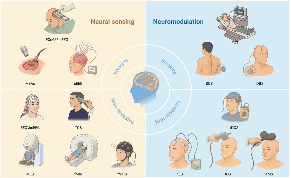
Figure 1.
Neural sensing and neuromodulation technology.
-
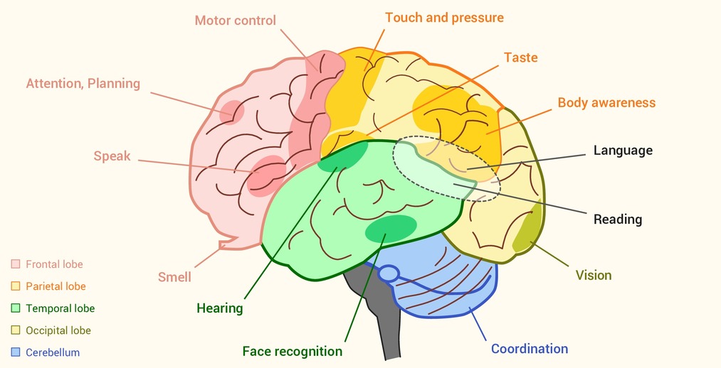
Figure 2.
Reference diagram for the specific functions of each region of the brain.
-
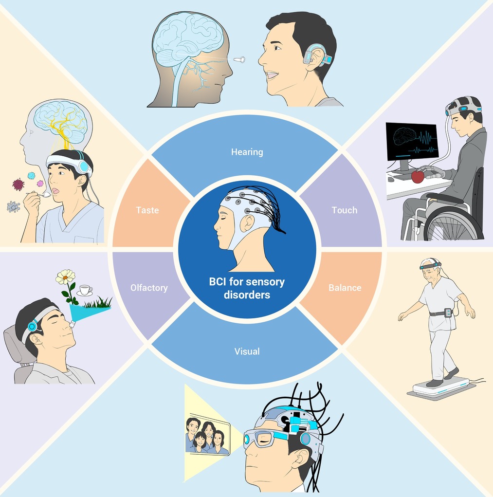
Figure 3.
Applications of brain computer interface in sensory disorders.
-
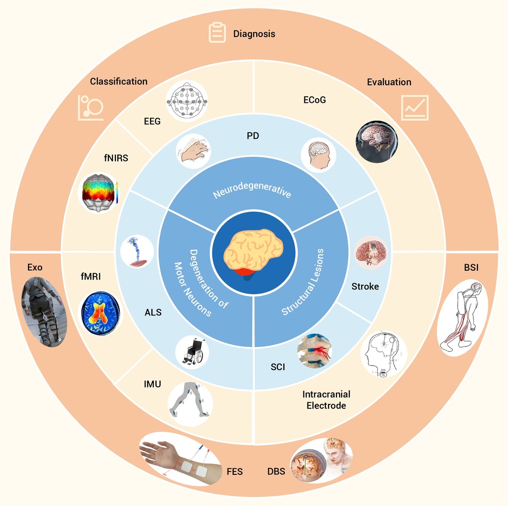
Figure 4.
Application of brain computer interface in motor disorders.
-
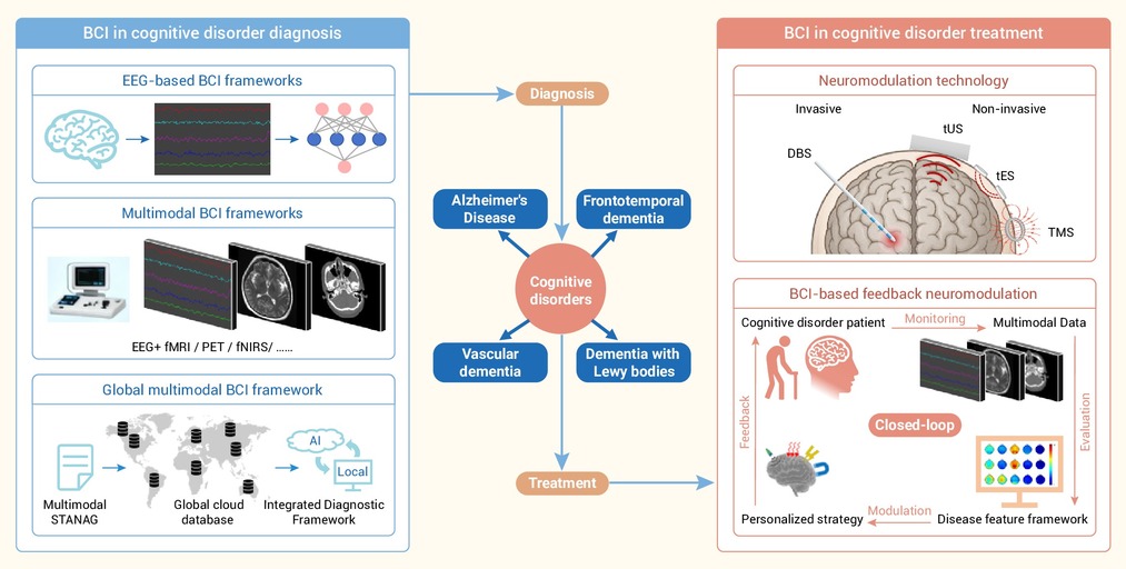
Figure 5.
Application of brain computer interface in cognitive disorders.
-
Figure 6.
Application of brain computer interface in mental disorders.
