
Absolute quantification of the microbiota spatial distribution in the murine large intestine
-
GRAPHICAL ABSTRACT
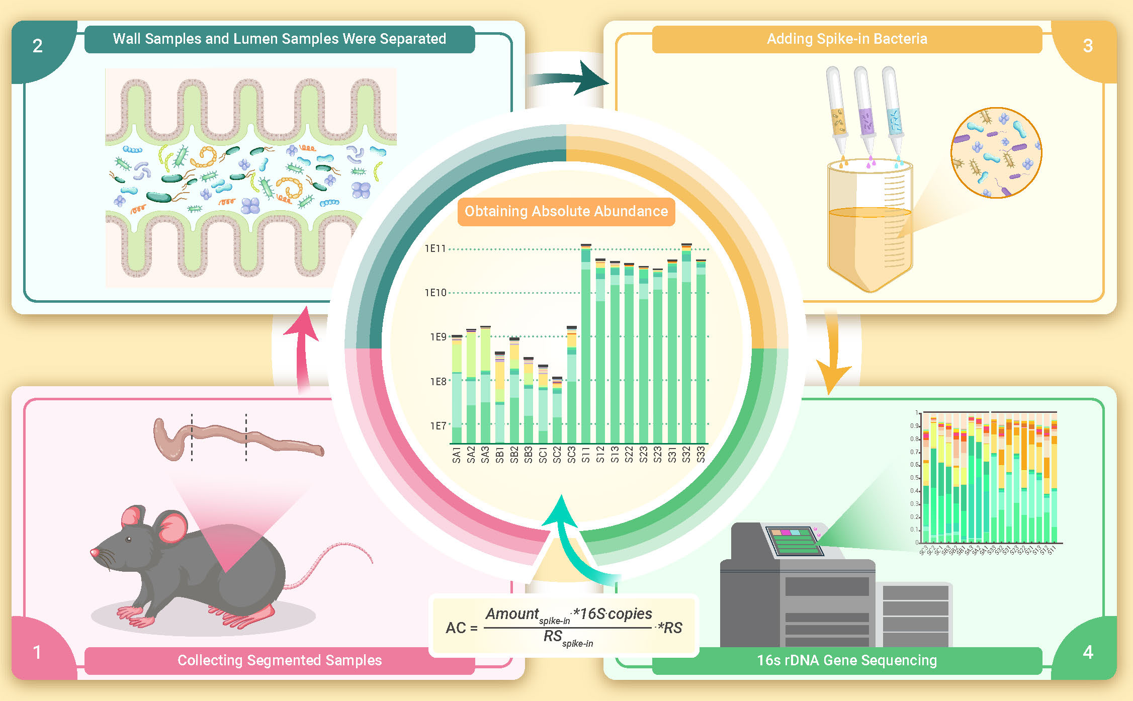
 DownLoad:
Full size image
DownLoad:
Full size image
-
PUBLIC SUMMARY
-
Reliable absolute quantification of microbiome based on triple spike-in bacteria.
Absolute quantification of total bacteria in cecum, distal colon, and proximal colon of mice.
Absolute quantification of bacteria in content and mucus layer in different part of the intestine.
-
-
ABSTRACT
The species diversity, abundance, and location of intestinal bacteria bear major consequences on host intestinal functions, affecting the balance between health and disease. Recent studies addressed the relative abundance of bacteria in feces and on the gut mucosal surface. However, few of these publications reported the absolute number of bacteria in different compartments of the gut, especially those associated with the intestinal epithelial surface, compared to the luminal content reflected by feces. In this study, the number of bacteria present in the lumen or associated with the mucosal surface of mice gut samples was calculated using the bacterial “internal standard method” during sequencing. Three spiking bacteria were used for mutual corroboration. The microbiota spatial distribution in the cecum, proximal and distal colon were studied in the lumen content and gut wall fixed niches. This study also provides an example to demonstrate that absolute and relative quantification may yield different conclusions. This study’s method may eliminate such misjudgments and provide insight into the distribution and quantity of gut microbiome. -

-
REFERENCES
[1] Luo M., Zhang D., Tian C., et al.(2023). The Innovation Life: An international journal of life sciences. The Innovation Life 1 : 100001. DOI: 10.59717/j.xinn-life.2023.100001. [2] Qin, J., Li, R., Raes, J., et al.(2010). A human gut microbial gene catalogue established by metagenomic sequencing. Nature 464 : 59-65. DOI: 10.1038/nature08821. [3] Kaźmierczak-Siedlecka, K., Dvořák, A., Folwarski, M., et al.(2020). Fungal gut microbiota dysbiosis and its role in colorectal, oral, and pancreatic carcinogenesis. Cancers (Basel) 12 : 1326. DOI: 10.3390/cancers12051326. [4] de Vos, W. M., Tilg, H., Van Hul, M., et al. (2022). Gut microbiome and health: mechanistic insights. Gut 71 : 1020−1032. DOI: 10.1136/gutjnl-2021-326789. [5] Bai, X., Sun, Y., Li, Y., et al.(2022). Landscape of the gut archaeome in association with geography, ethnicity, urbanization, and diet in the Chinese population. Microbiome 10 : 147. DOI: 10.1186/s40168-022-01335-7. [6] Du Y., Liu L., Ma W., et al. (2023). The role of extratumoral and intratumoral microorganisms in cancer immunotherapy. The Innovation Life 1 : 100016. DOI: 10.59717/j.xinn-life.2023.100016. [7] Chen, Z., Jin, W., Hoover, A., et al. (2023). Decoding the microbiome: Advances in genetic manipulation for gut bacteria. Trends Microbiol. 31 :1143-1161. [8] Zhu, B., Wang, X.,and Li, L. (2010). Human gut microbiome: The second genome of human body. Protein Cell. 1 : 718−725. DOI: 10.1007/s13238-010-0093-z. [9] Sender, R., Fuchs, S.,and Milo, R. (2016). Are we really vastly outnumbered. Revisiting the ratio of bacterial to host cells in humans. Cell 164 : 337−340. DOI: 10.1016/j.cell.2016.01.013. [10] Sender, R., Fuchs, S.,and Milo, R. (2016). Revised estimates for the number of human and bacteria cells in the body. PLoS Biology 14 : e1002533. DOI: 10.1371/journal.pbio.1002533. [11] Lewis, W. H., Tahon, G., Geesink, P., et al. (2021). Innovations to culturing the uncultured microbial majority. Nat. Rev. Microbiol. 19 : 225−240. DOI: 10.1038/s41579-020-00458-8. [12] Stephen, A. M. and Cummings, J. H. (1980). The microbial contribution to human faecal mass. J. Med. Microbiol. 13 : 45−56. DOI: 10.1099/00222615-13-1-45. [13] Thiel, R. and Blaut, M. (2005). An improved method for the automated enumeration of fluorescently labelled bacteria in human faeces. J. Med. Microbiol. 61 : 369−379. DOI: 10.1016/j.mimet.2004.12.014. [14] Marchesi, J. R. and Ravel, J. (2015). The vocabulary of microbiome research: a proposal. Microbiome 3 : 31. DOI: 10.1186/s40168-015-0094-5. [15] Xu, C., Kadosh, D., Sun, D., et al. (2023). Editorial: Omics-originated exploration of pathogenic patterns and molecular mechanisms in human and animal fungal pathogens. Front. Microbiol. 14 : 1243709. DOI: 10.3389/fmicb.2023.1243709. [16] Barlow, J. T., Bogatyrev, S. R., and Ismagilov, R. F. (2020). A quantitative sequencing framework for absolute abundance measurements of mucosal and lumenal microbial communities. Nat. Commun. 11 : 2590. DOI: 10.1038/s41467-020-16224-6. [17] Dreier, M., Meola, M., Berthoud, H., et al.(2022). High-throughput qPCR and 16S rRNA gene amplicon sequencing as complementary methods for the investigation of the cheese microbiota. BMC Microbiol. 22 : 48. DOI: 10.1186/s12866-022-02451-y. [18] Tettamanti Boshier, F.A., Srinivasan, S., Lopez, A, et al. (2020). Complementing 16S rRNA gene amplicon sequencing with total bacterial load to infer absolute species concentrations in the vaginal microbiome. mSystems 5 : e00777-19. DOI: 10.1128/mSystems.00777-19. [19] Wang, S., Wu, Q., Han, Y., et al.(2021). Gradient internal standard method for absolute quantification of microbial amplicon sequencing data. mSystems 6 : e-00964-20. DOI: 10.1128/mSystems.00964-20. [20] Tkacz, A., Hortala, M., and Poole, P. S. (2018). Absolute quantitation of microbiota abundance in environmental samples. Microbiome 6 : 110. DOI: 10.1186/s40168-018-0491-7. [21] Stämmler, F., Gläsner, J., Hiergeist, A., et al.(2016). Adjusting microbiome profiles for differences in microbial load by spike-in bacteria. Microbiome 4 : 28. DOI: 10.1186/s40168-016-0175-0. [22] Zoetendal, E. G., Rajilic-Stojanovic, M.,and de Vos, W. M. (2008). High-throughput diversity and functionality analysis of the gastrointestinal tract microbiota. Gut 57 : 1605−1615. DOI: 10.1136/gut.2007.133603. [23] Azkanaz, M., Corominas-Murtra, B., Ellenbroek, S.I.J., et al.(2022). Retrograde movements determine effective stem cell numbers in the intestine. Nature 607 : 548-554. DOI: 10.1038/s41586-022-04962-0. [24] Tang, R., Sansonetti, P. J., and Gao, Y. Z. (2023). Stem cell retrograde: A new reason why colorectal cancer is more common than small intestinal cancer. The Innovation 4 : 100387. DOI: 10.1016/j.xinn.2023.100387. [25] Pédron, T., Mulet, C., Dauga, C., et al.(2012). A crypt-specific core microbiota resides in the mouse colon. mBio 3 : e00116-12. DOI: 10.1128/mBio.00116-12. [26] Saffarian, A., Mulet, C., Regnault, B., et al.(2019). Crypt- and mucosa-associated core microbiotas in humans and their alteration in colon cancer patients. mBio 10 : e01315-19. DOI: 10.1128/mBio.01315-19. [27] Duncan, K., Carey-Ewend, K.,and Vaishnava, S. (2021). Spatial analysis of gut microbiome reveals a distinct ecological niche associated with the mucus layer. Gut Microbes 13 : 1874815. DOI: 10.1080/19490976.2021.1874815. [28] Motta, J. P., Wallace, J. L., Buret, A. G.,et al. (2021). Deraison, C. & Vergnolle, N. Gastrointestinal biofilms in health and disease. Nat. Rev. Gastroenterol. Hepatol. 18 : 314−334. DOI: 10.1038/s41575-020-00397-y. [29] Hu, L. X. et al.(2022). Poly-γ-glutamic acid production by engineering a DegU quorum-sensing circuit in bacillus subtilis. ACS Synth. Biol. 11 : 4156-4170. DOI: 10.1021/acssynbio.2c00464. [30] Jiang, X.W., Liu, H., Xu, Y., et al.(2009). Genetic and biochemical analyses of chlorobenzene degradation gene clusters in Pandoraea sp. strain MCB032. Arch. Microbiol. 191 : 485-492. DOI: 10.1007/s00203-009-0476-9. [31] Ramos-Gonzalez, M. I.,and Molin, S. (1998). Cloning, sequencing, and phenotypic characterization of the rpoS gene from Pseudomonas putida KT2440. J. Bacteriol. 180 : 3421−3431. DOI: 10.1128/JB.180.13.3421-3431.1998. [32] Gao, Y.-Z., Wang, Y., Ji, M., et al. (2023). A whole-cell hydrogen peroxide biosensor and its application in visual food analysis. The Innovation Life 1 : 100011. DOI: 10.59717/j.xinn-life.2023.100011. [33] Callahan, B.J., McMurdie, P.J., Rosen, M.J., et al.(2016). DADA2: High-resolution sample inference from Illumina amplicon data. Nat. Methods 13 , 581-583. DOI: 10.1038/nmeth.3869. [34] Bolyen, E., Rideout, J.R., Dillon, M.R., et al. (2019). Reproducible, interactive, scalable and extensible microbiome data science using QIIME 2. Nat. Biotechnol. 37 : 852-857. DOI: 10.1038/s41587-019-0252-6. [35] Schloss, P.D., Westcott, S.L., Ryabin, T., et al. (2009). Introducing mothur: Open-source, platform-independent, community-supported software for describing and comparing microbial communities. Appl. Environ. Microbiol. 75 : 7537-7541. DOI: 10.1128/AEM.01541-09. [36] Gao, Y. Z., Liu, H., Chao, H. J., et al. (2016). Constitutive expression of a nag-like dioxygenase gene through an internal promoter in the 2-chloronitrobenzene catabolism gene cluster of Pseudomonas stutzeri ZWLR2-1. Appl. Environ. Microbiol. 82 : 3461−3470. DOI: 10.1128/AEM.00197-16. [37] Stoddard, S. F., Smith, B. J., Hein, R., et al. (2015). rrnDB: Improved tools for interpreting rRNA gene abundance in bacteria and archaea and a new foundation for future development. Nucleic Acids Res. 43 : D593−598. DOI: 10.1093/nar/gku1201. [38] Feng, Y., Zhang, M., Liu, Y., et al. (2023). Quantitative microbiome profiling reveals the developmental trajectory of the chicken gut microbiota and its connection to host metabolism. iMeta 2 : e105. DOI: 10.1002/imt2.105. -
ABOUT THIS ARTICLE
Cite this article:
Jin W.-Y., Guo J.-X., Zhang M., et al., (2023). Absolute quantification of the microbiota spatial distribution in the murine large intestine. The Innovation Life 1(2), 100030. https://doi.org/10.59717/j.xinn-life.2023.100030
Welcome!
To request copyright permission to republish or share portions of our works, please visit Copyright Clearance Center's (CCC) Marketplace website at marketplace.copyright.com.

Article Metrics
Relative Articles
Cited by
Export File
Citation
Jin W.-Y., Guo J.-X., Zhang M., et al., (2023). Absolute quantification of the microbiota spatial distribution in the murine large intestine. The Innovation Life 1(2), 100030. https://doi.org/10.59717/j.xinn-life.2023.100030

Format
Content


-
Figure 1.
Flowchart for calculating bacterial abundance in different parts of mice intestinal tract (The figure was drawn using Medpeer)
-
Figure 2.
Relative abundance of different genera in the samples
-
Figure 3.
Verification of the spiked-in microbial cells added
-
Figure 4.
Application prospects of absolute quantitative analysis in intestinal bacteria
