
Sustained microglial activation and accelerated elimination of dendritic spines during acute sleep deprivation and restoration
-
GRAPHICAL ABSTRACT
 DownLoad:
Full size image
DownLoad:
Full size image
-
PUBLIC SUMMARY
-
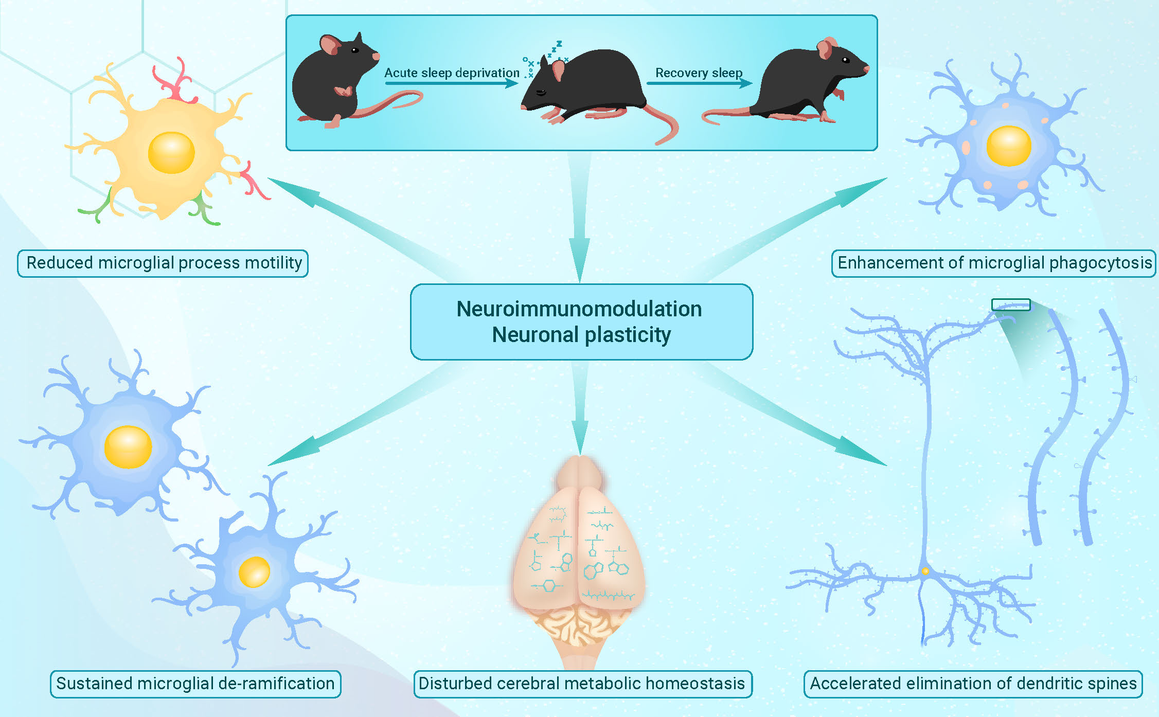
Microglial de-ramification occurs during acute sleep deprivation and restoration.
Reduced microglial processes mobility during sleep deprivation and restoration.
Acute sleep deprivation and restoration increase the phagocytosis of microglia.
Acute sleep deprivation and restoration induce elimination of dendritic spines.
A substantial number of metabolites and pathways fail to recover after recovery sleep.
-
-
ABSTRACT
Sleep is a fundamental physiological condition strongly regulated by a variety of neuroanatomical and neurochemical systems. Recent studies have indicated that catch-up sleep falls short of effectiveness to counteract the negative consequences of sleep debt however, the underlying mechanisms remain poorly understood. In this study, by using various transgenic fluorescent mouse models as well as techniques including intravital two-photon imaging and immunofluorescence staining of brain sections, we have documented morphological and functional changes of microglia during acute sleep deprivation (ASD) and subsequent short-term recovery sleep (RS). In these cerebral resident immune cells, we observed sustained microglial de-ramification, reduction of process motility and enhancement of microglial phagocytosis across brain regions. Given the intimate connections between microglia activity and neuronal plasticity, we also investigated synaptic plasticity and demonstrated an accelerated elimination of dendritic spines during both ASD and subsequent RS. Furthermore, untargeted metabolomic analyses revealed extensive whole-brain metabolic changes during ASD, and that a substantial number of metabolites and pathways failed to recover within a short period of RS. It is tempting to speculate that the disturbed cerebral metabolic homeostasis contributes to the sustained microglial activation and accelerated elimination of dendritic spines during this process. This study reveals the adverse effects of sleep loss on neuroimmunomodulation and neuronal plasticity, and implicates potential mechanisms underlying how irregular sleep schedules lead to neurological disorders. -

-
REFERENCES
[1] Karmanova, I.G. and Lazarev, S.G. (1979). Stages of sleep evolution (facts and hypotheses). Waking Sleeping 3 : 137−147. [2] Cappuccio, F.P., D’Elia, L., Strazzullo, P., et al. (2010). Quantity and quality of sleep and incidence of type 2 diabetes: A systematic review and meta-analysis. Diabetes Care 33 : 414−420. DOI: 10.2337/dc09-1124. [3] Huang, W., Ramsey, K.M., Marcheva, B., and Bass, J. (2011). Circadian rhythms, sleep, and metabolism. J. Clin. Invest. 121 : 2133−2141. DOI: 10.1172/JCI46043. [4] Romero-Corral, A., Caples, S.M., Lopez-Jimenez, F., and Somers, V.K. (2010). Interactions between obesity and obstructive sleep apnea: Implications for treatment. Chest 137 : 711−719. DOI: 10.1378/chest.09-0360. [5] Nagai, M., Hoshide, S., and Kario, K. (2010). Sleep duration as a risk factor for cardiovascular disease- a review of the recent literature. Curr. Cardiol. Rev. 6 : 54−61. DOI: 10.2174/157340310790231635. [6] Miller, M.A. (2015). The role of sleep and sleep disorders in the development, diagnosis, and management of neurocognitive disorders. Front. Neurol. 6 : 224. DOI: 10.3389/fneur.2015.00224. [7] Raven, F., Van der Zee, E.A., Meerlo, P., and Havekes, R. (2018). The role of sleep in regulating structural plasticity and synaptic strength: Implications for memory and cognitive function. Sleep Med. Rev. 39 : 3−11. DOI: 10.1016/j.smrv.2017.05.002. [8] Bolin, D.J. (2019). Sleep deprivation and its contribution to mood and performance deterioration in college athletes. Curr. Sports Med. Rep. 18 : 305−310. DOI: 10.1249/JSR.0000000000000621. [9] Lewis, K.S., Gordon-Smith, K., Forty, L., et al. (2017). Sleep loss as a trigger of mood episodes in bipolar disorder: Individual differences based on diagnostic subtype and gender. Br. J. Psychiatry 211 : 169−174. DOI: 10.1192/bjp.bp.117.202259. [10] Musiek, E.S., and Holtzman, D.M. (2016). Mechanisms linking circadian clocks, sleep, and neurodegeneration. Science 354 : 1004−1008. DOI: 10.1126/science.aah4968. [11] Puentes-Mestril, C., and Aton, S.J. (2017). Linking network activity to synaptic plasticity during sleep: Hypotheses and recent data. Front. Neural Circuits 11 : 61. DOI: 10.3389/fncir.2017.00061. [12] Puentes-Mestril, C., Roach, J., Niethard, N., et al. (2019). How rhythms of the sleeping brain tune memory and synaptic plasticity. Sleep 42 : zsz095. DOI: 10.1093/sleep/zsz095. [13] Thomas, C.W., Guillaumin, M.C.C., McKillop, L.E., et al. (2020). Global sleep homeostasis reflects temporally and spatially integrated local cortical neuronal activity. eLife 9 : 1−25. DOI: 10.7554/eLife.54148. [14] Delorme, J., Wang, L., Kuhn, F.R., et al. (2021). Sleep loss drives acetylcholine- And somatostatin interneuron-mediated gating of hippocampal activity to inhibit memory consolidation. Proc. Natl. Acad. Sci. USA 118 : e2019318118. DOI: 10.1073/pnas.2019318118. [15] Lyons, L.C., Chatterjee, S., Vanrobaeys, Y., et al. (2020). Translational changes induced by acute sleep deprivation uncovered by TRAP-Seq. Mol. Brain. 13 : 165. DOI: 10.1186/s13041-020-00702-5. [16] Noya, S.B., Colameo, D., Brüning, F., et al. (2019). The forebrain synaptic transcriptome is organized by clocks but its proteome is driven by sleep. Science 366 : eaav2642. DOI: 10.1126/science.aav2642. [17] Delorme, J., Wang, L., Kodoth, V., et al. (2021). Hippocampal neurons’ cytosolic and membrane-bound ribosomal transcript profiles are differentially regulated by learning and subsequent sleep. Proc. Natl. Acad. Sci. USA 118 : e2108534118. DOI: 10.1073/pnas.2108534118. [18] Li, Q., and Barres, B.A. (2018). Microglia and macrophages in brain homeostasis and disease. Nat. Rev. Immunol. 18 : 225−242. DOI: 10.1038/nri.2017.125. [19] Butovsky, O. and Weiner, H.L. (2018). Microglial signatures and their role in health and disease. Nat. Rev. Neurosci. 19 : 622−635. DOI: 10.1038/s41583-018-0057-5. [20] Corsi, G., Picard, K., di Castro, M.A., et al. (2022). Microglia modulate hippocampal synaptic transmission and sleep duration along the light/dark cycle. Glia 70 : 89−105. DOI: 10.1002/glia.24090. [21] Deurveilher, S., Golovin, T., Hall, S., and Semba, K. (2021). Microglia dynamics in sleep/wake states and in response to sleep loss. Neurochem. Int. 143 : 104944. DOI: 10.1016/j.neuint.2020.104944. [22] Wadhwa, M., Kumari, P., Chauhan, G., et al. (2017). Sleep deprivation induces spatial memory impairment by altered hippocampus neuroinflammatory responses and glial cells activation in rats. J. Neuroimmunol. 312 : 38−48. DOI: 10.1016/j.jneuroim.2017.09.003. [23] Bellesi, M., Vivo, L. De., Chini, M., et al. (2017). Sleep loss promotes astrocytic phagocytosis and microglial activation in mouse cerebral cortex. J. Neurosci. 37 : 5263−5273. DOI: 10.1523/JNEUROSCI.3981-16.2017. [24] Wadhwa, M., Prabhakar, A., Ray, K., et al. (2017). Inhibiting the microglia activation improves the spatial memory and adult neurogenesis in rat hippocampus during 48 h of sleep deprivation. J. Neuroinflammation. 14 : 222. DOI: 10.1186/s12974-017-0998-z. [25] Wisor, J.P., Schmidt, M.A., and Clegern, W.C. (2011). Evidence for neuroinflammatory and microglial changes in the cerebral response to sleep loss. Sleep. 34, 261−272. DOI: 10.1093/sleep/34.3.261. [26] Manchanda, S., Singh, H., Kaur, T., and Kaur, G. (2018). Low-grade neuroinflammation due to chronic sleep deprivation results in anxiety and learning and memory impairments. Mol. Cell Biochem. 449 : 63−72. DOI: 10.1007/s11010-018-3343-7. [27] Zielinski, M.R., Kim, Y., Karpova, S.A., et al. (2014). Chronic sleep restriction elevates brain interleukin-1 beta and tumor necrosis factor-alpha and attenuates brain-derived neurotrophic factor expression. Neurosci. Lett. 580 : 27−31. DOI: 10.1016/j.neulet.2014.07.043. [28] Hsu, J.C., Lee, Y.S., Chang, C.N., et al. (2003). Sleep deprivation inhibits expression of NADPH-d and NOS while activating microglia and astroglia in the rat hippocampus. Cells Tissues Organs 173 : 242−254. DOI: 10.1159/000070380. [29] Neniskyte, U., and Gross, C.T. (2017). Errant gardeners: Glial-cell-dependent synaptic pruning and neurodevelopmental disorders. Nat. Rev. Neurosci. 18 : 658−670. DOI: 10.1038/nrn.2017.110. [30] Mallya, A.P., Wang, H.D., Lee, H.NR., and Deutch, A.Y. (2019). Microglial Pruning of Synapses in the Prefrontal Cortex during Adolescence Cereb. Cortex 29 : 1634−1643. DOI: 10.1093/cercor/bhy061. [31] Kopec, A.M., Smith, C.J., Ayre, N.R., et al. (2018). Microglial dopamine receptor elimination defines sex-specific nucleus accumbens development and social behavior in adolescent rats. Nat. Commun. 9 : 3769. DOI: 10.1038/s41467-018-06118-z. [32] Stowell, R.D., Sipe, G.O., Dawes, R.P., et al. (2019). Noradrenergic signaling in the wakeful state inhibits microglial surveillance and synaptic plasticity in the mouse visual cortex. Nat. Neurosci. 22 : 1782−1792. DOI: 10.1038/s41593-019-0514-0. [33] Liu, Y.U., Ying, Y., Li, Y., et al. (2019). Neuronal network activity controls microglial process surveillance in awake mice via norepinephrine signaling. Nat. Neurosci. 22 : 1771−1781. DOI: 10.1038/s41593-019-0511-3. [34] Hristovska, I., Robert, M., Combet, K., et al. (2022). Sleep decreases neuronal activity control of microglial dynamics in mice. Nat. Commun. 13 : 1−15. DOI: 10.1038/s41467-022-34035-9. [35] Nadjar, A., Wigren, H.K.M., and Tremblay, M.E. (2017). Roles of microglial phagocytosis and inflammatory mediators in the pathophysiology of sleep disorders. Front. Cell Neurosci. 11 : 1−11. DOI: 10.3389/fncel.2017.00250. [36] Brüning, F., Noya, S.B., Bange, T., et al. (2019). Sleep-wake cycles drive daily dynamics of synaptic phosphorylation. Science 366 : eaav3617. DOI: 10.1126/science.aav3617. [37] Datta, S. (2010). Cellular and chemical neuroscience of mammalian sleep. Sleep Med. 11 : 431−440. DOI: 10.1016/j.sleep.2010.02.002. [38] Bellesi, M., Bushey, D., Chini, M., et al. (2016). Contribution of sleep to the repair of neuronal DNA double-strand breaks: Evidence from flies and mice. Sci. Rep. 6 : 36804. DOI: 10.1038/srep36804. [39] Vaccaro, A., Kaplan, Dor. Y., Nambara, K., et al. (2020). Sleep loss can cause death through accumulation of reactive oxygen species in the gut. Cell 181 : 1307−1328.e15. DOI: 10.1016/j.cell.2020.04.049. [40] Hines, D.J., Schmitt, L.I., Hines, R.M., et al. (2013). Antidepressant effects of sleep deprivation require astrocyte-dependent adenosine mediated signaling. Transl. Psychiatry 3 : e212−9. DOI: 10.1038/tp.2012.136. [41] Naidoo, N., Davis, J.G., Zhu, J., et al. (2014). Aging and sleep deprivation induce the unfolded protein response in the pancreas: Implications for metabolism. Aging Cell 13 : 131−141. DOI: 10.1111/acel.12158. [42] Dunn, W.B., Broad hurst, D., Begley, P., et al. (2011). Procedures for large-scale metabolic profiling of serum and plasma using gas chromatography and liquid chromatography coupled to mass spectrometry. Nat. Protoc. 6: 1060−1083. DOI: 10.1038/nprot.2011.335. [43] Want, E.J., Wilson, I.D., Gika, H., et al. (2010). Global metabolic profiling procedures for urine using UPLC-MS. Nat. Protoc. 5: 1005−1018. DOI: 10.1038/nprot.2010.50. [44] Wang, J., Zhang, T., Shen, X., et al. (2016). Serum metabolomics for early diagnosis of esophageal squamous cell carcinoma by UHPLC-QTOF/MS. Metabolomics 12: 116. DOI: 10.1007/s11306-016-1050-5. [45] Smith, C.A., Want, E.J., O’Maille, G., et al. XCMS: Processing mass spectrometry data for metabolite profiling using nonlinear peak alignment, matching, and identification. Anal. Chem. 78: 779−787. DOI: 10.1021/ac051437y. [46] Kuhl, C., Tautenhahn, R., Böttcher, C., et al. (). CAMERA: An integrated strategy for compound spectra extraction and annotation of liquid chromatography/mass spectrometry data sets. Anal. Chem. 84(): 283−289. [47] Chong, J., Wishart, D.S., and Xia, J. (2019). Using MetaboAnalyst 4.0 for comprehensive and integrative metabolomics data analysis. Curr. Protoc. Bioinforma. 68: e86. DOI: 10.1002/cpbi.86. [48] Zhu, L., Wang, L., Ju, F., et al. (2017). Transient global cerebral ischemia induces rapid and sustained reorganization of synaptic structures. J. Cereb. Blood Flow Metab. 37: 2756−2767. DOI: 10.1177/0271678X16674736. [49] Yang, G., Pan, F., Parkhurst, C.N., et al. (2010). Thinned-skull cranial window technique for long-term imaging of the cortex in live mice. Nat. Protoc. 5: 213−220. [50] Zuo, Y., Yang, G., Kwon, E., et al. (2005). Long-term sensory deprivation prevents dendritic spine loss in primary somatosensory cortex. Nature 436: 261−265. DOI: 10.1038/nature03715. [51] Ju, F., Ran, Y., Zhu, L., et al. (2018). Increased BBB permeability enhances activation of microglia and exacerbates loss of dendritic spines after transient global cerebral ischemia. Front. Cell Neurosci. 12: 1−14. DOI: 10.3389/fncel.2018.00236. [52] Sipe, G.O., Lowery, R.L., Tremblay, M., et al. (2016). Microglial P2Y12 is necessary for synaptic plasticity in mouse visual cortex. Nat. Commun. 7: 10905. DOI: 10.1038/ncomms10905. [53] Liu, Y.U., Ying, Y., Li, Y., et al. (2019). Neuronal network activity controls microglial process surveillance in awake mice via norepinephrine signaling. Nat. Neurosci. 22: 1771−1781. DOI: 1038/s41593-019-0511-3. [54] Morrison, H.W. and Filosa, J.A. (2013). A quantitative spatiotemporal analysis of microglia morphology during ischemic stroke and reperfusion. J. Neuroinflammation 10: 1−20. DOI: 10.1186/ 1742-2094-10-4. [55] York, E.M., Bernier, L.P., and MacVicar, B.A. (2018). Microglial modulation of neuronal activity in the healthy brain. Dev Neurobiol. 78 : 593−603. DOI: 10.1002/dneu.22571. [56] Wake, H., Horiuchi, H., Kato, D., et al. (2019). Physiological implications of microglia–synapse interactions. Methods Mol. Biol. 2034 : 69−80. DOI: 10.1007/978-1-4939-9658-2_6. [57] Socodato, R., Portugal, C.C., Domith, I., et al. (2015). c-Src function is necessary and sufficient for triggering microglial cell activation. Glia 63 : 497−511. DOI: 10.1002/glia.22767. [58] Li, T., Pang, S., Yu, Y., et al. (2013). Proliferation of parenchymal microglia is the main source of microgliosis after ischaemic stroke. Brain 136 : 3578−3588. DOI: 10.1093/brain/awt287. [59] Kyrargyri, V., Madry, C., Rifat, A., et al. (2020). P2Y(13) receptors regulate microglial morphology, surveillance, and resting levels of interleukin 1β release. Glia 68 : 328−344. DOI: 10.1002/glia.23719. [60] Kettenmann, H., Kirchhoff, F., and Verkhratsky, A. (2013). Microglia: new roles for the synaptic stripper. Neuron 77 : 10−18. DOI: 10.1016/j.neuron.2012.12.023. [61] Illouz, T., Madar, R., Biragyn, A., and Okun, E. (2019). Restoring microglial and astroglial homeostasis using DNA immunization in a Down Syndrome mouse model. Brain Behav. Immun. 75 : 163−180. DOI: 10.1016/j.bbi.2018.10.004. [62] Korzhevskii, D.E., and Kirik, O.V. (2016). Brain microglia and microglial markers. Neurosci. Behav. Physiol. 46 : 284−290. DOI: 10.1007/s11055-016-0231-z. [63] Liu, Y.J., Spangenberg, E.E., Tang, B., et al. (2021). Microglia elimination increases neural circuit connectivity and activity in adult mouse cortex. J. Neurosci. 41 : 1274−1287. DOI: 10.1523/JNEUROSCI.2140-20.2020. [64] Hong, S., Beja-Glasser, V.F., Nfonoyim, B.M., et al. (2016). Complement and microglia mediate early synapse loss in Alzheimer mouse models. Science 352 : 712−716. DOI: 10.1126/science.aad8373. [65] Wake, H., Moorhouse, A.J., Jinno, S., et al. (2009). Resting microglia directly monitor the functional state of synapses in vivo and determine the fate of ischemic terminals. J Neurosci. 29 : 3974−3980. DOI: 10.1523/JNEUROSCI.4363-08.2009. [66] Tremblay, M.Ě., Lowery, R.L., and Majewska, A.K. (2010). Microglial interactions with synapses are modulated by visual experience. PLoS Biol. 8 : e1000527. DOI: 10.1371/journal.pbio.1000527. [67] Schafer, D.P., Lehrman, E.K., Kautzman, A.G., et al. (2012). Microglia sculpt postnatal neural circuits in an activity and complement-dependent manner. Neuron 74 : 691−705. DOI: 10.1016/j.neuron.2012.03.026. [68] Irwin, M.R. (2019). Sleep and inflammation: partners in sickness and in health. Nat. Rev. Immunol. 19 : 702−715. DOI: 10.1038/s41577-019-0190-z. [69] Irwin, M.R. and Opp, M.R. (2017). Sleep health: Reciprocal regulation of sleep and innate immunity. Neuropsychopharmacology 42 : 129−155. DOI: 10.1038/npp.2016.148. [70] Garbarino, S., Lanteri, P., Bragazzi, N.L., et al. (2021). Role of sleep deprivation in immune-related disease risk and outcomes. Commun. Biol. 4 : 1304. DOI: 10.1038/s42003-021-02825-4. [71] Besedovsky, L., Lange, T., and Haack, M. (2019). The sleep-immune crosstalk in health and disease. Physiol. Rev. 99, 1325−1380. DOI: 10.1152/physrev.00010.2018. [72] Bellesi, M., de Vivo, L., Chini, M., et al. (2017). Sleep loss promotes astrocytic phagocytosis and microglial activation in mouse cerebral cortex. J. Neurosci. 37 : 5263−5273. DOI: 10.1523/JNEUROSCI.3981-16.2017. [73] Tuan, L.H. and Lee, L.J. (2019). Microglia-mediated synaptic pruning is impaired in sleep-deprived adolescent mice. Neurobiol. Dis. 130 : 104517. DOI: 10.1016/j.nbd.2019.104517. [74] Xie, Y., Ba, L., Wang, M., et al. (2020). Chronic sleep fragmentation shares similar pathogenesis with neurodegenerative diseases: Endosome-autophagosome-lysosome pathway dysfunction and microglia-mediated neuroinflammation. CNS Neurosci. Ther. 26 : 215−227. DOI: 10.1111/cns.13218. [75] Hall, S., Deurveilher, S., Robertson, G.S., and Semba, K. (2020). Homeostatic state of microglia in a rat model of chronic sleep restriction. Sleep 43 : zsaa108. DOI: 10.1093/sleep/zsaa108. [76] Parhizkar, S., Gent, G., Chen, Y., et al. (2023). Sleep deprivation exacerbates microglial reactivity and Aβ deposition in a TREM2-dependent manner in mice. Sci. Transl. Med. 15 : eade6285. DOI: 10.1126/scitranslmed.ade6285. [77] Zhu, B., Dong, Y., Xu, Z., et al. (2012). Sleep disturbance induces neuroinflammation and impairment of learning and memory. Neurobiol. Dis. 48 : 348−355. DOI: 10.1016/j.nbd.2012.06.022. [78] Yin, M., Chen, Y., Zheng, H., et al. (2017). Assessment of mouse cognitive and anxiety-like behaviors and hippocampal inflammation following a repeated and intermittent paradoxical sleep deprivation procedure. Behav. Brain Res. 321 : 69−78. DOI: 10.1016/j.bbr.2016.12.034. [79] Mishra, I., Pullum, K.B., Eads, K.N., et al. (2022). Peripheral sympathectomy alters neuroinflammatory and microglial responses to sleep fragmentation in female mice. Neuroscience 505 : 111−124. DOI: 10.1016/j.neuroscience.2022.09.022. [80] Faraut, B., Boudjeltia, K.Z., Vanhamme, L., and Kerkhofs, M. (2012). Immune, inflammatory and cardiovascular consequences of sleep restriction and recovery. Sleep Med. Rev. 16 : 137−149. DOI: 10.1016/j.smrv.2011.05.001. [81] Faraut, B., Boudjeltia, K.Z., Dyzma, M., et al. (2011). Benefits of napping and an extended duration of recovery sleep on alertness and immune cells after acute sleep restriction. Brain Behav. Immun. 25 : 16−24. DOI: 10.1016/j.bbi.2010.08.001. [82] Combs, C.K., Karlo, J.C., Kao, S.C., and Landreth, G.E. (2001). beta-Amyloid stimulation of microglia and monocytes results in TNFalpha-dependent expression of inducible nitric oxide synthase and neuronal apoptosis. J. Neurosci. 21 : 1179−1188. DOI: 10.1523/JNEUROSCI.21-04-01179.2001. [83] Perry, V.H., and Holmes, C. (2014). Microglial priming in neurodegenerative disease. Nat. Rev. Neurol. 10 : 217−224. DOI: 10.1038/nrneurol.2014.38. [84] Hu, Y., Cao, K., Wang, F., et al. (2022). Dual roles of hexokinase 2 in shaping microglial function by gating glycolytic flux and mitochondrial activity. Nat. Metab. 4 : 1756−1774. DOI: 10.1038/s42255-022-00707-5. [85] Miyamoto, A., Wake, H., Ishikawa, A.W., et al. (2016). Microglia contact induces synapse formation in developing somatosensory cortex. Nat. Commun. 7 : 12540. DOI: 10.1038/ncomms12540. [86] Akiyoshi, R., Wake, H., Kato, D., et al. (2018). Microglia enhance synapse activity to promote local network synchronization. eNeuro. 5 : ENEURO.0088−18.2018. DOI: 10.1523/ENEURO.0088-18.2018. [87] Eyo, U.B., Peng, J., Swiatkowski, P., et al. (2014). Neuronal hyperactivity recruits microglial processes via neuronal NMDA receptors and microglial P2Y12 receptors after status epilepticus. J. Neurosci. 34 : 10528−10540. DOI: 10.1523/JNEUROSCI.0416-14.2014. [88] Lou, N., Takano, T., Pei, Y., et al. (2016). Purinergic receptor P2RY12-dependent microglial closure of the injured blood-brain barrier. Proc. Natl. Acad. Sci. USA. 113 : 1074−1079. DOI: 10.1073/pnas.1520398113. [89] Paolicelli, R.C., Bolasco, G., Pagani, F., et al. (2011). Synaptic pruning by microglia is necessary for normal brain development. Science 333 : 1456−1458. DOI: 10.1126/science.1202529. [90] Filipello, F., Morini, R., Corradini, I., et al. (2018). The microglial innate immune receptor trem2 is required for synapse elimination and normal brain connectivity. Immunity 48 : 979−991. DOI: 10.1016/j.immuni.2018.04.016. [91] Wang, C., Yue, H., Hu, Z., et al. (2020). Microglia mediate forgetting via complement-dependent synaptic elimination. Science 367 : 688−694. DOI: 10.1126/science.aaz2288. [92] Damisah, E.C., Hill, R.A., Rai, A., et al. (2020). Astrocytes and microglia play orchestrated roles and respect phagocytic territories during neuronal corpse removal in vivo. Sci. Adv. 6 : eaba3239. DOI: 10.1126/sciadv.aba3239. [93] Salvany, S., Casanovas, A., Piedrafita, L., et al. (2021). Microglial recruitment and mechanisms involved in the disruption of afferent synaptic terminals on spinal cord motor neurons after acute peripheral nerve injury. Glia 69 : 1216−1240. DOI: 10.1002/glia.23959. [94] Zhu, M., Xiao, B., Xue, T., et al. (2023). Cdc42GAP deficiency contributes to the Alzheimer’s disease phenotype. Brain 146 : 4350−4365. DOI: 10.1093/brain/awad184. [95] Raamsdonk, L.M., Teusink, B., Broadhurst, D., et al. (2001). A functional genomics strategy that uses metabolome data to reveal the phenotype of silent mutations. Nat. Biotechnol. 19 : 45−50. DOI: 10.1038/83496. [96] Giskeødegård, G.F., Davies, S.K., Revell, V.L., et al. (2015). Diurnal rhythms in the human urine metabolome during sleep and total sleep deprivation. Sci. Rep. 5 : 14843. DOI: 10.1038/srep14843. [97] Davies, S.K., Ang, J.E., Revell, V.L., et al. (2014). Effect of sleep deprivation on the human metabolome. Proc. Natl. Acad. Sci. USA. 111 : 10761−10766. DOI: 10.1073/pnas.1402663111. [98] Akazawa, N., Kobayashi, N., Nakamura, Y., et al. (2019). Effect of sleep efficiency on salivary metabolite profile and cognitive function during exercise in volleyball athletes. Eur. J. Appl. Physiol. 119 : 2215−2223. DOI: 10.1007/s00421-019-04205-7. [99] Liu, D., Wang, Q., Li, Y., et al. (2023). Fructus gardeniae ameliorates anxiety-like behaviors induced by sleep deprivation via regulating hippocampal metabolomics and gut microbiota. Front. Cell Infect. Microbiol. 13 : 1167312. DOI: 10.3389/fcimb.2023.1167312. [100] An, Y., Varma, V.R., Varma, S., et al. (2018). Evidence for brain glucose dysregulation in Alzheimer’s disease. Alzheimers Dement. 14 : 318−329. DOI: 10.1016/j.jalz.2017.09.011. [101] Alam, Z.I., Jenner, A., Daniel, S.E., et al. (1997). Oxidative DNA damage in the parkinsonian brain: an apparent selective increase in 8-hydroxyguanine levels in substantia nigra. J. Neurochem. 69 : 1196−1203. DOI: 10.1046/j.1471-4159.1997.69031196.x. [102] Markesbery, W.R., and Lovell, M.A. (2006). DNA oxidation in Alzheimer’s disease. Antioxid Redox Signal 8 : 2039−2045. DOI: 10.1089/ars.2006.8.2039. [103] Barbieri, I., and Kouzarides, T. (2020). Role of RNA modifications in cancer. Nat. Rev. Cancer 20 : 303−322. DOI: 10.1038/s41568-020-0253-2. [104] Sheng, Z., Oka, S., Tsuchimoto, D., et al. (2012). 8-Oxoguanine causes neurodegeneration during MUTYH-mediated DNA base excision repair. J. Clin. Invest. 122 : 4344−4361. DOI: 10.1172/JCI65053. [105] Takahashi, T. and Suzuki, T. (2012). Role of sulfatide in normal and pathological cells and tissues. J. Lipid Res. 53 : 1437−1450. DOI: 10.1194/jlr.R026682. [106] Tsukahara, T., Matsuda, Y., and Haniu, H. (2017). Lysophospholipid-related diseases and pparγ signaling pathway. Int. J. Mol. Sci. 18 : 2730. DOI: 10.3390/ijms18122730. [107] Schilcher, I., Ledinski, G., Radulović, S., et al. (2019). Endothelial lipase increases antioxidative capacity of high-density lipoprotein. Biochim. Biophys. acta Mol. cell Biol. lipids 1864 : 1363−1374. DOI: 10.1016/j.bbalip.2019.06.011. [108] Radu, C.G., Yang, L.V., Riedinger, M., et al. (2004). T cell chemotaxis to lysophosphatidylcholine through the G2A receptor. Proc. Natl. Acad. Sci. USA 101 : 245−250. DOI: 10.1073/pnas.2536801100. [109] Ngwenya, B.Z. and Yamamoto, N. (1985). Activation of peritoneal macrophages by lysophosphatidylcholine. Biochim. Biophys. Acta 839 : 9−15. DOI: 10.1016/0304-4165(85)90175-8. [110] Qin, X., Qiu, C., and Zhao, L. (2014). Lysophosphatidylcholine perpetuates macrophage polarization toward classically activated phenotype in inflammation. Cell Immunol. 289 : 185−190. DOI: 10.1016/j.cellimm.2014.04.010. -
ABOUT THIS ARTICLE
Cite this article:
Liu J., Xu Y., Ji Y., et al., (2023). Sustained microglial activation and accelerated elimination of dendritic spines during acute sleep deprivation and restoration. The Innovation Life 1(3), 100037. https://doi.org/10.59717/j.xinn-life.2023.100037
Welcome!
To request copyright permission to republish or share portions of our works, please visit Copyright Clearance Center's (CCC) Marketplace website at marketplace.copyright.com.

Article Metrics
Relative Articles
Cited by
Export File
Citation
Liu J., Xu Y., Ji Y., et al., (2023). Sustained microglial activation and accelerated elimination of dendritic spines during acute sleep deprivation and restoration. The Innovation Life 1(3), 100037. https://doi.org/10.59717/j.xinn-life.2023.100037

Format
Content


-
Figure 1.
Acute sleep deprivation (ASD) model and experimental design in this study
-
Figure 2.
Microglia display sustained activation during ASD and subsequent RS
-
Figure 3.
In vivo imaging of microglial process motility during ASD and subsequent RS
-
Figure 4.
Phagocytic activity of microglia is persistently increased after ASD and subsequent RS
-
Figure 5.
Effects of ASD and subsequent RS on the turnover of dendritic spines
-
Figure 6.
Metabolic profiling of whole brain during ASD and subsequent RS
-
Figure 7.
Metabolites and pathways identified as recoverable/unrecoverable within 24 hours of RS
