
Exercise alleviates diabetic kidney disease through PPARδ-CPT1α pathway-dependent fatty acid β-oxidation
-
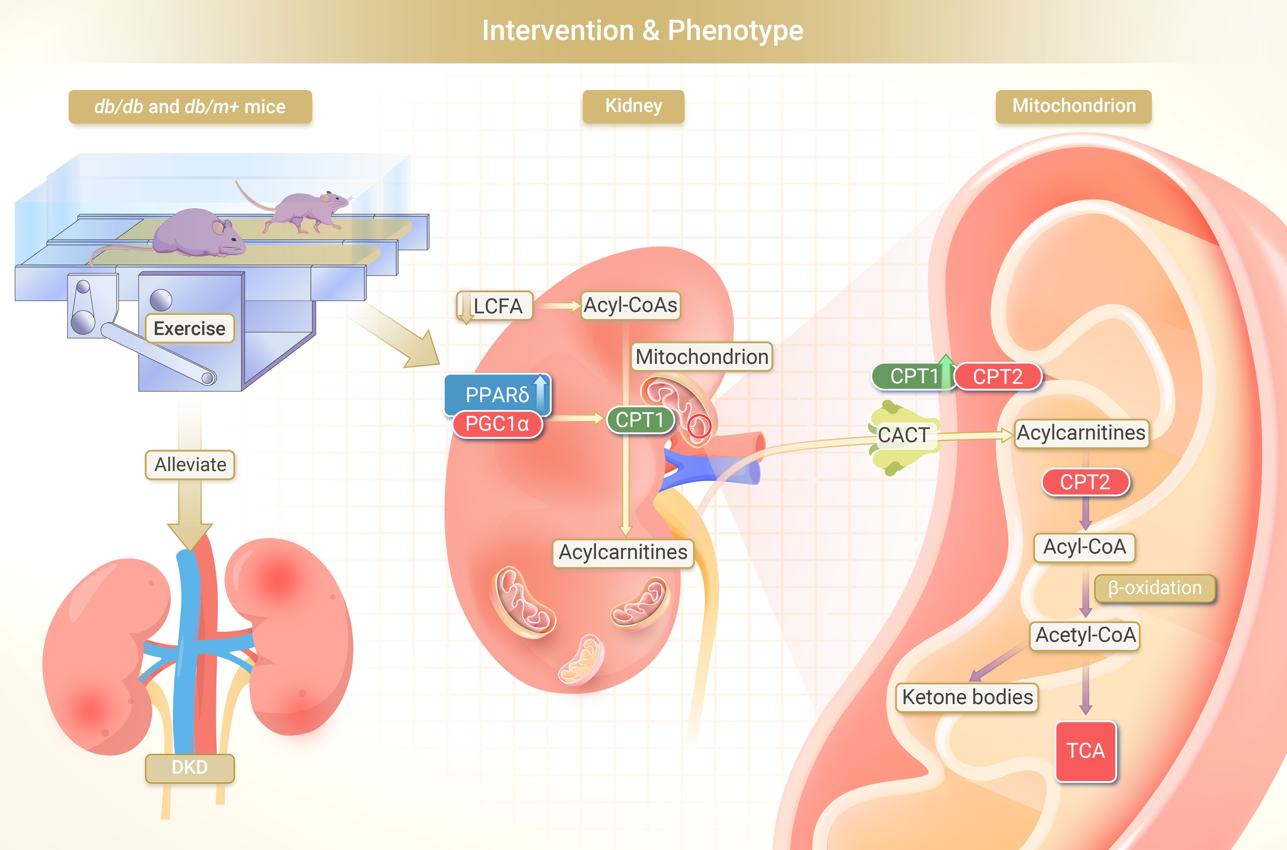
GRAPHICAL ABSTRACT
 DownLoad:
Full size image
DownLoad:
Full size image
-
PUBLIC SUMMARY
-
Regular physical exercise is an effective strategy in alleviating diabetic kidney disease.
Exercise restores renal function in diabetic mice through enhancing PPARδ-CPT1α-dependent fatty acid β-oxidation.
Reveal an unappreciated link between exercise and the restoration of renal function in diabetic kidney disease.
Could potentially become a new target for therapeutic intervention against diabetic kidney disease.
-
-
ABSTRACT
Diabetic kidney disease (DKD), a severe diabetic complication affecting approximately one-third of diabetic patients, is the leading cause of end-stage chronic kidney disease. The benefits of regular exercise for patients with DKD have been well documented, particularly in overweight patients with DKD. However, the underlying mechanisms are incompletely understood. The present study demonstrates that exercise improves kidney function in diabetic db/db mice through activating PPARδ-mediated fatty acid β-oxidation (FAO). Twelve-week treadmill running exercise improved kidney function in db/db mice. Metabolomics and transcriptomics profiling analysis collectively revealed that dysregulation of FAO in db/db mice was largely corrected by running exercise. KEGG pathway enrichment revealed that PPAR pathway, a critical signalling cascade in FAO and exercise, is involved in DKD, suggesting PPARδ activation protects renal function through promoting FAO. Which was confirmed by improved renal function and enhanced FAO in db/db mice treated with PPARδ agonist GW501516. Oppositely, PPARδ-specific inhibitor GSK0660 abolished exercise-mediated improvement in renal function and FAO. In combining with gene expression profiling data we identified CPT1α, the rate-limiting enzyme in fatty acid oxidation, was suppressed in the kidney of diabetic mice and reversed by exercise and the PPARδ agonist GW501516. The findings hint at a potential role for the PPARδ-CPT1α pathway in exercise-induced improvements in diabetic renal function and may warrant further exploration of the PPARδ-CPT1α pathway as a therapeutic target in DKD. -

-
REFERENCES
[1] Gross, J.L., de Azevedo, M.J., Silveiro, S.P., et al. (2005). Diabetic nephropathy: Diagnosis, prevention, and treatment. Diabetes Care 28: 164−176. DOI: 10.2337/diacare.28.1.164. [2] Packham, D.K., Alves, T.P., Dwyer, J.P., et al. (2012). Relative incidence of ESRD versus cardiovascular mortality in proteinuric type 2 diabetes and nephropathy: Results from the DIAMETRIC (Diabetes Mellitus Treatment for Renal Insufficiency Consortium) database. Am. J. Kidney Dis. 59: 75−83. DOI: 10.1053/j.ajkd.2011.09.017. [3] Knowler, W.C., Barrett-Connor, E., Fowler, S.E., et al. (2002). Reduction in the incidence of type 2 diabetes with lifestyle intervention or metformin. N. Engl. J. Med. 346: 393−403. DOI: 10.1056/NEJMOA012512. [4] Balducci, S., Iacobellis, G., Parisi, L., et al. (2006). Exercise training can modify the natural history of diabetic peripheral neuropathy. J. Diabetes Complications 20: 216−223. DOI: 10.1016/j.jdiacomp.2005.07.005. [5] Juszczak, F., Vlassembrouck, M., Botton, O., et al. (2020). Delayed exercise training improves obesity-induced chronic kidney disease by activating AMPK pathway in high-fat diet-fed mice. Int. J. Mol. Sci. 22: 350. DOI: 10.3390/ijms22010350. [6] Sietsema, K.E., Amato, A., Adler, S.G., et al. (2004). Exercise capacity as a predictor of survival among ambulatory patients with end-stage renal disease. Kidney Int. 65: 719−724. DOI: 10.1111/j.1523-1755.2004.00411.x. [7] Straznicky, N.E., Grima, M.T., Lambert, E.A., et al. (2011). Exercise augments weight loss induced improvement in renal function in obese metabolic syndrome individuals. J. Hypertens. 29: 553−564. DOI: 10.1097/HJH.0b013e3283418875. [8] Conn, V.S., Hafdahl, A.R., Mehr, D.R., et al. (2007). Metabolic effects of interventions to increase exercise in adults with type 2 diabetes. Diabetol 50: 913−921. DOI: 10.1007/s00125-007-0625-0. [9] Goldberg, A.P., Geltman, E.M., Gavin, 3rd, J.R., et al. (1986). Exercise training reduces coronary risk and effectively rehabilitates hemodialysis patients. Nephron 42: 311−316. DOI: 10.1159/000183694. [10] Anderson, J.E., Boivin, M.R., and Hatchett, L. (2004). Effect of exercise training on interdialytic ambulatory and treatment-related blood pressure in hemodialysis patients. JAMA Cardiol. 26: 539−544. DOI: 10.1081/JDI-200031735. [11] Brooks, G.A., and Mercier, J. (1994). Balance of carbohydrate and lipid utilization during exercise: The “crossover” concept. J. Appl. Physiol. 76: 2253−2261. DOI: 10.1152/jappl.1994.76.6.2253. [12] Kang, H.M., Ahn, S.H., Choi, P., et al. (2015). Defective fatty acid oxidation in renal tubular epithelial cells has a key role in kidney fibrosis development. Nat. Med. 21: 37−46. DOI: 10.1038/nm.3762. [13] Stadler, K., Goldberg, I.J., and Susztak, K. (2015). The evolving understanding of the contribution of lipid metabolism to diabetic kidney disease. Curr. Diab. Rep. 15: 40. DOI: 10.1007/s11892-015-0611-8. [14] Narkar, V.A., Downes, M., Yu, R.T., et al. (2008). AMPK and PPARδ agonists are exercise mimetics. Cell 134: 405−415. DOI: 10.1016/j.cell.2008.06.051. [15] Fan, W., Waizenegger, W., Lin, C.S., et al. (2017). PPARδ promotes running endurance by preserving glucose. Cell Metab. 25: 1186−1193.e4. DOI: 10.1016/j.cmet.2017.04.006. [16] Fritz, T., Krämer, D.K., Karlsson H.K.R., et al. (2006). Low-intensity exercise increases skeletal muscle protein expression of PPARδ and UCP3 in type 2 diabetic patients. Wiley Online Libr. 22: 492−498. DOI: 10.1002/dmrr.656. [17] Cheang, W.S., Wong, W.T., Zhao, L., et al. (2017). PPARδ is required for exercise to attenuate endoplasmic reticulum stress and endothelial dysfunction in diabetic mice. Diabetes 66: 519−528. DOI: 10.2337/db15-1657. [18] Idrovo, J.-P., Yang, W.-L., Nicastro, J., et al. (2012). Stimulation of carnitine palmitoyltransferase 1 improves renal function and attenuates tissue damage after ischemia/reperfusion. J. Surg. Res. 177: 157−164. DOI: 10.1016/j.jss.2012.05.053. [19] Miguel, V., Tituaña, J., Herrero, J.I., et al. (2021). Renal tubule Cpt1a overexpression protects from kidney fibrosis by restoring mitochondrial homeostasis. J. Clin. Invest. 131 : e140695. DOI: 10.1172/JCI140695. [20] Yuan, Q., Lv, Y., Ding, H., et al. (2021). CPT1α maintains phenotype of tubules via mitochondrial respiration during kidney injury and repair. Cell Death. Dis. 12: 792. DOI: 10.1038/s41419-021-04085-w. [21] Zhao, Y., Gao, P., Sun, F., et al. (2016). Sodium intake regulates glucose homeostasis through the PPARδ/Adiponectin-Mediated SGLT2 pathway. Cell Metab. 23: 699−711. DOI: 10.1016/j.cmet.2016.02.019. [22] Xiang, L., Zhang, H., Wei, J., et al. (2018). Metabolomics studies on db/db diabetic mice in skeletal muscle reveal effective clearance of overloaded intermediates by exercise. Anal. Chim. Acta. 1037: 130−139. DOI: 10.1016/j.aca.2017.11.082. [23] Xiang, L., Nie, J., Wang, L., et al. (2021). Integrated metabolomics analysis of the effect of PPARδ agonist GW501516 on catabolism of BCAAs and carboxylic acids in diabetic mice. Chinese Chem. Lett. 32: 2197−2202. DOI: 10.1016/j.cclet.2020.12.042. [24] Xiang, L., Wei, J., Tian, X.Y., et al. (2017). Comprehensive analysis of acylcarnitine species in db/db mouse using a novel method of high-resolution parallel reaction monitoring reveals widespread metabolic dysfunction induced by diabetes. Anal. Chem. 89: 10368−10375. DOI: 10.1021/acs.analchem.7b02283. [25] Kosmadakis, G.C., Bevington, A., Smith, A.C., et al. (2010). Physical exercise in patients with severe kidney disease. Nephron Clin. Pract. 115: c7−c16. DOI: 10.1159/000286344. [26] Sharma, K., McCue, P., and Dunn, S.R. (2003). Diabetic kidney disease in the db/db mouse. Am. J. Physiol. Physiol. 284: F1138−F1144. DOI: 10.1152/ajprenal.00315.2002. [27] Kriketos, A.D., Gan, S.K., Poynten, A.M., et al. (2004). Exercise increases adiponectin levels and insulin sensitivity in humans. Diabetes Care 27: 629−630. DOI: 10.2337/diacare.27.2.629. [28] Cui, S., Verroust, P.J., Moestrup, S.K., et al. (1996). Megalin/gp330 mediates uptake of albumin in renal proximal tubule. Am. J. Physiol. Ren. Physiol. 271 :F900-907 . DOI: 10.1152/AJPRENAL.1996.271.4.F900. [29] Brosius, F.C. (2008). New insights into the mechanisms of fibrosis and sclerosis in diabetic nephropathy. Rev. Endocr. Metab. Disord. 9: 245−254. DOI: 10.1007/S11154-008-9100-6. [30] Fan, Y., Yi, Z., D’Agati, V.D., et al. (2019). Comparison of kidney transcriptomic profiles of early and advanced diabetic nephropathy reveals potential new mechanisms for disease progression. Diabetes 68: 2301−2314. DOI: 10.2337/db19-0204. [31] Varga, T., Czimmerer, Z., and Nagy, L. (2011). PPARs are a unique set of fatty acid regulated transcription factors controlling both lipid metabolism and inflammation. Biochim. Biophys. Acta - Mol. Basis Dis. 1812: 1007−1022. DOI: 10.1016/j.bbadis.2011.02.014. [32] Tanaka, T., Yamamoto, J., Iwasaki, S., et al. (2003). Activation of peroxisome proliferator-activated receptor δ induces fatty acid β-oxidation in skeletal muscle and attenuates metabolic syndrome. Proc. Natl. Acad. Sci. USA. 100: 15924−15929. DOI: 10.1073/pnas.0306981100. [33] Chen, W., Gao, R., Xie, X., et al. (2015). A metabolomic study of the PPARδ agonist GW501516 for enhancing running endurance in Kunming mice. Sci. Rep. 5: 9884. DOI: 10.1038/srep09884. [34] Kasuga, J., Nakagome, I., Aoyama, A., et al. (2007). Design, synthesis, and evaluation of potent, structurally novel peroxisome proliferator-activated receptor (PPAR) δ-selective agonists. Bioorg. Med. Chem. 15: 5177−5190. DOI: 10.1016/j.bmc.2007.05.023. [35] Zhou, D., Jin, J., Liu, Q., et al. (2019). PPARδ agonist enhances colitis-associated colorectal cancer. Eur J Pharmacol 842: 248−254. DOI: 10.1016/j.ejphar.2018.10.050. [36] Gu, L., Shi, Y., Xu, W., et al. (2019). PPARβ/δ agonist GW501516 inhibits tumorigenesis and promotes apoptosis of the undifferentiated nasopharyngeal carcinoma C666-1 cells by regulating miR-206. Oncol. Res. 27: 923−933. DOI: 10.3727/096504019X15518706875814. [37] Dalla Vestra, M., Saller, A., Mauer, M., et al. (2001). Role of mesangial expansion in the pathogenesis of diabetic nephropathy. J. Nephrol. Suppl 4 : S51-7. [38] Wang, Y., Mohsen, A.-W., Mihalik, S.J., et al. (2010). Evidence for physical association of mitochondrial fatty acid oxidation and oxidative phosphorylation complexes. J. Biol. Chem. 285: 29834−29841. DOI: 10.1074/jbc.M110.139493. [39] Izquierdo-Lahuerta, A., Martínez-García, C., and Medina-Gómez, G. (2016). Lipotoxicity as a trigger factor of renal disease. J. Nephrol. 29: 603−610. DOI: 10.1007/S40620-016-0278-5. [40] Weinberg, J.M. (2006). Lipotoxicity. Kidney Int. 70: 1560−1566. DOI: 10.1038/sj.ki.5001834. [41] Estruch, R., Ros, E., Salas-Salvadó, J., et al. (2018). Primary prevention of cardiovascular disease with a mediterranean diet supplemented with extra-virgin olive oil or nuts. N. Engl. J. Med. 378: e34. DOI: 10.1056/NEJMoa1800389. [42] Beckerman, P., Qiu, C., Park, J., et al. (2017). Human kidney tubule-specific gene expression based dissection of chronic kidney disease traits. EBioMedicine 24: 267−276. DOI: 10.1016/j.ebiom.2017.09.014. [43] Zhou, D. and Liu, Y. (2016). Understanding the mechanisms of kidney fibrosis. Nat. Rev. Nephrol. 12: 68−70. DOI: 10.1038/nrneph.2015.215. [44] Wang, Y., Zhang, C., Yu, R., et al. (2005). Correction: Regulation of muscle fiber type and running endurance by PPARδ. PLoS Biol. 3: e61. DOI: 10.1371/journal.pbio.0030061. [45] Park, C., Zhang, Y., Zhang, X., et al. (2006). PPARα agonist fenofibrate improves diabetic nephropathy in db/db mice. Kidney Int. 69: 1511−1517. DOI: 10.1038/sj.ki.5000209. [46] Hu, G., Xu, L., Ma, Y., et al. (2020). Chronic exercise provides renal-protective effects with upregulation of fatty acid oxidation in the kidney of high fructose-fed rats. Am. J. Physiol. Renal Physiol. 318: F826−F834. DOI: 10.1152/ajprenal.00444.2019. [47] Braissant, O., Foufelle, F., Scotto, C., et al. (1996). Differential expression of peroxisome proliferator-activated receptors (PPARs): Tissue distribution of PPAR-alpha, -beta, and -gamma in the adult rat. Endocrinology 137: 354−366. DOI: 10.1210/endo.137.1.8536636. [48] Yang, J., Zhou, Y., and Guan, Y. (2012). PPARγ as a therapeutic target in diabetic nephropathy and other renal diseases. Curr. Opin. Nephrol. Hypertens. 21: 97−105. DOI: 10.1097/MNH.0b013e32834de526. -
ABOUT THIS ARTICLE
Cite this article:
Xiang L., Wang L., Xia Y., et al., (2024). Exercise alleviates diabetic kidney disease through PPARδ-CPT1α pathway-dependent fatty acid β-oxidation. The Innovation Life 2(2): 100065. https://doi.org/10.59717/j.xinn-life.2024.100065
Welcome!
To request copyright permission to republish or share portions of our works, please visit Copyright Clearance Center's (CCC) Marketplace website at marketplace.copyright.com.
Article Metrics
Relative Articles
Cited by
Export File
Citation
Xiang L., Wang L., Xia Y., et al., (2024). Exercise alleviates diabetic kidney disease through PPARδ-CPT1α pathway-dependent fatty acid β-oxidation. The Innovation Life 2(2): 100065. https://doi.org/10.59717/j.xinn-life.2024.100065

Format
Content
-
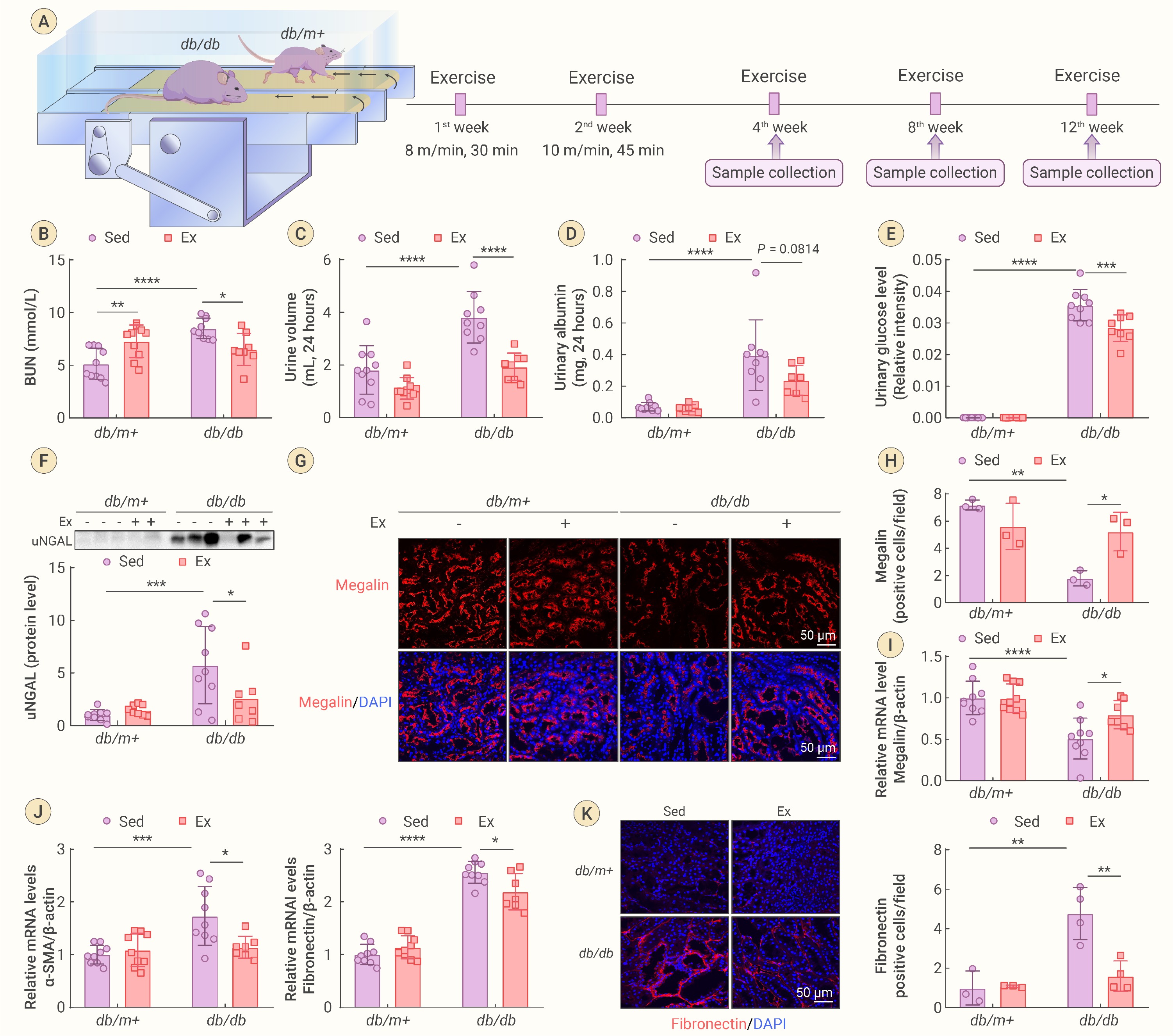
Figure 1.
Regular exercise training improves renal function in db/db mice
-
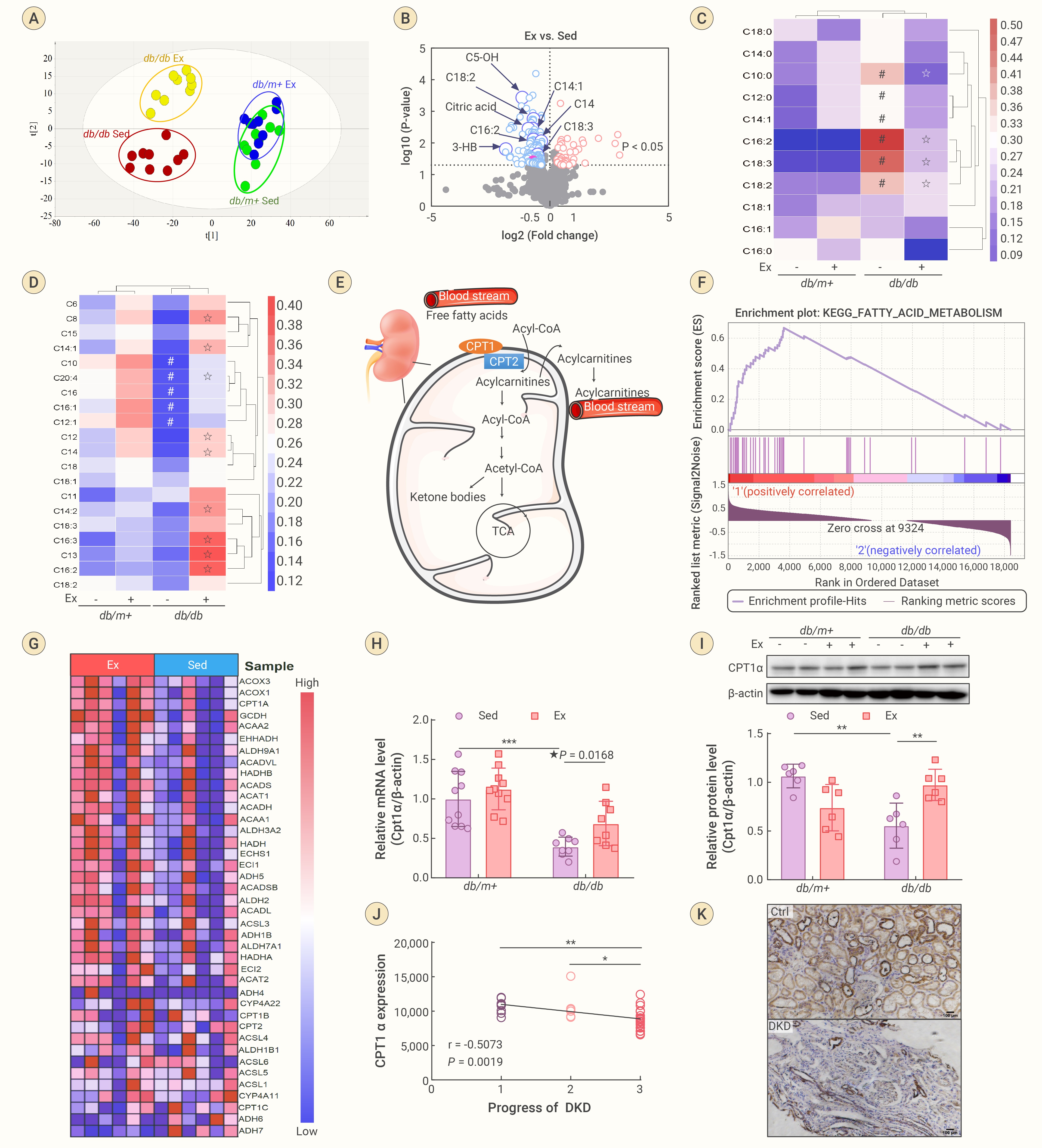
Figure 2.
Running exercise improves FAO efficiency in kidney of db/db mice
-
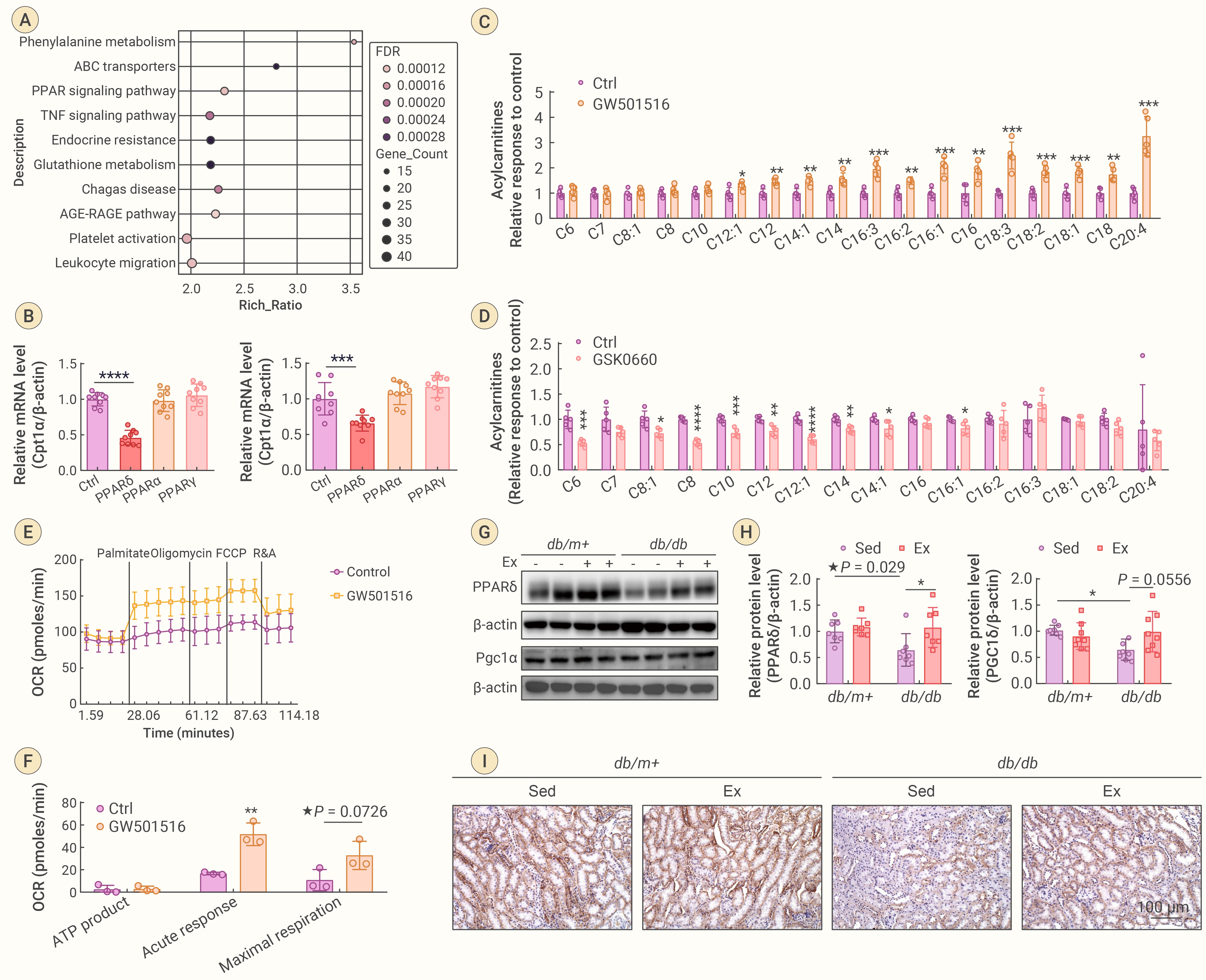
Figure 3.
PPARδ was enhanced by exercise and contribute to FAO efficiency
-
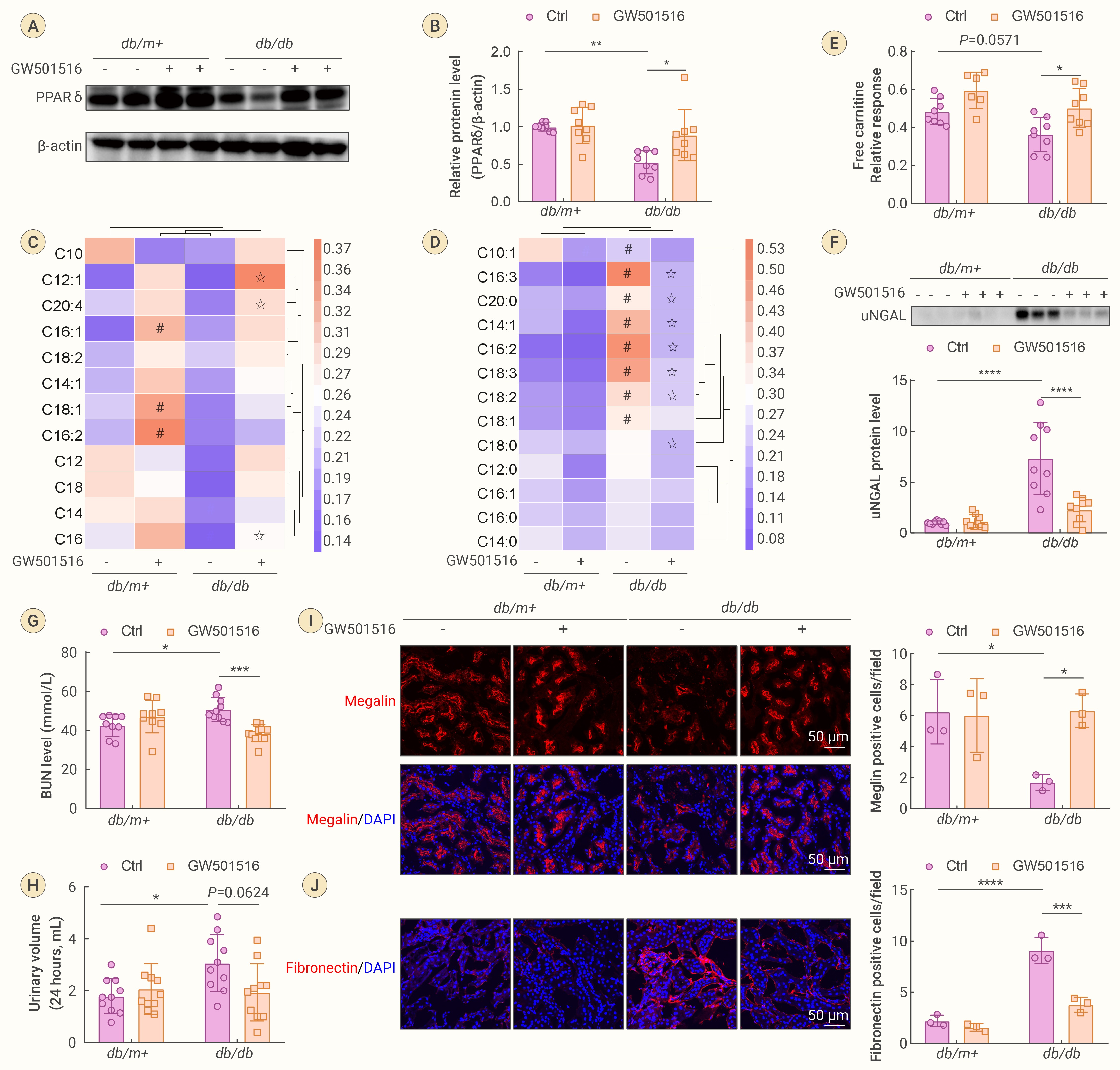
Figure 4.
Activation of PPARδ enhances FAO efficiency and renal function in db/db mice
-
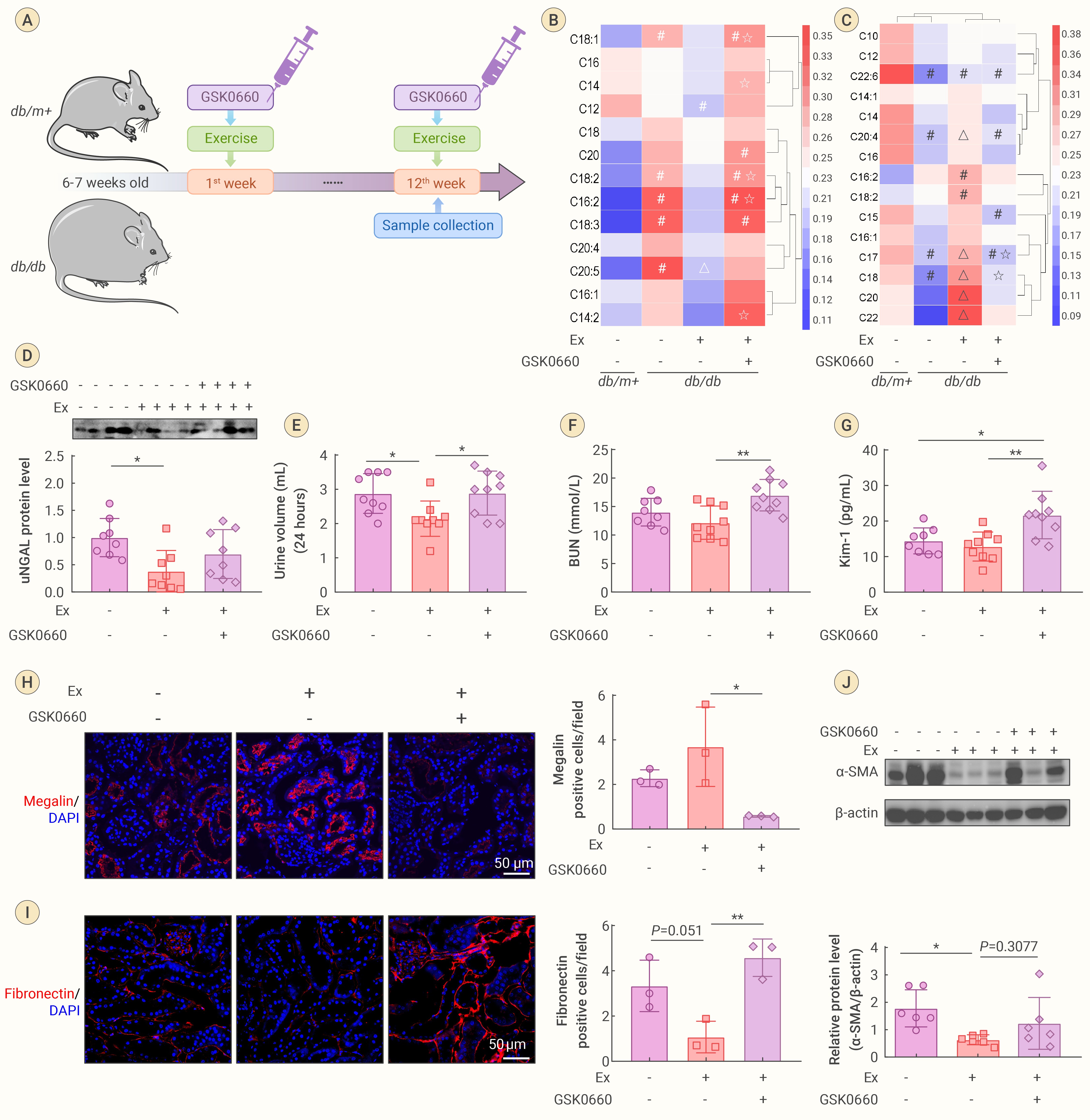
Figure 5.
Inhibition of PPARδ eliminates the beneficial effects of exercise on renal function and FAO efficiency in DKD mice
-
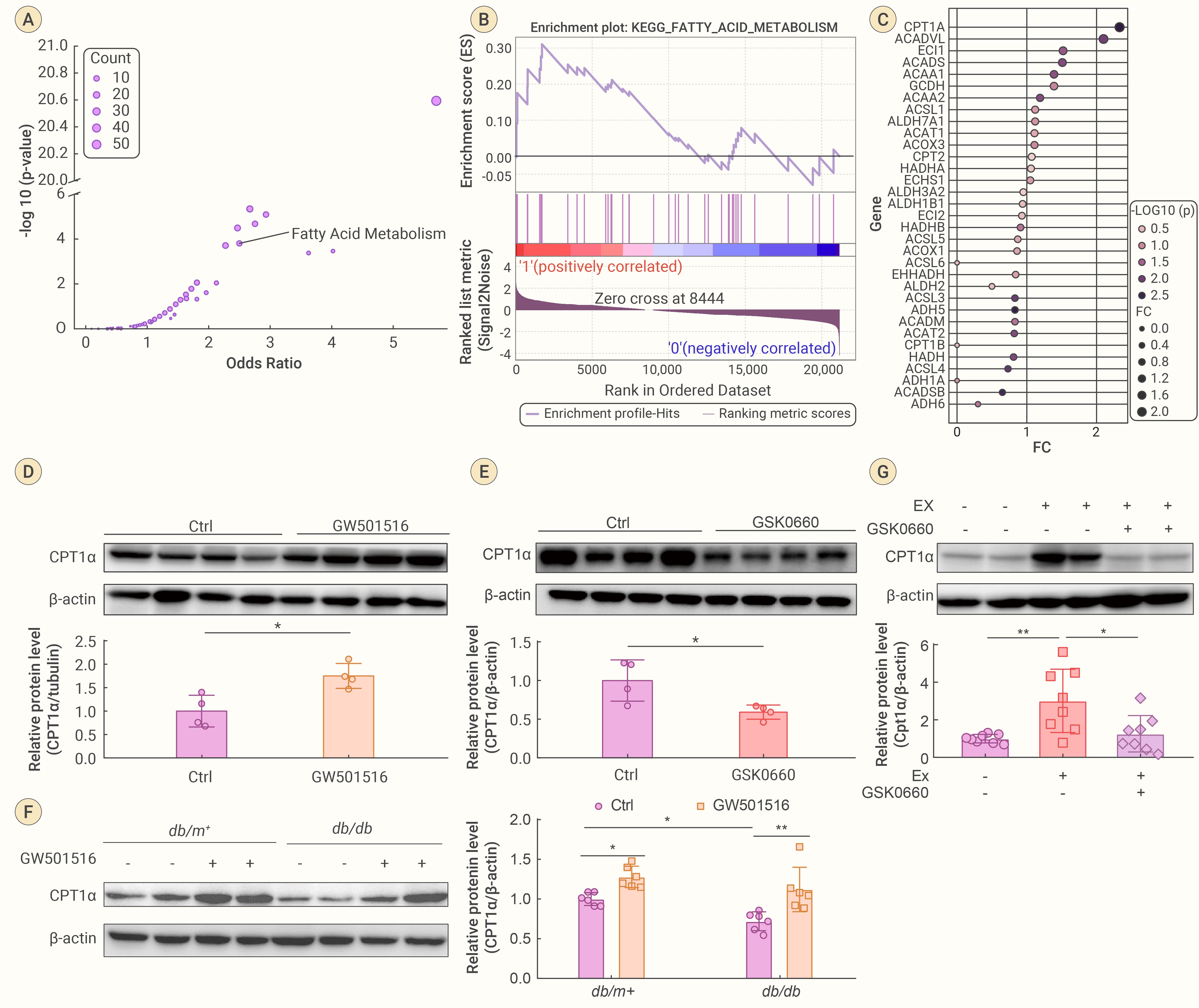
Figure 6.
PPARδ enhances CPT1α expression in kidney cells
