
-
GRAPHICAL ABSTRACT
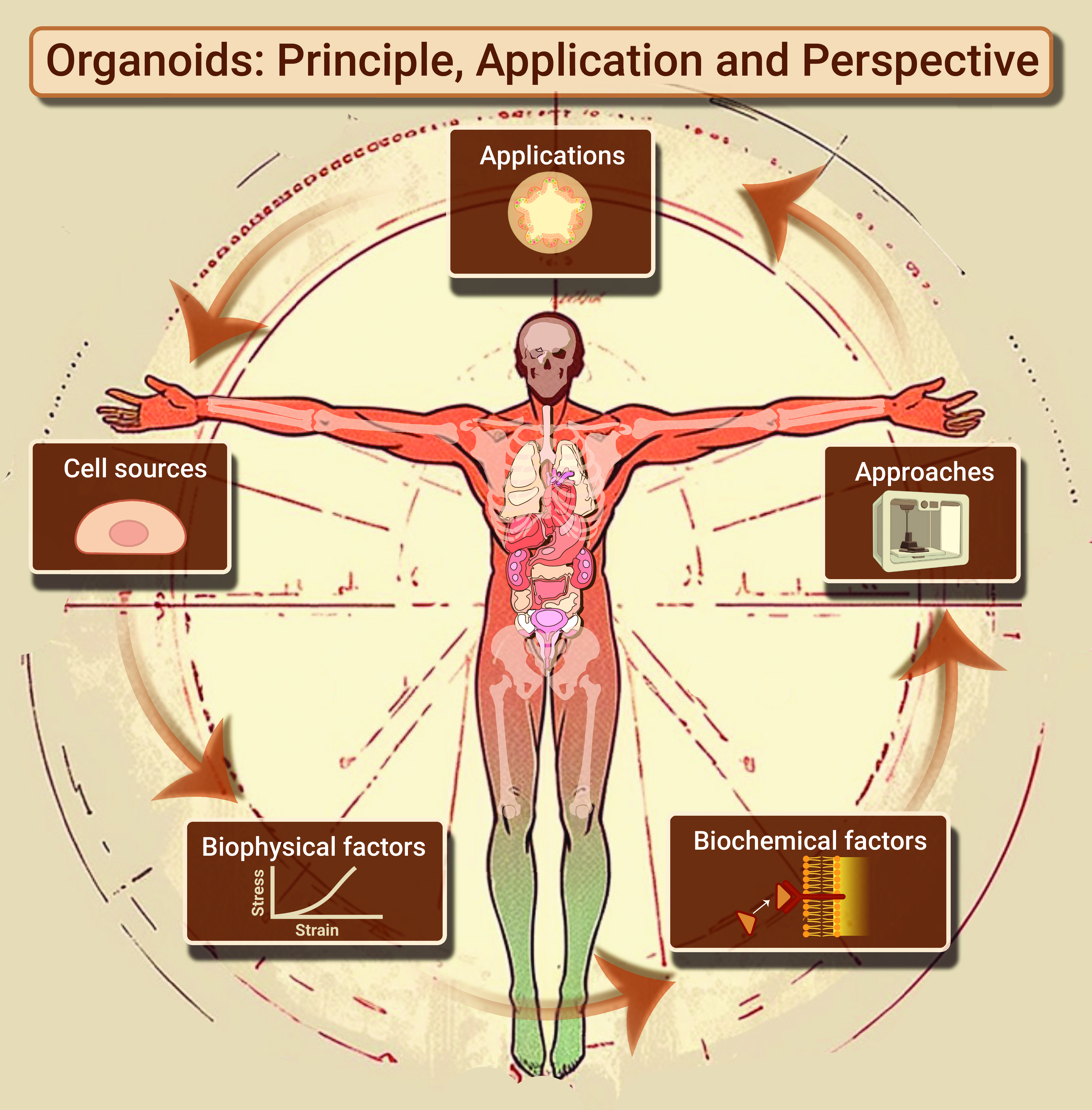
 DownLoad:
Full size image
DownLoad:
Full size image
-
PUBLIC SUMMARY
-
Organoids are in vitro systems that mimic the structures and functions of organs in vivo.
This review summarizes comprehensively the research updates on organoids.
Fundamental principles and organ-specific applications are discussed.
-
-
ABSTRACT
Organoid technology, a notable advancement in biomedical engineering, has emerged over the past decade, offering significant scientific and therapeutic potential. By accurately mimicking the structural and functional intricacies of human organs at a small scale, organoids have become a groundbreaking tool for exploring basic biological principles, understanding disease mechanisms, and progressing regenerative medicine. Despite the large number of relevant reports, a comprehensive summary of current organoid research updates is needed urgently for interdisciplinary researchers with an interest in constructing biomimetic tissue models. This review presents a thorough look at the diverse fields of organoid research, covering the fundamental principles guiding organoid formation, the broad applications in various biological systems, and the encouraging prospects they bring for future scientific and clinical pursuits. -

-
REFERENCES
[1] Rossi, G., Manfrin, A., and Lutolf, M. P. (2018). Progress and potential in organoid research. Nat. Rev. Genet. 19: 671−687. DOI: 10.1038/s41576-018-0051-9. [2] Rheinwatd, J. G. and Green, H. (1975). Seria cultivation of strains of human epidemal keratinocytes: The formation keratinizin colonies from single cells. Cell 6: 331−343. DOI: 10.1016/S0092-8674(75)80001-8. [3] Ambrosi, T. H., Longaker, M. T., and Chan, C. K. F. (2019). A revised perspective of skeletal stem cell biology. Front. Cell Dev. Biol. 7: 189. DOI: 10.3389/fcell.2019.00189. [4] Sato, T., Vries, R. G., Snippert, H. J., et al. (2009). Single Lgr5 stem cells build crypt-villus structures in vitro without a mesenchymal niche. Nature 459: 262−265. DOI: 10.1038/nature07935. [5] Zhao, Z., Chen, X., Dowbaj, A. M., et al. (2022). Organoids. Nat. Rev. Methods Primer 2: 94. DOI: 10.1038/s43586-022-00174-y. [6] Tuveson, D. and Clevers, H. (2019). Cancer modeling meets human organoid technology. Science 364: 952−955. DOI: 10.1126/science.aaw6985. [7] Li, H., Gao, L., Du, J., et al. (2021). To better generate organoids, what can we learn from teratomas. Front. Cell Dev. Biol. 9: 700482. DOI: 10.3389/fcell.2021.700482. [8] Lancaster, M. A. and Knoblich, J. A. (2014). Organogenesis in a dish: Modeling development and disease using organoid technologies. Science 345: 1247125. DOI: 10.1126/science.1247125. [9] Qing, J., Guo, Q., Lv, L., et al. (2023). Organoid culture development for skeletal systems. Tissue Eng. Part B Rev. 29: 545−557. DOI: 10.1089/ten.teb.2023.0022. [10] Saiding, Q., Ma, J., Ke, C., et al. (2022). From “organs on a chip” to “patient on a chip”. The Innovation 3: 100282. DOI: 10.1016/j.xinn.2022.100282. [11] Liu, H., Hu, L., Zhang, D., et al. (2023). Stem cell niches functionalized strategies for organ regeneration and manufacturing. The Innovation Medicine 1: 100037. DOI: 10.59717/j.xinn-med.2023.100037. [12] Hofer, M. and Lutolf, M. P. (2021). Engineering organoids. Nat. Rev. Mater. 6: 402−420. DOI: 10.1038/s41578-021-00279-y. [13] Han, X., Cai, C., Deng, W., et al. (2024). Landscape of human organoids: Ideal model in clinics and research. The Innovation 5 : 100620. DOI: 10.1016/j.xinn.2024.100620. [14] Günther, C., Winner, B., Neurath, M. F., et al. (2022). Organoids in gastrointestinal diseases: from experimental models to clinical translation. Gut 71: 1892−1908. DOI: 10.1136/gutjnl-2021-326560. [15] Serra, D., Mayr, U., Boni, A., et al. (2019). Self-organization and symmetry breaking in intestinal organoid development. Nature 569: 66−72. DOI: 10.1038/s41586-019-1146-y. [16] Lukonin, I., Serra, D., Challet Meylan, L., et al. (2020). Phenotypic landscape of intestinal organoid regeneration. Nature 586: 275−280. DOI: 10.1038/s41586-020-2776-9. [17] Serra, D., Mayr, U., Boni, A., et al. (2019). Self-organization and symmetry breaking in intestinal organoid development. Nature 569: 66−72. DOI: 10.1038/s41586-019-1146-y. [18] Clevers, H. (2013). The intestinal crypt, a prototype stem cell compartment. Cell 154: 274−284. DOI: 10.1016/j.cell.2013.07.004. [19] Taelman, J., Diaz, M., and Guiu, J. (2022). Human intestinal organoids: Promise and challenge. Front. Cell Dev. Biol. 10 : 854740. DOI: 10.3389/fcell.2022.854740. [20] Sato, T., van Es, J. H., Snippert, H. J., et al. (2011). Paneth cells constitute the niche for Lgr5 stem cells in intestinal crypts. Nature 469: 415−418. DOI: 10.1038/nature09637. [21] Snippert, H. J., van der Flier, L.G., Sato, T., et al. (2010). Intestinal crypt homeostasis results from neutral competition between symmetrically dividing Lgr5 stem cells. Cell 143: 134−144. DOI: 10.1016/j.cell.2010.09.016. [22] Li, V. S. W. (2021). Modelling intestinal inflammation and infection using ‘mini-gut’ organoids. Nat. Rev. Gastroenterol. Hepatol. 18: 89−90. DOI: 10.1038/s41575-020-00391-4. [23] Dekkers, J. F., Wiegerinck, C. L., de Jonge, H. R., et al. (2013). A functional CFTR assay using primary cystic fibrosis intestinal organoids. Nat. Med. 19: 939−945. DOI: 10.1038/nm.3201. [24] Cruz, N. M., Song, X., Czerniecki, S. M., et al. (2017). Organoid cystogenesis reveals a critical role of microenvironment in human polycystic kidney disease. Nat. Mater. 16: 1112−1119. DOI: 10.1038/nmat4994. [25] Toshimitsu, K., Takano, A., Fujii, M., et al. (2022). Organoid screening reveals epigenetic vulnerabilities in human colorectal cancer. Nat. Chem. Biol. 18: 605−614. DOI: 10.1038/s41589-022-00984-x. [26] van de Wetering, M., Francies, H. E., Francis, J. M., et al. (2015). Prospective derivation of a living organoid biobank of colorectal cancer patients. Cell 161: 933−945. DOI: 10.1016/j.cell.2015.03.053. [27] Kim, M. B., Hwangbo, S., Jang, S., et al. (2022). Bioengineered co-culture of organoids to recapitulate host-microbe interactions. Mater. Today Bio. 16: 100345. DOI: 10.1016/j.mtbio.2022.100345. [28] Rodansky, E. S., Johnson, L. A., Huang, S., et al. (2015). Intestinal organoids: A model of intestinal fibrosis for evaluating anti-fibrotic drugs. Exp. Mol. Pathol. 98: 346−351. DOI: 10.1016/j.yexmp.2015.03.033. [29] Hu, H., Gehart, H., Artegiani, B., et al. (2018). Long-term expansion of functional mouse and human hepatocytes as 3D organoids. Cell 175: 1591−1606. DOI: 10.1016/j.cell.2018.11.013. [30] Huch, M., Dorrell, C., Boj, S. F., et al. (2013). In vitro expansion of single Lgr5+ liver stem cells induced by Wnt-driven regeneration. Nature 494: 247−250. DOI: 10.1038/nature11826. [31] Huch, M., Gehart, H., van Boxtel, R., et al. (2015). Long-term culture of genome-stable bipotent stem cells from adult human liver. Cell 160: 299−312. DOI: 10.1016/j.cell.2014.11.050. [32] Nunez-Nescolarde, A. B., Nikolic-Paterson, D. J., and Combes, A. N. (2022). Human kidney organoids and tubuloids as models of complex kidney disease. Am. J. Pathol. 192: 738−749. DOI: 10.1016/j.ajpath.2022.01.009. [33] Xu, Y., Kuppe, C., Perales-Patón, J., et al. (2022). Adult human kidney organoids originate from CD24+ cells and represent an advanced model for adult polycystic kidney disease. Nat. Genet. 54: 1690−1701. DOI: 10.1038/s41588-022-01202-z. [34] Yousef Yengej, F. A., Jansen, J., Rookmaaker, M. B., et al. (2020). Kidney organoids and tubuloids. Cells 9: 1326. DOI: 10.3390/cells9061326. [35] Salahudeen, A. A., Choi, S. S., Rustagi, A., et al. (2020). Progenitor identification and SARS-CoV-2 infection in human distal lung organoids. Nature 588: 670−675. DOI: 10.1038/s41586-020-3014-1. [36] Gao, D., Vela, I., Sboner, A., et al. (2014). Organoid cultures derived from patients with advanced prostate cancer. Cell 159: 176−187. DOI: 10.1016/j.cell.2014.08.016. [37] Drost, J., Karthaus, W. R., Gao, D., et al. (2016). Organoid culture systems for prostate epithelial and cancer tissue. Nat. Protoc. 11: 347−358. DOI: 10.1038/nprot.2016.006. [38] Karthaus, W. R., Iaquinta, P. J., Drost, J., et al. (2014). Identification of multipotent luminal progenitor cells in human prostate organoid cultures. Cell 159: 163−175. DOI: 10.1016/j.cell.2014.08.017. [39] McCracken, K. W., Aihara, E., Martin, B., et al. (2017). Wnt/β-catenin promotes gastric fundus specification in mice and humans. Nature 541: 182−187. DOI: 10.1038/nature21021. [40] Barker, N., Huch, M., Kujala, P., et al. (2010). Lgr5+ve stem cells drive self-renewal in the stomach and build long-lived gastric units in vitro. Cell Stem Cell 6: 25−36. DOI: 10.1016/j.stem.2009.11.013. [41] LeSavage, B. L., Suhar, R. A., Broguiere, N., et al. (2022). Next-generation cancer organoids. Nat. Mater. 21: 143−159. DOI: 10.1038/s41563-021-01057-5. [42] Yuki, K., Cheng, N., Nakano, M., et al. (2020). Organoid models of tumor immunology. Trends Immunol. 41: 652−664. DOI: 10.1016/j.it.2020.06.010. [43] Rowe, R. G. and Daley, G. Q. (2019). Induced pluripotent stem cells in disease modelling and drug discovery. Nat. Rev. Genet. 20: 377−388. DOI: 10.1038/s41576-019-0100-z. [44] Karagiannis P., Takahashi K., Saito M., et al. (2019). Induced pluripotent stem cells and their use in human models of disease and development. Physiol. Rev. 99: 79−114. DOI: 10.1152/physrev.00039.2017. [45] Taguchi, A. and Nishinakamura, R. (2017). Higher-order kidney organogenesis from pluripotent stem cells. Cell Stem Cell 21: 730−746. DOI: 10.1016/j.stem.2017.10.011. [46] Taguchi, A., Kaku, Y., Ohmori, T., et al. (2014). Redefining the in vivo origin of metanephric nephron progenitors enables generation of complex kidney structures from pluripotent stem cells. Cell Stem Cell 14: 53−67. DOI: 10.1016/j.stem.2013.11.010. [47] Takasato, M., Er, P. X., Chiu, H. S., et al. (2015). Kidney organoids from human iPS cells contain multiple lineages and model human nephrogenesis. Nature 526: 564−568. DOI: 10.1038/nature15695. [48] Birey, F., Andersen, J., Makinson, C. D., et al. (2017). Assembly of functionally integrated human forebrain spheroids. Nature 545: 54−59. DOI: 10.1038/nature22330. [49] Lancaster, M. A., Renner, M., Martin, C. A., et al. (2013). Cerebral organoids model human brain development and microcephaly. Nature 501: 373−379. DOI: 10.1038/nature12517. [50] Wang, B., Iriguchi, S., Waseda, M., et al. (2021). Generation of hypoimmunogenic T cells from genetically engineered allogeneic human induced pluripotent stem cells. Nat. Biomed. Eng. 5: 429−440. DOI: 10.1038/s41551-021-00730-z. [51] Ludwig, A. L., Mayerl, S. J., Gao, Y., et al. (2023). Re-formation of synaptic connectivity in dissociated human stem cell-derived retinal organoid cultures. Proc. Natl. Acad. Sci. USA. 120: e2213418120. DOI: 10.1073/pnas.2213418120. [52] Wahle, P., Brancati, G., Harmel, C., et al. (2023). Multimodal spatiotemporal phenotyping of human retinal organoid development. Nat. Biotechnol. 41 : 1–11. DOI: 10.1038/s41587-023-01747-2. [53] Li, X., Zhang, L., Tang, F., et al. (2021). Retinal organoids: Cultivation, differentiation, and transplantation. Front. Cell. Neurosci. 15: 638439. DOI: 10.3389/fncel.2021.638439. [54] Takasato, M., Er, P. X., Chiu, H. S., et al. (2016). Generation of kidney organoids from human pluripotent stem cells. Nat. Protoc. 11: 1681−1692. DOI: 10.1038/nprot.2016.098. [55] Tk N., N N., M S., et al. (2015). Generation of stomach tissue from mouse embryonic stem cells. Nat. Cell Biol. 17 : 984-993. DOI: 10.1038/ncb3200. [56] Takasato M., Er P. X., Becroft M., et al. (2014). Directing human embryonic stem cell differentiation towards a renal lineage generates a self-organizing kidney. Nat. Cell Biol. 16: 118−126. DOI: 10.1038/ncb2894. [57] Nakano T., Ando S., Takata N., et al. (2012). Self-formation of optic cups and storable stratified neural retina from human ESCs. Cell Stem Cell 10 : 771–785. DOI: 10.1016/j.stem.2012.05.009. [58] Eiraku, M., Takata, N., Ishibashi, H., et al. (2011). Self-organizing optic-cup morphogenesis in three-dimensional culture. Nature 472: 51−56. DOI: 10.1038/nature09941. [59] Park, S. E., Georgescu, A., and Huh, D. (2019). Organoids-on-a-chip. Science 364: 960−965. DOI: 10.1126/science.aaw7894. [60] Kratochvil, M. J., Seymour, A. J., Li T. L., et al. (2019). Engineered materials for organoid systems. Nat. Rev. Mater. 4: 606−622. DOI: 10.1038/s41578-019-0129-9. [61] Engler, A. J., Sen, S., Sweeney, H. L., et al. (2006). Matrix elasticity directs stem cell lineage specification. Cell 126: 677−89. DOI: 10.1016/j.cell.2006.06.044. [62] Beydag-Tasöz, B. S., Yennek, S., and Grapin-Botton, A. (2023). Towards a better understanding of diabetes mellitus using organoid models. Nat. Rev. Endocrinol. 19 : 232-248. DOI: 10.1038/s41574-022-00797-x. [63] Storm, C., Pastore, J. J., MacKintosh, F. C., et al. (2005). Nonlinear elasticity in biological gels. Nature 435: 191−194. DOI: 10.1038/nature03521. [64] Zhang, Y., Tang, C., Span, P. N., et al. (2020). Polyisocyanide hydrogels as a tunable platform for mammary gland organoid formation. Adv. Sci. 7: 2001797. DOI: 10.1002/advs.202001797. [65] Ye, S., Boeter, J. W. B., Mihajlovic, M., et al. (2020). A chemically defined hydrogel for human liver organoid culture. Adv. Funct. Mater. 30: 2000893. DOI: 10.1002/adfm.202000893. [66] Chaudhuri, O., Cooper-White, J., Janmey, P. A., et al. (2020). Effects of extracellular matrix viscoelasticity on cellular behaviour. Nature 584: 535−546. DOI: 10.1038/s41586-020-2612-2. [67] Elosegui-Artola, A., Gupta, A., Najibi, A. J., et al. (2023). Matrix viscoelasticity controls spatiotemporal tissue organization. Nat. Mater. 22: 117−127. DOI: 10.1038/s41563-022-01400-4. [68] Homan, K. A., Gupta, N., Kroll, K. T., et al. (2019). Flow-enhanced vascularization and maturation of kidney organoids in vitro. Nat. Methods 16: 255−262. DOI: 10.1038/s41592-019-0325-y. [69] Fathollahipour, S., Patil, P. S., and Leipzig, N. D. (2018). Oxygen regulation in development: Lessons from embryogenesis towards tissue engineering. Cells Tissues Organs 205: 350−371. DOI: 10.1159/000493162. [70] Walaas, G. A., Gopalakrishnan S., Bakke I., et al. (2023). Physiological hypoxia improves growth and functional differentiation of human intestinal epithelial organoids. Front. Immunol. 14: 1095812. DOI: 10.3389/fimmu.2023.1095812. [71] Suarez-Martinez, E., Suazo-Sanchez, I., Celis-Romero, M., et al. (2022). 3D and organoid culture in research: Physiology, hereditary genetic diseases and cancer. Cell Biosci. 12: 39. DOI: 10.1186/s13578-022-00775-w. [72] Li, C., Zhang, Y., Du, Y., et al. (2023). A review of advanced biomaterials and cells for the production of bone organoid. Small Sci. 3: 2300027. DOI: 10.1002/smsc.202300027. [73] Ornitz, D. M. (2000). FGFs, heparan sulfate and FGFRs: Complex interactions essential for development. BioEssays 22: 108−112. DOI: 3.0.CO;2-M">10.1002/(SICI)1521-1878(200002)22:2<108::AID-BIES2>3.0.CO;2-M. [74] Goonoo, N. and Bhaw-Luximon, A. (2019). Mimicking growth factors: Role of small molecule scaffold additives in promoting tissue regeneration and repair. RSC Adv. 9: 18124−18146. DOI: 10.1039/C9RA02765C. [75] Janda, C. Y., Dang, L. T., You, C., et al. (2017). Surrogate Wnt agonists that phenocopy canonical Wnt and β-catenin signalling. Nature 545: 234−237. DOI: 10.1038/nature22306. [76] Merenda, A., Fenderico, N., and Maurice, M. M. (2020). Wnt signaling in 3D: Recent advances in the applications of intestinal organoids. Trends Cell Biol. 30: 60−73. DOI: 10.1016/j.tcb.2019.10.003. [77] VanDussen, K. L., Carulli, A. J., Keeley, T. M., et al. (2012). Notch signaling modulates proliferation and differentiation of intestinal crypt base columnar stem cells. Development 139: 488−497. DOI: 10.1242/dev.070763. [78] Dou, Y., Pizarro, T., and Zhou, L. (2022). Organoids as a model system for studying Notch signaling in intestinal epithelial homeostasis and intestinal cancer. Am. J. Pathol. 192: 1347−1357. DOI: 10.1016/j.ajpath.2022.06.008. [79] Qin, H., Zhao, A., and Fu, X. (2017). Small molecules for reprogramming and transdifferentiation. Cell. Mol. Life Sci. 74: 3553−3575. DOI: 10.1007/s00018-017-2586-x. [80] Zhang, Q., Yang, J., Hu, N., et al. (2023). Small-molecule amines: A big role in the regulation of bone homeostasis. BONE Res. 11 : 40. DOI: 10.1038/s41413-023-00262-z. [81] Zhang, Z., O’Laughlin, R., Song, H., et al. (2022). Patterning of brain organoids derived from human pluripotent stem cells. Curr. Opin. Neurobiol. 74: 102536. DOI: 10.1016/j.conb.2022.102536. [82] Tanaka, Y. and Park, I. H. (2021). Regional specification and complementation with non-neuroectodermal cells in human brain organoids. J. Mol. Med. 99: 489−500. DOI: 10.1007/s00109-021-02051-9. [83] Gjorevski, N., Sachs, N., Manfrin, A., et al. (2016). Designer matrices for intestinal stem cell and organoid culture. Nature 539: 560−564. DOI: 10.1038/nature20168. [84] Farin, H. F., Jordens, I., Mosa, M. H., et al. (2016). Visualization of a short-range Wnt gradient in the intestinal stem-cell niche. Nature 530: 340−343. DOI: 10.1038/nature16937. [85] Ng-Blichfeldt, J. P., De Jong, T., Kortekaas, R. K., et al. (2019). TGF-β activation impairs fibroblast ability to support adult lung epithelial progenitor cell organoid formation. Am. J. Physiol.-Lung Cell. Mol. Physiol. 317: L14−28. DOI: 10.1152/ajplung.00400.2018. [86] Chernokal, B., Gonyea, C. R., and Gleghorn, J. P. (2023). Lung development in a dish: Models to interrogate the cellular niche and the role of mechanical forces in Development. Magin, C. M. (ed). Adv. Exp. Med. Biol. (Springer, Cham) 1413 : pp: 29–48. [87] Little, M. H., Hale, L. J., Howden, S. E., et al. (2019). Generating kidney from stem cells. Annu. Rev. Physiol. 81: 335−357. DOI: 10.1146/annurev-physiol-020518-114331. [88] Van Sprang, J. F., De Jong, S. M. J., and Dankers, P. Y. W. (2022). Biomaterial-driven kidney organoid maturation. Curr. Opin. Biomed. Eng. 21: 100355. DOI: 10.1016/j.cobme.2021.100355. [89] Zhu, X., Zhang, B., He, Y., et al. (2021). Liver organoids: Formation strategies and biomedical applications. Tissue Eng. Regen. Med. 18: 573−585. DOI: 10.1007/s13770-021-00357-w. [90] Wu, J. Y., and Vunjak-Novakovic, G. (2022). Bioengineering human cartilage–bone tissues for modeling of osteoarthritis. Stem Cells Dev. 31: 399−405. DOI: 10.1089/scd.2021.0317. [91] Huang, J., Zhang, L., Lu, A., et al. (2023). Organoids as innovative models for bone and joint diseases. Cells 12: 1590. DOI: 10.3390/cells12121590. [92] Hoang, P., Wang, J., Conklin, B. R., et al. (2018). Generation of spatial-patterned early-developing cardiac organoids using human pluripotent stem cells. Nat. Protoc. 13: 723−737. DOI: 10.1038/nprot.2018.006. [93] Kim, H., Kamm, R. D., Vunjak-Novakovic, G., et al. (2022). Progress in multicellular human cardiac organoids for clinical applications. Cell Stem Cell 29: 503−514. DOI: 10.1016/j.stem.2022.03.012. [94] Gjorevski, N. and Lutolf, M. P. (2017). Synthesis and characterization of well-defined hydrogel matrices and their application to intestinal stem cell and organoid culture. Nat. Protoc. 12: 2263−2274. DOI: 10.1038/nprot.2017.095. [95] Poudel, H., Sanford, K., Szwedo, P. K., et al. (2022). Synthetic matrices for intestinal organoid culture: Implications for better performance. ACS Omega 7: 38−47. DOI: 10.1021/acsomega.1c05136. [96] Aisenbrey, E. A. and Murphy, W. L. (2020). Synthetic alternatives to Matrigel. Nat. Rev. Mater. 5: 539−551. DOI: 10.1038/s41578-020-0199-8. [97] Prince, E., Chen, Z., Khuu, N., et al. (2021). Nanofibrillar hydrogel recapitulates changes occurring in the fibrotic extracellular matrix. Biomacromolecules 22: 2352−2362. DOI: 10.1021/acs.biomac.0c01714. [98] Chen, Z., Ezzo, M., Zondag, B., et al. (2024). Intrafibrillar crosslinking enables decoupling of mechanical properties and structure of a composite fibrous hydrogel. Adv. Mater. 36: 2305964. DOI: 10.1002/adma.202305964. [99] Lutolf, M.P., Gilbert, P.M., Blau, H.M. (2009). Designing materials to direct stem-cell fate. Nature 462 : 433–441. DOI: 10.1038/nature08602. [100] Liu, K., Vandaele, J., Yuan, H., et al. (2024). Structure and applications of PIC-based polymers and hydrogels. Cell Rep. Phys. Sci. 5: 101834. DOI: 10.1016/j.xcrp.2024.101834. [101] Zimoch, J., Padial, J. S., Klar, A. S., et al. (2018). Polyisocyanopeptide hydrogels: A novel thermo-responsive hydrogel supporting pre-vascularization and the development of organotypic structures. Acta Biomater. 70: 129−139. DOI: 10.1016/j.actbio.2018.01.042. [102] Schaafsma, P., Kracht, L., Baanstra, M., et al. (2023). Role of immediate early genes in the development of salivary gland organoids in polyisocyanopeptide hydrogels. Front. Mol. Biosci. 10: 1100541. DOI: 10.3389/fmolb.2023.1100541. [103] Chen, Z., Sugimura, R., Zhang, Y. S., et al. (2024). Organoids in concert: engineering in vitro models toward enhanced fidelity. Aggregate 5 : e478. DOI: 10.1002/agt2.478. [104] Chuva De Sousa Lopes, S. M. (2019). Accelerating maturation of kidney organoids. Nat. Mater. 18: 303−304. DOI: 10.1038/s41563-019-0326-3. [105] Zeng, Z., Huang, B., Parvez, R. K., et al. (2021). Generation of patterned kidney organoids that recapitulate the adult kidney collecting duct system from expandable ureteric bud progenitors. Nat. Commun. 12: 3641. DOI: 10.1038/s41467-021-23911-5. [106] Nishinakamura, R. (2019). Human kidney organoids: Progress and remaining challenges. Nat. Rev. Nephrol. 15: 613−624. DOI: 10.1038/s41581-019-0176-x. [107] Chen, Z., Kheiri, S., Young, E. W. K., et al. (2022). Trends in droplet microfluidics: From droplet generation to biomedical applications. Langmuir 38: 6233−6248. DOI: 10.1021/acs.langmuir.2c00491. [108] Li, S. R., Gulieva, R. E., Helms, L., et al. (2022). Glucose absorption drives cystogenesis in a human organoid-on-chip model of polycystic kidney disease. Nat. Commun. 13: 7918. DOI: 10.1038/s41467-022-35537-2. [109] Qu, H., Gao, C., Liu, K., et al. (2024). Gradient matters via filament diameter-adjustable 3D printing. Nat. Commun. 15: 2930. DOI: 10.1038/s41467-024-47360-y. [110] Yang, J., He, H., Li, D., et al. (2023). Advanced strategies in the application of gelatin-based bioink for extrusion bioprinting. Bio-Des. Manuf. 6: 586−608. DOI: 10.1007/s42242-023-00236-4. [111] Li, X., Liu, B., Pei, B., et al. (2020). Inkjet bioprinting of biomaterials. Chem. Rev. 120: 10793−10833. DOI: 10.1021/acs.chemrev.0c00008. [112] Jia, S., Yang, J., Lau, A., et al. (2023). Digital light processing-bioprinted poly-NAGA-GelMA-based hydrogel lenticule for precise refractive errors correction. Biofabrication 15 : 035011. DOI: 10.1088/1758-5090/accaab. [113] Zheng, Z., Lian, L., and Xie, M. (2024). Ultrasound volumetric bioprinting: Opportunities and challenges. The Innovation Life 2: 100053. DOI: 10.59717/j.xinn-life.2024.100053. [114] Gao, C., Tang, L., Qu, H., et al. (2024). A small‐molecule polycationic crosslinker boosts alginate‐based bioinks for extrusion bioprinting. Adv. Funct. Mater. 34: 2310369. DOI: 10.1002/adfm.202310369. [115] Qu, H., Han, Z., Chen, Z., et al. (2021). Fractal design boosts extrusion-based 3D printing of bone-mimicking radial-gradient scaffolds. Research 2021 : 9892689. DOI: 10.34133/2021/9892689. [116] Zhai, X., Ruan, C., Ma,Y., et al. (2018). 3D‐bioprinted osteoblast‐laden nanocomposite hydrogel constructs with induced microenvironments promote cell viability, differentiation, and osteogenesis both in vitro and in vivo. Adv. Sci. 5: 1700550. DOI: 10.1002/advs.201700550. [117] Zhang, Y., Li, D., Liu, Y., et al. (2024). 3D-bioprinted anisotropic bicellular living hydrogels boost osteochondral regeneration via reconstruction of cartilage–bone interface. The Innovation 5: 100542. DOI: 10.1016/j.xinn.2023.100542. [118] Wang, P., Rui, H., Gao, C., et al. (2023). Bioprinting living organs: The next milestone in organ transplantation. The Innovation Life 1: 100019. DOI: 10.59717/j.xinn-life.2023.100019. [119] Yang, J., Chen, Z., Gao, C., et al. (2024). A mechanical-assisted post-bioprinting strategy for challenging bone defects repair. Nat. Commun. 15: 3565. DOI: 10.1038/s41467-024-48023-8. [120] Vijayavenkataraman, S., Yan, W. C., Lu, W. F., et al. (2018). 3D bioprinting of tissues and organs for regenerative medicine. Adv. Drug Deliv. Rev. 132: 296−332. DOI: 10.1016/j.addr.2018.07.004. [121] Daly, A. C., Freeman, F. E., Gonzalez‐Fernandez, T., et al. (2017). 3D bioprinting for cartilage and osteochondral tissue engineering. Adv. Healthc. Mater. 6: 1700298. DOI: 10.1002/adhm.201700298. [122] Chakraborty, J., Chawla, S., and Ghosh, S. (2022). Developmental biology-inspired tissue engineering by combining organoids and 3D bioprinting. Curr. Opin. Biotechnol. 78: 102832. DOI: 10.1016/j.copbio.2022.102832. [123] Zhang, Y., Li, G., Wang, J., et al. (2023). Small joint organoids 3D bioprinting: Construction strategy and application. Small 20 : e2302506. DOI: 10.1002/smll.202302506. [124] Juraski, A. C., Sharma, S., Sparanese, S., et al. (2023). 3D bioprinting for organ and organoid models and disease modeling. Expert Opin. Drug Discov. 18: 1043−1059. DOI: 10.1080/17460441.2023.2234280. [125] Babaeeghazvini, P., Rueda-Delgado, L. M., Gooijers, J., et al. (2021). Brain structural and functional connectivity: A review of combined works of diffusion magnetic resonance imaging and electro-encephalography. Front. Hum. Neurosci. 15 : 721206. DOI: 10.3389/fnhum.2021.721206. [126] Mahumane, G. D., Kumar, P., du Toit, L. C., et al. (2018). 3D scaffolds for brain tissue regeneration: architectural challenges. Biomater. Sci. 6: 2812−2837. DOI: 10.1039/C8BM00422F. [127] Lebel, C. and Deoni, S. (2018). The development of brain white matter microstructure. Microstruct. Imaging 182: 207−218. DOI: 10.1016/j.neuroimage.2017.12.097. [128] Timmler, S. and Simons, M. (2019). Grey matter myelination. Glia 67: 2063−2070. DOI: 10.1002/glia.23614. [129] Yao, Z., van Velthoven, C. T. J., Kunst, M., et al. (2023). A high-resolution transcriptomic and spatial atlas of cell types in the whole mouse brain. Nature 624: 317−332. DOI: 10.1038/s41586-023-06812-z. [130] Garreta, E., Kamm, R. D., Chuva De Sousa Lopes, S. M., et al. (2021). Rethinking organoid technology through bioengineering. Nat. Mater. 20: 145−155. DOI: 10.1038/s41563-020-00804-4. [131] Chiaradia, I. and Lancaster, M. A. (2020). Brain organoids for the study of human neurobiology at the interface of in vitro and in vivo. Nat. Neurosci. 23: 1496−1508. DOI: 10.1038/s41593-020-00730-3. [132] Turner, D. A., Girgin, M., Alonso-Crisostomo, L., et al. (2017). Anteroposterior polarity and elongation in the absence of extraembryonic tissues and spatially localised signalling in Gastruloids, mammalian embryonic organoids. Development 144 : 3894-3906. DOI: 10.1242/dev.150391. [133] Qian, X., Nguyen, H. N., Song, M. M., et al. (2016). Brain-region-specific organoids using mini-bioreactors for modeling ZIKV exposure. Cell 165: 1238−1254. DOI: 10.1016/j.cell.2016.04.032. [134] Krefft O., Jabali A., Iefremova V., et al. (2018). Generation of Standardized and Reproducible Forebrain-type Cerebral Organoids from Human Induced Pluripotent Stem Cells. J. Vis. Exp. 131 : 56768. DOI: 10.3791/56768. [135] Heide, M., Huttner, W. B., and Mora-Bermúdez, F. (2018). Brain organoids as models to study human neocortex development and evolution. Curr. Opin. Cell Biol. 55: 8−16. DOI: 10.1016/j.ceb.2018.06.006. [136] Ciarpella, F., Zamfir, R. G., Campanelli, A., et al. (2023). Generation of mouse hippocampal brain organoids from primary embryonic neural stem cells. STAR Protoc. 4: 102413. DOI: 10.1016/j.xpro.2023.102413. [137] Heydari, Z., Moeinvaziri, F., Agarwal, T., et al. (2021). Organoids: A novel modality in disease modeling. Bio-Des. Manuf. 4: 689−716. DOI: 10.1007/s42242-021-00150-7. [138] Agarwal, T., Celikkin, N., Costantini, M., et al. (2021). Recent advances in chemically defined and tunable hydrogel platforms for organoid culture. Bio-Des. Manuf. 4: 641−674. DOI: 10.1007/s42242-021-00126-7. [139] Han, S., Okawa, S., Wilkinson, G. A., et al. (2021). Proneural genes define ground-state rules to regulate neurogenic patterning and cortical folding. Neuron 109: 2847−2863. DOI: 10.1016/j.neuron.2021.07.007. [140] Szebényi, K., Wenger, L. M. D., Sun, Y., et al. (2021). Human ALS/FTD brain organoid slice cultures display distinct early astrocyte and targetable neuronal pathology. Nat. Neurosci. 24: 1542−1554. DOI: 10.1038/s41593-021-00923-4. [141] Zhang, K., Chen, S., Yang, Q., et al. (2022). The Oligodendrocyte Transcription Factor 2 OLIG2 regulates transcriptional repression during myelinogenesis in rodents. Nat. Commun. 13: 1423. DOI: 10.1038/s41467-022-29068-z. [142] Cakir, B., Tanaka, Y., Kiral, F. R., et al. (2022). Expression of the transcription factor PU.1 induces the generation of microglia-like cells in human cortical organoids. Nat. Commun. 13 : 430. DOI: 10.1038/s41467-022-28043-y. [143] Schafer, S. T., Mansour, A. A., Schlachetzki, J. C. M., et al. (2023). An in vivo neuroimmune organoid model to study human microglia phenotypes. Cell 186: 2111−2126. DOI: 10.1016/j.cell.2023.04.022. [144] Nimtz, L., Hartmann, J., Tigges, J., et al. (2020). Characterization and application of electrically active neuronal networks established from human induced pluripotent stem cell-derived neural progenitor cells for neurotoxicity evaluation. Stem Cell Res. 45: 101761. DOI: 10.1016/j.scr.2020.101761. [145] Qian, X., Su, Y., Adam, C. D., et al. (2020). Sliced human cortical organoids for modeling distinct cortical layer formation. Cell Stem Cell 26: 766−781. DOI: 10.1016/j.stem.2020.02.002. [146] Albanese, A., Swaney, J. M., Yun, D. H., et al. (2020). Multiscale 3D phenotyping of human cerebral organoids. Sci. Rep. 10: 21487. DOI: 10.1038/s41598-020-78130-7. [147] Völkner, M., Zschätzsch, M., Rostovskaya, M., et al. (2016). Retinal organoids from pluripotent stem cells efficiently recapitulate retinogenesis. Stem Cell Rep. 6: 525−538. DOI: 10.1016/j.stemcr.2016.03.001. [148] Li, G., Liu, S., Chen, W., et al. (2022). Acellularized uvea hydrogel as novel injectable platform for cell‐based delivering treatment of retinal degeneration and optimizing retinal organoids inducible system. Adv. Healthc. Mater. 11: 2202114. DOI: 10.1002/adhm.202202114. [149] Andersen, J., Revah, O., Miura, Y., et al. (2020). Generation of functional human 3D cortico-motor assembloids. Cell 183: 1913−1929. DOI: 10.1016/j.cell.2020.11.017. [150] Miura, Y., Li, M. Y., Birey, F., et al. (2020). Generation of human striatal organoids and cortico-striatal assembloids from human pluripotent stem cells. Nat. Biotechnol. 38: 1421−1430. DOI: 10.1038/s41587-020-00763-w. [151] Bauersachs, H. G., Bengtson, C. P., Weiss, U., et al. (2022). N-methyl-d-aspartate receptor-mediated preconditioning mitigates excitotoxicity in human induced pluripotent stem cell-derived brain organoids. Neuroscience 484: 83−97. DOI: 10.1016/j.neuroscience.2021.12.026. [152] O’Brien, B. S., Mokry, R. L., Schumacher, M. L., et al. (2022). Downregulation of neurodevelopmental gene expression in iPSC-derived cerebral organoids upon infection by human cytomegalovirus. iScience 25: 104098. DOI: 10.1016/j.isci.2022.104098. [153] Park, J. C., Jang, S. Y., Lee, D., et al. (2021). A logical network-based drug-screening platform for Alzheimer’s disease representing pathological features of human brain organoids. Nat. Commun. 12: 280. DOI: 10.1038/s41467-020-20440-5. [154] Lee, S. E., Choi, H., Shin, N., et al. (2022). Zika virus infection accelerates Alzheimer’s disease phenotypes in brain organoids. Cell Death Discov. 8: 153. DOI: 10.1038/s41420-022-00958-x. [155] Smits, L. M., Reinhardt, L., Reinhardt, P., et al. (2019). Modeling Parkinson’s disease in midbrain-like organoids. NPJ Parkinsons Dis. 5: 5. DOI: 10.1038/s41531-019-0078-4. [156] Patikas, N., Ansari, R., and Metzakopian, E. (2023). Single-cell transcriptomics identifies perturbed molecular pathways in midbrain organoids using α-synuclein triplication Parkinson’s disease patient-derived iPSCs. Neurosci. Res. 195: 13−28. DOI: 10.1016/j.neures.2023.06.001. [157] Li, C., Fleck, J. S., Martins-Costa, C., et al. (2023). Single-cell brain organoid screening identifies developmental defects in autism. Nature 621: 373−380. DOI: 10.1038/s41586-023-06473-y. [158] Quadrato, G. and Birtele, M. (2023). Using 3D human brain organoids to perform longitudinal modeling and functional characterization of a top risk gene for autism spectrum disorder. Biol. Psychiatry 93: S56−S57. DOI: 10.1016/j.biopsych.2023.02.155. [159] Wang, Y., Wang, L., Zhu, Y., et al. (2018). Human brain organoid-on-a-chip to model prenatal nicotine exposure. Lab. Chip 18: 851−860. DOI: 10.1039/C7LC01084B. [160] Shaji, M., Tamada, A., Fujimoto, K., et al. (2024). Deciphering potential vascularization factors of on-chip co-cultured hiPSC-derived cerebral organoids. Lab. Chip 24: 680−696. DOI: 10.1039/D3LC00930K. [161] Aazmi, A., Zhou, H., Lv, W., et al. (2022). Vascularizing the brain in vitro. iScience 25 : 104110. DOI: 10.1016/j.isci.2022.104110. [162] Smirnova, L., Caffo, B. S., Gracias, D. H., et al. (2023). Organoid intelligence (OI): The new frontier in biocomputing and intelligence-in-a-dish. Front. Sci. 1: 1017235. DOI: 10.3389/fsci.2023.1017235. [163] Huang, Q., Tang, B., Romero, J. C., et al. (2022). Shell microelectrode arrays (MEAs) for brain organoids. Sci. Adv. 8: eabq5031. DOI: 10.1126/sciadv.abq5031. [164] Park, Y., Franz, C. K., Ryu, H., et al. (2021). Three-dimensional, multifunctional neural interfaces for cortical spheroids and engineered assembloids. Sci. Adv. 7: eabf9153. DOI: 10.1126/sciadv.abf9153. [165] Johnson, L. R. (2006). Physiology of the gastrointestinal tract. (Elsevier Academic Press). [166] Nightingale, J. M. D. and Spiller, R. (2023). Normal intestinal anatomy and physiology. Nightingale, J. M. D. (ed). Intest. Fail. (Springer International Publishing), pp: 13–33. DOI: 10.1007/978-3-031-22265-8_2. [167] Rahmani, S., Breyner, N. M., Su, H. M., et al. (2019). Intestinal organoids: A new paradigm for engineering intestinal epithelium in vitro. Biomaterials 194: 195−214. DOI: 10.1016/j.biomaterials.2018.12.006. [168] Adeniyi-Ipadeola, G., Nwanosike, H., and Ramani, S. (2023). Human intestinal organoids as models to study enteric bacteria and viruses. Curr. Opin. Microbiol. 75: 102362. DOI: 10.1016/j.mib.2023.102362. [169] Lechuga, S., Braga-Neto, M. B., Naydenov, N. G., et al. (2023). Understanding disruption of the gut barrier during inflammation: Should we abandon traditional epithelial cell lines and switch to intestinal organoids. Front. Immunol. 14: 1108289. DOI: 10.3389/fimmu.2023.1108289. [170] Miedzybrodzka, E. L., Foreman, R. E., Lu, V. B., et al. (2021). Stimulation of motilin secretion by bile, free fatty acids, and acidification in human duodenal organoids. Mol. Metab. 54: 101356. DOI: 10.1016/j.molmet.2021.101356. [171] Meran, L., Massie, I., Campinoti, S., et al. (2020). Engineering transplantable jejunal mucosal grafts using patient-derived organoids from children with intestinal failure. Nat. Med. 26: 1593−1601. DOI: 10.1038/s41591-020-1024-z. [172] Niklinska-Schirtz, B. J., Venkateswaran, S., Anbazhagan, M., et al. (2021). Ileal derived organoids from Crohn’s disease patients show unique transcriptomic and secretomic signatures. Cell. Mol. Gastroenterol. Hepatol. 12: 1267−1280. DOI: 10.1016/j.jcmgh.2021.06.018. [173] Liang, J., Li, X., Dong, Y., et al. (2022). Modeling human organ development and diseases with fetal tissue–derived organoids. Cell Transplant. 31 : 096368972211244. DOI: 10.1177/09636897221124481. [174] Sato, T., Stange, D. E., Ferrante, M., et al. (2011). Long-term expansion of epithelial organoids from human colon, adenoma, adenocarcinoma, and barrett’s epithelium. Gastroenterology 141: 1762−1772. DOI: 10.1053/j.gastro.2011.07.050. [175] Matano, M., Date, S., Shimokawa, M., et al. (2015). Modeling colorectal cancer using CRISPR-Cas9–mediated engineering of human intestinal organoids. Nat. Med. 21: 256−262. DOI: 10.1038/nm.3802. [176] Kraiczy, J., Nayak, K. M., Howell, K. J., et al. (2019). DNA methylation defines regional identity of human intestinal epithelial organoids and undergoes dynamic changes during development. Gut 68: 49−61. DOI: 10.1136/gutjnl-2017-314817. [177] Lucafò, M., Muzzo, A., Marcuzzi, M., et al. (2022). Patient-derived organoids for therapy personalization in inflammatory bowel diseases. World J. Gastroenterol. 28: 2636−2653. DOI: 10.3748/wjg.v28.i24.2636. [178] Schreurs, R. R. C. E., Baumdick, M. E., Drewniak, A., et al. (2021). In vitro co-culture of human intestinal organoids and lamina propria-derived CD4+ T cells. STAR Protoc. 2: 100519. DOI: 10.1016/j.xpro.2021.100519. [179] Wu, N., Sun, H., Zhao, X., et al. (2021). MAP3K2-regulated intestinal stromal cells define a distinct stem cell niche. Nature 592: 606−610. DOI: 10.1038/s41586-021-03283-y. [180] Gao, Y., Bi, D., Xie, R., et al. (2021). Fusobacterium nucleatum enhances the efficacy of PD-L1 blockade in colorectal cancer. Signal Transduct. Target. Ther. 6: 398. DOI: 10.1038/s41392-021-00795-x. [181] Puschhof, J., Pleguezuelos-Manzano, C., Martinez-Silgado, A., et al. (2021). Intestinal organoid cocultures with microbes. Nat. Protoc. 16: 4633−4649. DOI: 10.1038/s41596-021-00589-z. [182] Elmentaite, R., Ross, A. D. B., Roberts, K., et al. (2020). Single-cell sequencing of developing human gut reveals transcriptional links to childhood Crohn’s disease. Dev. Cell 55: 771−783. DOI: 10.1016/j.devcel.2020.11.010. [183] Aknouch, I., García-Rodríguez, I., Giugliano, F. P., et al. (2023). Amino acid variation at VP1-145 of enterovirus A71 determines the viral infectivity and receptor usage in a primary human intestinal model. Front. Microbiol. 14: 1045587. DOI: 10.3389/fmicb.2023.1045587. [184] Spence, J. R., Mayhew, C. N., Rankin, S. A., et al. (2011). Directed differentiation of human pluripotent stem cells into intestinal tissue in vitro. Nature 470: 105−109. DOI: 10.1038/nature09691. [185] Bouffi, C., Wikenheiser-Brokamp, K. A., Chaturvedi, P., et al. (2023). In vivo development of immune tissue in human intestinal organoids transplanted into humanized mice. Nat. Biotechnol. 41: 824−831. DOI: 10.1038/s41587-022-01558-x. [186] Krüger, J., Groß, R., Conzelmann, C., et al. (2021). Drug inhibition of SARS-CoV-2 Replication in human pluripotent stem cell–derived intestinal organoids. Cell. Mol. Gastroenterol. Hepatol. 11: 935−948. DOI: 10.1016/j.jcmgh.2020.11.003. [187] Wilson, S. S., Mayo, M., Melim, T., et al. (2021). Optimized culture conditions for improved growth and functional differentiation of mouse and human colon organoids. Front. Immunol. 11: 547102. DOI: 10.3389/fimmu.2020.547102. [188] Fordham, R. P., Yui, S., Hannan, N. R. F., et al. (2013). Transplantation of expanded fetal intestinal progenitors contributes to colon regeneration after injury. Cell Stem Cell 13: 734−744. DOI: 10.1016/j.stem.2013.09.015. [189] Múnera, J. O., Sundaram, N., Rankin, S. A., et al. (2017). Differentiation of human pluripotent stem cells into colonic organoids via transient activation of BMP signaling. Cell Stem Cell 21: 51−64. DOI: 10.1016/j.stem.2017.05.020. [190] Han, X., Lee, A., Huang, S., et al. (2019). Lactobacillus rhamnosus GG prevents epithelial barrier dysfunction induced by interferon-gamma and fecal supernatants from irritable bowel syndrome patients in human intestinal enteroids and colonoids. Gut Microbes 10: 59−76. DOI: 10.1080/19490976.2018.1479625. [191] Heo, I., Dutta, D., Schaefer, D. A., et al. (2018). Modelling Cryptosporidium infection in human small intestinal and lung organoids. Nat. Microbiol. 3: 814−823. DOI: 10.1038/s41564-018-0177-8. [192] Van Kampen, J. J. A., Dalm, V. A. S. H., Fraaij, P. L. A., et al. (2022). Clinical and in vitro evidence favoring immunoglobulin treatment of a chronic norovirus infection in a patient with common variable immunodeficiency. J. Infect. Dis. 226: 1781−1789. DOI: 10.1093/infdis/jiac085. [193] Cieza, R. J., Golob, J. L., Colacino, J. A., et al. (2021). Comparative analysis of public RNA-sequencing data from human intestinal enteroid (HIEs) Infected with enteric RNA viruses identifies universal and virus-specific epithelial responses. Viruses 13: 1059. DOI: 10.3390/v13061059. [194] Moshiri, J., Craven, A. R., Mixon, S. B., et al. (2023). Mechanosensitive extrusion of Enterovirus A71-infected cells from colonic organoids. Nat. Microbiol. 8: 629−639. DOI: 10.1038/s41564-023-01339-5. [195] Mykytyn, A. Z., Breugem, T. I., Geurts, M. H., et al. (2023). SARS-CoV-2 Omicron entry is type II transmembrane serine protease-mediated in human airway and intestinal organoid models. J. Virol. 97: e00851−23. DOI: 10.1128/jvi.00851-23. [196] Cloherty, A. P. M., Rader, A. G., Patel, K. S., et al. (2023). Berbamine suppresses intestinal SARS-CoV-2 infection via a BNIP3-dependent autophagy blockade. Emerg. Microbes Infect. 12: 2195020. DOI: 10.1080/22221751.2023.2195020. [197] Zang, R., Castro, M. F. G., McCune, B. T., et al. (2020). TMPRSS2 and TMPRSS4 promote SARS-CoV-2 infection of human small intestinal enterocytes. Sci. Immunol. 5: eabc3582. DOI: 10.1126/sciimmunol.abc3582. [198] Masmoudi, F., Santos-Ferreira, N., Pajkrt, D., et al. (2023). Evaluation of 3D human intestinal organoids as a platform for EV-A71 antiviral drug discovery. Cells 12: 1138. DOI: 10.3390/cells12081138. [199] Kourula, S., Derksen, M., Jardi, F., et al. (2023). Intestinal organoids as an in vitro platform to characterize disposition, metabolism, and safety profile of small molecules. Eur. J. Pharm. Sci. 188: 106481. DOI: 10.1016/j.ejps.2023.106481. [200] Berkers, G., Van Mourik, P., Vonk, A. M., et al. (2019). Rectal organoids enable personalized treatment of cystic fibrosis. Cell Rep. 26: 1701−1708. DOI: 10.1016/j.celrep.2019.01.068. [201] Chaudhry, R. and Bordoni, B. (2023). Anatomy, thorax, lungs. StatPearls (StatPearls Publishing). [202] Amador, C., Weber, C., and Varacallo, M. (2023). Anatomy, thorax, bronchial. StatPearls (StatPearls Publishing). [203] Liao, D. and Li, H. (2020). Dissecting the niche for alveolar type II cells with alveolar organoids. Front. Cell Dev. Biol. 8 : 419. DOI: 10.3389/fcell.2020.00419. [204] Hiemstra, P. S., Tetley, T. D., and Janes, S. M. (2019). Airway and alveolar epithelial cells in culture. Eur. Respir. J. 54 : 1900742. DOI: 10.1183/13993003.00742-2019. [205] Kong, J., Wen, S., Cao, W., et al. (2021). Lung organoids, useful tools for investigating epithelial repair after lung injury. Stem Cell Res. Ther. 12: 95. DOI: 10.1186/s13287-021-02172-5. [206] Kühl, L., Graichen, P., Von Daacke, N., et al. (2023). Human lung organoids—a novel experimental and precision medicine approach. Cells 12: 2067. DOI: 10.3390/cells12162067. [207] Bosáková, V., De Zuani, M., Sládková, L., et al. (2022). Lung organoids—the ultimate tool to dissect pulmonary diseases. Front. Cell Dev. Biol. 10: 899368. DOI: 10.3389/fcell.2022.899368. [208] Ebisudani, T., Hamamoto, J., Togasaki, K., et al. (2023). Genotype-phenotype mapping of a patient-derived lung cancer organoid biobank identifies NKX2-1-defined Wnt dependency in lung adenocarcinoma. Cell Rep. 42: 112212. DOI: 10.1016/j.celrep.2023.112212. [209] Lee, J., Baek, H., Jang, J., et al. (2023). Establishment of a human induced pluripotent stem cell derived alveolar organoid for toxicity assessment. Toxicol. In Vitro 89: 105585. DOI: 10.1016/j.tiv.2023.105585. [210] Barkauskas, C. E., Cronce, M. J., Rackley, C. R., et al. (2013). Type 2 alveolar cells are stem cells in adult lung. J. Clin. Invest. 123: 3025−3036. DOI: 10.1172/JCI68782. [211] Hoareau, L., Engelsen, A. S. T., Aanerud, M., et al. (2021). Induction of alveolar and bronchiolar phenotypes in human lung organoids. Physiol. Rep. 9 : 1900742. DOI: 10.14814/phy2.14857. [212] Hashimoto, R., Tamura, T., Watanabe, Y., et al. (2023). Evaluation of broad anti-coronavirus activity of autophagy-related compounds using human airway organoids. Mol. Pharm. 20: 2276−2287. DOI: 10.1021/acs.molpharmaceut.3c00114. [213] Hynds, R. E., Butler, C. R., Janes, S. M., et al. (2016). Expansion of human airway basal stem cells and their differentiation as 3D tracheospheres. Turksen, K. (ed). Organoids (Springer New York) 1576 , pp: 43–53. DOI: 10.1007/7651_2016_5. [214] Kishimoto, K. and Morimoto, M. (2021). Mammalian tracheal development and reconstruction: insights from in vivo and in vitro studies. Development 148: dev198192. DOI: 10.1242/dev.198192. [215] Sano, E., Suzuki, T., Hashimoto, R., et al. (2022). Cell response analysis in SARS-CoV-2 infected bronchial organoids. Commun. Biol. 5: 516. DOI: 10.1038/s42003-022-03499-2. [216] Demchenko, A., Kondrateva, E., Tabakov, V., et al. (2023). Airway and lung organoids from human-induced pluripotent stem cells can be used to assess CFTR conductance. Int. J. Mol. Sci. 24: 6293. DOI: 10.3390/ijms24076293. [217] Danahay, H., Pessotti, A. D., Coote, J., et al. (2015). Notch2 is required for inflammatory cytokine-driven goblet cell metaplasia in the lung. Cell Rep. 10: 239−252. DOI: 10.1016/j.celrep.2014.12.017. [218] Boecking, C. A., Walentek, P., Zlock, L. T., et al. (2022). A simple method to generate human airway epithelial organoids with externally orientated apical membranes. Am. J. Physiol.-Lung Cell. Mol. Physiol. 322: L420−L437. DOI: 10.1152/ajplung.00536.2020. [219] Dye, B. R., Hill, D. R., Ferguson, M. A., et al. (2015). In vitro generation of human pluripotent stem cell derived lung organoids. eLife 2015: 1−25. DOI: 10.7554/eLife.05098. [220] Kunisaki, S. M., Jiang, G., Biancotti, J. C., et al. (2021). Human induced pluripotent stem cell-derived lung organoids in an ex vivo model of the congenital diaphragmatic hernia fetal lung. Stem Cells Transl. Med. 10: 98−114. DOI: 10.1002/sctm.20-0199. [221] Chen, Y. W., Huang, S. X., De Carvalho, A. L. R. T., et al. (2017). A three-dimensional model of human lung development and disease from pluripotent stem cells. Nat. Cell Biol. 19: 542−549. DOI: 10.1038/ncb3510. [222] Youk, J., Kim, T., Evans, K. V., et al. (2020). Three-dimensional human alveolar stem cell culture models reveal infection response to SARS-CoV-2. Cell Stem Cell 27: 905−919. DOI: 10.1016/j.stem.2020.10.004. [223] Tamai, K., Sakai, K., Yamaki, H., et al. (2022). iPSC-derived mesenchymal cells that support alveolar organoid development. Cell Rep. Methods 2: 100314. DOI: 10.1016/j.crmeth.2022.100314. [224] Lamers, M. M., Van Der Vaart, J., Knoops, K., et al. (2021). An organoid‐derived bronchioalveolar model for SARS‐CoV‐2 infection of human alveolar type II‐like cells. EMBO J. 40: e105912. DOI: 10.15252/embj.2020105912. [225] Ekanger, C. T., Zhou, F., Bohan, D., et al. (2022). Human organotypic airway and lung organoid cells of bronchiolar and alveolar differentiation are permissive to infection by influenza and SARS-CoV-2 respiratory virus. Front. Cell. Infect. Microbiol. 12: 841447. DOI: 10.3389/fcimb.2022.841447. [226] Peng, L., Gao, L., Wu, X., et al. (2022). Lung organoids as model to study SARS-CoV-2 infection. Cells 11: 2758. DOI: 10.3390/cells11172758. [227] Heo, H. R. and Hong, S. H. (2021). Generation of macrophage containing alveolar organoids derived from human pluripotent stem cells for pulmonary fibrosis modeling and drug efficacy testing. Cell Biosci. 11: 216. DOI: 10.1186/s13578-021-00721-2. [228] Vazquez-Armendariz, A. I. and Herold, S. (2021). From clones to buds and branches: The use of lung organoids to model branching morphogenesis ex vivo. Front. Cell Dev. Biol. 9 : 631579. DOI: 10.3389/fcell.2021.631579. [229] Barkauskas, C. E., Chung, M. I., Fioret, B., et al. (2017). Lung organoids: Current uses and future promise. Dev. Camb. 144: 986−997. DOI: 10.1242/dev.140103. [230] Alysandratos, K. D., Garcia-de-Alba, C., Yao, C., et al. (2023). Culture impact on the transcriptomic programs of primary and iPSC-derived human alveolar type 2 cells. JCI Insight 8: e158937. DOI: 10.1172/jci.insight.158937. [231] Seo, H. R., Han, H. J., Lee, Y., et al. (2022). Human pluripotent stem cell-derived alveolar organoid with macrophages. Int. J. Mol. Sci. 23: 9211. DOI: 10.3390/ijms23169211. [232] Baptista, D., Tahmasebi Birgani, Z., Widowski, H., et al. (2023). Polymer film-based microwell array platform for long-term culture and research of human bronchial organoids. Mater. Today Bio 19 : 100603. DOI: 10.1016/j.mtbio.2023.100603. [233] Saran, R., Robinson, B., Abbott, K. C., et al. (2019). US renal data system 2018 annual data report: Epidemiology of kidney disease in the United States. Am. J. Kidney Dis. 73: A7−8. DOI: 10.1053/j.ajkd.2019.01.001. [234] Zahmatkesh, E., Khoshdel-Rad, N., Mirzaei, H., et al. (2021). Evolution of organoid technology: Lessons learnt in co-culture systems from developmental biology. Dev. Biol. 475: 37−53. DOI: 10.1016/j.ydbio.2021.03.001. [235] Tam, W. L., Freitas, Mendes L., Chen, X., et al. (2021). Human pluripotent stem cell-derived cartilaginous organoids promote scaffold-free healing of critical size long bone defects. Stem Cell Res. Ther. 12: 513. DOI: 10.1186/s13287-021-02580-7. [236] Morizane, R., Lam, A. Q., Freedman, B. S., et al. (2015). Nephron organoids derived from human pluripotent stem cells model kidney development and injury. Nat. Biotechnol. 33: 1193−1200. DOI: 10.1038/nbt.3392. [237] Little, M. H. and Combes, A. N. (2019). Kidney organoids: Accurate models or fortunate accidents. Genes Dev. 33: 1319−1345. DOI: 10.1101/gad.329573.119. [238] Little, M. H., Kumar, S. V., and Forbes, T. (2019). Recapitulating kidney development: Progress and challenges. Semin. Cell Dev. Biol. 91: 153−168. DOI: 10.1016/j.semcdb.2018.08.015. [239] Wu, H., Uchimura, K., Donnelly, E. L., et al. (2018). Comparative analysis and refinement of human PSC-derived kidney organoid differentiation with single-cell transcriptomics. Cell Stem Cell 23: 869−881. DOI: 10.1016/j.stem.2018.10.010. [240] Freedman, B. S., Brooks, C. R., Lam, A. Q., et al. (2015). Modelling kidney disease with CRISPR-mutant kidney organoids derived from human pluripotent epiblast spheroids. Nat. Commun. 6: 8715. DOI: 10.1038/ncomms9715. [241] Garreta, E., Prado, P., Tarantino, C., et al. (2019). Fine tuning the extracellular environment accelerates the derivation of kidney organoids from human pluripotent stem cells. Nat. Mater. 18: 397−405. DOI: 10.1038/s41563-019-0287-6. [242] Nerger, B. A., Sinha, S., Lee, N. N., et al. (2024). 3D hydrogel encapsulation regulates nephrogenesis in kidney organoids. Adv. Mater. 36 : e2308325. DOI: 10.1002/adma.202308325. [243] Ruiter, F. A. A., Morgan, F. L. C., Roumans, N., et al. (2022). Soft, dynamic hydrogel confinement improves kidney organoid lumen morphology and reduces epithelial–mesenchymal transition in culture. Adv. Sci. 9: 2200543. DOI: 10.1002/advs.202200543. [244] Czerniecki, S. M., Cruz, N. M., Harder, J. L., et al. (2018). High-throughput screening enhances kidney organoid differentiation from human pluripotent stem cells and enables automated multidimensional phenotyping. Cell Stem Cell 22: 929−940. DOI: 10.1016/j.stem.2018.04.022. [245] Combes, A. N., Zappia, L., Er, P. X., et al. (2019). Single-cell analysis reveals congruence between kidney organoids and human fetal kidney. Genome Med. 11: 3. DOI: 10.1186/s13073-019-0615-0. [246] Yoshimura, Y., Taguchi, A., Tanigawa, S., et al. (2019). Manipulation of nephron-patterning signals enables selective induction of podocytes from human pluripotent stem cells. J. Am. Soc. Nephrol. 30: 304−321. DOI: 10.1681/ASN.2018070747. [247] Wang, G., Heijs, B., Kostidis, S., et al. (2022). Spatial dynamic metabolomics identifies metabolic cell fate trajectories in human kidney differentiation. Cell Stem Cell 29: 1580−1593. DOI: 10.1016/j.stem.2022.10.008. [248] Karp, S., Pollak, M., and Subramanian, B. (2022). Disease modeling with kidney organoids. Micromachines 13: 1384. DOI: 10.3390/mi13091384. [249] Kim, J. W., Nam, S. A., Yi, J., et al. (2022). Kidney decellularized extracellular matrix enhanced the vascularization and maturation of human kidney organoids. Adv. Sci. 9: 2103526. DOI: 10.1002/advs.202103526. [250] Schutgens, F., Rookmaaker, M. B., Margaritis, T., et al. (2019). Tubuloids derived from human adult kidney and urine for personalized disease modeling. Nat. Biotechnol. 37: 303−313. DOI: 10.1038/s41587-019-0048-8. [251] Guo, H., Deng, N., Dou, L., et al. (2020). 3-D human renal tubular organoids generated from urine-derived stem cells for nephrotoxicity screening. ACS Biomater. Sci. Eng. 6: 6701−6709. DOI: 10.1021/acsbiomaterials.0c01468. [252] Ding, B., Sun, G., Liu, S., et al. (2020). Three-dimensional renal organoids from whole kidney cells: Generation, optimization, and potential application in nephrotoxicology in vitro. Cell Transplant. 29 : 096368971989706. DOI: 10.1177/0963689719897066. [253] Czerniecki, S. M., Cruz, N. M., Harder, J. L., et al. (2018). High-throughput screening enhances kidney organoid differentiation from human pluripotent stem cells and enables automated multidimensional phenotyping. Cell Stem Cell 22: 929−940. DOI: 10.1016/j.stem.2018.04.022. [254] Lawrence, M. L., Elhendawi, M., Morlock, M., et al. (2022). Human iPSC-derived renal organoids engineered to report oxidative stress can predict drug-induced toxicity. iScience 25: 103884. DOI: 10.1016/j.isci.2022.103884. [255] Digby, J. L. M., Vanichapol, T., Przepiorski, A., et al. (2020). Evaluation of cisplatin-induced injury in human kidney organoids. Am. J. Physiol. Renal Physiol. 318: F971−978. DOI: 10.1152/ajprenal.00597.2019. [256] Dilz, J., Auge, I., Groeneveld, K., et al. (2023). A proof-of-concept assay for quantitative and optical assessment of drug-induced toxicity in renal organoids. Sci. Rep. 13: 6167. DOI: 10.1038/s41598-023-33110-5. [257] Morizane, R., Lam, A. Q., Freedman, B. S., et al. (2015). Nephron organoids derived from human pluripotent stem cells model kidney development and injury. Nat. Biotechnol. 33: 1193−1200. DOI: 10.1038/nbt.3392. [258] Guo, H., Deng, N., Dou, L., et al. (2020). 3-D human renal tubular organoids generated from urine-derived stem cells for nephrotoxicity screening. ACS Biomater. Sci. Eng. 6: 6701−6709. DOI: 10.1021/acsbiomaterials.0c01468. [259] Homan, K. A., Gupta, N., Kroll, K. T., et al. (2019). Flow-enhanced vascularization and maturation of kidney organoids in vitro. Nat. Methods 16: 255−262. DOI: 10.1038/s41592-019-0325-y. [260] Wang, Q., Xiong, Y., Zhang, S., et al. (2021). The dynamics of metabolic characterization in iPSC-derived kidney organoid differentiation via a comparative omics approach. Front. Genet. 12: 632810. DOI: 10.3389/fgene.2021.632810. [261] Lampart, F. L., Iber, D., and Doumpas, N. (2023). Organoids in high-throughput and high-content screenings. Front. Chem. Eng. 5: 1120348. DOI: 10.3389/fceng.2023.1120348. [262] Kang, H. M. (2023). Kidney organoid derived from human pluripotent and adult stem cells for disease modeling. Dev. Reprod. 27: 57−65. DOI: 10.12717/DR.2023.27.2.57. [263] Forbes, T. A., Howden, S. E., Lawlor, K., et al. (2018). Patient-iPSC-derived kidney organoids show functional validation of a ciliopathic renal phenotype and reveal underlying pathogenetic mechanisms. Am. J. Hum. Genet. 102: 816−831. DOI: 10.1016/j.ajhg.2018.03.014. [264] Hirayama, R., Toyohara, K., Watanabe, K., et al. (2023). iPSC-derived type IV collagen α5-expressing kidney organoids model Alport syndrome. Commun. Biol. 6: 854. DOI: 10.1038/s42003-023-05203-4. [265] Freedman, B. S., Brooks, C. R., Lam, A. Q., et al. (2015). Modelling kidney disease with CRISPR-mutant kidney organoids derived from human pluripotent epiblast spheroids. Nat. Commun. 6: 8715. DOI: 10.1038/ncomms9715. [266] Hiratsuka, K., Miyoshi, T., Kroll, K. T., et al. (2022). Organoid-on-a-chip model of human ARPKD reveals mechanosensing pathomechanisms for drug discovery. Sci. Adv. 8: eabq0866. DOI: 10.1126/sciadv.abq0866. [267] Kim, J. W., Kim, H. W., Nam, S. A., et al. (2021). Human kidney organoids reveal the role of glutathione in Fabry disease. Exp. Mol. Med. 53: 1580−1591. DOI: 10.1038/s12276-021-00683-y. [268] Safi, W., Marco, A., Moya, D., et al. (2022). Assessing kidney development and disease using kidney organoids and CRISPR engineering. Front. Cell Dev. Biol. 10: 948395. DOI: 10.3389/fcell.2022.948395. [269] Garreta, E., Prado, P., Stanifer, M. L., et al. (2022). A diabetic milieu increases ACE2 expression and cellular susceptibility to SARS-CoV-2 infections in human kidney organoids and patient cells. Cell Metab. 34: 857−873. DOI: 10.1016/j.cmet.2022.04.009. [270] Zhang, W., Qi, R., Li, T., et al. (2021). Kidney organoids as a novel platform to evaluate lipopolysaccharide-Induced oxidative stress and apoptosis in acute kidney injury. Front. Med. 8: 766073. DOI: 10.3389/fmed.2021.766073. [271] Gupta, N., Matsumoto, T., Hiratsuka, K., et al. (2022). Modeling injury and repair in kidney organoids reveals that homologous recombination governs tubular intrinsic repair. Sci. Transl. Med. 14: eabj4772. DOI: 10.1126/scitranslmed.abj4772. [272] Przepiorski, A., Vanichapol, T., Espiritu, E. B., et al. (2022). Modeling oxidative injury response in human kidney organoids. Stem Cell Res. Ther. 13: 76. DOI: 10.1186/s13287-022-02752-z. [273] Nakao, Y., Mori, Y., Mori, M., et al. (2024). Recapitulation of cellular senescence, inflammation, and fibrosis in human kidney-derived tubuloids by repeated cisplatin treatment. medRxiv DOI: 10.1101/2024.03.17.24304404. [274] Jansen, J., Reimer, K. C., Nagai, J. S., et al. (2022). SARS-CoV-2 infects the human kidney and drives fibrosis in kidney organoids. Cell Stem Cell 29: 217−231. DOI: 10.1016/j.stem.2021.12.010. [275] Vanslambrouck, J. M., Neil, J. A., Rudraraju, R., et al. (2024). Kidney organoids reveal redundancy in viral entry pathways during ACE2-dependent SARS-CoV-2 infection. J. Virol. 98: e0180223. DOI: 10.1128/jvi.01802-23. [276] van den Berg, C. W., Koudijs, A., Ritsma, L., et al. (2020). In vivo assessment of size-selective glomerular sieving in transplanted human induced pluripotent stem cell-derived kidney organoids. J. Am. Soc. Nephrol. JASN 31: 921−929. DOI: 10.1681/ASN.2019060573. [277] Garreta, E., Kamm, R. D., Chuva De Sousa Lopes, S. M., et al. (2021). Rethinking organoid technology through bioengineering. Nat. Mater. 20: 145−155. DOI: 10.1038/s41563-020-00804-4. [278] Godoy, P., Hewitt, N. J., Albrecht, U., et al. (2013). Recent advances in 2D and 3D in vitro systems using primary hepatocytes, alternative hepatocyte sources and non-parenchymal liver cells and their use in investigating mechanisms of hepatotoxicity, cell signaling and ADME. Arch. Toxicol. 87: 1315−1530. DOI: 10.1007/s00204-013-1078-5. [279] Fagiuoli, S., Daina, E., D’Antiga, L., et al. (2013). Monogenic diseases that can be cured by liver transplantation. J. Hepatol. 59: 595−612. DOI: 10.1016/j.jhep.2013.04.004. [280] Marquardt, J. U. and Andersen, J. B. (2015). Liver cancer oncogenomics: opportunities and dilemmas for clinical applications. Hepatic Oncol. 2: 79−93. DOI: 10.2217/hep.14.24. [281] Seino, T., Kawasaki, S., Shimokawa, M., et al. (2018). Human pancreatic tumor organoids reveal loss of stem cell niche factor dependence during disease progression. Cell Stem Cell 22: 454−467.e6. DOI: 10.1016/j.stem.2017.12.009. [282] Bray, F., Ferlay, J., Soerjomataram, I., et al. (2018). Global cancer statistics 2018: GLOBOCAN estimates of incidence and mortality worldwide for 36 cancers in 185 countries. CA. Cancer J. Clin. 68: 394−424. DOI: 10.3322/caac.21492. [283] Skett, P. (1994). Problems in using isolated and cultured hepatocytes for xenobiotic metabolism/metabolism-based toxicity testing—solutions. Toxicol. In Vitro 8: 491−504. DOI: 10.1016/0887-2333(94)90174-0. [284] Caldwell, J. (1992). Problems and opportunities in toxicity testing arising from species differences in xenobiotic metabolism. Toxicol. Lett. 64–65 : 651–659. DOI: 10.1016/0378-4274(92)90244-E. [285] Zeigerer, A., Wuttke, A., Marsico, G., et al. (2017). Functional properties of hepatocytes in vitro are correlated with cell polarity maintenance. Exp. Cell Res. 350: 242−252. DOI: 10.1016/j.yexcr.2016.11.027. [286] Müsch, A. (2014). The unique polarity phenotype of hepatocytes. Exp. Cell Res. 328: 276−283. DOI: 10.1016/j.yexcr.2014.06.006. [287] Randell, S. H. and Fulcher, M. L. (2013). Epithelial cell culture protocols: Second edition (Humana Press). [288] Baker, B. M. and Chen, C. S. (2012). Deconstructing the third dimension – how 3D culture microenvironments alter cellular cues. J. Cell Sci. 125 : jcs.079509. DOI: 10.1242/jcs.079509. [289] Eiraku, M. and Sasai, Y. (2012). Self-formation of layered neural structures in three-dimensional culture of ES cells. Curr. Opin. Neurobiol. 22: 768−777. DOI: 10.1016/j.conb.2012.02.005. [290] Workman, M. J., Mahe, M. M., Trisno, S., et al. (2017). Engineered human pluripotent-stem-cell-derived intestinal tissues with a functional enteric nervous system. Nat. Med. 23: 49−59. DOI: 10.1038/nm.4233. [291] Weaver, V. M., Lelièvre, S., Lakins, J. N., et al. (2002). β4 integrin-dependent formation of polarized three-dimensional architecture confers resistance to apoptosis in normal and malignant mammary epithelium. Cancer Cell 2: 205−216. DOI: 10.1016/S1535-6108(02)00125-3. [292] Li, L., Knutsdottir, H., Hui, K., et al. (2019). Human primary liver cancer organoids reveal intratumor and interpatient drug response heterogeneity. JCI Insight 4: e121490. DOI: 10.1172/jci.insight.121490. [293] Cao, W., Liu, J., Wang, L., et al. (2019). Modeling liver cancer and therapy responsiveness using organoids derived from primary mouse liver tumors. Carcinogenesis 40: 145−154. DOI: 10.1093/carcin/bgy129. [294] Sorrentino, G., Rezakhani, S., Yildiz, E., et al. (2020). Mechano-modulatory synthetic niches for liver organoid derivation. Nat. Commun. 11: 3416. DOI: 10.1038/s41467-020-17161-0. [295] Ouchi, R., Togo, S., Kimura, M., et al. (2019). Modeling steatohepatitis in humans with pluripotent stem cell-derived organoids. Cell Metab. 30: 374−384. DOI: 10.1016/j.cmet.2019.05.007. [296] Nguyen, R., Bae, S. D. W., Zhou, G., et al. (2020). Application of organoids in translational research of human diseases with a particular focus on gastrointestinal cancers. Biochim. Biophys. Acta. Rev. Cancer 1873: 188350. DOI: 10.1016/j.bbcan.2020.188350. [297] Nuciforo, S., Fofana, I., Matter, M. S., et al. (2018). Organoid models of human liver cancers derived from tumor needle biopsies. Cell Rep. 24: 1363−1376. DOI: 10.1016/j.celrep.2018.07.001. [298] Shinozawa, T., Kimura, M., Cai, Y., et al. (2021). High-fidelity drug-induced liver injury screen using human pluripotent stem cell–derived organoids. Gastroenterology 160: 831−846. DOI: 10.1053/j.gastro.2020.10.002. [299] De Crignis, E., Hossain, T., Romal, S., et al. (2021). Application of human liver organoids as a patient-derived primary model for HBV infection and related hepatocellular carcinoma. eLife 10: e60747. DOI: 10.7554/eLife.60747. [300] Au, S. H., Chamberlain, M. D., Mahesh, S., et al. (2014). Hepatic organoids for microfluidic drug screening. Lab. Chip 14: 3290. DOI: 10.1039/C4LC00531G. [301] Achberger, K., Probst, C., Haderspeck, J., et al. (2019). Merging organoid and organ-on-a-chip technology to generate complex multi-layer tissue models in a human retina-on-a-chip platform. eLife 8: e46188. DOI: 10.7554/eLife.46188. [302] Prodanov, L., Jindal, R., Bale, S. S., et al. (2016). Long‐term maintenance of a microfluidic 3D human liver sinusoid. Biotechnol. Bioeng. 113: 241−246. DOI: 10.1002/bit.25700. [303] Bhise, N. S., Manoharan, V., Massa, S., et al. (2016). A liver-on-a-chip platform with bioprinted hepatic spheroids. Biofabrication 8: 014101. DOI: 10.1088/1758-5090/8/1/014101. [304] Rajan, S. A. P., Aleman, J., Wan, M., et al. (2020). Probing prodrug metabolism and reciprocal toxicity with an integrated and humanized multi-tissue organ-on-a-chip platform. Acta Biomater. 106: 124−135. DOI: 10.1016/j.actbio.2020.02.015. [305] Baktash, Y., Madhav, A., Coller, K. E., et al. (2018). Single particle imaging of polarized hepatoma organoids upon hepatitis C virus infection reveals an ordered and sequential entry process. Cell Host Microbe 23: 382−394. DOI: 10.1016/j.chom.2018.02.005. [306] Ortega-Prieto, A. M., Skelton, J. K., Wai, S. N., et al. (2018). 3D microfluidic liver cultures as a physiological preclinical tool for hepatitis B virus infection. Nat. Commun. 9: 682. DOI: 10.1038/s41467-018-02969-8. [307] Lee, W. M. (1993). Acute liver failure. N. Engl. J. Med. 329: 1862−1872. DOI: 10.1056/NEJM199312163292508. [308] Assy, N., Adams, P. C., Myers, P., et al. (2007). Randomized controlled trial of total immunosuppression withdrawal in liver transplant recipients: Role of ursodeoxycholic acid. Transplantation 83: 1571−1576. DOI: 10.1097/01.tp.0000266678.32250.76. [309] Nicolas, C. T., Hickey, R. D., Chen, H. S., et al. (2017). Concise review: Liver regenerative medicine: From hepatocyte transplantation to bioartificial livers and bioengineered grafts. Stem Cells 35: 42−50. DOI: 10.1002/stem.2500. [310] Takebe T., Sekine K., Kimura M., et al. (2017). Massive and Reproducible Production of Liver Buds Entirely from Human Pluripotent Stem Cells. Cell Rep. 21: 2661−2670. DOI: 10.1016/j.celrep.2017.11.005. [311] Wu, Y., Ravnic, D. J., and Ozbolat, I. T. (2020). Intraoperative bioprinting: Repairing tissues and organs in a surgical setting. Trends Biotechnol. 38: 594−605. DOI: 10.1016/j.tibtech.2020.01.004. [312] Wu, Y., Su, H., Li, M., et al. (2023). Digital light processing‐based multi‐material bioprinting: Processes, applications, and perspectives. J. Biomed. Mater. Res. A 111: 527−542. DOI: 10.1002/jbm.a.37473. [313] Grix, T., Ruppelt, A., Thomas, A., et al. (2018). Bioprinting perfusion-enabled liver equivalents for advanced organ-on-a-chip applications. Genes 9: 176. DOI: 10.3390/genes9040176. [314] Goulart, E., De Caires-Junior, L. C., Telles-Silva, K. A., et al. (2019). 3D bioprinting of liver spheroids derived from human induced pluripotent stem cells sustain liver function and viability in vitro. Biofabrication 12: 015010. DOI: 10.1088/1758-5090/ab4a30. [315] Hart, N. H., Newton, R. U., Tan, J., et al. (2020). Biological basis of bone strength: Anatomy, physiology and measurement. J. Musculoskelet. Neuronal Interact. 20: 347−371. [316] Saul, D. and Khosla, S. (2022). Fracture healing in the setting of endocrine diseases, aging, and cellular senescence. Endocr. Rev. 43: 984−1002. DOI: 10.1210/endrev/bnac008. [317] Shapiro, F. and Wu, J. Y. (2019). Woven bone overview: Structural classification based on its integral role in developmental, repair and pathological bone formation throughout vertebrate groups. Eur. Cell. Mater. 38: 137−167. DOI: 10.22203/eCM.v038a11. [318] Morgan, E. F., Unnikrisnan, G. U., and Hussein, A. I. (2018). Bone mechanical properties in healthy and diseased states. Annu. Rev. Biomed. Eng. 20: 119−143. DOI: 10.1146/annurev-bioeng-062117-121139. [319] Sims, N. A. and Vrahnas, C. (2014). Regulation of cortical and trabecular bone mass by communication between osteoblasts, osteocytes and osteoclasts. Arch. Biochem. Biophys. 561: 22−28. DOI: 10.1016/j.abb.2014.05.015. [320] Derecka, M. and Crispino, J. D. (2023). Bone marrow avatars: Mimicking hematopoiesis in a dish. Cancer Discov. 13: 263−265. DOI: 10.1158/2159-8290.CD-22-1303. [321] Mabuchi, Y., Okawara, C., Méndez-Ferrer, S., et al. (2021). Cellular heterogeneity of mesenchymal stem/stromal cells in the bone marrow. Front. Cell Dev. Biol. 9: 689366. DOI: 10.3389/fcell.2021.689366. [322] Muraglia, A., Corsi, A., Riminucci, M., et al. (2003). Formation of a chondro-osseous rudiment in micromass cultures of human bone-marrow stromal cells. J. Cell Sci. 116: 2949−2955. DOI: 10.1242/jcs.00527. [323] Bahney, C. S., Zondervan, R. L., Allison, P., et al. (2019). Cellular biology of fracture healing. J. Orthop. Res. 37: 35−50. DOI: 10.1002/jor.24170. [324] Muraglia, A., Corsi, A., Riminucci, M., et al. (2003). Formation of a chondro-osseous rudiment in micromass cultures of human bone-marrow stromal cells. J. Cell Sci. 116: 2949−2955. DOI: 10.1242/jcs.00527. [325] Akiva, A., Melke, J., Ansari, S., et al. (2021). An organoid for woven bone. Adv. Funct. Mater. 31: 2010524. DOI: 10.1002/adfm.202010524. [326] Bahmaee, H., Owen, R., Boyle, L., et al. (2020). Design and evaluation of an osteogenesis-on-a-chip microfluidic device incorporating 3D cell culture. Front. Bioeng. Biotechnol. 8: 557111. DOI: 10.3389/fbioe.2020.557111. [327] Park, Y., Cheong, E., Kwak, J. G., et al. (2021). Trabecular bone organoid model for studying the regulation of localized bone remodeling. Sci. Adv. 7: eabd6495. DOI: 10.1126/sciadv.abd6495. [328] Iordachescu, A., Hughes, E. A. B., Joseph, S., et al. (2021). Trabecular bone organoids: A micron-scale ‘humanised’ prototype designed to study the effects of microgravity and degeneration. NPJ Microgravity 7: 17. DOI: 10.1038/s41526-021-00146-8. [329] Bolander, J., Mota, C., Ooi, H. W., et al. (2021). Bioinspired development of an in vitro engineered fracture callus for the treatment of critical long bone defects. Adv. Funct. Mater. 31: 2104159. DOI: 10.1002/adfm.202104159. [330] Xie, C., Liang, R., Ye, J., et al. (2022). High-efficient engineering of osteo-callus organoids for rapid bone regeneration within one month. Biomaterials 288: 121741. DOI: 10.1016/j.biomaterials.2022.121741. [331] Nilsson Hall, G., Mendes, L. F., Gklava, C., et al. (2020). Developmentally engineered callus organoid bioassemblies exhibit predictive in vivo long bone healing. Adv. Sci. 7: 1902295. DOI: 10.1002/advs.201902295. [332] Olijnik, A. A., Rodriguez-Romera, A., Wong, Z. C., et al. (2024). Generating human bone marrow organoids for disease modeling and drug discovery. Nat. Protoc. 19 : 2117-2146. DOI: 10.1038/s41596-024-00971-7. [333] Anastasaki, C., Wilson, A. F., Chen, A. S., et al. (2022). Generation of human induced pluripotent stem cell-derived cerebral organoids for cellular and molecular characterization. STAR Protoc. 3: 101173. DOI: 10.1016/j.xpro.2022.101173. [334] Vallmajo‐Martin, Q., Broguiere, N., Millan, C., et al. (2020). PEG/HA hybrid hydrogels for biologically and mechanically tailorable bone marrow organoids. Adv. Funct. Mater. 30: 1910282. DOI: 10.1002/adfm.201910282. [335] Giger, S., Hofer, M., Miljkovic-Licina, M., et al. (2022). Microarrayed human bone marrow organoids for modeling blood stem cell dynamics. APL Bioeng. 6: 036101. DOI: 10.1063/5.0092860. [336] Khan, A., Rodriguez-Romera, A., Colombo, M., et al. (2022). Human bone marrow organoids enable the study of hematopoietic cell-stromal interactions and support the survival of malignant cells from patients. Blood 140: 1679−1681. DOI: 10.1182/blood-2022-164881. [337] Lin, W., Wang, M., Xu, L., et al. (2023). Cartilage organoids for cartilage development and cartilage-associated disease modeling. Front. Cell Dev. Biol. 11: 1125405. DOI: 10.3389/fcell.2023.1125405. [338] Ma, Z., Wu, Y., Li, G., et al. (2023). Extracellular vesicles-loaded DNA hydrogels: A promising candidate for cartilage organoids engineering. Chem. Eng. J. 477: 147146. DOI: 10.1016/j.cej.2023.147146. [339] Zhou, Z., Song, P., Wu, Y., et al. (2024). Dual-network DNA–silk fibroin hydrogels with controllable surface rigidity for regulating chondrogenic differentiation. Mater. Horiz. 11: 1465−1483. DOI: 10.1039/D3MH01581E. [340] Shen, C., Wang, J., Li, G., et al. (2024). Boosting cartilage repair with silk fibroin-DNA hydrogel-based cartilage organoid precursor. Bioact. Mater. 35: 429−444. DOI: 10.1016/j.bioactmat.2024.02.016. [341] Li, M., Abrahante, J. E., Vegoe, A., et al. (2021). Self-organized emergence of hyaline cartilage in hiPSC-derived multi-tissue organoids. bioRxiv DOI: 10.1101/2021.09.21.461213. [342] Foltz, L., Levy, T., Possemato, A., et al. (2021). Craniofacial cartilage organoids from human embryonic stem cells via a neural crest cell intermediate. bioRxiv DOI: 10.1101/2021.05.31.446459. [343] Paggi, C. A., Hendriks, J., Karperien, M., et al. (2022). Emulating the chondrocyte microenvironment using multi-directional mechanical stimulation in a cartilage-on-chip. Lab. Chip 22: 1815−1828. DOI: 10.1039/D1LC01069G. [344] Occhetta, P., Mainardi, A., Votta, E., et al. (2019). Hyperphysiological compression of articular cartilage induces an osteoarthritic phenotype in a cartilage-on-a-chip model. Nat. Biomed. Eng. 3: 545−557. DOI: 10.1038/s41551-019-0406-3. [345] Iordachescu, A., Hughes, E. A. B., Joseph, S., et al. (2021). Trabecular bone organoids: A micron-scale “humanised” prototype designed to study the effects of microgravity and degeneration. NPJ Microgravity 7: 17. DOI: 10.1038/s41526-021-00146-8. [346] Li, D., Li, P., Xu, Y., et al. (2022). Progress in montmorillonite functionalized artificial bone scaffolds: Intercalation and interlocking, nanoenhancement, and controlled drug release. J. Nanomater. 2022: 7900382. DOI: 10.1155/2022/7900382. [347] Li, C., Tan, R., Guo, Y., et al. (2018). Using 3D finite element models verified the importance of callus material and microstructure in biomechanics restoration during bone defect repair. Comput. Methods Biomech. Biomed. Engin. 21: 83−90. DOI: 10.1080/10255842.2018.1425404. [348] Ketenjian, A. Y., Jafri, A. M., and Arsenis, C. (1978). Studies on the mechanism of callus cartilage differentiation and calcification during fracture healing. Orthop. Clin. North Am. 9: 43−65. DOI: 10.1016/S0030-5898(20)30880-4. [349] Wu, Y., Ye, W., Gao, Y., et al. (2023). Application of organoids in regenerative medicine. Stem Cells 41: 1101−1112. DOI: 10.1093/stmcls/sxad072. [350] Khan, A. O., Rodriguez-Romera, A., Reyat, J. S., et al. (2023). Human bone marrow organoids for disease modeling, discovery, and validation of therapeutic targets in hematologic malignancies. Cancer Discov. 13: 364−385. DOI: 10.1158/2159-8290.CD-22-0199. [351] Griffin, K. H., Thorpe, S. W., Sebastian, A., et al. (2023). Engineered bone marrow as a clinically relevant ex vivo model for primary bone cancer research and drug screening. Proc. Natl. Acad. Sci. USA. 120: e2302101120. DOI: 10.1073/pnas.2302101120. [352] Tsukagoshi, Y. and Matsushita, Y. (2022). Bone regeneration: A message from clinical medicine and basic science. Clin. Anat. 35: 808−819. DOI: 10.1002/ca.23917. [353] Abd Radzak, N., Murali, M. R., and Kamarul, T. K. T. (2021). Assessing the potential of bone marrow concentrate for cartilage repair and regeneration in animal models: A systemic review. Sains Malays. 50: 1727−1744. DOI: 10.17576/jsm-2021-5006-19. [354] Martin, F., Lehmann, M., Sack, U., et al. (2017). Featured article: In vitro development of personalized cartilage microtissues uncovers an individualized differentiation capacity of human chondrocytes. Exp. Biol. Med. Maywood NJ 242: 1746−1756. DOI: 10.1177/1535370217728498. [355] Wu, Y., Ye, W., Gao, Y., et al. (2023). Application of organoids in regenerative medicine. Stem Cells 41: 1101−1112. DOI: 10.1093/stmcls/sxad072. [356] Maly, K., Andres Sastre, E., Farrell, E., et al. (2021). COMP and TSP-4: Functional roles in articular cartilage and relevance in osteoarthritis. Int. J. Mol. Sci. 22: 2242. DOI: 10.3390/ijms22052242. [357] Virani, S. S., Alonso, A., Aparicio, H. J., et al. (2021). Heart disease and stroke statistics—2021 update: A report from the American Heart Association. Circulation 143 : e254-e743. DOI: 10.1161/CIR.0000000000000950. [358] He Y., Li Q., Chen P., et al. (2022). A smart adhesive Janus hydrogel for non-invasive cardiac repair and tissue adhesion prevention. Nat. Commun. 13: 7666. DOI: 10.1038/s41467-022-35437-5. [359] Shen, S., Zhang, J., Han, Y., et al. (2023). A core–shell nanoreinforced ion‐conductive implantable hydrogel bioelectronic patch with high sensitivity and bioactivity for real‐time synchronous heart monitoring and repairing. Adv. Healthc. Mater. 12: 2301990. DOI: 10.1002/adhm.202301990. [360] Song, X., Zhang, J., Shen, S., et al. (2023). Cardiac-adaptive conductive hydrogel patch enabling construction of mechanical–electrical anisotropic microenvironment for heart repair. Research 6: 0161. DOI: 10.34133/research.0161. [361] Kupfer, M. E., Lin, W. H., Ravikumar, V., et al. (2020). In situ expansion, differentiation, and electromechanical coupling of human cardiac muscle in a 3D bioprinted, chambered organoid. Circ. Res. 127: 207−224. DOI: 10.1161/CIRCRESAHA.119.316155. [362] Lewis-Israeli, Y. R., Wasserman, A. H., Gabalski, M. A., et al. (2021). Self-assembling human heart organoids for the modeling of cardiac development and congenital heart disease. Nat. Commun. 12: 5142. DOI: 10.1038/s41467-021-25329-5. [363] Voges, H. K., Foster, S. R., Reynolds, L., et al. (2023). Vascular cells improve functionality of human cardiac organoids. Cell Rep. 42: 112322. DOI: 10.1016/j.celrep.2023.112322. [364] Tan, Y., Coyle, R. C., Barrs, R. W., et al. (2023). Nanowired human cardiac organoid transplantation enables highly efficient and effective recovery of infarcted hearts. Sci. Adv. 9: eadf2898. DOI: 10.1126/sciadv.adf2898. [365] Srivastava, D. (2021). Modeling human cardiac chambers with organoids. N. Engl. J. Med. 385: 847−849. DOI: 10.1056/NEJMcibr2108627. [366] Hofbauer, P., Jahnel, S. M., Papai, N., et al. (2021). Cardioids reveal self-organizing principles of human cardiogenesis. Cell 184: 3299−3317. DOI: 10.1016/j.cell.2021.04.034. [367] Drakhlis, L., Biswanath, S., Farr, C. M., et al. (2021). Human heart-forming organoids recapitulate early heart and foregut development. Nat. Biotechnol. 39: 737−746. DOI: 10.1038/s41587-021-00815-9. [368] Lee, S. G., Kim, Y. J., Son, M. Y., et al. (2022). Generation of human iPSCs derived heart organoids structurally and functionally similar to heart. Biomaterials 290: 121860. DOI: 10.1016/j.biomaterials.2022.121860. [369] Richards, D. J., Li, Y., Kerr, C. M., et al. (2020). Human cardiac organoids for the modelling of myocardial infarction and drug cardiotoxicity. Nat. Biomed. Eng. 4: 446−462. DOI: 10.1038/s41551-020-0539-4. [370] Richards, D. J., Coyle, R. C., Tan, Y., et al. (2017). Inspiration from heart development: Biomimetic development of functional human cardiac organoids. Biomaterials 142: 112−123. DOI: 10.1016/j.biomaterials.2017.07.021. [371] Hoang, P., Kowalczewski, A., Sun, S., et al. (2021). Engineering spatial-organized cardiac organoids for developmental toxicity testing. Stem Cell Rep. 16: 1228−1244. DOI: 10.1016/j.stemcr.2021.03.013. [372] Lyu, Q., Gong, S., Lees, J. G., et al. (2022). A soft and ultrasensitive force sensing diaphragm for probing cardiac organoids instantaneously and wirelessly. Nat. Commun. 13: 7259. DOI: 10.1038/s41467-022-34860-y. [373] Lee, J., Sutani, A., Kaneko, R., et al. (2020). In vitro generation of functional murine heart organoids via FGF4 and extracellular matrix. Nat. Commun. 11: 4283. DOI: 10.1038/s41467-020-18031-5. [374] Mills, R. J., Parker, B. L., Quaife-Ryan, G. A., et al. (2019). Drug screening in human PSC-cardiac organoids identifies pro-proliferative compounds acting via the mevalonate pathway. Cell Stem Cell 24: 895−907. DOI: 10.1016/j.stem.2019.03.009. [375] Pham H. T. M., Nguyen D. L., Kim H. S., et al. (2023). A novel and cost-effective method for high-throughput 3D culturing and rhythmic assessment of hiPSC-derived cardiomyocytes using retroreflective Janus microparticles. Biomater. Res. 27: 79. DOI: 10.1186/s40824-023-00416-4. [376] Sallam, K., Thomas, D., Gaddam, S., et al. (2022). Modeling effects of immunosuppressive drugs on human hearts using induced pluripotent stem cell–derived cardiac organoids and single-cell RNA sequencing. Circulation 145: 1367−1369. DOI: 10.1161/CIRCULATIONAHA.121.054317. [377] Ghosheh, M., Ehrlich, A., Ioannidis, K., et al. (2023). Electro-metabolic coupling in multi-chambered vascularized human cardiac organoids. Nat. Biomed. Eng. 7: 1493−1513. DOI: 10.1038/s41551-023-01071-9. [378] Page, R. L., O’Bryant, C. L., Cheng, D., et al. (2016). Drugs that may cause or exacerbate heart failure: A scientific statement from the American Heart Association. Circulation 134 : e32-69. DOI: 10.1161/CIR.0000000000000426. [379] Nugraha, B., Buono, M. F., Von Boehmer, L., et al. (2019). Human cardiac organoids for disease modeling. Clin. Pharmacol. Ther. 105: 79−85. DOI: 10.1002/cpt.1286. [380] Zhang, J., Shen, S., Lin, R., et al. (2023). Highly stretchable and biocompatible wrinkled nanoclay‐composite hydrogel with enhanced sensing capability for precise detection of myocardial infarction. Adv. Mater. 35: 2209497. DOI: 10.1002/adma.202209497. [381] He, Y., Hou, H., Wang, S., et al. (2021). From waste of marine culture to natural patch in cardiac tissue engineering. Bioact. Mater. 6: 2000−2010. DOI: 10.1016/j.bioactmat.2020.12.011. [382] Zhao, D., Lei, W., and Hu, S. (2021). Cardiac organoid-a promising perspective of preclinical model. Stem Cell Res. Ther. 12: 272. DOI: 10.1186/s13287-021-02340-7. [383] Wimmer, R. A., Leopoldi, A., Aichinger, M., et al. (2019). Human blood vessel organoids as a model of diabetic vasculopathy. Nature 565: 505−510. DOI: 10.1038/s41586-018-0858-8. [384] Xu, J., Fang, S., Deng, S., et al. (2022). Generation of neural organoids for spinal-cord regeneration via the direct reprogramming of human astrocytes. Nat. Biomed. Eng. 7: 253−269. DOI: 10.1038/s41551-022-00963-6. [385] Romitti, M., Tourneur, A., De Faria Da Fonseca, B., et al. (2022). Transplantable human thyroid organoids generated from embryonic stem cells to rescue hypothyroidism. Nat. Commun. 13: 7057. DOI: 10.1038/s41467-022-34776-7. [386] Isla-Magrané, H., Zufiaurre-Seijo, M., García-Arumí, J., et al. (2022). All-trans retinoic acid modulates pigmentation, neuroretinal maturation, and corneal transparency in human multiocular organoids. Stem Cell Res. Ther. 13: 376. DOI: 10.1186/s13287-022-03053-1. [387] Cowan, C. S., Renner, M., De Gennaro, M., et al. (2020). Cell types of the human retina and its organoids at single-cell resolution. Cell 182: 1623−1640. DOI: 10.1016/j.cell.2020.08.013. [388] Turco, M. Y., Gardner, L., Kay, R. G., et al. (2018). Trophoblast organoids as a model for maternal–fetal interactions during human placentation. Nature 564: 263−267. DOI: 10.1038/s41586-018-0753-3. [389] Tsakmaki, A., Fonseca Pedro, P., and Bewick, G. A. (2020). Diabetes through a 3D lens: Organoid models. Diabetologia 63: 1093−1102. DOI: 10.1007/s00125-020-05126-3. [390] Saha, A., Capowski, E., Fernandez Zepeda, M. A., et al. (2022). Cone photoreceptors in human stem cell-derived retinal organoids demonstrate intrinsic light responses that mimic those of primate fovea. Cell Stem Cell 29: 460−471. DOI: 10.1016/j.stem.2022.01.002. [391] Bannier-Hélaouët, M., Post, Y., Korving, J., et al. (2021). Exploring the human lacrimal gland using organoids and single-cell sequencing. Cell Stem Cell 28: 1221−1232. DOI: 10.1016/j.stem.2021.02.024. [392] Sheridan, M. A., Fernando, R. C., Gardner, L., et al. (2020). Establishment and differentiation of long-term trophoblast organoid cultures from the human placenta. Nat. Protoc. 15: 3441−3463. DOI: 10.1038/s41596-020-0381-x. [393] Huang, L., Tu, Z., Wei, L., et al. (2023). Generating functional multicellular organoids from human placenta villi. Adv. Sci. 10: 2301565. DOI: 10.1002/advs.202301565. [394] Fitzgerald, H. C., Dhakal, P., Behura, S. K., et al. (2019). Self-renewing endometrial epithelial organoids of the human uterus. Proc. Natl. Acad. Sci. USA. 116: 23132−23142. DOI: 10.1073/pnas.1915389116. [395] Nie, J., Ueda, Y., Solivais, A. J., et al. (2022). CHD7 regulates otic lineage specification and hair cell differentiation in human inner ear organoids. Nat. Commun. 13: 7053. DOI: 10.1038/s41467-022-34759-8. [396] Zhu, N., Wang, W., Liu, Z., et al. (2020). Morphogenesis and cytopathic effect of SARS-CoV-2 infection in human airway epithelial cells. Nat. Commun. 11: 3910. DOI: 10.1038/s41467-020-17796-z. [397] Liu, Z., Anderson, J. D., Deng, L., et al. (2020). Human nasal epithelial organoids for therapeutic development in cystic fibrosis. Genes 11: 603. DOI: 10.3390/genes11060603. [398] Stroulios, G., Brown, T., Moreni, G., et al. (2022). Apical-out airway organoids as a platform for studying viral infections and screening for antiviral drugs. Sci. Rep. 12: 7673. DOI: 10.1038/s41598-022-11700-z. [399] Li, L., Jiao, L., Feng, D., et al. (2024). Human apical-out nasal organoids reveal an essential role of matrix metalloproteinases in airway epithelial differentiation. Nat. Commun. 15: 143. DOI: 10.1038/s41467-023-44488-1. [400] Aazmi, A., Zhou, H., Lv, W., et al. (2022). Vascularizing the brain in vitro. iScience 25: 104110. DOI: 10.1016/j.isci.2022.104110. [401] Shi, Y., Sun, L., Wang, M., et al. (2020). Vascularized human cortical organoids (vOrganoids) model cortical development in vivo. PLoS Biol. 18: e3000705. DOI: 10.1371/journal.pbio.3000705. [402] Rawal, P., Tripathi, D. M., Ramakrishna, S., et al. (2021). Prospects for 3D bioprinting of organoids. Bio-Des. Manuf. 4: 627−640. DOI: 10.1007/s42242-020-00124-1. [403] Bein, A., Kim, S., Goyal, G., et al. (2021). Enteric coronavirus infection and treatment modeled with an immunocompetent human intestine-on-a-chip. Front. Pharmacol. 12: 718484. DOI: 10.3389/fphar.2021.718484. [404] Du, X., Chen, Z., Li, Q., et al. (2023). Organoids revealed: Morphological analysis of the profound next generation in-vitro model with artificial intelligence. Bio-Des. Manuf. 6: 319−339. DOI: 10.1007/s42242-022-00226-y. [405] Zou, Z., Luo, X., Chen, Z., et al. (2022). Emerging microfluidics-enabled platforms for osteoarthritis management: from benchtop to bedside. Theranostics 12: 891−909. DOI: 10.7150/thno.62685. -
ABOUT THIS ARTICLE
Cite this article:
Liu K., Fang X., Aazmi A., et al., (2024). Organoids: Principle, application and perspective. The Innovation Life 2(3): 100088. https://doi.org/10.59717/j.xinn-life.2024.100088
Welcome!
To request copyright permission to republish or share portions of our works, please visit Copyright Clearance Center's (CCC) Marketplace website at marketplace.copyright.com.

Export File
Citation
Liu K., Fang X., Aazmi A., et al., (2024). Organoids: Principle, application and perspective. The Innovation Life 2(3): 100088. https://doi.org/10.59717/j.xinn-life.2024.100088

Format
Content
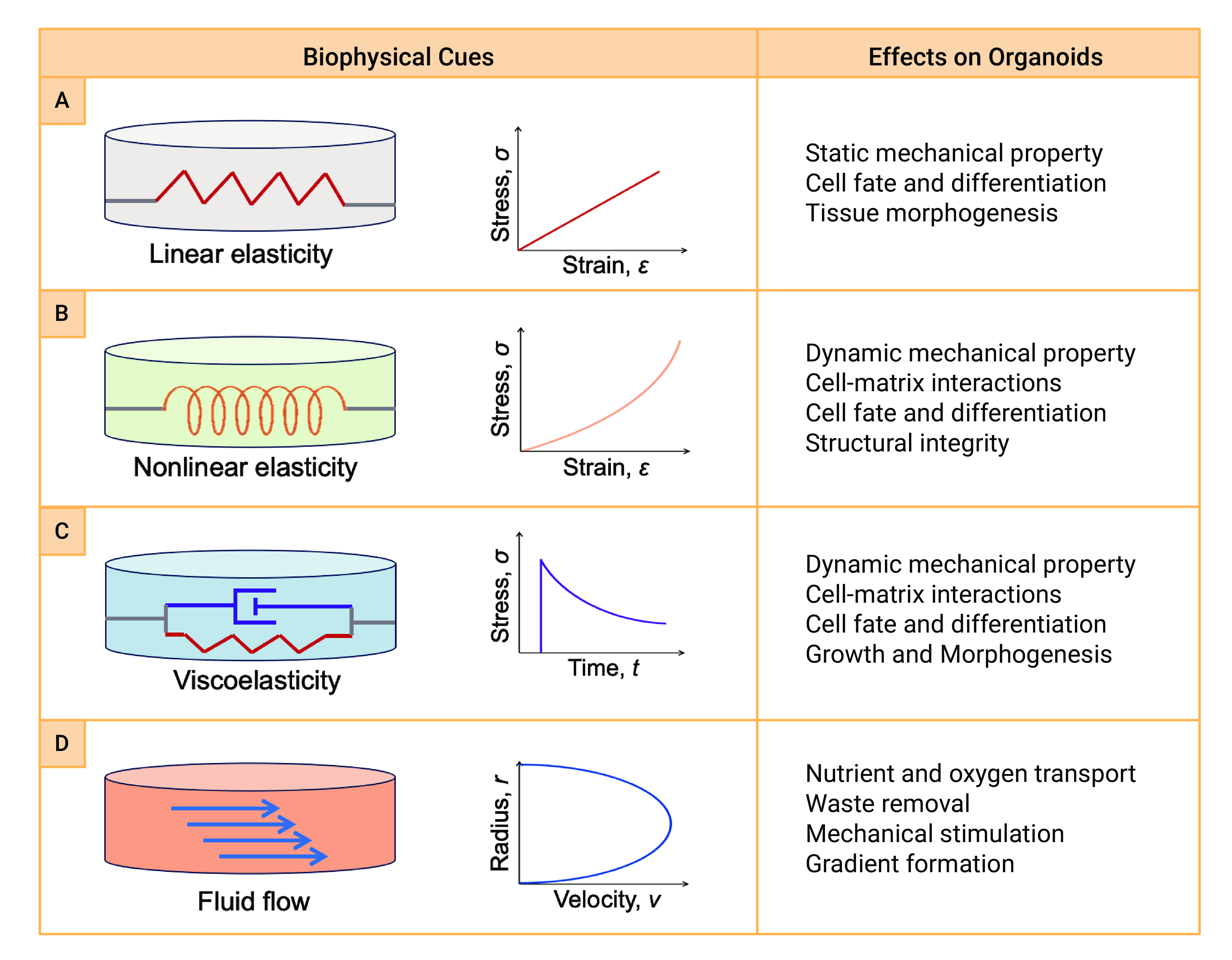

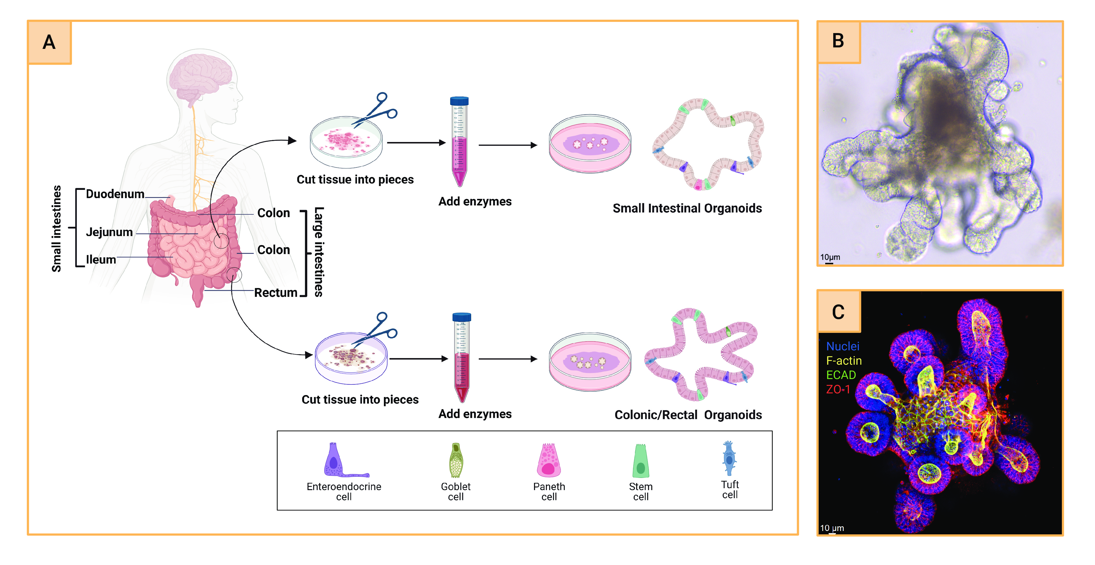


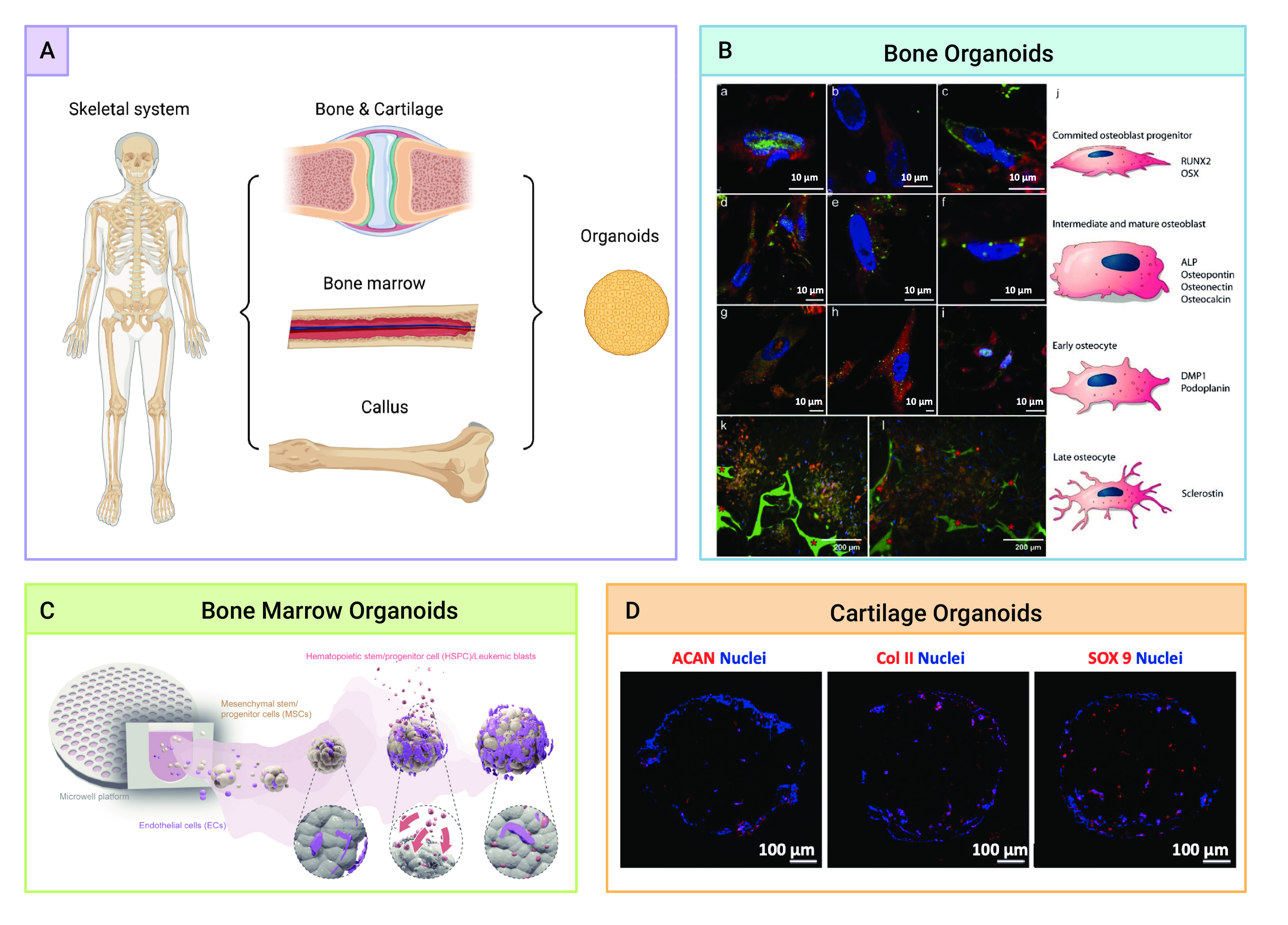
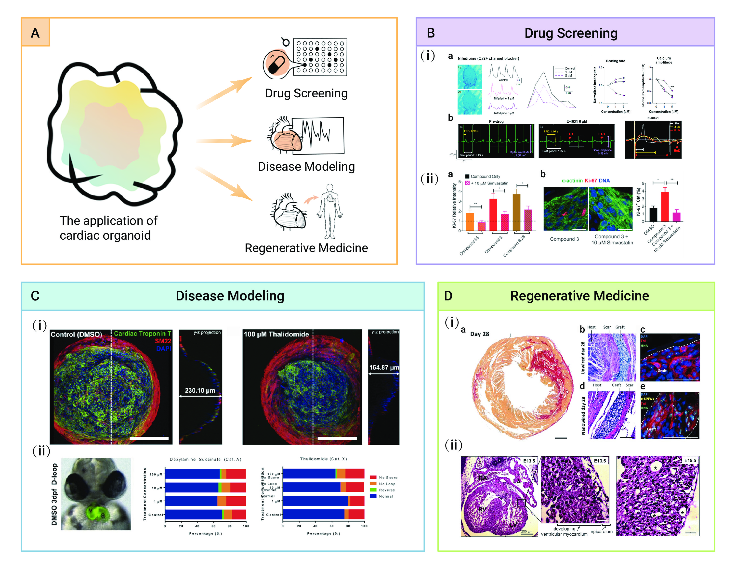
-
Figure 1.
Statistics and general principles of organoids
-
Figure 2.
Different mechanical properties and their effects on organoid development
-
Figure 3.
Applications of Brain organoids
-
Figure 4.
Intestinal organoids
-
Figure 5.
Illustration of the human lung anatomy and classification of human lung organoids
-
Figure 6.
Studies on liver organoids and bioprinting
-
Figure 7.
Skeletal organoids
-
Figure 8.
Cardiac organoids
