
Exploring the epidemiology, pathogenesis, and immunotherapeutic advancements in Clostridium difficile infection
-
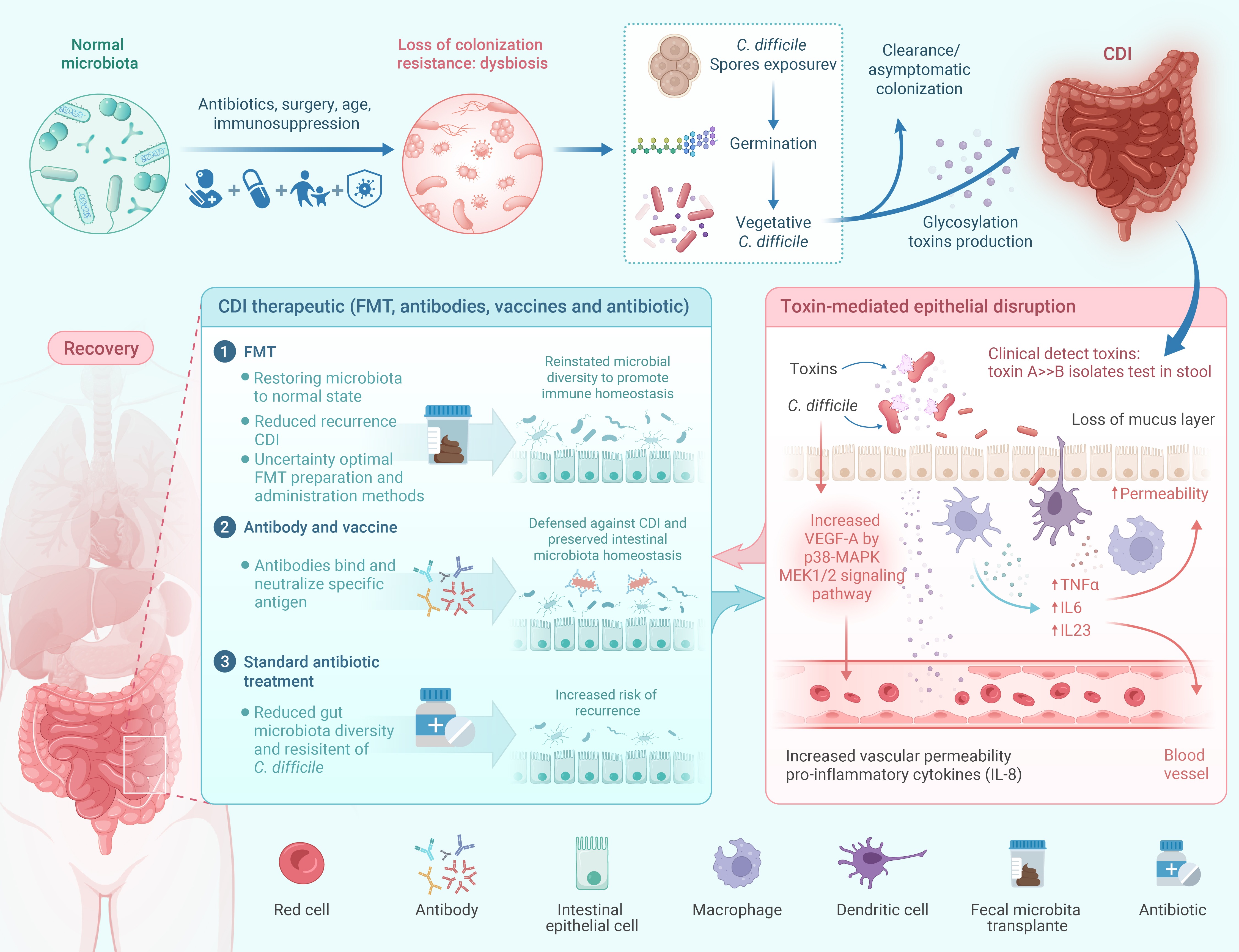
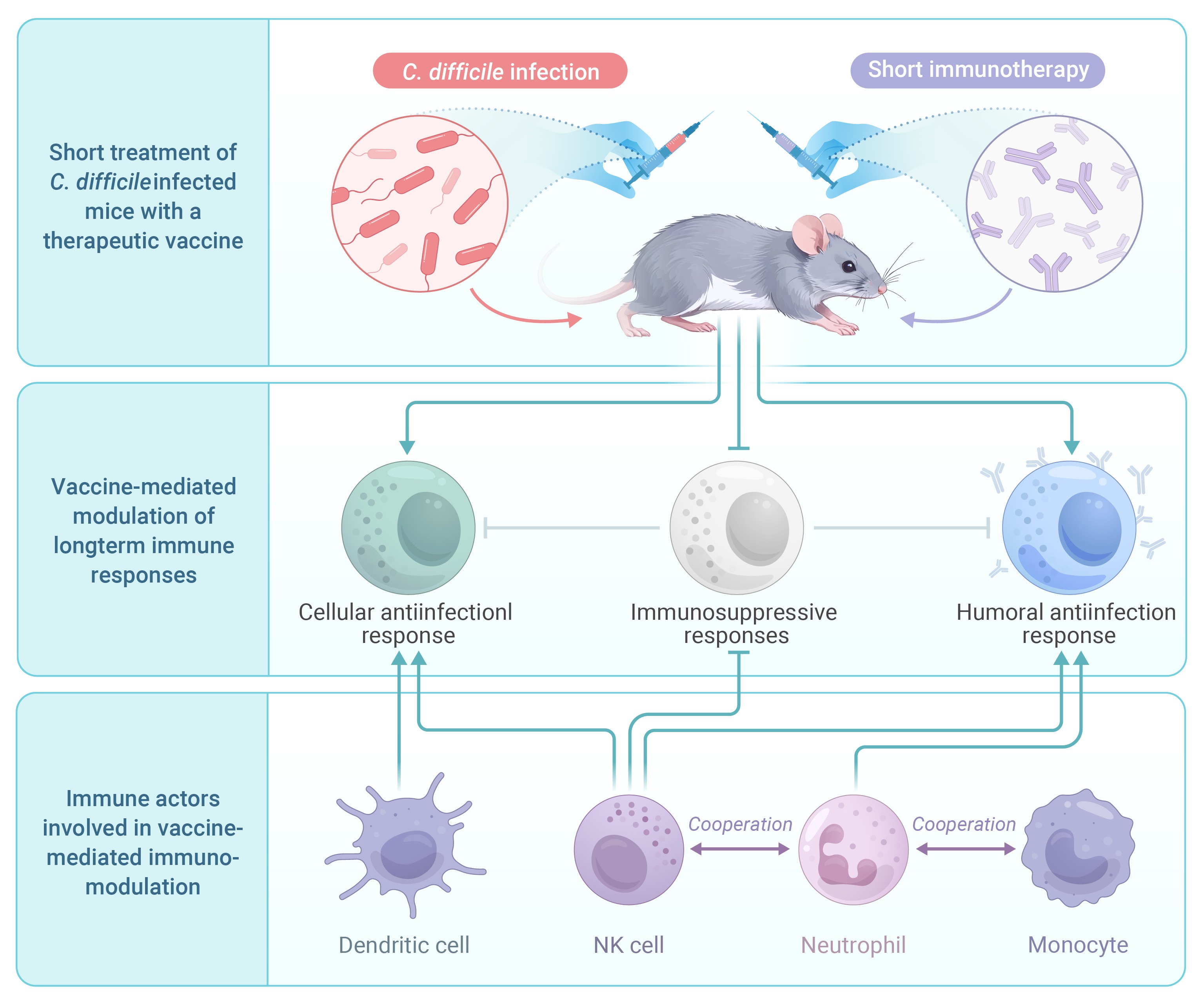
GRAPHICAL ABSTRACT

 DownLoad:
Full size image
DownLoad:
Full size image
-
PUBLIC SUMMARY
-
Impact of C. difficile: C. difficile is a leading cause of severe gastrointestinal disease with significant complications.
Traditional antibiotics disrupt the gut microbiota, increasing the risk of recurrent C. difficile infection (CDI).
Emerging immunotherapies aim to reduce CDI recurrence.
Long-term immunity strategies aim to promote lasting immunity, improving outcomes and preventing recurrent CDI.
-
-
ABSTRACT
Clostridium difficile (C. difficile) is a gram-positive, spore-forming bacillus that thrives in anaerobic conditions. It serves as the primary causative agent for various diseases globally, including nosocomial antibiotic-associated diarrhea and pseudomembranous colitis, potentially leading to fatal outcomes. The production of up to three toxins by this bacterium is considered its primary virulence mechanism in C. difficile infection (CDI). These toxins initiate inflammation, cause tissue damage, and result in diarrhea. Conventional antibiotic treatments for CDI not only substantially reduce intestinal microbiota but also increase CDI recurrence rates. Immunotherapy has emerged as a promising approach for combating CDI, offering a novel strategy to target this challenging pathogen. Various immunotherapeutic modalities, such as monoclonal antibodies targeting specific C. difficile toxins, fecal microbiota transplantation (FMT) to restore microbial balance, and vaccines to stimulate protective immune responses, have shown potential in preclinical and clinical studies. This review examines the current landscape of immunotherapy for CDI, highlighting significant advancements, challenges, and future directions in utilizing the immune system to address this substantial healthcare burden. -

-
REFERENCES
[1] McFee R.B. and Abdelsayed G.G. (2009). Clostridium difficile. Dis. Mon. 55:439−470. DOI:10.1016/j.disamonth.2009.04.010 [2] Kordus S.L., Thomas A.K. and Lacy D.B. (2021). Clostridioides difficile toxins: Mechanisms of action and antitoxin therapeutics. Nat. Rev. Microbiol. 20:285−298. DOI:10.1038/s41579-021-00660-2 [3] Buddle J.E. and Fagan, R.P. (2023). Pathogenicity and virulence of Clostridioides difficile. Virulence 14:2150452. DOI:10.1080/21505594.2022.2150452 [4] Davies K., Lawrence J., Berry C., et al. (2020). Risk factors for primary Clostridium difficile infection; results from the observational study of risk factors for Clostridium difficile infection in hospitalized patients with infective diarrhea (ORCHID). Front. Pub. Heal. 8:293. DOI:10.3389/fpubh.2020.00293 [5] Eyre D.W., Walker S., Wyllie D., et al. (2012). Predictors of first recurrence of Clostridium difficile infection: Implications for initial management. Clin Infect Dis. 55:S77−S87. DOI:10.1093/cid/cis356 [6] Etienne-Mesmin L., Chassaing B., Adekunle O., et al. (2018). Clostridium difficile toxin-positive latently infect mouse colonies and protect against highly pathogenic. Gut 67:860−871. DOI:10.1136/gutjnl-2016-313510 [7] Lessa F.C., Mu Y., Bamberg W.M., et al. (2015). Burden of Clostridium difficile infection in the United States. N. Engl. J. Med. 372:825−834. DOI:10.1056/NEJMoa1408913 [8] Guh A.Y., Mu Y., Winston L.G., et al. (2020). Trends in U. S. burden of Clostridioides difficile infection and outcomes. N. Engl. J. Med. 382:1320−1330. DOI:10.1056/NEJMoa1910215 [9] Dubberke E.R. and Olsen M.A. (2012). Burden of Clostridium difficile on the healthcare system. Clin Infect Dis. 55:S88−S92. DOI:10.1093/cid/cis335 [10] Collins D.A., Sohn K.M., Wu Y., et al. (2020). Clostridioides difficile infection in the Asia-Pacific region. Emerg. Microbes Infect. 9:42−52. DOI:10.1080/22221751.2019.1702480 [11] Warny M., Pepin J., Fang A., et al. (2005). Toxin production by an emerging strain of Clostridium difficile associated with outbreaks of severe disease in North America and Europe. Lancet 366:1079−1084. DOI:10.1016/S0140-6736(05)67420-X [12] Tamez-Torres K.M., Torres-González P., Leal-Vega F., et al. (2017). Impact of Clostridium difficile infection caused by the NAP1/RT027 strain on severity and recurrence during an outbreak and transition to endemicity in a Mexican tertiary care center. Int. J. Infect. Dis. 65:44−49. DOI:10.1016/j.ijid.2017.09.022 [13] Gateau C., Deboscker S., Couturier J., et al. (2019). Local outbreak of Clostridioides difficile PCR-Ribotype 018 investigated by multi locus variable number tandem repeat analysis, whole genome multi locus sequence typing and core genome single nucleotide polymorphism typing. Anaerobe. 60:102087−102087. DOI:10.1016/j.anaerobe.2019.102087 [14] Kim J., Kim Y. and Pai H. (2016). Clinical characteristics and treatment outcomes of Clostridium difficile infections by PCR ribotype 017 and 018 strains. PloS One. 11:e0168849−e0168849. DOI:10.1371/journal.pone.0168849 [15] Hung Y.-P., Huang I.H., Lin H.-J., et al. (2016). Predominance of Clostridium difficile ribotypes 017 and 078 among toxigenic clinical isolates in southern Taiwan. PloS One 11:e0166159−e0166159. DOI:10.1371/journal.pone.0166159 [16] Shin B.-M., Kuak E.Y., Yoo S.J., et al. (2008). Emerging toxin A−B+ variant strain of Clostridium difficile responsible for pseudomembranous colitis at a tertiary care hospital in Korea. Diagn. Microbiol. Infect. Dis. 60:333−337. DOI:10.1016/j.diagmicrobio.2007.10.022 [17] Drudy D., Harnedy N., Fanning S., et al. (2007). Emergence and control of fluoroquinolone‐resistant, toxin A–aegative, toxin B–positive Clostridium difficile. Infect. Control Hosp. Epidemiol. 28:932−940. DOI:10.1086/519181 [18] Cheng V.C.C., Yam W.C., Chan J.F.W., et al. (2009). Clostridium difficile ribotype 027 arrives in Hong Kong. Int. J. Antimicrob. Agents. 34:492−493. DOI:10.1016/j.ijantimicag.2009.04.004 [19] Cao Y., Wang L., Ke S., et al. (2021). Fecal mycobiota combined with host immune factors distinguish Clostridioides difficile infection from asymptomatic carriage. Gastroenterology. 160:2328−2339.e2326. DOI:10.1053/j.gastro.2021.02.069 [20] Webb Brandon, J., Subramanian, A., Lopansri, B., et al. (2020). Antibiotic exposure and risk for hospital-associated Clostridioides difficile infection. Antimicrob. Agents Chemother. 64 :10.1128/aac.02169-02119. DOI:10.1128/aac.02169-19. [21] Fletcher J.R., Erwin S., Lanzas C., et al. (2018). Shifts in the gut metabolome and Clostridium difficile transcriptome throughout colonization and infection in a mouse model. mSphere 3:e00089−18. DOI:10.1128/mSphere.00089-18 [22] Allegretti J.R., Kearney S., Li, N., et al. (2016). 95 Clostridium difficile infection associates with distinct bile acid and microbiome profiles. Gastroenterology 150:S24. DOI:10.1016/S0016-5085(16)30206-2 [23] Schubert Alyxandria M., Sinani H. and Schloss Patrick, D. (2015). Antibiotic-induced alterations of the murine gut microbiota and subsequent effects on colonization resistance against Clostridium difficile. mBio 6 : 10.1128/mbio.00974-00915. DOI:10.1128/mbio.00974-15. [24] Lin C.-Y., Cheng, H.-T., Kuo C.-J., et al. (2022). Proton pump inhibitor-induced gut dysbiosis increases mortality rates for patients with Clostridioides difficile infection. Microbiol. Spectr. 10:e00486−00422. DOI:10.1128/spectrum.00486-22 [25] Dalal R.S. and Allegretti J.R. (2021). Diagnosis and management of Clostridioides difficile infection in patients with inflammatory bowel disease. Curr. Opin. Gastroenterol. 37:336−343. DOI:10.1097/mog.0000000000000739 [26] Sokol H., Jegou S., McQuitty C., et al. (2018). Specificities of the intestinal microbiota in patients with inflammatory bowel disease and Clostridium difficile infection. Gut Microbes 9:55−60. DOI:10.1080/19490976.2017.1361092 [27] Abernathy-Close L., Barron Madeline R., George James M., et al. (2021). Intestinal inflammation and altered gut microbiota associated with inflammatory bowel disease render mice susceptible to Clostridioides difficile colonization and infection. mBio 12 :10.1128/mbio.02733-02720. DOI:10.1128/mbio.02733-20. [28] Ananthakrishnan A.N., Oxford E.C., Nguyen D.D., et al. (2013). Genetic risk factors for Clostridium difficile infection in ulcerative colitis. Aliment. Pharmacol. Ther. 38:522−530. DOI:10.1111/apt.12425 [29] Spigaglia P. (2020). COVID-19 and Clostridioides difficile infection (CDI): Possible implications for elderly patients. Anaerobe 64:102233. DOI:10.1016/j.anaerobe.2020.102233 [30] Imlay H., Kaul D. and Rao K. (2016). Risk factors for Clostridium difficile infection in HIV-infected patients. SAGE Open Med. 4 :2050312116684295. DOI:10.1177/2050312116684295. [31] Miller A.C., Sewell D.K., Segre A.M., et al. (2021). Risk for Clostridioides difficile iinfection among hospitalized patients associated with multiple healthcare exposures prior to admission. J. Infect. Dis. 224:684−694. DOI:10.1093/infdis/jiaa773 [32] Tschudin-Sutter S., Kuijper E.J., Durovic A., et al. (2018). Guidance document for prevention of Clostridium difficile infection in acute healthcare settings. Clin. Microbiol. Infect. 24:1051−1054. DOI:10.1016/j.cmi.2018.02.020 [33] Asempa, T.E., and Nicolau, D.P. (2017). Clostridium difficile infection in the elderly: an update on management. Clin. Interv. Aging 12:1799−1809. DOI:10.2147/CIA.S149089 [34] Yoshikawa T.T. and Norman, D.C. (2017). Geriatric infectious diseases: current concepts on diagnosis and management. J. Am. Geriatr. Soc. 65:631−641. DOI:10.1111/jgs.14731 [35] Abernathy-Close L., Dieterle Michael G., Vendrov Kimberly C., et al. (2020). Aging dampens the intestinal innate immune response during severe Clostridioides difficile infection and is associated with altered cytokine levels and granulocyte mobilization. Infect. Immun. 88:e00960−19. DOI:10.1128/iai.00960-19 [36] Gregory A.L., Pensinger D.A. and Hryckowian A.J. (2021). A short chain fatty acid–centric view of Clostridioides difficile pathogenesis. PLoS Pathog. 17:e1009959. DOI:10.1371/journal.ppat.1009959 [37] Lv J., Da R., Cheng Y., et al. (2020). Mechanism of antibacterial activity of bacillus amyloliquefaciens C-1 lipopeptide toward anaerobic Clostridium difficile. Biomed. Res. Int. 2020:3104613. DOI:10.1155/2020/3104613 [38] Nagao-Kitamoto H., Leslie J.L., Kitamoto S., et al. (2020). Interleukin-22-mediated host glycosylation prevents Clostridioides difficile infection by modulating the metabolic activity of the gut microbiota. Nat. Med. 26:608−617. DOI:10.1038/s41591-020-0764-0 [39] Schumacher J., Nienhaus A., Heber S., et al. (2023). Exploring the inhibitory potential of the antiarrhythmic drug amiodarone against Clostridioides difficile toxins TcdA and TcdB. Gut Microbes 15:2256695. DOI:10.1080/19490976.2023.2256695 [40] Chandrasekaran R., Kenworthy A.K. and Lacy, D.B. (2016). Clostridium difficile toxin a undergoes clathrin-independent, PACSIN2-dependent endocytosis. PLoS Pathog. 12:e1006070. DOI:10.1371/journal.ppat.1006070 [41] Tao L., Tian S., Zhang J., et al. (2019). Sulfated glycosaminoglycans and low-density lipoprotein receptor contribute to Clostridium difficile toxin A entry into cells. Nat. Microbiol. 4:1760−1769. DOI:10.1038/s41564-019-0464-z [42] Tian S., Xiong X., Zeng J., et al. (2022). Identification of TFPI as a receptor reveals recombination-driven receptor switching in Clostridioides difficile toxin B variants. Nat. Commun. 13:6786. DOI:10.1038/s41467-022-33964-9 [43] Luo J., Yang Q., Zhang X., et al. (2022). TFPI is a colonic crypt receptor for TcdB from hypervirulent clade 2 C. difficile. Cell 185:980−994.e915. DOI:10.1016/j.cell.2022.02.010 [44] Chen P., Zeng J., Liu Z., et al. (2021). Structural basis for CSPG4 as a receptor for TcdB and a therapeutic target in Clostridioides difficile infection. Nat. Commun. 12:3748. DOI:10.1038/s41467-021-23878-3 [45] Schöttelndreier D., Langejürgen A., Lindner R., et al (2020). Low density lipoprotein receptor-related protein-1 (LRP1) is involved in the uptake of Clostridioides difficile toxin A and serves as an internalizing receptor. Front. Cell. Infect. Microbiol. 10 :565465. DOI:10.3389/fcimb.2020.565465. [46] Castro-Córdova P., Otto-Medina M., Montes-Bravo N., et al. (2023). Redistribution of the novel Clostridioides difficile spore adherence receptor E-cadherin by TcdA and TcdB increases spore binding to adherens junctions. Infect. Immun. 91:e00476−00422. DOI. DOI:10.1128/iai.00476-22 [47] Chen X., Yang X., de Anda J., et al. (2020). Clostridioides difficile toxin A remodels membranes and mediates DNA entry into cells to activate toll-like receptor 9 signaling. Gastroenterology 159:2181−2192.e2181. DOI:10.1053/j.gastro.2020.08.038 [48] Carter Glen, P., Chakravorty, A., Pham Nguyen Tu, A., et al. (2015). Defining the roles of TcdA and TcdB in localized gastrointestinal disease, systemic organ damage, and the host response during Clostridium difficile infections. mBio 6 : 10.1128/mbio.00551-00515. DOI:10.1128/mbio.00551-15. [49] Yu, H., Chen, K., Sun, Y., et al. (2017). Cytokines are markers of the Clostridium difficile-induced inflammatory response and predict disease severity. Clin Vaccine Immunol. 24:e00037−00017. DOI:10.1128/CVI.00037-17 [50] Huang J., Kelly C.P., Bakirtzi K., et al. (2019). Clostridium difficile toxins induce VEGF-A and vascular permeability to promote disease pathogenesis. Nat. Microbiol. 4:269−279. DOI:10.1038/s41564-018-0300-x [51] Gerding D.N., Johnson S., Rupnik M. et al. (2014). Clostridium difficile binary toxin CDT: Mechanism, epidemiology, and potential clinical importance. Gut Microbes 5:15−27. DOI:10.4161/gmic.26854 [52] Boraschi D. and Tagliabue, A. (2023). Harnessing the power of inflammation in immunoprevention and immunotherapy. The Innovation Life 1:100025. DOI:10.59717/j.xinn-life.2023.100025 [53] Chen X. and Kelly C. (2018). C. difficile on and off: a dual role for cysteine protease autoprocessing of toxin B on cytotoxicity vs proinflammatory toxin actions? Cell. Mol. Gastroenterol. Hepatol. 5 :654-655. DOI:10.1016/j.jcmgh.2018.02.011. [54] Xu H., Yang J., Gao W., et al. (2014). Innate immune sensing of bacterial modifications of Rho GTPases by the Pyrin inflammasome. Nature 513:237−241. DOI:10.1038/nature13449 [55] Saavedra P.H.V., Huang L., Ghazavi F., et al. (2018). Apoptosis of intestinal epithelial cells restricts Clostridium difficile infection in a model of pseudomembranous colitis. Nat. Commun. 9:4846. DOI:10.1038/s41467-018-07386-5 [56] Lyerly D.M., Saum K.E., Macdonald D.K., et al. (1985). Effects of Clostridium difficile toxins given intragastrically to animals. Infect. Immun. 47:349−352. DOI:10.1128/IAI.47.2.349-352.1985 [57] Sambol S.P., Johnson S., Gerding D.N., et al. (2009). Toxin B is essential for virulence of Clostridium difficile. Nature 458:1176−1179. DOI:10.1038/nature07822 [58] Komatsu M., Kato H., Aihara M., et al. (2003). High frequency of antibiotic-associated diarrhea due to toxin A-negative, toxin B-positive Clostridium difficile in a hospital in Japan and risk factors for infection. Eur. J. Clin. Microbiol. Infect. Dis. 22:525−529. DOI:10.1007/s10096-003-0992-5 [59] Carter G.P., Rood J.I. and Lyras, D. (2010). The role of toxin A and toxin B in Clostridium difficile-associated disease. Gut Microbes 1:58−64. DOI:10.4161/gmic.1.1.10768 [60] Heap J.T., Kelly M.L., Minton N.P., et al. (2010). The role of toxin A and toxin B in Clostridium difficile infection. Nature 467:711−713. DOI:10.1038/nature09397 [61] Lin Q., Pollock N.R., Banz A., et al. (2020). Toxin A–predominant pathogenic Clostridioides difficile: a novel clinical phenotype. Clin. Infect. Dis. 70:2628−2633. DOI:10.1093/cid/ciz727 [62] Wang L., Cao Y., Lou, E., et al. (2023). The role of gut fungi in Clostridioides difficile infection. Biomed. J. 47:100686. DOI:10.1016/j.bj.2023.100686 [63] van Nood E., Vrieze A., Nieuwdorp M., et al. (2013). Duodenal infusion of donor feces for recurrent Clostridium difficile. N. Engl. J. Med. 368:407−415. DOI:10.1056/NEJMoa1205037 [64] Danne C., Rolhion N. and Sokol, H. (2021). Recipient factors in faecal microbiota transplantation: one stool does not fit all. Nat. Rev. Gastroenterol. Hepatol. 18:503−513. DOI:10.1038/s41575-021-00441-5 [65] Hamilton M.J., Weingarden A.R., Unno T., et al. (2013). High-throughput DNA sequence analysis reveals stable engraftment of gut microbiota following transplantation of previously frozen fecal bacteria. Gut Microbes 4:125−135. DOI:10.4161/gmic.23571 [66] Sehgal K. and Khanna, S. (2021). Gut microbiome and Clostridioides difficile infection: A closer look at the microscopic interface. Ther. Adv. Gastroenterol. 14 :1756284821994736. DOI:10.1177/1756284821994736. [67] Fachi J.L., Sécca C., Rodrigues P.B., et al. (2019). Acetate coordinates neutrophil and ILC3 responses against C. difficile through FFAR2. J. Exp. Med. 217 :e20190489. DOI:10.1084/jem.20190489. [68] Yang C., Mogno I., Contijoch E.J., et al. (2020). Fecal IgA levels are determined by strain-level differences in bacteroides ovatus and are modifiable by gut microbiota manipulation. Cell Host Microbe. 27:467−475.e466. DOI:10.1016/j.chom.2020.01.016 [69] Frisbee A.L. and Petri W.A. (2020). Considering the immune system during fecal microbiota transplantation for Clostridioides difficile infection. Trends Mol. Med. 26:496−507. DOI:10.1016/j.molmed.2020.01.009 [70] Yadegar A., Pakpoor S., Ibrahim F.F., et al. (2023). Beneficial effects of fecal microbiota transplantation in recurrent Clostridioides difficile infection. Cell Host Microbe. 31:695−711. DOI:10.1016/j.chom.2023.03.019 [71] Seekatz A.M., Aas J., Gessert C.E., et al. (2014). Recovery of the gut microbiome following fecal microbiota transplantation. mBio 5:e00893−00814. DOI:10.1128/mBio.00893-14 [72] Moreau G.B., Naz F. and Petri, W.A. (2024). Fecal microbiota transplantation stimulates type 2 and tolerogenic immune responses in a mouse model. Anaerobe 86:102841. DOI:10.1016/j.anaerobe.2024.102841 [73] Cook L., Rees W.D., Wong M.Q., et al. (2021). Fecal microbiota transplantation for recurrent Clostridioides difficile infection enhances adaptive immunity to C. difficile Toxin B. Gastroenterology 160:2155−2158.e2154. DOI:10.1053/j.gastro.2021.01.009 [74] Buffie C.G., Bucci V., Stein R.R., et al. (2015). Precision microbiome reconstitution restores bile acid mediated resistance to Clostridium difficile. Nature 517:205−208. DOI:10.1038/nature13828 [75] Jan N., Hays R.A., Oakland D.N., et al. (2021). Fecal microbiota transplantation increases colonic IL-25 and dampens tissue inflammation in patients with recurrent Clostridioides difficile. mSphere 6:e0066921. DOI:10.1128/msphere.00669-21 [76] Ghani R., Mullish B.H., Roberts L.A., et al. (2022). The potential utility of fecal (or intestinal) microbiota transplantation in controlling infectious diseases. Gut Microbes 14:2038856. DOI:10.1080/19490976.2022.2038856 [77] Burrello C., Garavaglia F., Cribiù F.M., et al. (2018). Therapeutic faecal microbiota transplantation controls intestinal inflammation through IL10 secretion by immune cells. Nat. Commun. 9:5184. DOI:10.1038/s41467-018-07359-8 [78] Nakajima A., Sasaki T., Itoh K., et al. (2020). A soluble fiber diet increases bacteroides fragilis group abundance and immunoglobulin a production in the Gut. Appl. Environ. Microbiol. 86:e00405−00420. DOI:10.1128/AEM.00405-20 [79] Segal J.P., Mullish B.H., Quraishi M.N., et al. (2020). Mechanisms underpinning the efficacy of faecal microbiota transplantation in treating gastrointestinal disease. Ther. Adv. Gastroenterol. 13 :1756284820946904. DOI: 10.1177/1756284820946904. [80] Johal S.S., Lambert C.P., Hammond J., et al. (2004). Colonic IgA producing cells and macrophages are reduced in recurrent and non-recurrent Clostridium difficile associated diarrhoea. J. Clin. Pathol. 57:973−979. DOI:10.1136/jcp.2003.015875 [81] Warny M., Vaerman J.P., Avesani V., et al (1994). Human antibody response to Clostridium difficile toxin A in relation to clinical course of infection. Infect. Immun. 62 :384-389. DOI:10.1128/IAI.62.2.384-389.1994. [82] Kyne L., Warny M., Qamar A., et al. (2001). Association between antibody response to toxin A and protection against recurrent Clostridium difficile diarrhoea. Lancet 357:189−193. DOI:10.1016/S0140-6736(00)03592-3 [83] Qiu H., Cassan R., Johnstone D., et al. (2016). Novel Clostridium difficile anti-toxin (TcdA and TcdB) humanized monoclonal antibodies demonstrate in vitro neutralization across a broad spectrum of clinical strains and in vivo potency in a hamster spore challenge model. Plos One 11:e0157970. DOI:10.1371/journal.pone.0157970 [84] Humphreys David P. and Wilcox Mark, H. (2014). Antibodies for treatment of Clostridium difficile infection. Clin. Vaccine Immunol. 21:913−923. DOI:10.1128/CVI.00116-14 [85] Israel L., Deborah C M., Brett A L., et al. (2010). Treatment with monoclonal antibodies against Clostridium difficile toxins. N. Engl. J. Med. 362:197−205. DOI:10.1056/NEJMoa0907635 [86] Warn P., Thommes P., Sattar A., et al. (2016). Disease progression and resolution in rodent models of Clostridium difficile infection and impact of antitoxin antibodies and vancomycin. Antimicrob. Agents Chemother. 60:6471−6482. DOI:10.1128/aac.00974-16 [87] Marozsan A.J., Ma D., Nagashima K.A., et al. (2012). Protection against Clostridium difficile infection with broadly neutralizing antitoxin monoclonal antibodies. J. Infect. Dis. 206:706−713. DOI:10.1093/infdis/jis416 [88] Davies N.L., Compson J.E., MacKenzie B., et al. (2013). A mixture of functionally oligoclonal humanized monoclonal antibodies that neutralize Clostridium difficile TcdA and TcdB with high levels of in vitro potency shows in vivo protection in a hamster infection model. Clin. Vaccine Immunol. 20:377−390. DOI:10.1128/CVI.00625-12 [89] Wilcox M.H., Gerding D.N., Poxton I.R., et al. (2017). Bezlotoxumab for prevention of recurrent Clostridium difficile infection. N. Engl. J. Med. 376:305−317. DOI:10.1056/NEJMoa1602615 [90] Yang Z., Shi L., Yu H., et al. (2016). Intravenous adenovirus expressing a multi-specific, single-domain antibody neutralizing TcdA and TcdB protects mice from Clostridium difficile infection. FEMS Pathog. Dis. 74:ftw078. DOI:10.1093/femspd/ftw078 [91] Kelly C.P., Poxton I.R., Shen J., et al. (2019). Effect of Endogenous Clostridioides difficile Toxin Antibodies on Recurrence of C. difficile Infection. . Clin. Infect. Dis. 71:81−86. DOI:10.1093/cid/ciz809 [92] Hernandez L., Kroh H., Hsieh E., et al. (2017). Epitopes and mechanism of action of the Clostridium difficile toxin A-neutralizing antibody actoxumab. J. Mol. Biol. 429:1030−1044. DOI:10.1016/j.jmb.2017.02.010 [93] Babcock Gregory J., Broering Teresa J., Hernandez Hector J., et al. (2006). Human monoclonal antibodies directed against toxins A and B prevent Clostridium difficile-induced mortality in hamsters. Infect. Immun. 74:6339−6347. DOI:10.1128/iai.00982-06 [94] Zhang Q., Widmer G. and Tzipori S. (2013). A pig model of the human gastrointestinal tract. Gut Microbes 4:193−200. DOI:10.4161/gmic.23867 [95] Steele J., Mukherjee J., Parry N., et al. (2013). Antibody against TcdB, but not TcdA, prevents development of gastrointestinal and systemic Clostridium difficile disease. J. Infect. Dis. 207:323−330. DOI:10.1093/infdis/jis669 [96] Abramowicz M., Zuccotti G. and Pflomm, J.-M. (2017). Bezlotoxumab (Zinplava) for prevention of recurrent Clostridium difficile infection. JAMA 318:659−660. DOI:10.1001/jama.2017.10092 [97] Mullard A. (2016). FDA approves antitoxin antibody. Nat. Rev. Drug Discov. 15:811. DOI:10.1038/nrd.2016.257 [98] Donald R.G.K., Flint M., Kalyan N., et al. (2013). A novel approach to generate a recombinant toxoid vaccine against Clostridium difficile. Microbiology 159:1254−1266. DOI:10.1099/mic.0.066712-0 [99] Kitchin N., Remich S.A., Peterson J., et al. (2020). A phase 2 study evaluating the safety, tolerability, and immunogenicity of two 3-dose regimens of a Clostridium difficile vaccine in healthy US adults aged 65 to 85 years. Clin. Infect. Dis. 70:1−10. DOI:10.1093/cid/ciz153 [100] de Bruyn G., Gordon D.L., Steiner T., et al. (2021). Safety, immunogenicity, and efficacy of a Clostridioides difficile toxoid vaccine candidate: A phase 3 multicentre, observer-blind, randomised, controlled trial. Lancet Infect. Dis. 21:252−262. DOI:10.1016/S1473-3099(20)30331-5 [101] de Bruyn G., Saleh J., Workman D., et al. (2016). Defining the optimal formulation and schedule of a candidate toxoid vaccine against Clostridium difficile infection: A randomized Phase 2 clinical trial. Vaccine 34:2170−2178. DOI:10.1016/j.vaccine.2016.03.028 [102] Foglia G., Shah S., Luxemburger C., et al. (2012). Clostridium difficile: Development of a novel candidate vaccine. Vaccine 30:4307−4309. DOI:10.1016/j.vaccine.2012.01.056 [103] Feuerstadt P., Louie T.J., Lashner B., et al. (2022). SER-109, an oral microbiome therapy for recurrent Clostridioides difficile infection. N. Engl. J. Med. 386:220−229. DOI:10.1056/NEJMoa2106516 [104] Chandrabali G., Janneke M.V., Xinhua C., et al. (2013). Toll-like receptor 5-dependent immunogenicity and protective efficacy of a recombinant fusion protein vaccine containing the nontoxic domains of Clostridium difficile toxins A and B and salmonella enterica serovar typhimurium flagellin in a mouse model of Clostridium difficile disease. Infect. Immun. 81:2190−2196. DOI:10.1128/IAI.01074-12 [105] Wang S., Heuler J., Wickramage I., et al. (2022). Genomic and phenotypic characterization of the nontoxigenic Clostridioides difficile strain CCUG37785 and demonstration of its therapeutic potential for the prevention of C. difficile infection. Microbiol. Spectr. 10:e0178821−e0178821. DOI:10.1128/spectrum.01788-21 [106] Zheng L., Kelly C.J., Battista K.D., et al. (2017). Microbial-derived butyrate promotes epithelial barrier function through IL-10 receptor-dependent repression of claudin-2. J. Immunol. 199:2976−2984. DOI:10.4049/jimmunol.1700105 [107] Inan M.S., Rasoulpour R.J., Yin L., et al. (2000). The luminal short-chain fatty acid butyrate modulates NF-κB activity in a human colonic epithelial cell line. Gastroenterology 118:724−734. DOI:10.1016/S0016-5085(00)70142-9 [108] Hayashi A., Nagao-Kitamoto H., Kitamoto S., et al. (2021). The butyrate-producing bacterium Clostridium butyricum suppresses Clostridioides difficile infection via neutrophiland antimicrobial cytokine-dependent but GPR43/109a-independent mechanisms. J. Immunol. 206:1576−1585. DOI:10.4049/jimmunol.2000353 [109] Best E.L., Freeman J. and Wilcox, M.H. (2012). Models for the study of Clostridium difficile infection. Gut Microbes 3:145−167. DOI:10.4161/gmic.19526 [110] Gardiner D.F., Rosenberg T., Zaharatos J., et al. (2009). A DNA vaccine targeting the receptor-binding domain of Clostridium difficile toxin A. Vaccine 27:3598−3604. DOI:10.1016/j.vaccine.2009.03.058 [111] Matchett W.E., Anguiano-Zarate S., Malewana G.B.R., et al. (2020). A replicating single-cycle adenovirus vaccine effective against Clostridium difficile. Vaccines 8:470. DOI:10.3390/vaccines8030470 [112] Zhang B.-Z., Cai J., Yu B., et al. (2016). A DNA vaccine targeting TcdA and TcdB induces protective immunity against Clostridium difficile. BMC Infect. Dis. 16:596. DOI:10.1186/s12879-016-1924-1 [113] Dieterle M.G., Rao K. and Young, V.B. (2019). Novel therapies and preventative strategies for primary and recurrent Clostridium difficile infections: Novel therapies for C. difficile infections. Ann. N. Y. Acad. Sci. 1435:110−138. DOI:10.1111/nyas.13958 [114] Cammarota G., Ianiro G., Tilg H., et al. (2017). European consensus conference on faecal microbiota transplantation in clinical practice. Gut 66:569. DOI:10.1136/gutjnl-2016-313017 [115] Kelly C.R., Khoruts A., Staley C., et al. (2016). Effect of fecal microbiota transplantation on recurrence in multiply recurrent Clostridium difficile infectionAnn. Intern. Med. 165:609−616. DOI:10.7326/m16-0271%m27547925 [116] Bryce K P., Brendan C., Emmalee P., et al. (2020). Long-term efficacy and safety of fecal microbiota transplantation for treatment of recurrent Clostridioides difficile infection. J. Clin. Gastroenterol. 54:701−706. DOI:10.1097/MCG.0000000000001281 [117] Zhu G., Hu J. and Xi R. (2021). The cellular niche for intestinal stem cells: A team effort. Cell Regen. 10:1. DOI:10.1186/s13619-020-00061-5 [118] Barker N. (2014). Adult intestinal stem cells: Critical drivers of epithelial homeostasis and regeneration. Nat. Rev. Mol. Cell Biol. 15:19−33. DOI:10.1038/nrm3721 [119] Cribas E.S. (2023). Intestinal immune and epithelial responses to Clostridioides difficile infection. [120] Mileto S.J., Jardé T., Childress K.O., et al. (2020). Clostridioides difficile infection damages colonic stem cells via TcdB, impairing epithelial repair and recovery from disease. Proc. Natl. Acad. Sci. USA 117:8064−8073. DOI:10.1073/pnas.1915255117 [121] Holmberg F.E.O., Seidelin J.B., Yin X., et al. (2017). Culturing human intestinal stem cells for regenerative applications in the treatment of inflammatory bowel disease. EMBO Mol. Med. 9:558−570-570. DOI:10.15252/emmm.201607260 [122] Hanash, Alan M., Dudakov, Jarrod A., Hua, G., et al. (2012). Interleukin-22 protects intestinal stem cells from immune-mediated tissue damage and regulates sensitivity to graft versus host disease. Immunity. 37:339−350. DOI:10.1016/j.immuni.2012.05.028 [123] Uddin Md J., Thompson B., Leslie Jhansi L., et al. (2024). Investigating the impact of antibiotic-induced dysbiosis on protection from Clostridium difficile colitis by mouse colonic innate lymphoid cells. mBio 15:e03338−03323. DOI:10.1128/mbio.03338-23 [124] Xie J., Li L.-f., Dai T.-y., et al. (2022). Short-chain fatty acids produced by ruminococcaceae mediate α-linolenic acid promote intestinal stem cells proliferation. Mol. Nutr. Food Res. 66:2100408. DOI:10.1002/mnfr.202100408 [125] Markandey M., Bajaj A., Ilott N.E., et al. (2021). Gut microbiota: Sculptors of the intestinal stem cell niche in health and inflammatory bowel disease. Gut Microbes 13:1990827. DOI:10.1080/19490976.2021.1990827 [126] Nigro G. and Sansonetti P.J. (2015). Microbiota and gut stem cells cross-talks: A new view of epithelial homeostasis. Curr. Stem Cell Rep. 1:48−52. DOI:10.1007/s40778-014-0005-x -
ABOUT THIS ARTICLE
Cite this article:
Wang L., Lee C., Gálvez J., et al. (2025). Exploring the epidemiology, pathogenesis, and immunotherapeutic advancements in Clostridium difficile infection. The Innovation Life 3:100107. https://doi.org/10.59717/j.xinn-life.2024.100107
Welcome!
To request copyright permission to republish or share portions of our works, please visit Copyright Clearance Center's (CCC) Marketplace website at marketplace.copyright.com.
Export File
Citation
Wang L., Lee C., Gálvez J., et al. (2025). Exploring the epidemiology, pathogenesis, and immunotherapeutic advancements in Clostridium difficile infection. The Innovation Life 3:100107. https://doi.org/10.59717/j.xinn-life.2024.100107

Format
Content


-
Figure 1.
Pathogenesis of CDI and emerging treatment approaches for CDI
-
Figure 2.
Fecal Microbiota transplantation (FMT) for patients with recalcitrant CDI by modulating immune response
-
Figure 3.
Schematic of monoclonal antibodies for immunotherapy
-
Figure 4.
Mechanisms Involved in the Induction of Vaccinal Effects
