
Exploration of glycogen deposition disorders in low-body-weight fetuses in a pig model
-
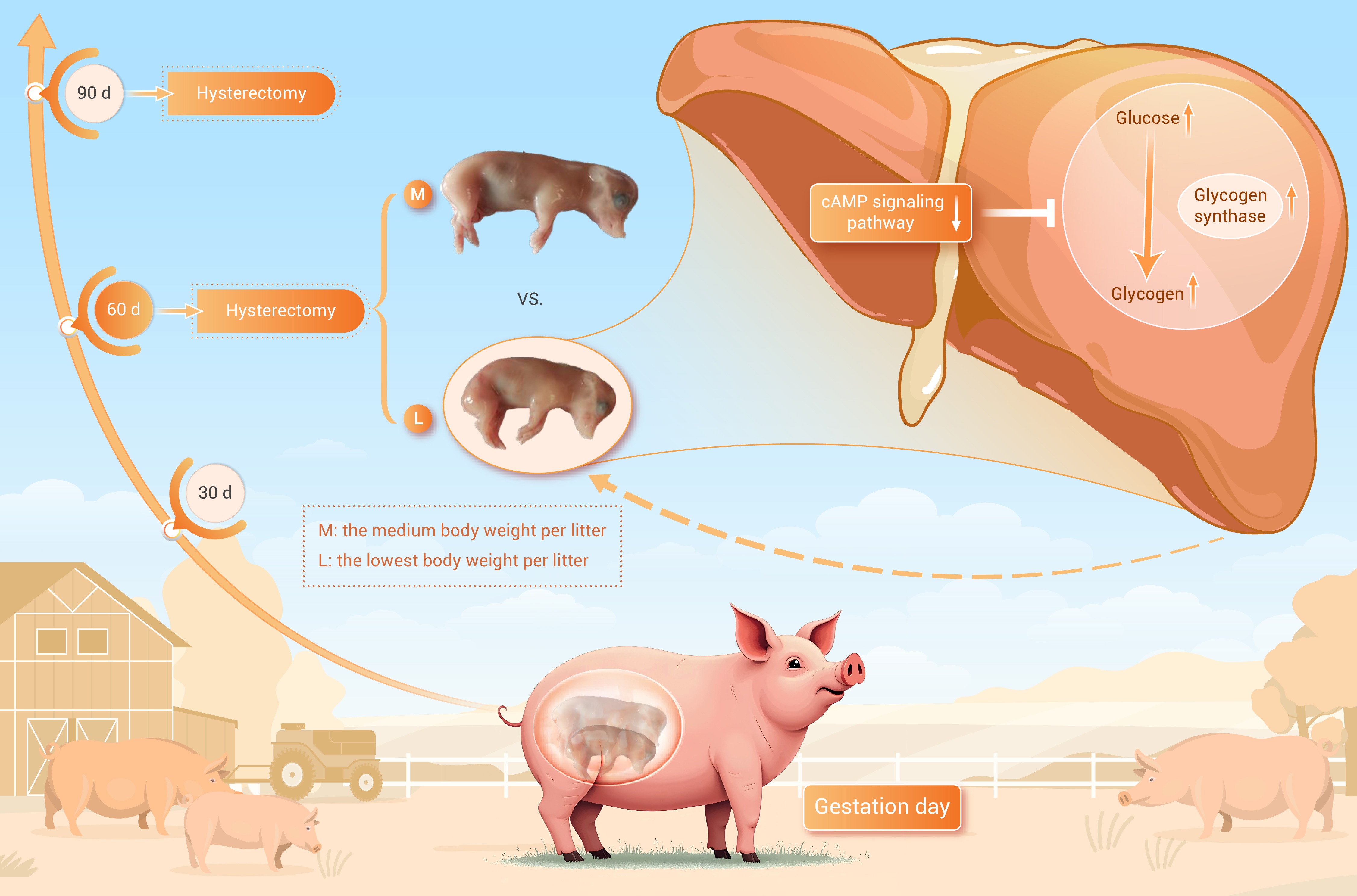
GRAPHICAL ABSTRACT
 DownLoad:
Full size image
DownLoad:
Full size image
-
PUBLIC SUMMARY
-
Hepatic glycogen deposition disorders happened in low-body-weight fetal pigs at mid-gestation but not at late gestation.
Abnormal glucose deposition in the liver may impair the development of low-birth-weight fetal pigs at mid-gestation.
The cAMP signaling pathway is crucial for regulating hepatic glycogen deposition in fetal pigs at mid-gestation.
-
-
ABSTRACT
Glucose is a vital energy source for the fetus, storing any surplus as glycogen. Neonates experiencing intrauterine growth restriction exhibit decreased hepatic glycogen levels. Consequently, the regulation of glycogen deposition may hold immense significance in fetal development, yet the intricacies of glycogen deposition homeostasis remain poorly understood. Here, we aimed to explore the differences in fetal glycogen deposition and its potential mechanisms using fetal pigs with the lowest (L) and medium (M) body weights on gestation days 60 (GD 60) and 90 (GD 90). We observed higher hepatic glycogen concentrations in the L group on GD 60, which correlated with higher hepatic glucose levels and glycogen synthase activity. In addition, our investigation using RNA-seq and immortalized fetal pig liver cells revealed that the cAMP signaling pathway ranked prominently in the KEGG enrichment analysis of differentially expressed genes. Notably, this pathway was relatively suppressed in the livers of L group pigs on GD 60, compared to the M group. The cAMP signaling pathway suppression resulted in glycogen concentration upregulation in our in vitro model. Overall, our findings suggest that glucose needed for fetal development deposited abnormally in the liver may hinder the growth of low-body-weight fetuses at mid-gestation. Furthermore, we have identified the cAMP signaling pathway as a pivotal regulator of hepatic glycogen deposition homeostasis in fetuses at mid-gestation, offering a novel perspective on the factors contributing to delayed fetal growth. -

-
REFERENCES
[1] Suhag A. and Berghella V. (2013). Intrauterine Growth Restriction (IUGR): Etiology and Diagnosis. Current Obstetrics and Gynecology Reports 2:102−111. DOI:10.1007/s13669-013-0041-z [2] Wang J., Feng C., Liu T., et al. (2017). Physiological alterations associated with intrauterine growth restriction in fetal pigs: Causes and insights for nutritional optimization. Mol. Reprod. Dev. 84:897−904. DOI:10.1002/mrd.22842 [3] Almeida, F. and Dias, A.A. (2022). Pregnancy in pigs: The journey of an early life. Domestic Animal Endocrinology 78:106656. DOI:10.1016/j.domaniend.2021.106656 [4] Ergaz Z., Avgil M. and Ornoy A. (2005). Intrauterine growth restriction—etiology and consequences: what do we know about the human situation and experimental animal models. Reprod. Toxicol. 20:301−322. DOI:10.1016/j.reprotox.2005.04.007 [5] Smith J., Murphy M., and Kandasamy Y. (2013). The IUGR infant: A case study and associated problems with IUGR infants. Journal of Neonatal Nursing 19:46−53. DOI:10.1016/j.jnn.2012.12.005 [6] Lawrence E.J. (2006). Part 1: a matter of size: evaluating the growth-restricted neonate. Adv. Neonatal. Care 6 :313-322. DOI:10.1016/j.adnc.2006.08.006 [7] Schook L.B., Collares T.V., Darfour-Oduro K.A., et al. (2015). Unraveling the swine genome: Implications for human health. Annu. Rev. Anim. Biosci. 3:219−244. DOI:10.1146/annurev-animal-022114-110815 [8] Wei Y.S., Tang W.J., Mao P.Y., et al. (2024). Sexually dimorphic response to hepatic injury in newborn suffering from intrauterine growth restriction.Adv. Sci. (Weinh) 11 :e2403095. DOI:10.1002/advs.202403095 [9] Long B.S., Yin C., Fan Q.W., et al. (2016). Global liver proteome analysis using iTRAQ reveals AMPK-mTOR-autophagy signaling is altered by intrauterine growth restriction in newborn piglets. J. Proteome Res. 15 :1262-1273. DOI:10.1021/acs.jproteome.6b00001 [10] Amdi C., Krogh U., Flummer C., et al. (2013). Intrauterine growth restricted piglets defined by their head shape ingest insufficient amounts of colostrum. J. Anim. Sci. 91 :5605-5613. DOI:10.2527/jas.2013-6824 [11] Čapková A. and Jirasek J. (1968). Glycogen reserves in organs of human foetuses in the first half of pregnancy. Neonatology 13:129−142. DOI:10.1159/000240140 [12] Cowett R.M. (2012). Principles of perinatal-neonatal metabolism (Springer Science & Business Media). [13] Mota-Rojas D., Orozco-Gregorio H., Villanueva-Garcia D., et al. (2011). Foetal and neonatal energy metabolism in pigs and humans: a review. Veterinární Medicína 56 :215-225. DOI:10.17221/1565-vetmed [14] Shoham-Vardi I., Leiberman J.R., and Kopernik G. (1994). The association of primiparity with intrauterine growth retardation. Eur. J. Obstet. Gynecol. Reprod. Biol. 53:95−101. DOI:10.1016/0028-2243(94)90214-3 [15] Council, N.R. (2012). Nutrient requirements of swine (The National Academies Press, Washington, DC). [16] Baxter E.M., Jarvis S., D’Eath R.B., et al. (2008). Investigating the behavioural and physiological indicators of neonatal survival in pigs. Theriogenology 69:773−783. DOI:10.1016/j.theriogenology.2007.12.007 [17] Qi M., Tan B., Wang J., et al. (2021). Postnatal growth retardation is associated with deteriorated intestinal mucosal barrier function using a porcine model. J. Cell Physiol. 236 :2631-2648. DOI:10.1002/jcp.30028 [18] Trauth J.A., Seidler F.J. and Slotkin, T.A. (2000). An animal model of adolescent nicotine exposure: effects on gene expression and macromolecular constituents in rat brain regions. Brain Res. 867:29−39. DOI:10.1016/S0006-8993(00)02208-3 [19] Shao Y., Xiong X., Wang K., et al. (2022). Early weaning leads to the remodeling of lipid profile in piglet jejunal crypt cells during post-weaning days. Anim. Nutr. 11:102−111. DOI:10.1016/j.aninu.2022.07.001 [20] Andersen C.L., Jensen J.L. and Ørntoft, T.F. (2004). Normalization of real-time quantitative reverse transcription-PCR data: a model-based variance estimation approach to identify genes suited for normalization, applied to bladder and colon cancer data sets. Cancer Res. 64 :5245-5250. DOI:10.1158/0008-5472.Can-04-0496 [21] Livak K.J. and Schmittgen T.D. (2001). Analysis of relative gene expression data using real-time quantitative PCR and the 2− ΔΔCT method. Methods 25:402−408. DOI:10.1006/meth.2001.1262 [22] Ding L., Xu Z.-J., Shi H.-H., et al. (2021). Sterol sulfate alleviates atherosclerosis via mediating hepatic cholesterol metabolism in ApoE−/− mice. Food & Function 12 :4887-4896. DOI:10.1039/D0FO03266B [23] Motulsky H.J. and Brown R.E. (2006). Detecting outliers when fitting data with nonlinear regression – a new method based on robust nonlinear regression and the false discovery rate. BMC Bioinformatics 7 :123. DOI:10.1186/1471-2105-7-123 [24] Lents C.A. and Freking B.A. (2019). Intrauterine position and adjacent fetal sex affects fetal and placental growth throughout gestation, but not embryonic viability, in pigs selected for component traits of litter size. Anim. Reprod. Sci. 209 :106139. DOI:10.1016/j.anireprosci.2019.106139 [25] Studer R.K., Snowdowne K.W., and Borle A.B. (1984). Regulation of hepatic glycogenolysis by glucagon in male and female rats. Role of cAMP and Ca2+ and interactions between epinephrine and glucagon. J. Biol. Chem. 259 :3596-3604. [26] Knight J.W., Bazer F.W., Thatcher W.W., et al. (1977). Conceptus development in intact and unilaterally hysterectomized-ovariectomized gilts: interrelations among hormonal status, placental development, fetal fluids and fetal growth. J. Anim. Sci. 44 :620-637. DOI:10.2527/jas1977.444620x [27] Wu G., Bazer F.W., Johnson G.A., et al. (2011). TRIENNIAL GROWTH SYMPOSIUM: Important roles for L-glutamine in swine nutrition and production. J. Anim. Sci. 89 :2017-2030. DOI:10.2527/jas.2010-3614 [28] Limesand S.W., Rozance P.J., Smith D., et al (2007). Increased insulin sensitivity and maintenance of glucose utilization rates in fetal sheep with placental insufficiency and intrauterine growth restriction. Am. J. Physiol. Endocrinol. Metab. 293 :E1716-1725. DOI:10.1152/ajpendo.00459.2007 [29] Roza S.J., Steegers E.A.P., Verburg B.O., et al. (2008). What is spared by fetal brain-sparing? Fetal circulatory redistribution and behavioral problems in the general population. Am. J. Epidemiol. 168 :1145-1152. DOI:10.1093/aje/kwn233 [30] Swanson K., Reynolds L. and Caton J. (2000). Influence of dietary intake and lasalocid on serum hormones and metabolites and visceral organ growth and morphology in wether lambs. Small Rumin. Res. 35:235−247. DOI:10.1016/S0921-4488(99)00092-9 [31] Agius L. (2008). Glucokinase and molecular aspects of liver glycogen metabolism. Biochem. J. 414 :1-18. DOI:10.1042/bj20080595 [32] Hung T.-H., Wu C.-P., Li M.-J. et al. (2017). Mammalian target of rapamycin signaling is a mechanistic link between increased endoplasmic reticulum stress and autophagy in the placentas of pregnancies complicated by growth restriction. Placenta 60:9−20. DOI:10.1016/j.placenta.2017.10.001 [33] Doherty M.J., Moorhead G., Morrice N., et al. (1995). Amino acid sequence and expression of the hepatic glycogen-binding (GL-subunit of protein phosphatase-1. FEBS Letters 375:294−298. DOI:10.1016/0014-5793(95)01184-G [34] Doherty M.J., Young P.R. and Cohen, P.T. (1996). Amino acid sequence of a novel protein phosphatase 1 binding protein (R5) which is related to the liver-and muscle-specific glycogen binding subunits of protein phosphatase 1. FEBS Letters 399:339−343. DOI:10.1016/S0014-5793(96)01357-9 [35] Green A.R., Aiston S., Greenberg C.C., et al. (2004). The glycogenic action of protein targeting to glycogen in hepatocytes involves multiple mechanisms including phosphorylase inactivation and glycogen synthase translocation. J. Biol. Chem. 279 :46474-46482. DOI:10.1074/jbc.M405660200 [36] Roach P.J. (2002). Glycogen and its metabolism. Curr. Mol. Med. 2:101−120. DOI:10.2174/1566524024605761 [37] Agius L. (2015). Role of glycogen phosphorylase in liver glycogen metabolism. Mol. Aspects Med. 46 :34-45. DOI:10.1016/j.mam.2015.09.002 [38] Frame S., Cohen P. and Biondi R.M. (2001). A common phosphate binding site explains the unique substrate specificity of GSK3 and its inactivation by phosphorylation. Mol. Cell 7:1321−1327. DOI:10.1016/S1097-2765(01)00253-2 [39] Fang, X., Yu, S.X., Lu, Y., Bast, R.C., Woodgett, J.R., and Mills, G.B. (2000). Phosphorylation and inactivation of glycogen synthase kinase 3 by protein kinase A. Proc. Natl. Acad. Sci. USA 97:11960−11965. DOI:10.1073/pnas.220413597 [40] Bultot L., Guigas B., Von Wilamowitz-Moellendorff A., et al. (2012). AMP-activated protein kinase phosphorylates and inactivates liver glycogen synthase. Biochem. J. 443 :193-203. DOI:10.1042/bj20112026 [41] Muñoz-Alonso M.J., Guillemain G., Kassis N., et al. (2000). A novel cytosolic dual specificity phosphatase, interacting with glucokinase, increases glucose phosphorylation rate. J. Biol. Chem. 275 :32406-32412. DOI:10.1074/jbc.m000841200. -
ABOUT THIS ARTICLE
Cite this article:
Shao Y., Bai M., Wang S., et al. (2025). Exploration of glycogen deposition disorders in low-body-weight fetuses in a pig model. The Innovation Life 3:100119. https://doi.org/10.59717/j.xinn-life.2024.100119
Welcome!
To request copyright permission to republish or share portions of our works, please visit Copyright Clearance Center's (CCC) Marketplace website at marketplace.copyright.com.
Export File
Citation
Shao Y., Bai M., Wang S., et al. (2025). Exploration of glycogen deposition disorders in low-body-weight fetuses in a pig model. The Innovation Life 3:100119. https://doi.org/10.59717/j.xinn-life.2024.100119

Format
Content
-
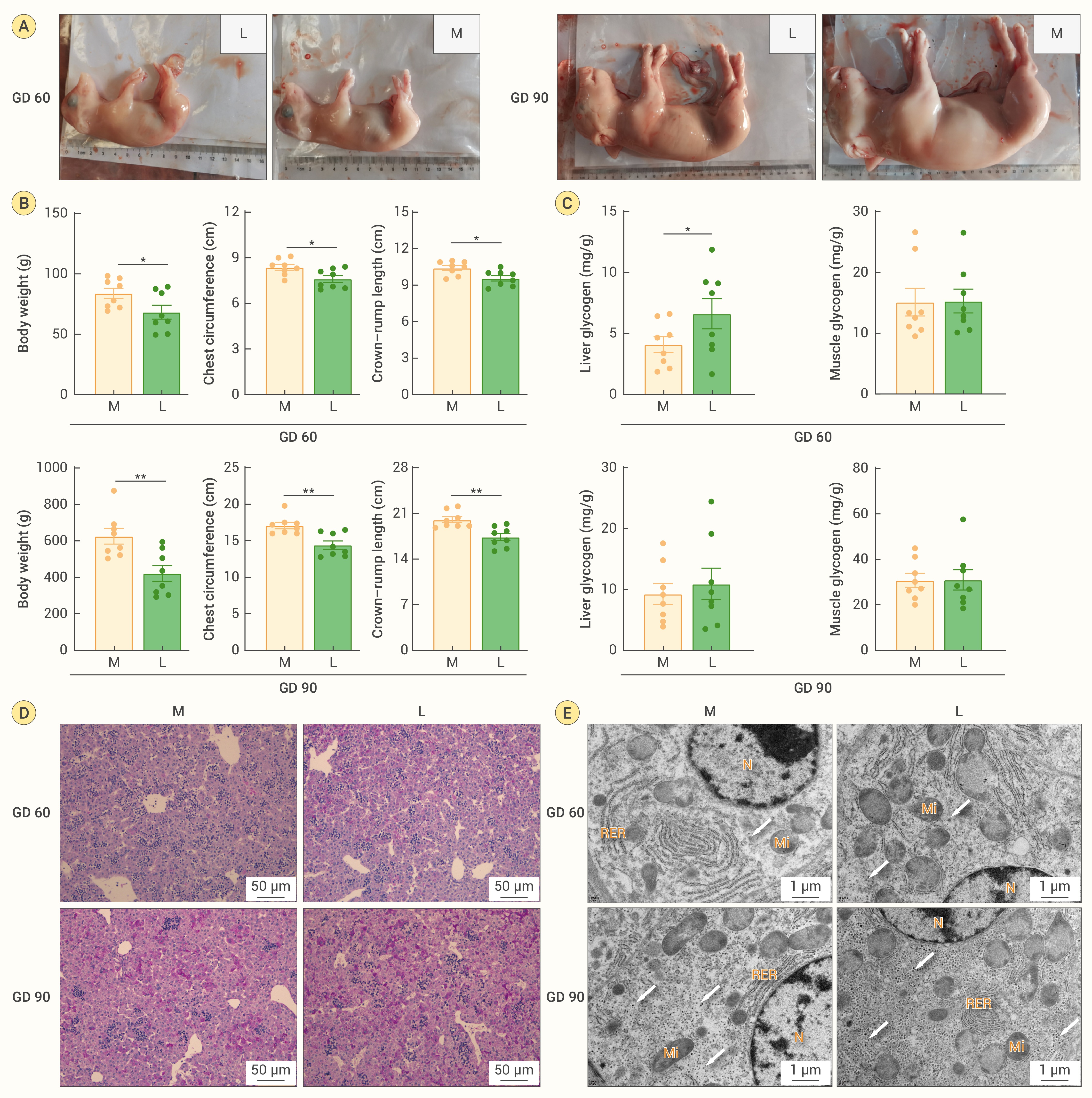
Figure 1.
Low-body-weight fetal pigs have higher hepatic glycogen concentration
-
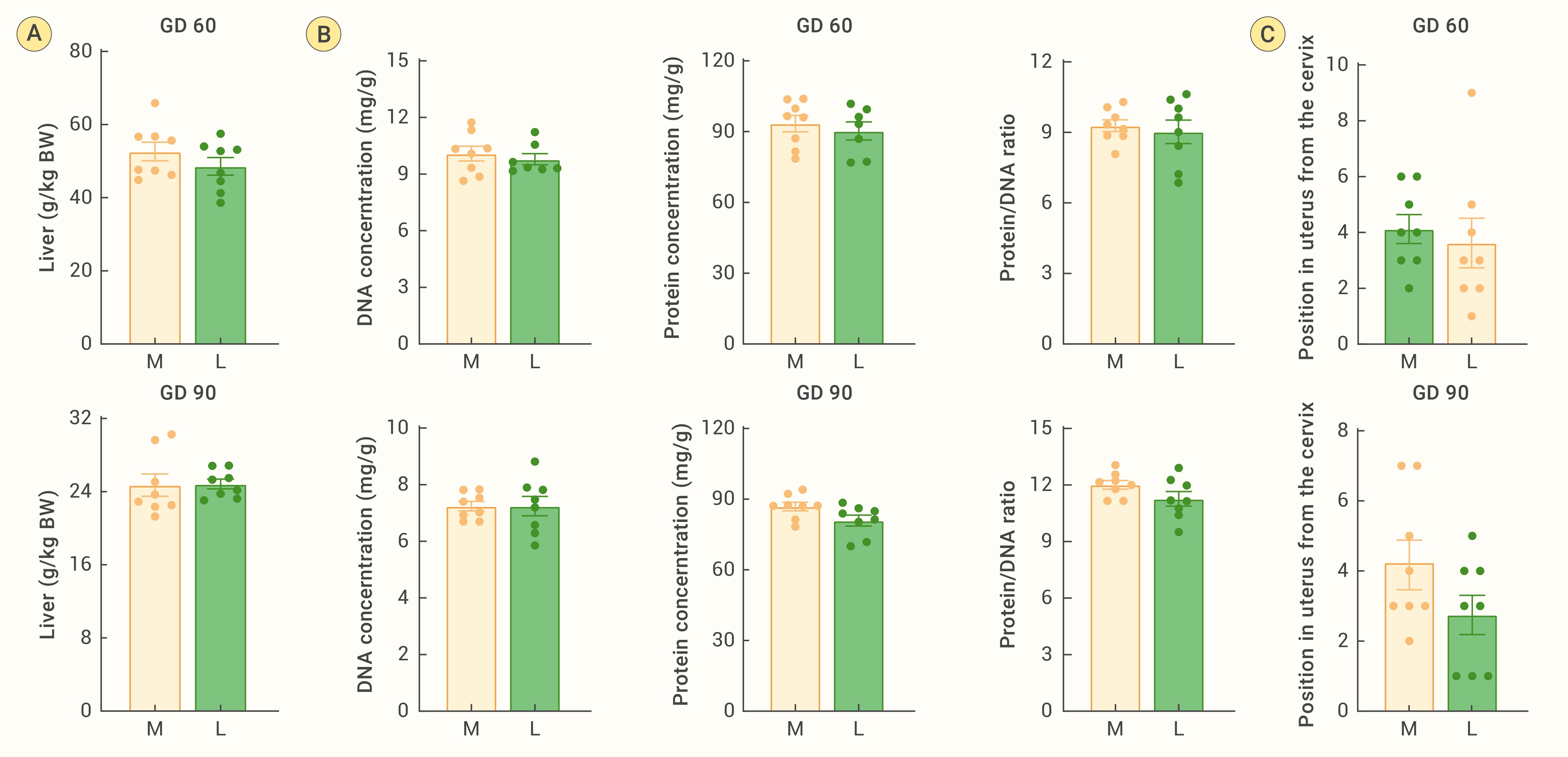
Figure 2.
Higher hepatic glycogen concentration is not related to liver development and intrauterine position
-
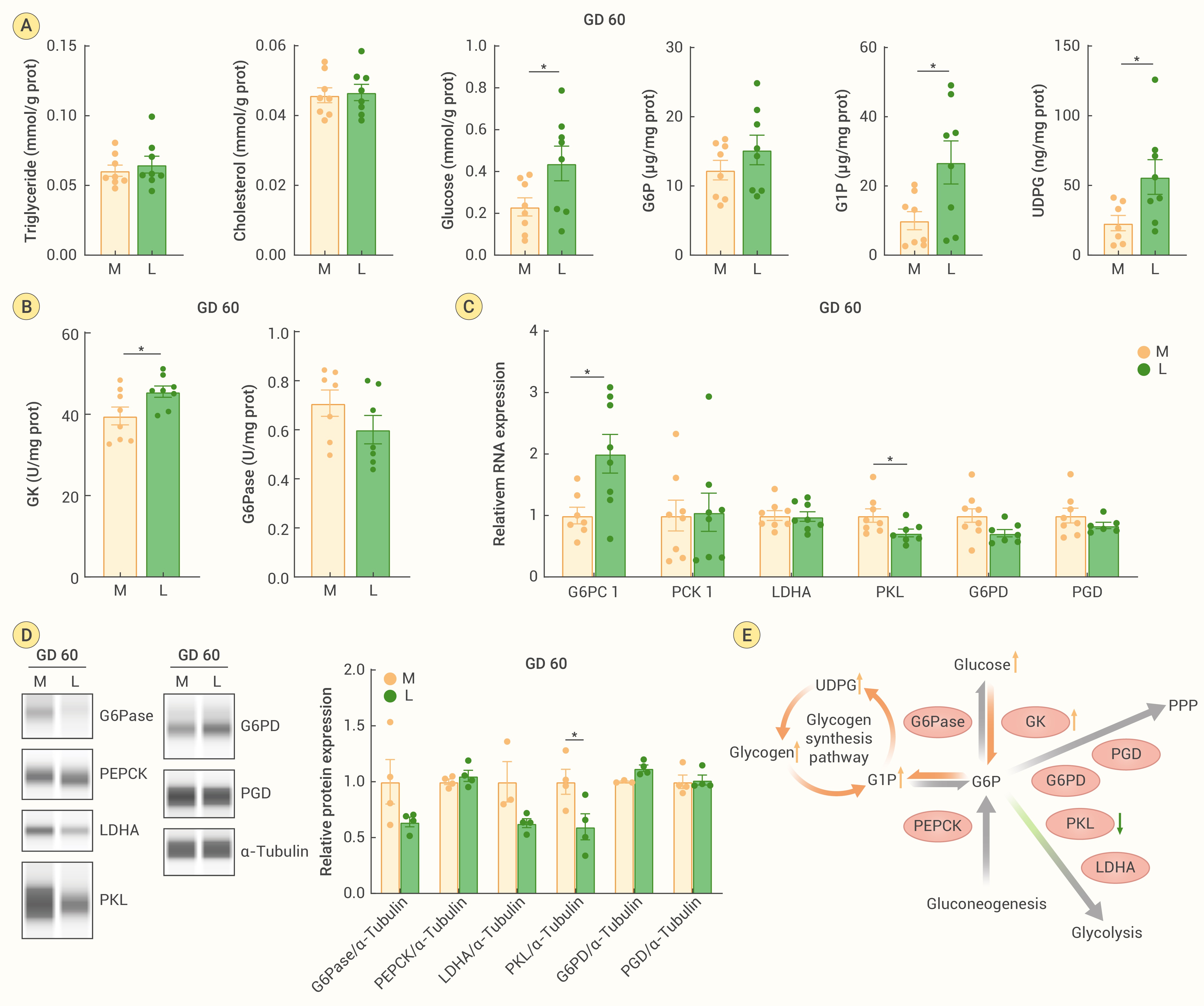
Figure 3.
Increased hepatic glucose flux into the glycogen synthesis pathway contributes to glycogen accumulation in the L group on GD 60
-
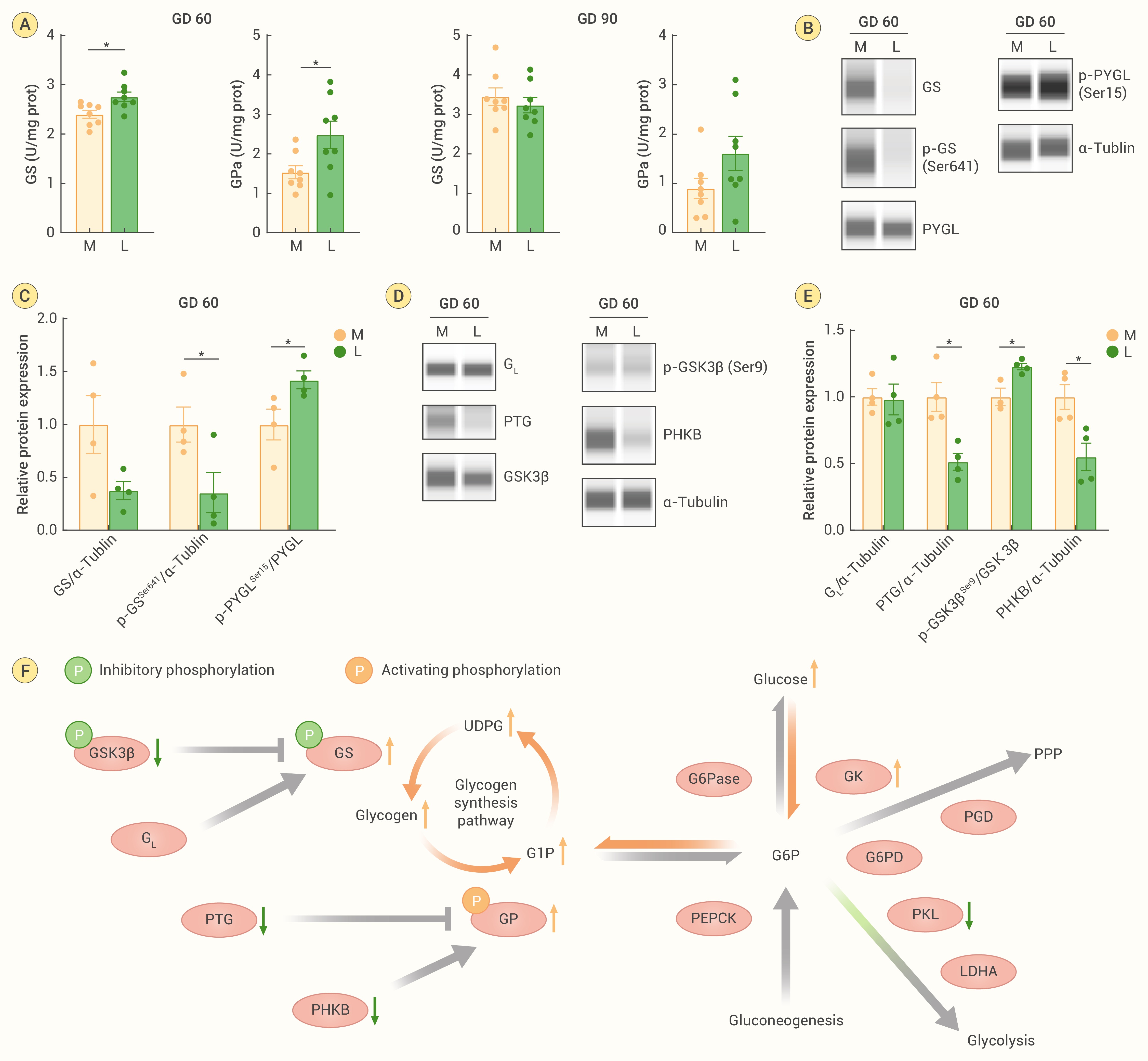
Figure 4.
Protein expression levels of regulators involved in modulating GS and GPa activity on GD 60
-
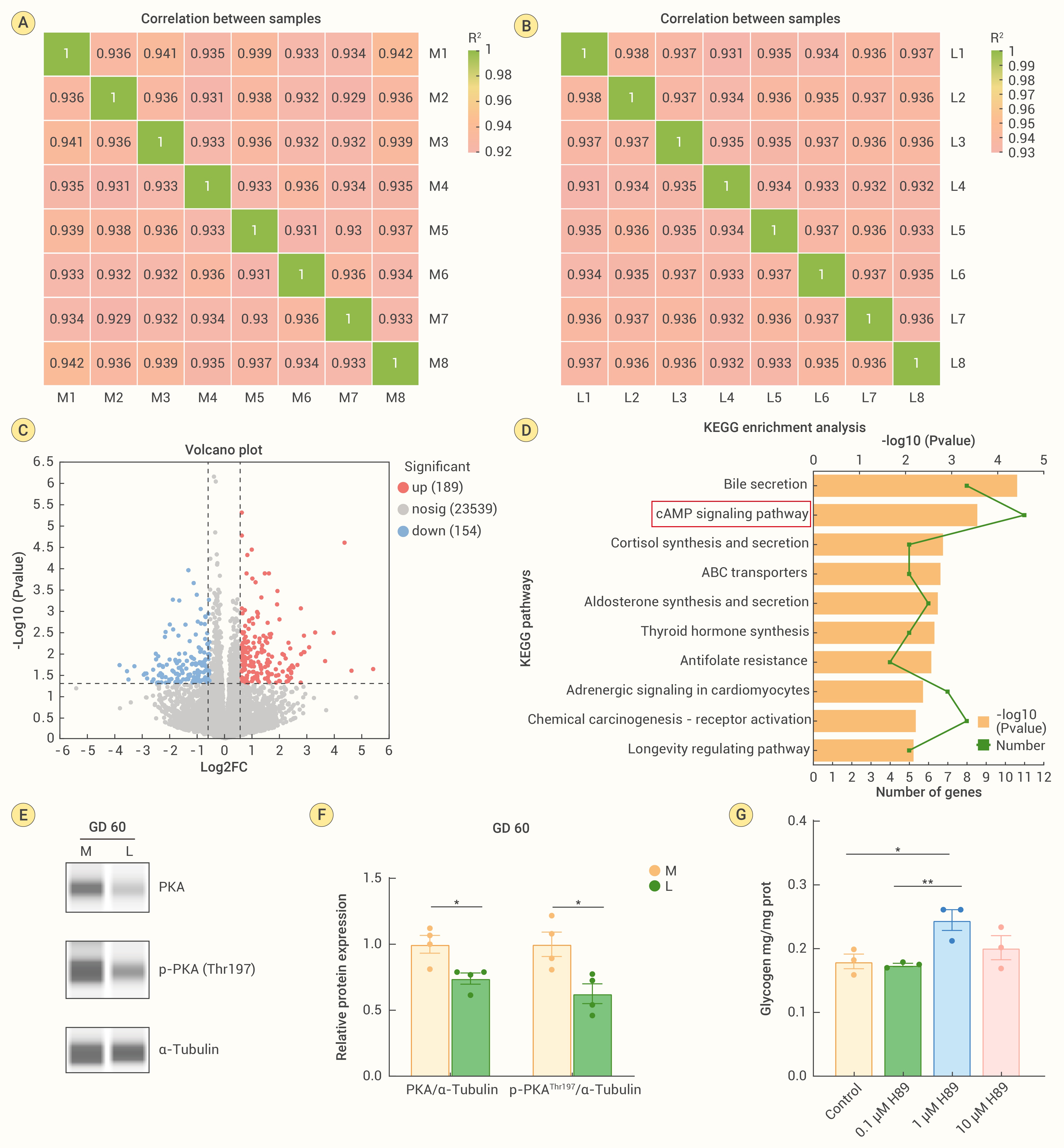
Figure 5.
RNA-seq analysis reveals potential targets involved in hepatic glycogen deposition regulation on GD 60
