
Expression pattern of glutaminase informs the dynamics of glutamine metabolism
-
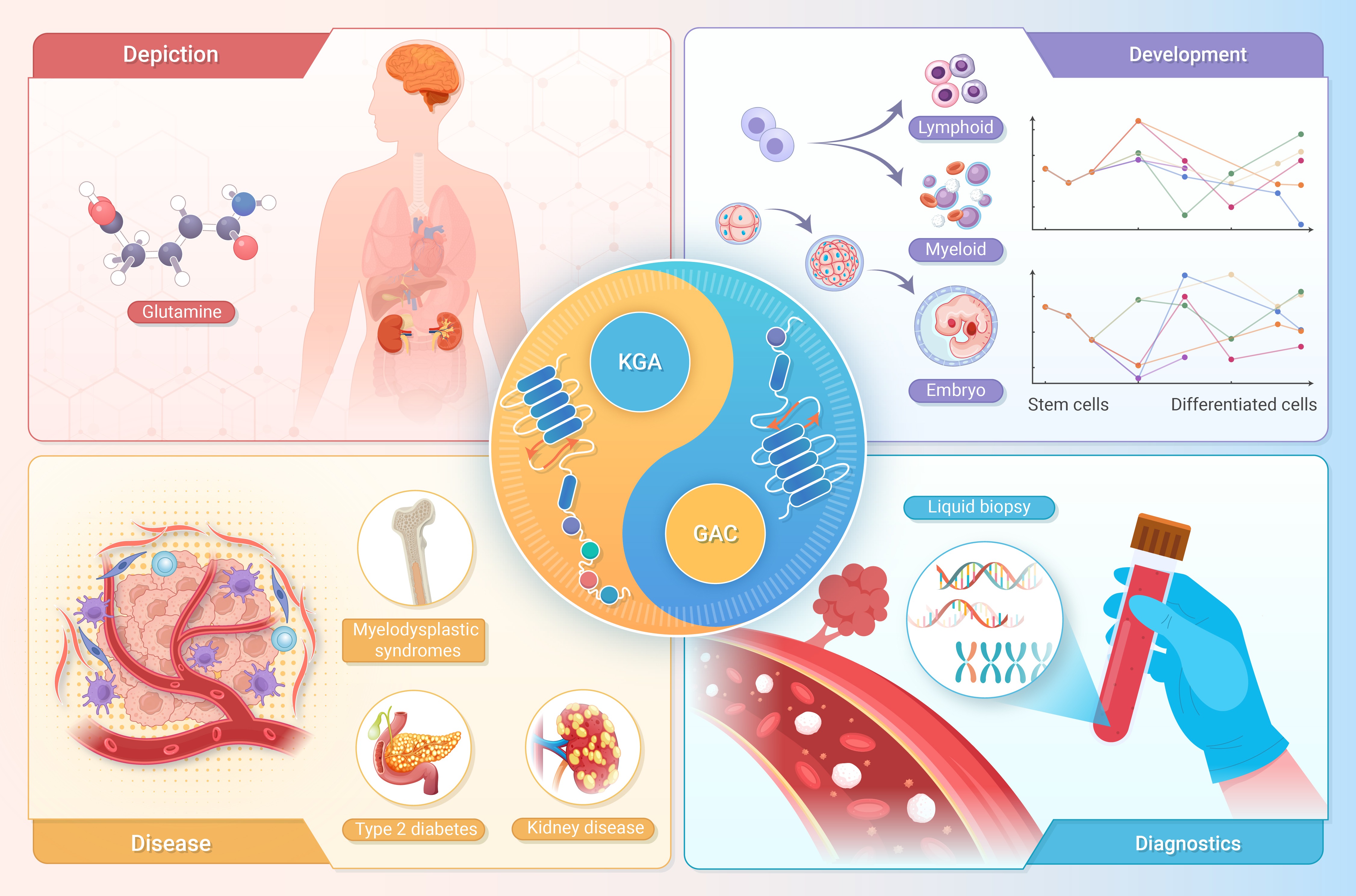
GRAPHICAL ABSTRACT
 DownLoad:
Full size image
DownLoad:
Full size image
-
PUBLIC SUMMARY
-
We propose a universal approach to quantify glutamine metabolism via glutaminase expression patterns.
We reveal key timings and insights into glutamine metabolism dynamics in development, diseases, and cancer.
Altered glutamine metabolism in cancer patients' plasma (PBMCs, platelets) aids noninvasive diagnosis.
Glutaminase expression patterns track glutamine metabolism dynamics, demonstrating translational value.
-
-
ABSTRACT
Glutamine is an essential nutrient that plays critical roles in both normal physiological processes and disease pathogenesis. In this study, we introduce a quantitative approach to trace the dynamics of glutamine metabolism by monitoring the expression patterns of glutaminase (GLS) and its two splicing isoforms, whose coding products exhibit distinct catalytic activities. This approach can be seamlessly integrated with quantitative PCR (qPCR), conventional bulk RNA sequencing, and emerging single-cell RNA sequencing technologies. We identified key temporal dynamics of glutamine metabolism during embryonic development and stem cell differentiation. Additionally, we characterized alterations in glutamine metabolism across various disease states and in response to drug treatments. In cancer, glutamine metabolism is highly variable across tumor types and is associated with T-cell exhaustion within the tumor microenvironment, as well as the metastatic potential of circulating tumor cells. Furthermore, aberrant glutamine metabolism was detected in various peripheral blood analytes from cancer patients, suggesting the potential for non-invasive cancer diagnosis. Our findings demonstrate the utility of quantitative glutamine metabolism tracing in investigating cellular processes, disease mechanisms, therapeutic responses, and its potential application in translational medicine. -

-
REFERENCES
[1] Yang L., Venneti S. and Nagrath D. (2017). Glutaminolysis: A hallmark of cancer metabolism. Annu. Rev. Biomed. Eng. 19:163−194. DOI:10.1146/annurev-bioeng-071516-044546 [2] Yang W. H., Qiu Y., Stamatatos O., et al. (2021). Enhancing the efficacy of glutamine metabolism inhibitors in cancer therapy. Trends Cancer 7:790−804. DOI:10.1016/j.trecan.2021.04.003 [3] Li X., Peng X., Li Y., et al. (2024). Glutamine addiction in tumor cell: oncogene regulation and clinical treatment. Cell Commun. Signal 22:12. DOI:10.1186/s12964-023-01449-x [4] Dollet L., Kuefner M., Caria E., et al. (2022). Glutamine regulates skeletal muscle immunometabolism in type 2 diabetes. Diabetes 71:624−636. DOI:10.2337/db20-0814 [5] Cruzat V., Macedo Rogero M., Noel Keane K., et al. (2018). Glutamine: metabolism and immune function, supplementation and clinical translation. Nutrients 10. DOI:10.3390/nu10111564 [6] Smith B., Schafer X. L., Ambeskovic A., et al. (2016). Addiction to coupling of the Warburg Effect with glutamine catabolism in cancer cells. Cell Rep. 17:821−836. DOI:10.1016/j.celrep.2016.09.045 [7] Vaupel P., and Multhoff G. (2021). Revisiting the Warburg effect: historical dogma versus current understanding. J. Physiol. 599:1745−1757. DOI:10.1113/JP278810 [8] Simon J., Nunez-Garcia M., Fernandez-Tussy P., et al. (2020). Targeting hepatic glutaminase 1 ameliorates non-alcoholic steatohepatitis by restoring very-low-density lipoprotein triglyceride assembly. Cell Metab. 31:605-622 e610. DOI:10.1016/j.cmet.2020.01.013 [9] Tu H., Yin X., Wen J., et al. (2022). Glutaminase 1 blockade alleviates nonalcoholic steatohepatitis via promoting proline metabolism. Biochem. Biophys. Res. Commun. 634:1−9. DOI:10.1016/j.bbrc.2022.10.007 [10] Bayraktar A., Li X., Kim W., et al. (2023). Drug repositioning targeting glutaminase reveals drug candidates for the treatment of Alzheimer's disease patients. J. Transl. Med. 21:332. DOI:10.1186/s12967-023-04192-6 [11] Okabe S., Moriyama M., Arai Y., et al. (2024). Glutaminase 1 plays critical roles in myelodysplastic syndrome and acute myeloid leukemia cells. Cancer Biomark. DOI:10.3233/CBM-230454 [12] Jin J., Byun J. K., Choi Y. K., et al. (2023). Targeting glutamine metabolism as a therapeutic strategy for cancer. Exp. Mol. Med. 55:706−715. DOI:10.1038/s12276-023-00971-9 [13] Patel C. H., Leone R. D., Horton M. R., et al. (2019). Targeting metabolism to regulate immune responses in autoimmunity and cancer. Nat. Rev. Drug Discov. 18:669−688. DOI:10.1038/s41573-019-0032-5 [14] Ansari R. E., Craze M. L., Althobiti M., et al. (2020). Enhanced glutamine uptake influences composition of immune cell infiltrates in breast cancer. Br. J. Cancer 122:94−101. DOI:10.1038/s41416-019-0626-z [15] Xu L., Yin Y., Li Y., et al. (2021). A glutaminase isoform switch drives therapeutic resistance and disease progression of prostate cancer. Proc. Natl. Acad. Sci. USA 118. DOI:10.1073/pnas.2012748118 [16] Varghese S., Pramanik S., Williams L. J., et al. (2021). The glutaminase inhibitor CB-839 (Telaglenastat) enhances the antimelanoma activity of T-Cell-mediated immunotherapies. Mol. Cancer Ther. 20:500−511. DOI:10.1158/1535-7163.MCT-20-0430 [17] Wang J. J., Siu M. K., Jiang Y. X., et al. (2021). A combination of glutaminase inhibitor 968 and PD-L1 blockade boosts the immune response against ovarian cancer. Biomolecules 11. DOI:10.3390/biom11121749 [18] Sun K. (2020). Ktrim: an extra-fast and accurate adapter- and quality-trimmer for sequencing data. Bioinformatics 36:3561−3562. DOI:10.1093/bioinformatics/btaa171 [19] Zhao Y., Yang M., Gong F., et al. (2024). Accelerating 3D genomics data analysis with Microcket. Commun. Biol. 7:675. DOI:10.1038/s42003-024-06382-4 [20] Li B., and Dewey C. N. (2011). RSEM: accurate transcript quantification from RNA-Seq data with or without a reference genome. BMC Bioinformatics 12:323. DOI:10.1186/1471-2105-12-323 [21] Sayers E. W., Beck J., Bolton E. E., et al. (2024). Database resources of the National Center for Biotechnology Information. Nucleic Acids Res. 52:D33−D43. DOI:10.1093/nar/gkad1044 [22] Hao Y., Hao S., Andersen-Nissen E., et al. (2021). Integrated analysis of multimodal single-cell data. Cell 184:3573-3587 e3529. DOI:10.1016/j.cell.2021.04.048 [23] Korsunsky I., Millard N., Fan J., et al. (2019). Fast, sensitive and accurate integration of single-cell data with Harmony. Nat. Methods 16:1289−1296. DOI:10.1038/s41592-019-0619-0 [24] Hay S. B., Ferchen K., Chetal K., et al. (2018). The Human Cell Atlas bone marrow single-cell interactive web portal. Exp. Hematol. 68:51−61. DOI:10.1016/j.exphem.2018.09.004 [25] Aran D., Looney A. P., Liu L., et al. (2019). Reference-based analysis of lung single-cell sequencing reveals a transitional profibrotic macrophage. Nat. Immunol. 20:163−172. DOI:10.1038/s41590-018-0276-y [26] Miao Z., Deng K., Wang X., et al. (2018). DEsingle for detecting three types of differential expression in single-cell RNA-seq data. Bioinformatics 34:3223−3224. DOI:10.1093/bioinformatics/bty332 [27] Picelli S., Björklund Å. K., Faridani O. R., et al. (2013). Smart-seq2 for sensitive full-length transcriptome profiling in single cells. Nat. Methods 10:1096−1098. DOI:10.1038/nmeth.2639 [28] Zheng G. X., Terry J. M., Belgrader P., et al. (2017). Massively parallel digital transcriptional profiling of single cells. Nat. Commun. 8:14049. DOI:10.1038/ncomms14049 [29] Cassago A., Ferreira A. P., Ferreira I. M., et al. (2012). Mitochondrial localization and structure-based phosphate activation mechanism of Glutaminase C with implications for cancer metabolism. Proc. Natl. Acad. Sci. USA 109:1092−1097. DOI:10.1073/pnas.1112495109 [30] Adamoski D., Dias M. M., Quesnay J. E. N., et al. (2023). Molecular mechanism of glutaminase activation through filamentation and the role of filaments in mitophagy protection. Nat. Struct. Mol. Biol. 30:1902−1912. DOI:10.1038/s41594-023-01118-0 [31] The GTEx Consortium (2013). The Genotype-Tissue Expression (GTEx) project. Nat. Genet. 45:580-585 [32] The ENCODE Project Consortium (2012). An integrated encyclopedia of DNA elements in the human genome. Nature 489:57-74. DOI:10.1038/nature11247 [33] Katt W. P., Lukey M. J., and Cerione R. A. (2017). A tale of two glutaminases: homologous enzymes with distinct roles in tumorigenesis. Future Med. Chem. 9:223−243. DOI:10.4155/fmc-2016-0190 [34] Zhang J., Zhao J., Dahan P., et al. (2018). Metabolism in pluripotent stem cells and early mammalian development. Cell Metab. 27:332−338. DOI:10.1016/j.cmet.2018.01.008 [35] Oburoglu L., Tardito S., Fritz V., et al. (2014). Glucose and glutamine metabolism regulate human hematopoietic stem cell lineage specification. Cell Stem Cell 15:169−184. DOI:10.1016/j.stem.2014.06.002 [36] Meynial-Denis D. (2016). Glutamine metabolism in advanced age. Nutr. Rev. 74:225−236. DOI:10.1093/nutrit/nuv052 [37] Yan L., Yang M., Guo H., et al. (2013). Single-cell RNA-Seq profiling of human preimplantation embryos and embryonic stem cells. Nat. Struct. Mol. Biol. 20:1131−1139. DOI:10.1038/nsmb.2660 [38] Xue Z., Huang K., Cai C., et al. (2013). Genetic programs in human and mouse early embryos revealed by single-cell RNA sequencing. Nature 500:593−597. DOI:10.1038/nature12364 [39] Deng Q., Ramskold D., Reinius B., et al. (2014). Single-cell RNA-seq reveals dynamic, random monoallelic gene expression in mammalian cells. Science 343:193−196. DOI:10.1126/science.1245316 [40] Mazid M. A., Ward C., Luo Z., et al. (2022). Rolling back human pluripotent stem cells to an eight-cell embryo-like stage. Nature 605:315−324. DOI:10.1038/s41586-022-04625-0 [41] Macfarlan T. S., Gifford W. D., Driscoll S., et al. (2012). Embryonic stem cell potency fluctuates with endogenous retrovirus activity. Nature 487:57−63. DOI:10.1038/nature11244 [42] Taubenschmid-Stowers J., Rostovskaya M., Santos F., et al. (2022). 8C-like cells capture the human zygotic genome activation program in vitro. Cell Stem Cell 29:449-459 e446. DOI:10.1016/j.stem.2022.01.014 [43] Liu Y., Fan X., Wang R., et al. (2018). Single-cell RNA-seq reveals the diversity of trophoblast subtypes and patterns of differentiation in the human placenta. Cell Res. 28:819−832. DOI:10.1038/s41422-018-0066-y [44] Chen L., Kostadima M., Martens J. H. A., et al. (2014). Transcriptional diversity during lineage commitment of human blood progenitors. Science 345:1251033. DOI:10.1126/science.1251033 [45] Zhang Y., Li T., Preissl S., et al. (2019). Transcriptionally active HERV-H retrotransposons demarcate topologically associating domains in human pluripotent stem cells. Nat. Genet. 51:1380−1388. DOI:10.1038/s41588-019-0479-7 [46] Salabei J.K., Lorkiewicz P.K., Holden C.R., et al. (2015). Glutamine regulates cardiac progenitor cell metabolism and proliferation. Stem Cells 33:2613−2627. DOI:10.1002/stem.2047 [47] Wiedenmann S., Breunig M., Merkle J., et al. (2021). Single-cell-resolved differentiation of human induced pluripotent stem cells into pancreatic duct-like organoids on a microwell chip. Nat. Biomed. Eng. 5:897−913. DOI:10.1038/s41551-021-00757-2 [48] Bornstein R., Mulholland M. T., Sedensky M., et al. (2023). Glutamine metabolism in diseases associated with mitochondrial dysfunction. Mol. Cell Neurosci. 126:103887. DOI:10.1016/j.mcn.2023.103887 [49] Segerstolpe A., Palasantza A., Eliasson P., et al. (2016). Single-cell transcriptome profiling of human pancreatic islets in health and type 2 diabetes. Cell Metab. 24:593−607. DOI:10.1016/j.cmet.2016.08.020 [50] Govaere O., Cockell S., Tiniakos D., et al. (2020). Transcriptomic profiling across the nonalcoholic fatty liver disease spectrum reveals gene signatures for steatohepatitis and fibrosis. Sci. Transl. Med. 12. DOI:10.1126/scitranslmed.aba4448 [51] Masoodi M., Gastaldelli A., Hyotylainen T., et al. (2021). Metabolomics and lipidomics in NAFLD: biomarkers and non-invasive diagnostic tests. Nat. Rev. Gastroenterol. Hepatol. 18:835−856. DOI:10.1038/s41575-021-00502-9 [52] Bennett D. A., Schneider J. A., Buchman A. S., et al. (2012). Overview and findings from the rush memory and aging project. Curr. Alzheimer Res. 9:646−663. DOI:10.2174/156720512801322663 [53] Crimins J. L., Pooler A., Polydoro M., et al. (2013). The intersection of amyloid beta and tau in glutamatergic synaptic dysfunction and collapse in Alzheimer's disease. Ageing Res. Rev. 12:757−763. DOI:10.1016/j.arr.2013.03.002 [54] Dussiau C., Boussaroque A., Gaillard M., et al. (2022). Hematopoietic differentiation is characterized by a transient peak of entropy at a single-cell level. BMC Biol. 20:60. DOI:10.1186/s12915-022-01264-9 [55] Steensma D. P. (2018). Myelodysplastic syndromes current treatment algorithm 2018. Blood Cancer J. 8:47. DOI:10.1038/s41408-018-0085-4 [56] Steensma D. P. (2015). Myelodysplastic syndromes: diagnosis and treatment. Mayo. Clin. Proc. 90:969−983. DOI:10.1016/j.mayocp.2015.04.001 [57] Thai M., Graham N. A., Braas D., et al. (2014). Adenovirus E4ORF1-induced MYC activation promotes host cell anabolic glucose metabolism and virus replication. Cell Metab. 19:694−701. DOI:10.1016/j.cmet.2014.03.009 [58] Herwanto V., Tang B., Wang Y., et al. (2021). Blood transcriptome analysis of patients with uncomplicated bacterial infection and sepsis. BMC Res. Notes 14:76. DOI:10.1186/s13104-021-05488-w [59] Mueller H., Wildum S., Luangsay S., et al. (2018). A novel orally available small molecule that inhibits hepatitis B virus expression. J. Hepatol. 68:412−420. DOI:10.1016/j.jhep.2017.10.014 [60] Yoo H. C., Yu Y. C., Sung Y., et al. (2020). Glutamine reliance in cell metabolism. Exp. Mol. Med. 52:1496−1516. DOI:10.1038/s12276-020-00504-8 [61] Demicco M., Liu X. Z., Leithner K., et al. (2024). Metabolic heterogeneity in cancer. Nat. Metab. 6:18−38. DOI:10.1038/s42255-023-00963-z [62] Zhang J., Pavlova N. N. and Thompson C. B. (2017). Cancer cell metabolism: the essential role of the nonessential amino acid, glutamine. EMBO J. 36:1302−1315. DOI:10.15252/embj.201696151 [63] Weinstein J. N., Collisson E. A., Mills G. B., et al. (2013). The Cancer Genome Atlas Pan-Cancer analysis project. Nat. Genet. 45:1113−1120. DOI:10.1038/ng.2764 [64] Schnepp P. M., Shelley G., Dai J., et al. (2020). Single-cell transcriptomics analysis identifies nuclear protein 1 as a regulator of docetaxel resistance in prostate cancer cells. Mol. Cancer Res. 18:1290−1301. DOI:10.1158/1541-7786.MCR-20-0051 [65] Gomes A. P., Ilter D., Low V., et al. (2020). Age-induced accumulation of methylmalonic acid promotes tumour progression. Nature 585:283−287. DOI:10.1038/s41586-020-2630-0 [66] Zhong X., He Z., Yin L., et al. (2023). Glutamine metabolism in tumor metastasis: Genes, mechanisms and the therapeutic targets. Heliyon 9:e20656. DOI:10.1016/j.heliyon.2023.e20656 [67] Fan F., Chen D., Zhao Y., et al. (2020). Rapid preliminary purity evaluation of tumor biopsies using deep learning approach. Comput. Struct. Biotechnol. J. 18:1746−1753. DOI:10.1016/j.csbj.2020.06.007 [68] Che L. H., Liu J. W., Huo J. P., et al. (2021). A single-cell atlas of liver metastases of colorectal cancer reveals reprogramming of the tumor microenvironment in response to preoperative chemotherapy. Cell Discov. 7:80. DOI:10.1038/s41421-021-00312-y [69] Laughney A. M., Hu J., Campbell N. R., et al. (2020). Regenerative lineages and immune-mediated pruning in lung cancer metastasis. Nat. Med. 26:259−269. DOI:10.1038/s41591-019-0750-6 [70] Lin W., Noel P., Borazanci E. H., et al. (2020). Single-cell transcriptome analysis of tumor and stromal compartments of pancreatic ductal adenocarcinoma primary tumors and metastatic lesions. Genome Med. 12:80. DOI:10.1186/s13073-020-00776-9 [71] Blank C. U., Haining W. N., Held W., et al. (2019). Defining 'T cell exhaustion'. Nat. Rev. Immunol 19:665−674. DOI:10.1038/s41577-019-0221-9 [72] Ma G., Zhang Z., Li P., et al. (2022). Reprogramming of glutamine metabolism and its impact on immune response in the tumor microenvironment. Cell Commun Signal 20:114. DOI:10.1186/s12964-022-00909-0 [73] Kaymak I., Williams K. S., Cantor J. R., et al. (2021). Immunometabolic interplay in the tumor microenvironment. Cancer Cell 39:28−37. DOI:10.1016/j.ccell.2020.09.004 [74] Sun Y. F., Wu L., Liu S. P., et al. (2021). Dissecting spatial heterogeneity and the immune-evasion mechanism of CTCs by single-cell RNA-seq in hepatocellular carcinoma. Nat. Commun. 12:4091. DOI:10.1038/s41467-021-24386-0 [75] Franses J. W., Philipp J., Missios P., et al. (2020). Pancreatic circulating tumor cell profiling identifies LIN28B as a metastasis driver and drug target. Nat. Commun. 11:3303. DOI:10.1038/s41467-020-17150-3 [76] Aceto N., Bardia A., Miyamoto D. T., et al. (2014). Circulating tumor cell clusters are oligoclonal precursors of breast cancer metastasis. Cell 158:1110−1122. DOI:10.1016/j.cell.2014.07.013 [77] Schuster E., Taftaf R., Reduzzi C., et al. (2021). Better together: circulating tumor cell clustering in metastatic cancer. Trends Cancer 7:1020−1032. DOI:10.1016/j.trecan.2021.07.001 [78] Diamantopoulou Z., Castro-Giner F., Schwab F. D., et al. (2022). The metastatic spread of breast cancer accelerates during sleep. Nature 607:156−162. DOI:10.1038/s41586-022-04875-y [79] Han Z., Feng W., Hu R., et al. (2021). RNA-seq profiling reveals PBMC RNA as a potential biomarker for hepatocellular carcinoma. Sci. Rep. 11:17797. DOI:10.1038/s41598-021-96952-x [80] Best M. G., Sol N., Kooi I., et al. (2015). RNA-seq of tumor-educated platelets enables blood-based pan-cancer, multiclass, and molecular pathway cancer diagnostics. Cancer Cell 28:666−676. DOI:10.1016/j.ccell.2015.09.018 [81] Liu T., Wang X., Guo W., et al. (2022). RNA sequencing of tumor-educated platelets reveals a three-gene diagnostic signature in esophageal squamous cell carcinoma. Front. Oncol. 12:824354. DOI:10.3389/fonc.2022.824354 [82] Yu S., Li Y., Liao Z., et al. (2020). Plasma extracellular vesicle long RNA profiling identifies a diagnostic signature for the detection of pancreatic ductal adenocarcinoma. Gut 69:540−550. DOI:10.1136/gutjnl-2019-318860 [83] Wang Y., Li Y., Zhao R., et al. (2017). Glutaminase C overexpression in the brain induces learning deficits, synaptic dysfunctions, and neuroinflammation in mice. Brain Behav. Immun. 66:135−145. DOI:10.1016/j.bbi.2017.06.007 [84] Gao G., Zhao S., Xia X., et al. (2019). Glutaminase C regulates microglial activation and pro-inflammatory exosome release: relevance to the pathogenesis of Alzheimer's Disease. Front. Cell Neurosci. 13:264. DOI:10.3389/fncel.2019.00264 [85] Soomro I., Sun Y., Li Z., et al. (2018). Glutamine metabolism via glutaminase 1 in autosomal-dominant polycystic kidney disease. Nephrol. Dial Transplant 33:1343−1353. DOI:10.1093/ndt/gfx349 [86] Rodriguez-Terrones D., Hartleben G., Gaume X., et al. (2020). A distinct metabolic state arises during the emergence of 2-cell-like cells. EMBO Rep. 21:e48354. DOI:10.15252/embr.201948354 [87] Marsboom G., Zhang G. F., Pohl-Avila N., et al. (2016). Glutamine metabolism regulates the pluripotency transcription factor OCT4. Cell Rep. 16:323−332. DOI:10.1016/j.celrep.2016.05.089 [88] Lu V., Roy I. J., Torres A., Jr., et al. (2022). Glutamine-dependent signaling controls pluripotent stem cell fate. Dev. Cell 57:610-623 e618. DOI:10.1016/j.devcel.2022.02.003 [89] Devreker F., Winston R. M., and Hardy K. (1998). Glutamine improves human preimplantation development in vitro. Fertil. Steril. 69:293−299. DOI:10.1016/s0015-0282(97)00463-9 [90] Wang Y., Huang Y., Zhao L., et al. (2014). Glutaminase 1 is essential for the differentiation, proliferation, and survival of human neural progenitor cells. Stem Cells Dev. 23:2782−2790. DOI:10.1089/scd.2014.0022 [91] Kaiser L., Weinschrott H., Quint I., et al. (2020). Metabolite patterns in human myeloid hematopoiesis result from lineage-dependent active metabolic pathways. Int. J. Mol. Sci. 21. DOI:10.3390/ijms21176092 [92] Carr E. L., Kelman A., Wu G. S., et al. (2010). Glutamine uptake and metabolism are coordinately regulated by ERK/MAPK during T lymphocyte activation. J. Immunol. 185:1037−1044. DOI:10.4049/jimmunol.0903586 [93] Lyu J., Gu Z., Zhang Y., et al. (2024). A glutamine metabolic switch supports erythropoiesis. Science 386:eadh9215. DOI:10.1126/science.adh9215 [94] Cluntun A. A., Lukey M. J., Cerione R. A., et al. (2017). Glutamine metabolism in cancer: understanding the heterogeneity. Trends Cancer 3:169−180. DOI:10.1016/j.trecan.2017.01.005 [95] Altman B. J., Stine Z. E., and Dang C. V. (2016). From Krebs to clinic: glutamine metabolism to cancer therapy. Nat. Rev. Cancer 16:619−634. DOI:10.1038/nrc.2016.71 [96] Njei B., Al-Ajlouni Y. A., Ameyaw P., et al. (2024). Role of ammonia and glutamine in the pathogenesis and progression of metabolic dysfunction-associated steatotic liver disease: A systematic review. J. Gastroenterol. Hepatol. 39:1788−1808. DOI:10.1111/jgh.16603 [97] Salcedo C., Pozo Garcia V., Garcia-Adan B., et al. (2023). Increased glucose metabolism and impaired glutamate transport in human astrocytes are potential early triggers of abnormal extracellular glutamate accumulation in hiPSC-derived models of Alzheimer's disease. J. Neurochem. DOI:10.1111/jnc.16014 [98] Andersen J. V., Schousboe A., and Verkhratsky A. (2022). Astrocyte energy and neurotransmitter metabolism in Alzheimer's Disease: integration of the glutamate/GABA-glutamine cycle. Prog. Neurobiol. 217:102331. DOI:10.1016/j.pneurobio.2022.102331 [99] Ward P. S., Patel J., Wise D. R., et al. (2010). The common feature of leukemia-associated IDH1 and IDH2 mutations is a neomorphic enzyme activity converting alpha-ketoglutarate to 2-hydroxyglutarate. Cancer Cell 17:225−234. DOI:10.1016/j.ccr.2010.01.020 [100] Hu D., Zhang Z., Liu X., et al. (2024). Generalizable transcriptome-based tumor malignant level evaluation and molecular subtyping towards precision oncology. J. Transl. Med. 22:512. DOI:10.1186/s12967-024-05326-0 [101] Shajahan-Haq A. N., Cook K. L., Schwartz-Roberts J. L., et al. (2014). MYC regulates the unfolded protein response and glucose and glutamine uptake in endocrine resistant breast cancer. Mol. Cancer 13:239. DOI:10.1186/1476-4598-13-239 [102] Momcilovic M., Bailey S. T., Lee J. T., et al. (2018). The GSK3 signaling axis regulates adaptive glutamine metabolism in lung squamous cell carcinoma. Cancer Cell 33:905-921 e905. DOI:10.1016/j.ccell.2018.04.002 [103] Nicolay B. N., Gameiro P. A., Tschop K., et al. (2013). Loss of RBF1 changes glutamine catabolism. Genes Dev. 27:182−196. DOI:10.1101/gad.206227.112 [104] McGuirk S., Gravel S. P., Deblois G., et al. (2013). PGC-1alpha supports glutamine metabolism in breast cancer. Cancer Metab. 1:22. DOI:10.1186/2049-3002-1-22 [105] Zhao J., Hu Z., Zheng X., et al. (2024). Blood biomarkers of hepatocellular carcinoma: a critical review. Front. Cell Dev. Biol. 12:1489836. DOI:10.3389/fcell.2024.1489836 [106] van der Pol Y., and Mouliere F. (2019). Toward the early detection of cancer by decoding the epigenetic and environmental fingerprints of cell-free DNA. Cancer Cell 36:350−368. DOI:10.1016/j.ccell.2019.09.003 [107] An Y., Zhao X., Zhang Z., et al. (2023). DNA methylation analysis explores the molecular basis of plasma cell-free DNA fragmentation. Nat. Commun. 14:287. DOI:10.1038/s41467-023-35959-6 [108] Sun K., Jiang P., Chan K. C. A., et al. (2015). Plasma DNA tissue mapping by genome-wide methylation sequencing for noninvasive prenatal, cancer, and transplantation assessments. Proc. Natl. Acad. Sci. USA 112:E5503−5512. DOI:10.1073/pnas.1508736112 [109] Ju J., Zhao X., An Y., et al. (2024). Cell-free DNA end characteristics enable accurate and sensitive cancer diagnosis. Cell Rep. Methods 4:100877. DOI:10.1016/j.crmeth.2024.100877 -
ABOUT THIS ARTICLE
Cite this article:
Hu D., Wang W., Zhao X., et al. (2025). Expression pattern of glutaminase informs the dynamics of glutamine metabolism. The Innovation Life 3:100128. https://doi.org/10.59717/j.xinn-life.2024.100128
Welcome!
To request copyright permission to republish or share portions of our works, please visit Copyright Clearance Center's (CCC) Marketplace website at marketplace.copyright.com.
Article Metrics
Relative Articles
Cited by
Export File
Citation
Hu D., Wang W., Zhao X., et al. (2025). Expression pattern of glutaminase informs the dynamics of glutamine metabolism. The Innovation Life 3:100128. https://doi.org/10.59717/j.xinn-life.2024.100128

Format
Content
-
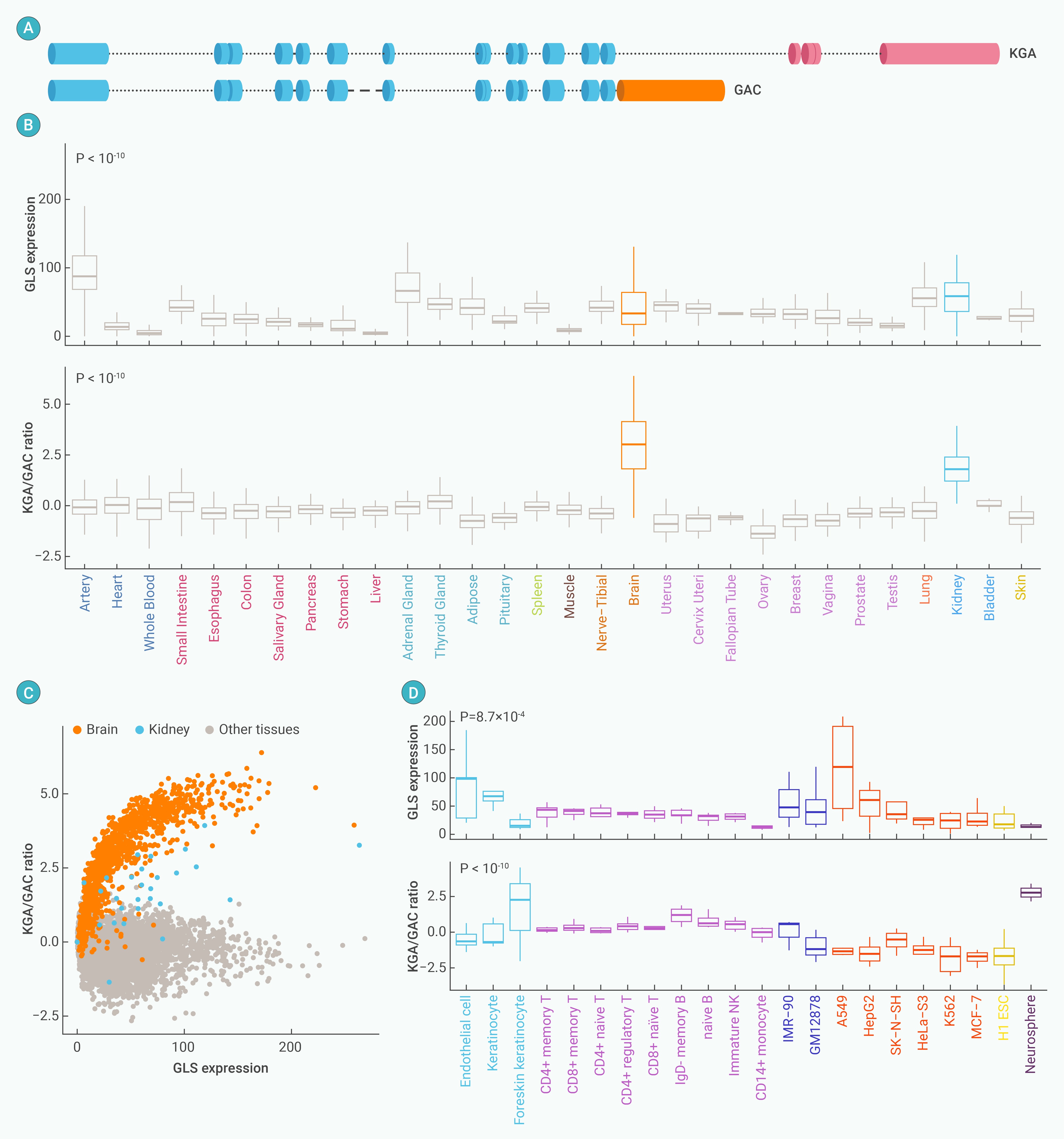
Figure 1.
Glutamine metabolism patterns in human tissues and cell lines
-
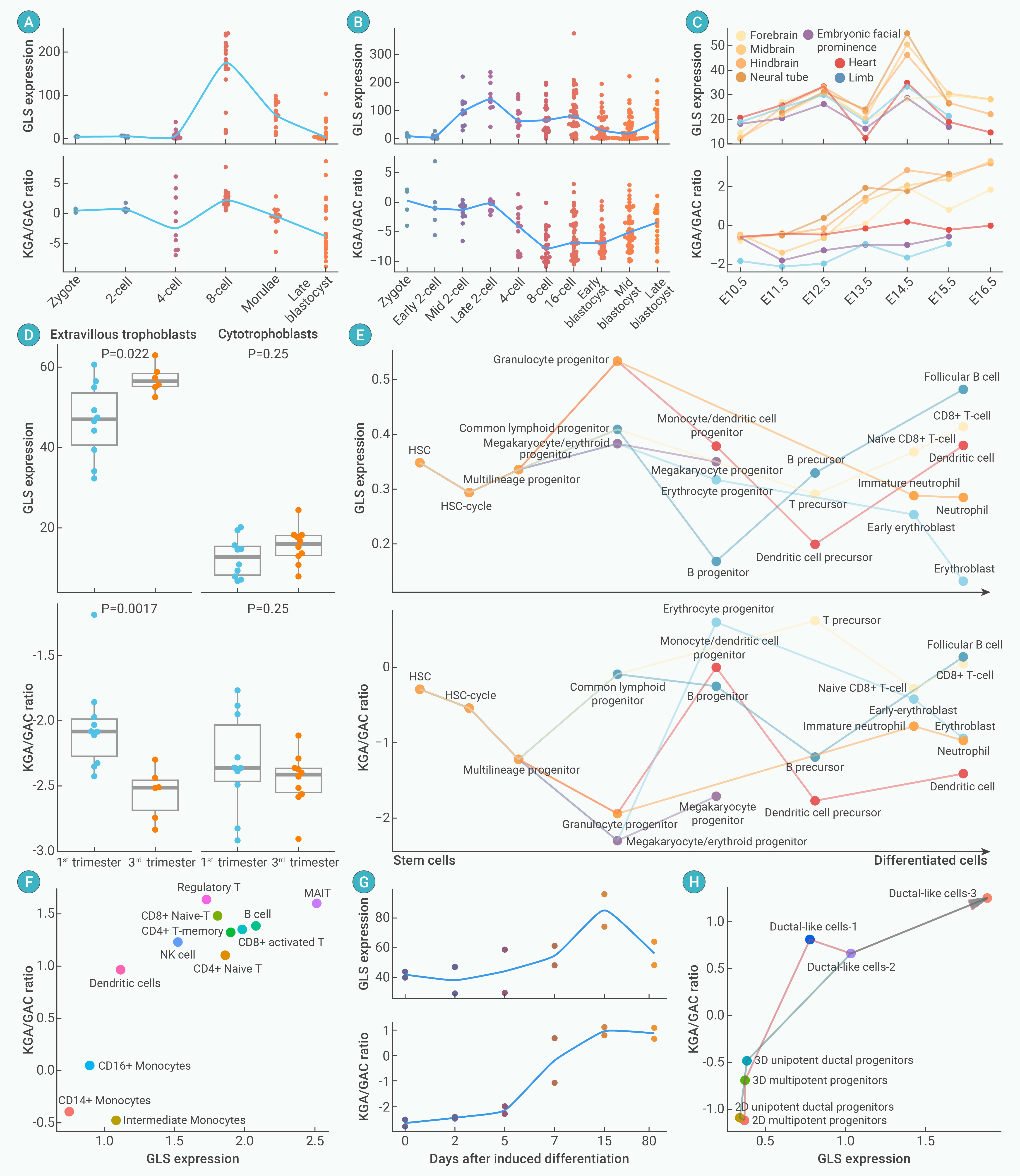
Figure 2.
Glutamine metabolism dynamics in development and differentiation
-
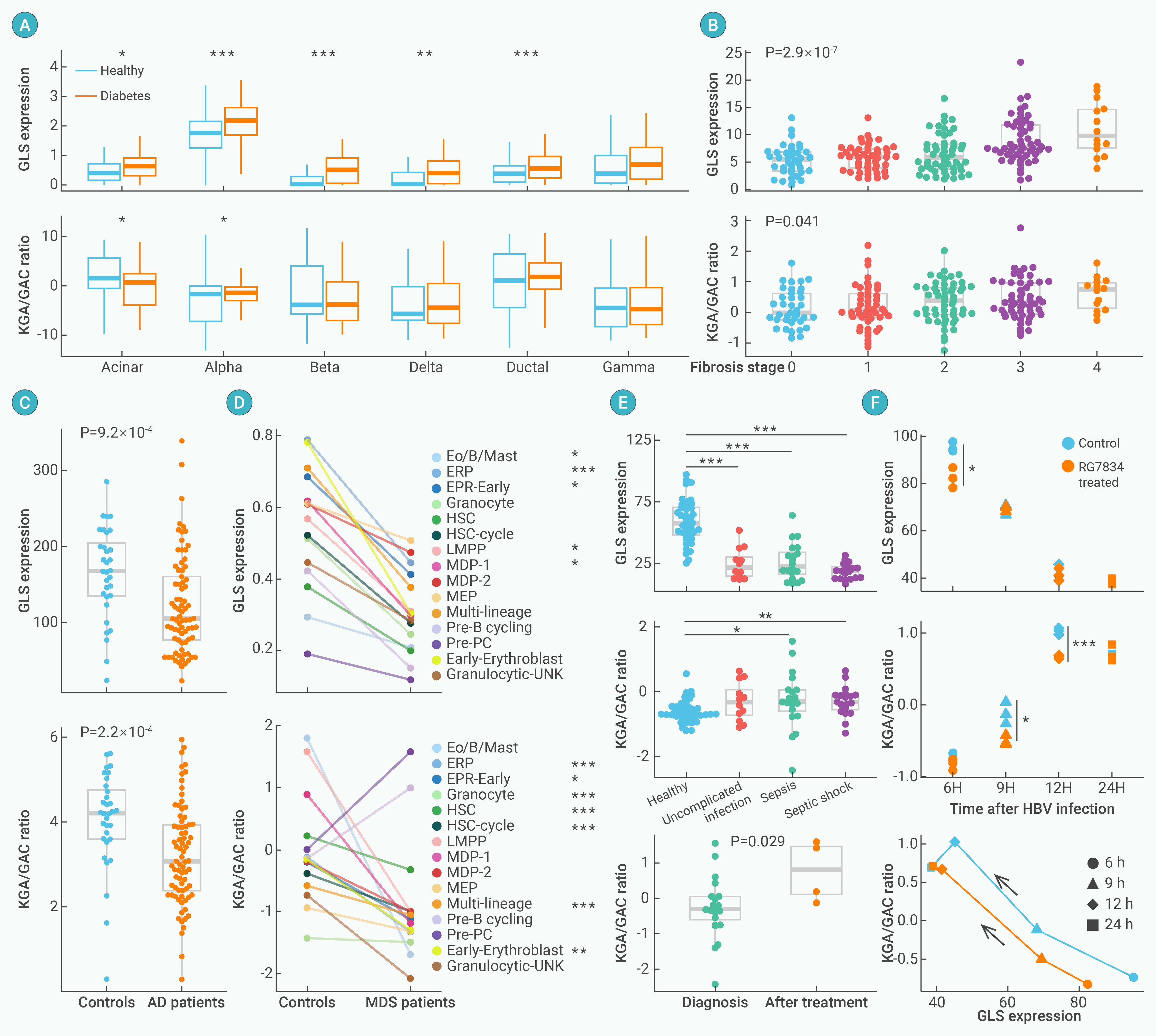
Figure 3.
Glutamine metabolism in diseases
-
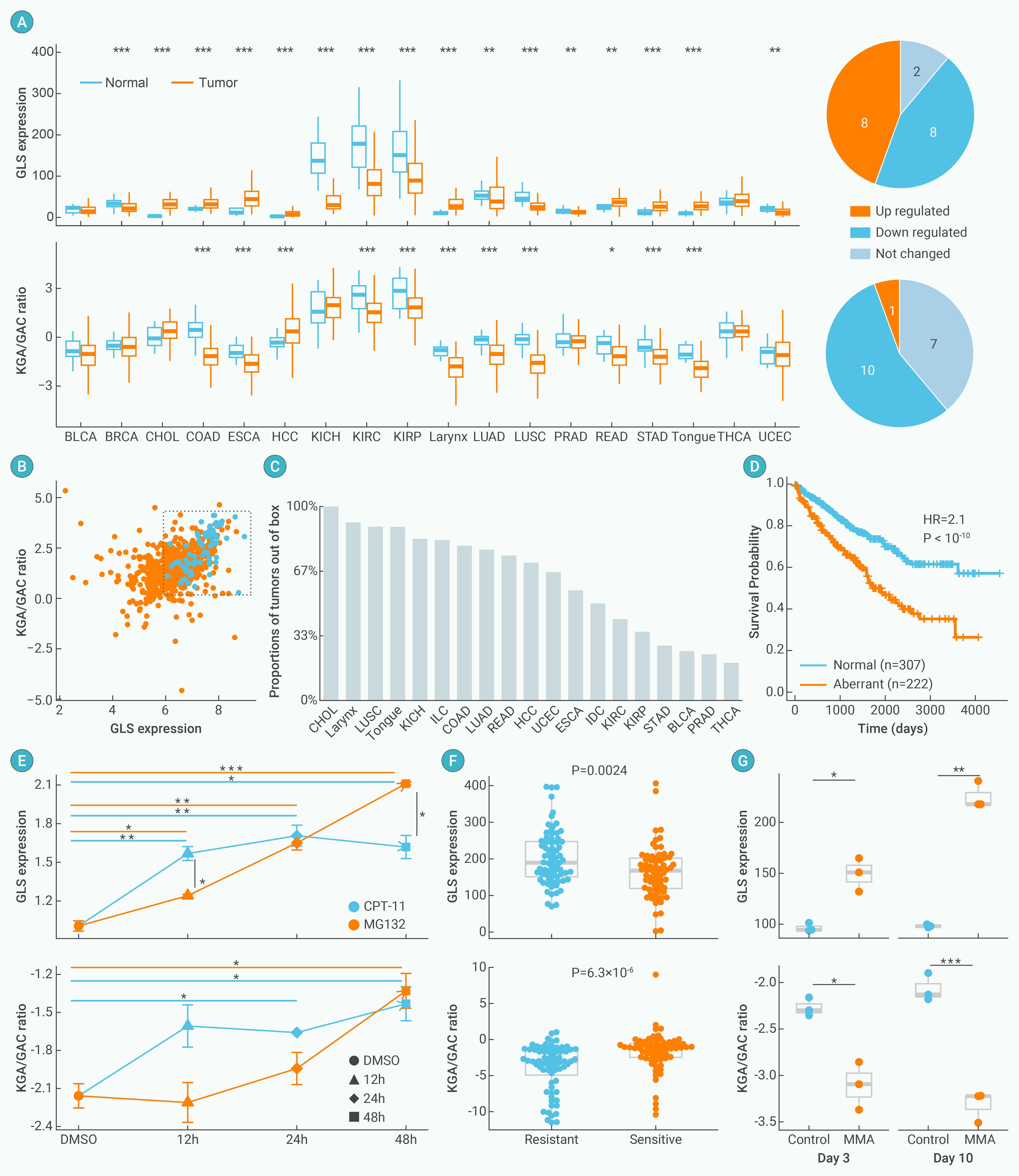
Figure 4.
Glutamine metabolism in cancer
-
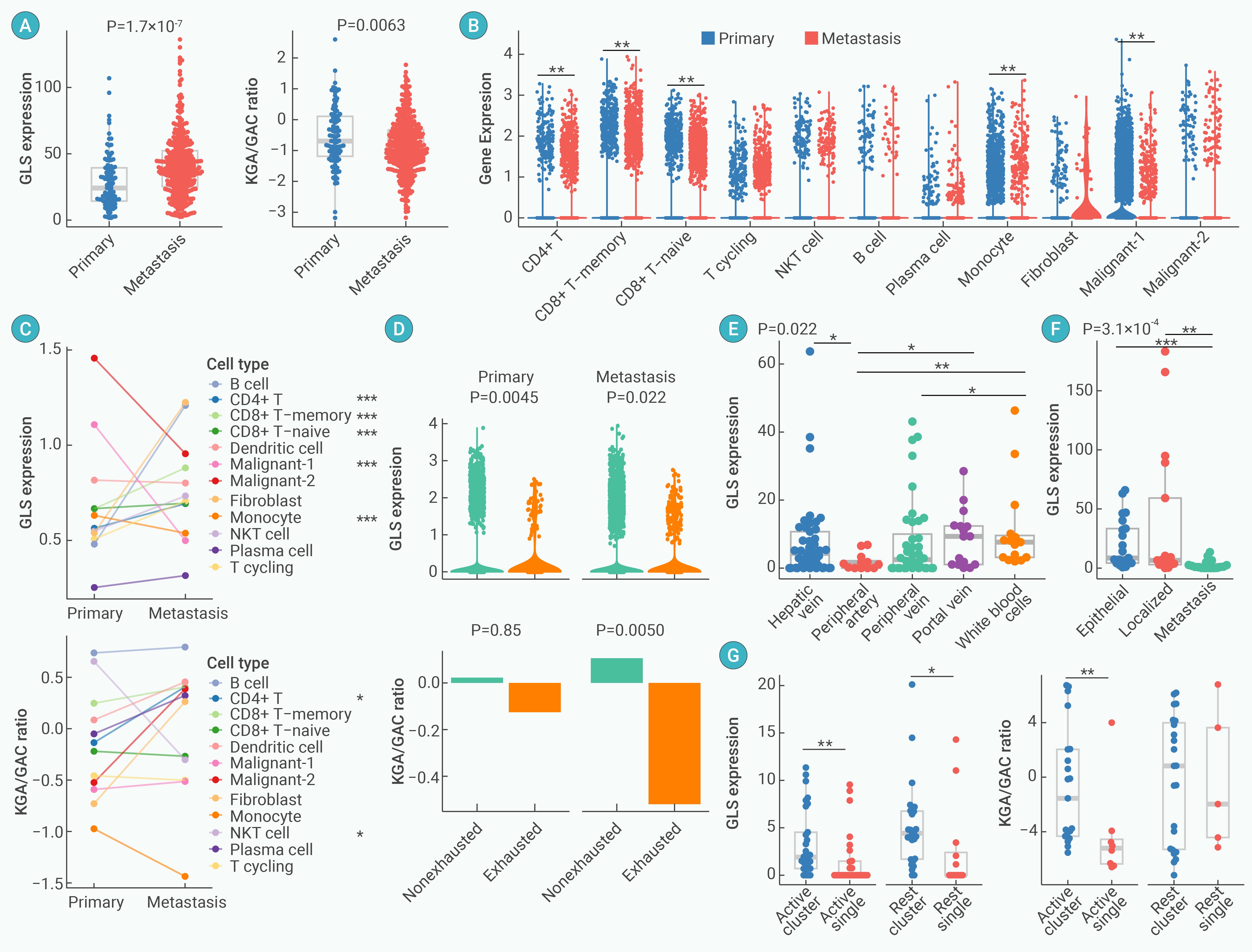
Figure 5.
Glutamine metabolism dynamics in cancer metastasis
-
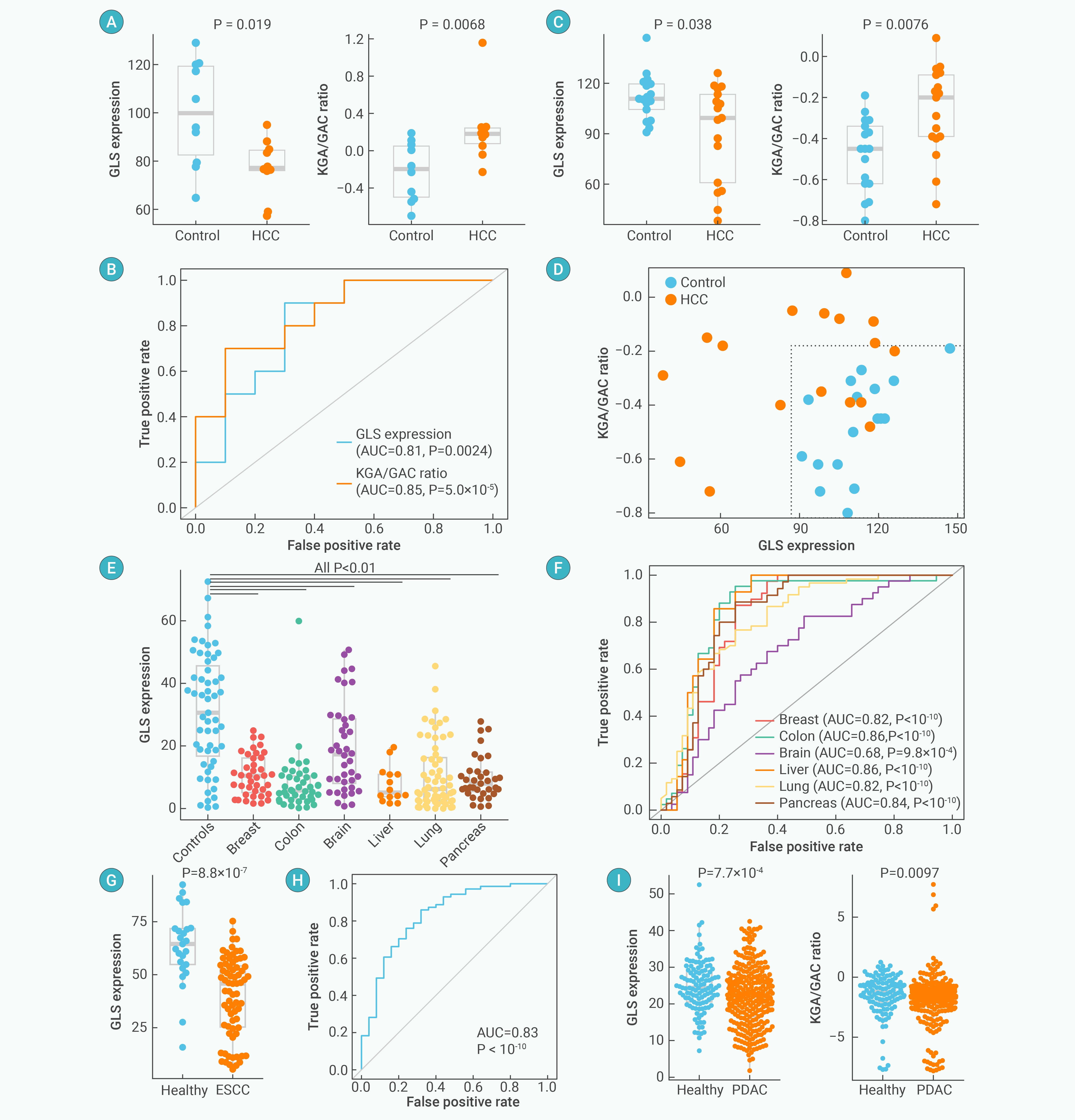
Figure 6.
Glutamine metabolism in peripheral blood for non-invasive cancer diagnosis
