
Radiation damage behavior of soft matter in ultrafast cryo-electron microscopy (cryo-UEM)
-
GRAPHICAL ABSTRACT
 DownLoad:
Full size image
DownLoad:
Full size image
-
PUBLIC SUMMARY
-
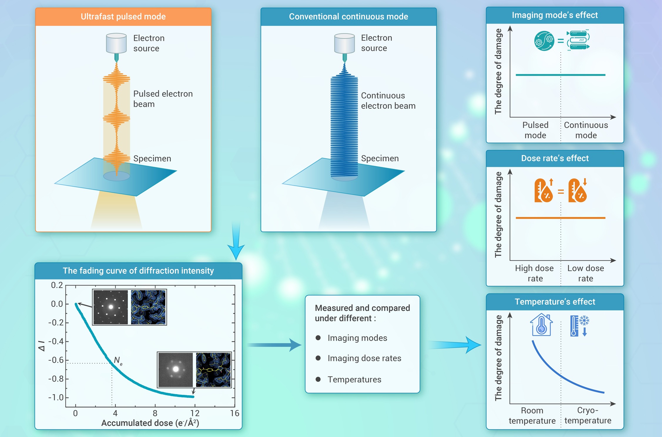
An ultrafast cryo-electron microscopy (cryo-UEM) system based on an ultrafast laser was established.
Diffraction-intensity fading curves and critical doses of samples were measured under different conditions.
The radiation damage in soft-matter samples did not show any dependence on the imaging electron dose rates.
The pulsed electron beams cannot mitigate the radiation damage in room-temperature and cryogenic samples.
The physical mechanisms behind the experimental results of radiation damage were systematically analyzed.
-
-
ABSTRACT
Whether time-modulated pulsed-electron imaging can mitigate sample radiation damage is still controversial. The effectiveness of such mitigation and relevant potential applications in cryo-EM remain to be explored. Herein, we built an ultrafast cryo-EM system based on an ultrafast laser. Using such system and the saturated aliphatic hydrocarbon compounds (C44H90), the diffraction-intensity fading curves and corresponding critical electron doses (Ne) of samples were carefully measured under different imaging modes, temperatures, imaging dose rates and pulsed repetition rates. Our experimental results demonstrate that the fading curves and Ne values of C44H90 crystals show no correlation with the imaging electron dose rates. As the temperature decreased, the Ne values of the sample increased, indicating a cryoprotective effect on sample radiation damage. Interestingly, at constant temperature, the fading curves and Ne values of the sample in multi-electron-packet and near-single-electron-packet pulsed modes are all approximately the same as those in conventional continuous electron-beam mode, even when obtained at different pulsed repetition rates. These results show that the time-modulated pulsed electron beams do not appear to mitigate the electron radiation damage that occurs in samples. The physical mechanisms underlying the radiation damage behavior under different conditions were also carefully analyzed. Our findings provide new insights and an experimental basis for understanding sample radiation damage under electron beams, offering guidance and inspiration for elucidating the fundamental principles of radiation damage. -

-
REFERENCES
[1] Li X., Mooney P., Zheng S., et al. (2013). Electron counting and beam-induced motion correction enable near-atomic-resolution single-particle cryo-EM. Nat. Methods 10:584−590. DOI:10.1038/nmeth.2472 [2] Kühlbrandt W. (2014). The resolution revolution. Science 343:1443−1444. DOI:10.1126/science.1251652 [3] Tian B., Zhou M., Feng F., et al. (2022). Cryogenic superresolution correlative light and electron microscopy of vitreous sections. Biophys. Rep. 8:193−204. DOI:10.52601/bpr.2022.220005 [4] Dubochet J. (2018). On the development of electron cryo-microscopy (nobel lecture). Angew. Chem. Int. Ed. 57:10842−10846. DOI:10.1002/anie.201804280 [5] Glaeser R.M., and Taylor K.A. (1978). Radiation damage relative to transmission electron microscopy of biological specimens at low temperature: a review. J. Microsc. 112:127−138. DOI:10.1111/j.1365-2818.1978.tb01160.x [6] Zhang D., Zhu Y., Liu L., et al. (2018). Atomic-resolution transmission electron microscopy of electron beam–sensitive crystalline materials. Science 359:675−679. DOI:10.1126/science.aao0865 [7] Cheng Y. (2018). Single-particle cryo-EM—How did it get here and where will it go. Science 361:876−880. DOI:10.1126/science.aat4346 [8] Chen Q., Dwyer C., Sheng G., et al. (2020). Imaging beam-sensitive materials by electron microscopy. Adv. Mater. 32:1907619. DOI:10.1002/adma.201907619 [9] Jiang N. (2016). Electron beam damage in oxides: a review. Rep. Prog. Phys. 79:016501. DOI:10.1088/0034-4885/79/1/016501 [10] Garman E.F., and Weik M. (2023). Radiation damage to biological macromolecules. Curr. Opin. Struct. Biol. 82:102662. DOI:10.1016/j.sbi.2023.102662 [11] Egerton R.F. (2012). Mechanisms of radiation damage in beam-sensitive specimens, for TEM accelerating voltages between 10 and 300 kV. Microsc. Res. Tech. 75:1550−1556. DOI:10.1002/jemt.22099 [12] Egerton R.F. (2019). Radiation damage to organic and inorganic specimens in the TEM. Micron 119:72−87. DOI:10.1016/j.micron.2019.01.005 [13] Glaeser R.M., Mcmullan G., Faruqi A.R., and Henderson R. (2011). Images of paraffin monolayer crystals with perfect contrast: Minimization of beam-induced specimen motion. Ultramicroscopy 111:90−100. DOI:10.1016/j.ultramic.2010.10.010 [14] Sun F. (2018). Orienting the future of bio-macromolecular electron microscopy. Chinese Phys. B 27:063601. DOI:10.1088/1674-1056/27/6/063601 [15] Baker L.A., and Rubinstein J.L. (2010). Chapter fifteen - radiation damage in electron cryomicroscopy. Jensen G.J. (ed). Methods in enzymology (Academic Press), pp. 371-388. DOI:10.1016/S0076-6879(10)81015-8. [16] Kato K., Miyazaki N., Hamaguchi T., et al. (2021). High-resolution cryo-EM structure of photosystem II reveals damage from high-dose electron beams. Commun. Biol. 4:382. DOI:10.1038/s42003-021-01919-3 [17] Vandenbussche E.J., and Flannigan D.J. (2019). Reducing radiation damage in soft matter with femtosecond-timed single-electron packets. Nano Lett. 19:6687−6694. DOI:10.1021/acs.nanolett.9b03074 [18] Xue H., Zhang M., Liu J., et al. (2022). Cryo-electron tomography related radiation-damage parameters for individual-molecule 3D structure determination. Front. Chem. 10:889203. DOI:10.3389/fchem.2022.889203 [19] Bonham R.A., and Kennerly R.E. (1978). Pulsed electron beams to reduce radiation damage. Ann. N. Y. Acad. Sci. 306:85−94. DOI:10.1111/j.1749-6632.1978.tb25641.x [20] Glaeser R.M. (2025). Commonsense and common nonsense opinions: prospects for further reducing beam damage in electron microscopy of radiation-sensitive specimens. Ultramicroscopy 271:114118. DOI:10.1016/j.ultramic.2025.114118 [21] Grubb D.T. (1974). Radiation damage and electron microscopy of organic polymers. J. Mater. Sci. 9:1715−1736. DOI:10.1007/BF00540772 [22] Flannigan D.J., and Vandenbussche E.J. (2023). Pulsed-beam transmission electron microscopy and radiation damage. Micron 172:103501. DOI:10.1016/j.micron.2023.103501 [23] Flannigan D.J., Curtis W.A., Vandenbussche E.J., and Zhang Y. (2022). Low repetition-rate, high-resolution femtosecond transmission electron microscopy. J. Chem. Phys. 157:180903. DOI:10.1063/5.0128109 [24] Kisielowski C., Specht P., Freitag B., et al. (2019). Discovering hidden material properties of MgCl2 at atomic resolution with structured temporal electron illumination of picosecond time resolution. Adv. Funct. Mater. 29:1807818. DOI:10.1002/adfm.201807818 [25] Kisielowski C., Specht P., Rozeveld S., et al. (2022). Exploring functional materials by understanding beam-sample interactions. Adv. Funct. Mater. 32:2201112. DOI:10.1002/adfm.202201112 [26] Glaeser R.M. (2008). Retrospective: Radiation damage and its associated “Information Limitations”. J. Struct. Biol. 163:271−276. DOI:10.1016/j.jsb.2008.06.001 [27] Glaeser R.M. (1971). Limitations to significant information in biological electron microscopy as a result of radiation damage. J. Ultrastruct. Res. 36:466−482. DOI:10.1016/S0022-5320(71)80118-1 [28] Taylor K.A., and Glaeser R.M. (1976). Electron microscopy of frozen hydrated biological specimens. J. Ultrastruct. Res. 55:448−456. DOI:10.1016/S0022-5320(76)80099-8 [29] Glaeser R.M. (1979). Prospects for extending the resolution limit of the electron microscope. J. Microsc. 117:77−91. DOI:10.1111/j.1365-2818.1979.tb00232.x [30] Jeng T.-W., and Chiu W. (1984). Quantitative assessment of radiation damage in a thin protein crystal. J. Microsc. 136:35−44. DOI:10.1111/j.1365-2818.1984.tb02544.x [31] Sliz P., Harrison S.C., and Rosenbaum G. (2003). How does radiation damage in protein crystals depend on X-ray dose. Structure 11:13−19. DOI:10.1016/S0969-2126(02)00910-3 [32] Nannenga B.L., Bu G., and Shi D. (2018). The evolution and the advantages of MicroED. Front. Mol. Biosci. 5:DOI:10.3389/fmolb.2018.00114 [33] Chen J.Z., Sachse C., Xu C., et al. (2008). A dose-rate effect in single-particle electron microscopy. J. Struct. Biol. 161:92−100. DOI:10.1016/j.jsb.2007.09.017 [34] Jiang N., and Spence J.C.H. (2012). On the dose-rate threshold of beam damage in TEM. Ultramicroscopy 113:77−82. DOI:10.1016/j.ultramic.2011.11.016 [35] Matsuya Y., Mcmahon S.J., Tsutsumi K., et al. (2018). Investigation of dose-rate effects and cell-cycle distribution under protracted exposure to ionizing radiation for various dose-rates. Sci . Rep. 8:8287. DOI:10.1038/s41598-018-26556-5 [36] Dubochet J., Adrian M., Chang J.-J., et al. (1988). Cryo-electron microscopy of vitrified specimens. Q . Rev. Biophys. 21:129−228. DOI:10.1017/S0033583500004297 [37] Lobastov V.A., Weissenrieder J., Tang J., and Zewail A.H. (2007). Ultrafast electron microscopy (UEM): Four-dimensional imaging and diffraction of nanostructures during phase transitions. Nano Lett. 7:2552−2558. DOI:10.1021/nl071341e [38] Shorokhov D., and Zewail A.H. (2016). Perspective: 4D ultrafast electron microscopy—Evolutions and revolutions. J. Chem. Phys. 144:080901. DOI:10.1063/1.4941375 [39] Kim Y.-J., Park W.-W., Nho H.-W., and Kwon O.-H. (2024). High-resolution correlative imaging in ultrafast electron microscopy. Adv. Phys-X 9:2316710. DOI:10.1080/23746149.2024.2316710 [40] Nabben D., Kuttruff J., Stolz L., et al. (2023). Attosecond electron microscopy of sub-cycle optical dynamics. Nature 619:63−67. DOI:10.1038/s41586-023-06074-9 [41] Vanacore G.M., Fitzpatrick A.W.P., and Zewail A.H. (2016). Four-dimensional electron microscopy: Ultrafast imaging, diffraction and spectroscopy in materials science and biology. Nano Today 11:228−249. DOI:10.1016/j.nantod.2016.04.009 [42] Armstrong M.R., Boyden K., Browning N.D., et al. (2007). Practical considerations for high spatial and temporal resolution dynamic transmission electron microscopy. Ultramicroscopy 107:356−367. DOI:10.1016/j.ultramic.2006.09.005 [43] Kuwahara M., and Agemura T. (2023). Temporal resolution in transmission electron microscopy using a photoemission electron source. Microscopy 72:97−110. DOI:10.1093/jmicro/dfac069 [44] Du D.X., Simjanoska M., and Fitzpatrick A.W.P. (2023). Four-dimensional microED of conformational dynamics in protein microcrystals on the femto-to-microsecond timescales. J. Struct. Biol. 215:107941. DOI:10.1016/j.jsb.2023.107941 [45] Peet M.J., Henderson R., and Russo C.J. (2019). The energy dependence of contrast and damage in electron cryomicroscopy of biological molecules. Ultramicroscopy 203:125−131. DOI:10.1016/j.ultramic.2019.02.007 [46] Naydenova K., Kamegawa A., Peet M.J., et al. (2022). On the reduction in the effects of radiation damage to two-dimensional crystals of organic and biological molecules at liquid-helium temperature. Ultramicroscopy 237:113512. DOI:10.1016/j.ultramic.2022.113512 [47] Cao G., Sun S., Li Z., et al. (2015). Clocking the anisotropic lattice dynamics of multi-walled carbon nanotubes by four-dimensional ultrafast transmission electron microscopy. Sci. Rep. 5:8404. DOI:10.1038/srep08404 [48] Yan R., Edwards T.J., Pankratz L.M., et al. (2015). Simultaneous determination of sample thickness, tilt, and electron mean free path using tomographic tilt images based on Beer–Lambert law. J. Struct. Biol. 192:287−296. DOI:10.1016/j.jsb.2015.09.019 [49] Baker L.A., Smith E.A., Bueler S.A., and Rubinstein J.L. (2010). The resolution dependence of optimal exposures in liquid nitrogen temperature electron cryomicroscopy of catalase crystals. J. Struct. Biol. 169:431−437. DOI:10.1016/j.jsb.2009.11.014 [50] Unwin P.N.T., and Henderson R. (1975). Molecular structure determination by electron microscopy of unstained crystalline specimens. J. Mol. Biol. 94:425−440. DOI:10.1016/0022-2836(75)90212-0 [51] Hayward S.B., and Glaeser R.M. (1979). Radiation damage of purple membrane at low temperature. Ultramicroscopy 4:201−210. DOI:10.1016/S0304-3991(79)90211-0 [52] Siegel G. (1972). The influence of very low temperature on the radiation damage of organic crystals irradiated by 100 keV-electrons. Z. Naturforsch. 27:325−332. DOI:10.1515/zna-1972-0219 [53] Li Y., Kang D.-D., Dai J.-Y., and Wang L.-W. (2024). The cage effect of electron beam irradiation damage in cryo-electron microscopy. npj Comput. Mater. 10:115. DOI:10.1038/s41524-024-01299-2 [54] Konieczny K.A., Paul I., Rodriguez J.A., and Garcia-Garibay M.A. (2024). From beam damage to massive reaction amplification under the electron microscope: an ionization-induced chain reaction in crystals of a dewar benzene. ACS Cent. Sci. DOI:10.1021/acscentsci.4c01429 [55] Dertinger H., and Jung H. (1970). The hit theory. Dertinger H., and Jung H. (ed). Molecular radiation biology (Springer US), pp. 12-23. DOI:10.1007/978-1-4684-6247-0_2. [56] Isaacson M., Johnson D., and Crewe A.V. (1973). Electron beam excitation and damage of biological molecules; Its implications for specimen damage in electron microscopy. Radiat. Res. 55:205−224. DOI:10.2307/3573678 [57] Drenth J. (1999). The theory of X-ray diffraction by a crystal. Drenth J. (ed). Principles of protein X-ray crystallography (Springer New York), pp. 70-116. DOI:10.1007/978-1-4757-3092-0_4 [58] Woolfson M.M. (1997). An introduction to X-ray crystallography (Cambridge University Press). DOI:10.1017/CBO9780511622557 [59] Holton J. (2009). A beginner's guide to radiation damage. J. Synchrotron Radiat. 16:133−142. DOI:10.1107/S0909049509004361 [60] Thornburg D.D., and Wayman C.M. (1973). Specimen temperature increases during transmission electron microscopy. phys. stat. sol. (a) 15:449−453. DOI:10.1002/pssa.2210150209 [61] Stenn K., and Bahr G.F. (1970). Specimen damage caused by the beam of the transmission electron microscope, a correlative reconsideration. J. Ultrastruct. Res. 31:526−550. DOI:10.1016/S0022-5320(70)90167-X [62] Li Z., and Peng G. (2022). Spatial transcriptomics: new dimension of understanding biological complexity. Biophys. Rep. 8:119−135. DOI:10.52601/bpr.2021.210037 -
ABOUT THIS ARTICLE
Cite this article:
Zhao Y., Qi C., Zhu C., et al. (2025). Radiation damage behavior of soft matter in ultrafast cryo-electron microscopy (cryo-UEM). The Innovation Life 3:100145. https://doi.org/10.59717/j.xinn-life.2025.100145
Welcome!
To request copyright permission to republish or share portions of our works, please visit Copyright Clearance Center's (CCC) Marketplace website at marketplace.copyright.com.
Supplementary Information
-
download_supplementary
Script_S1
Article Metrics
Relative Articles
Cited by
Export File
Citation
Zhao Y., Qi C., Zhu C., et al. (2025). Radiation damage behavior of soft matter in ultrafast cryo-electron microscopy (cryo-UEM). The Innovation Life 3:100145. https://doi.org/10.59717/j.xinn-life.2025.100145

Format
Content

-
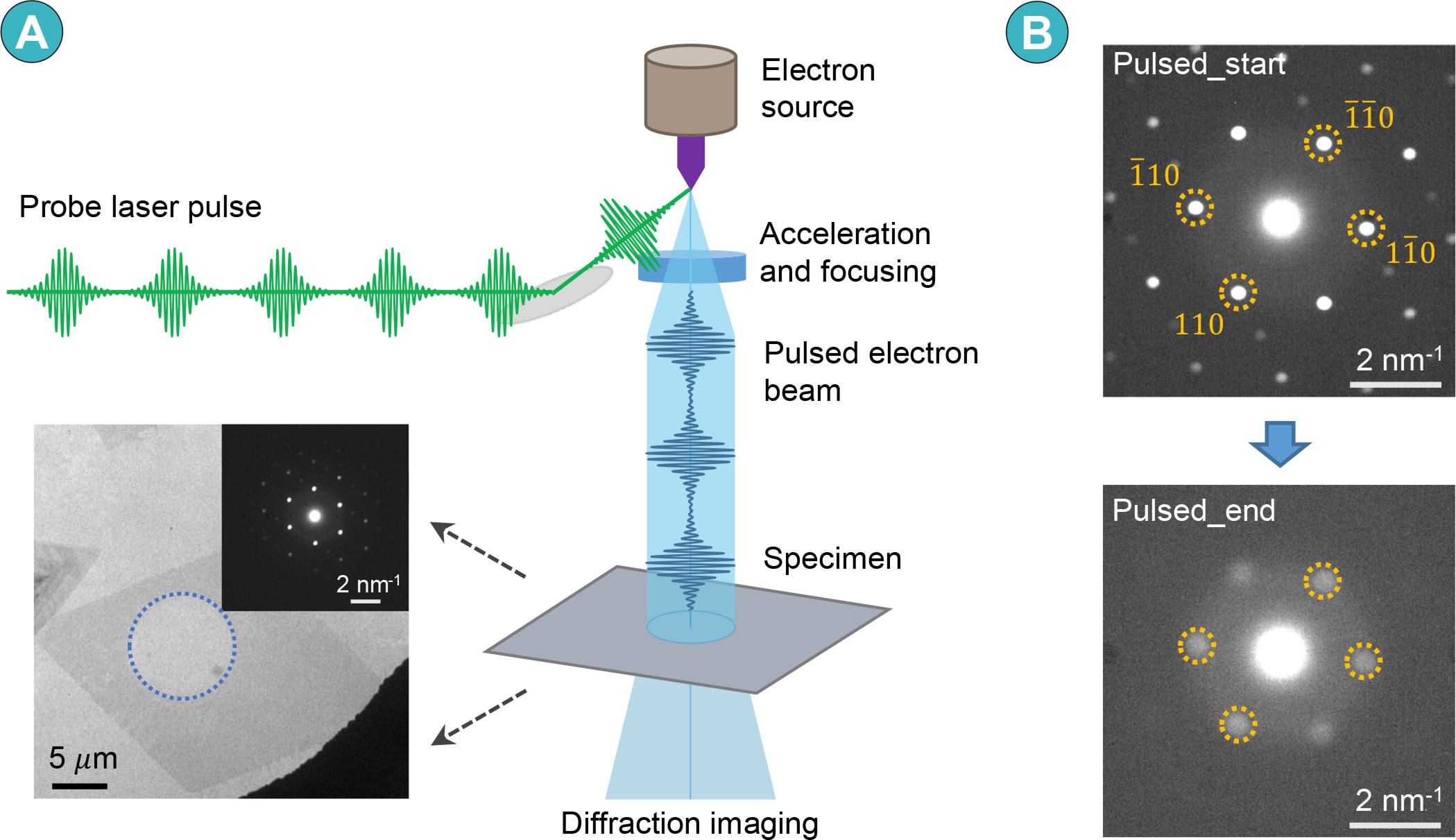
Figure 1.
Experimental procedures for ultrafast pulsed electron imaging on C44H90 crystals.
-
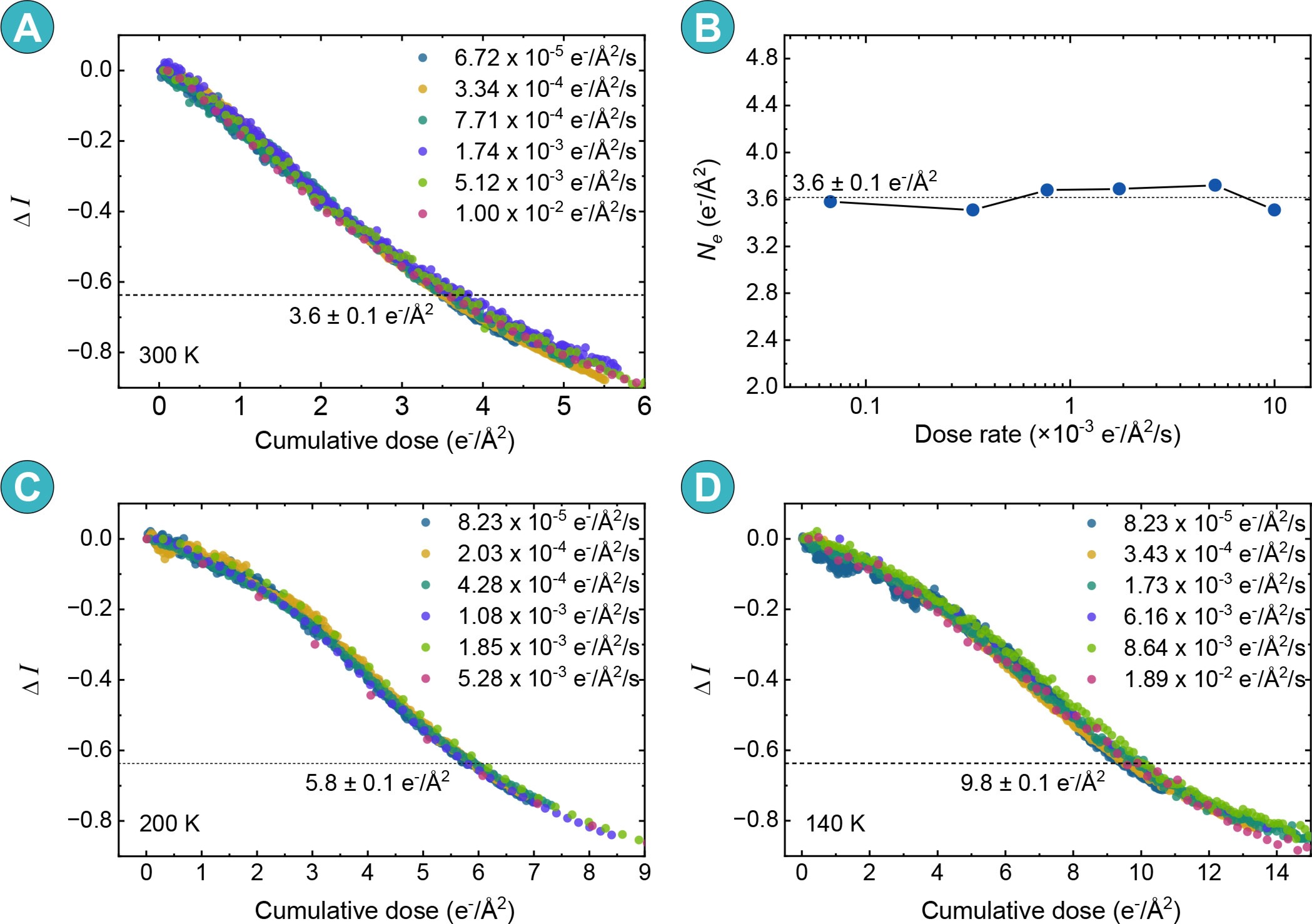
Figure 2.
Electron radiation damage of C44H90 crystals in conventional continuous electron-beam mode and at different temperatures (300 K, 200 K and 140 K)
-
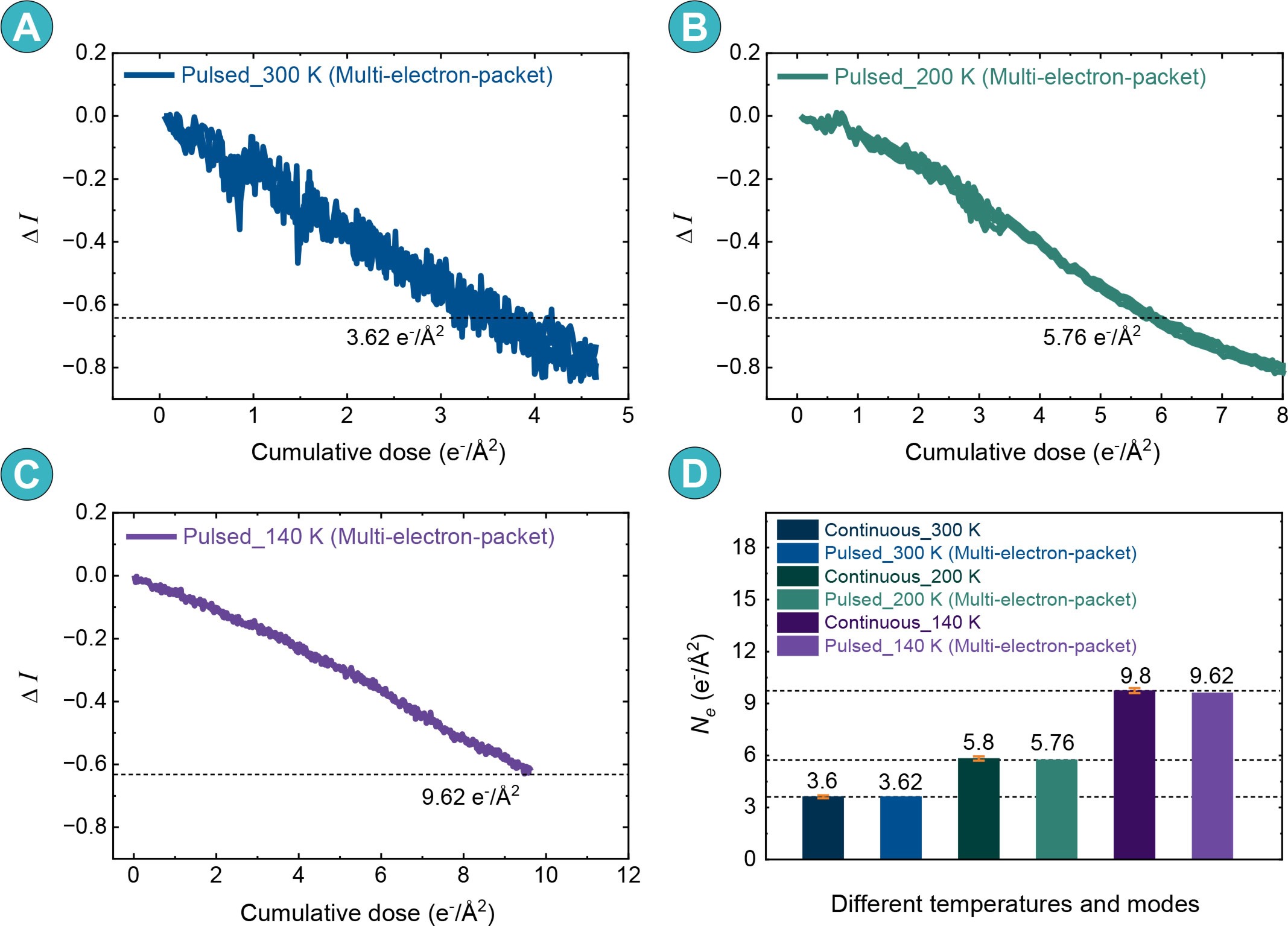
Figure 3.
Electron radiation damage of C44H90 crystals in ultrafast multi-electron-packet pulsed electron-beam mode at different temperatures
-
Figure 4.
Electron radiation damage of C44H90 crystals in ultrafast near-single-electron-packet pulsed electron-beam mode at different temperatures
-
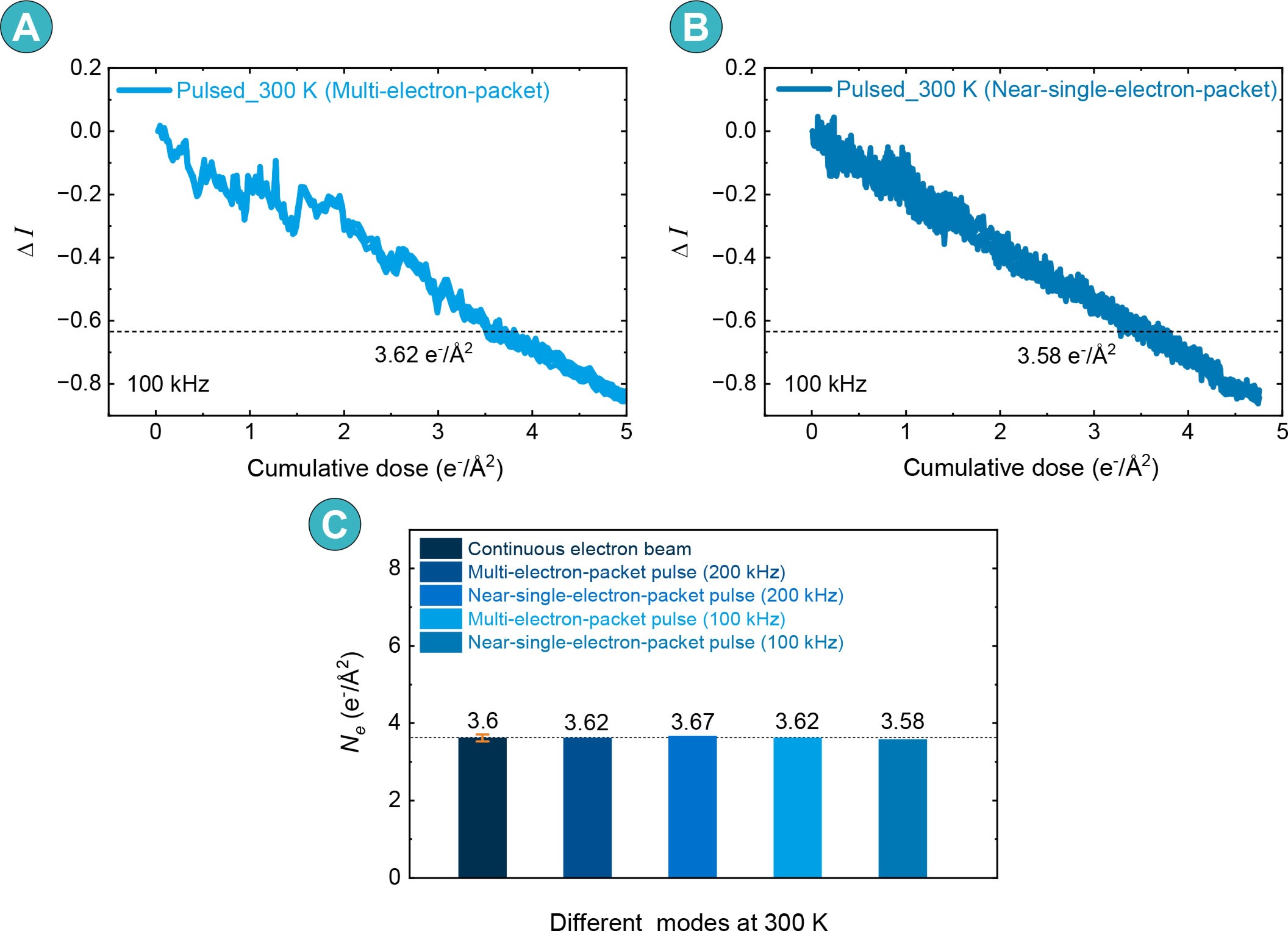
Figure 5.
Electron radiation damage of C44H90 crystals at 100 kHz laser repetition rate and 300 K
