
Dynamic keratinocyte Piezo2 expression drives itch and nerve remodeling in atopic dermatitis
-
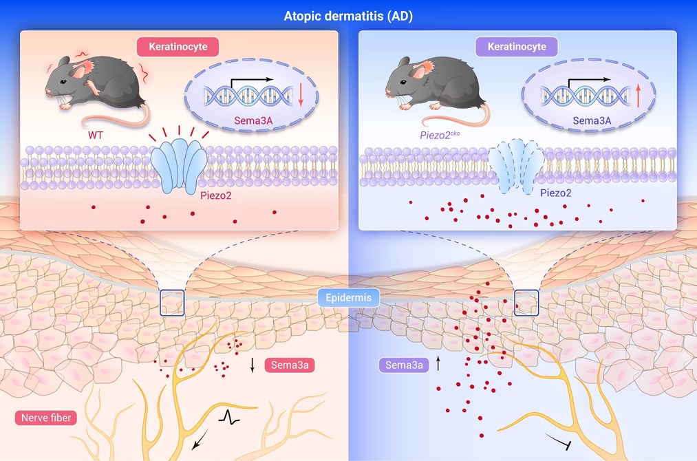
GRAPHICAL ABSTRACT
 DownLoad:
Full size image
DownLoad:
Full size image
-
PUBLIC SUMMARY
-
Keratinocyte Piezo2 is selectively upregulated in AD.
Piezo2-Sema3a axis drives itch and nerve remodeling.
Piezo2 is involved in AD itch.
-
-
ABSTRACT
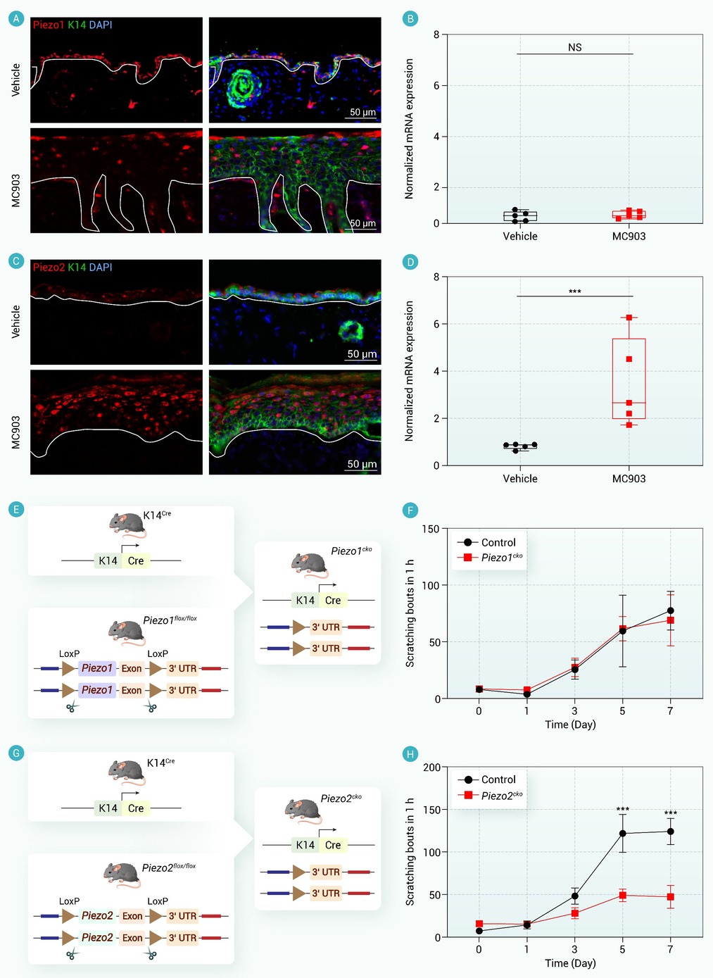
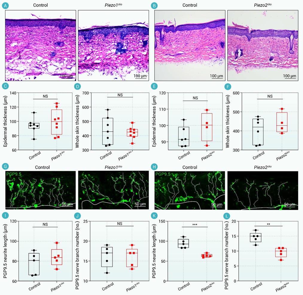
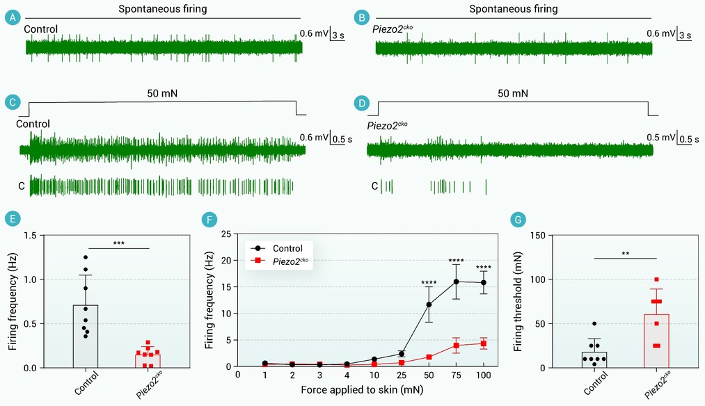
Keratinocytes actively contribute to somatosensory signaling in chronic skin diseases, yet the mechanotransduction mechanisms involved in atopic dermatitis (AD)-associated itch remain unclear. Here we identify keratinocyte-expressed mechanosensitive ion channel Piezo2 as a critical driver of chronic itch in AD. While Piezo1 was constitutively expressed and dispensable for chronic itch in the MC903-induced AD model, Piezo2 expression was markedly upregulated in keratinocytes of lesional AD skin. Keratinocyte-specific Piezo2 deletion attenuated spontaneous scratching and spontaneous C fiber activity, while having minimal impact on skin inflammation. Mechanistically, Piezo2 suppressed the neurorepulsive factor Semaphorin 3A (Sema3a), a key regulator of sensory nerve architecture. Loss of Piezo2 restored Sema3a expression in the epidermis, reduced nerve fiber branching, and dampened C fiber hyperexcitability. Furthermore, keratinocyte-specific ablation of Sema3a exacerbated AD-related itch and nerve fiber outgrowth. These findings establish a non-neuronal epithelial mechanism wherein keratinocyte Piezo2 modulates sensory nerve remodeling and pruritus in chronic skin inflammation, offering potential targets for therapeutic intervention. -

-
REFERENCES
[1] Meledathu S., Naidu M. P. and Brunner P. M. (2025). Update on atopic dermatitis. J. Allergy Clin. Immunol. 155:1124−1132. DOI:10.1016/j.jaci.2025.01.013 [2] Sroka-Tomaszewska J. and Trzeciak M. (2021). Molecular mechanisms of atopic dermatitis pathogenesis. Int. J. Mol. Sci. 22:4130. DOI:10.3390/ijms22084130 [3] Yang G., Seok J. K., Kang H. C., et al. (2020). Skin barrier abnormalities and immune dysfunction in atopic dermatitis. Int. J. Mol. Sci. 21:2867. DOI:10.3390/ijms21082867 [4] Langan S. M., Irvine A. D. and Weidinger S. (2020). Atopic dermatitis. Lancet 396:345−360. DOI:10.1016/s0140-6736(20)31286-1 [5] Wilson S. R., Thé L., Batia L. M., et al. (2013). The epithelial cell-derived atopic dermatitis cytokine TSLP activates neurons to induce itch. Cell 155:285−295. DOI:10.1016/j.cell.2013.08.057 [6] Li H., Zhang Z., Zhang H., et al. (2021). Update on the pathogenesis and therapy of atopic dermatitis. Clin. Rev. Allergy Immunol. 61:324−338. DOI:10.1007/s12016-021-08880-3 [7] Jiang H., Harris M. B. and Rothman P. (2000). IL-4/IL-13 signaling beyond JAK/STAT. J. Allergy Clin. Immunol. 105:1063−1070. DOI:10.1067/mai.2000.107604 [8] Runtsch M. C., Angiari S., Hooftman A., et al. (2022). Itaconate and itaconate derivatives target JAK1 to suppress alternative activation of macrophages. Cell Metab. 34:487−501.e488. DOI:10.1016/j.cmet.2022.02.002 [9] Goleva E., Berdyshev E. and Leung D. Y. (2019). Epithelial barrier repair and prevention of allergy. J. Clin. Invest. 129:1463−1474. DOI:10.1172/jci124608 [10] Yosipovitch G., Rosen J. D. and Hashimoto T. (2018). Itch: From mechanism to (novel) therapeutic approaches. J. Allergy Clin. Immunol. 142:1375−1390. DOI:10.1016/j.jaci.2018.09.005 [11] Ji R. R., Chamessian A. and Zhang Y. Q. (2016). Pain regulation by non-neuronal cells and inflammation. Science 354:572−577. DOI:10.1126/science.aaf8924 [12] Baumbauer K. M., DeBerry J. J., Adelman P. C., et al. (2015). Keratinocytes can modulate and directly initiate nociceptive responses. Elife 4:e09674. DOI:10.7554/eLife.09674 [13] Moehring F., Cowie A. M., Menzel A. D., et al. (2018). Keratinocytes mediate innocuous and noxious touch via ATP-P2X4 signaling. Elife 7:e31684. DOI:10.7554/eLife.31684 [14] Gouin O., L'Herondelle K., Lebonvallet N., et al. (2017). TRPV1 and TRPA1 in cutaneous neurogenic and chronic inflammation: Pro-inflammatory response induced by their activation and their sensitization. Protein. Cell 8:644−661. DOI:10.1007/s13238-017-0395-5 [15] Kefauver J. M., Ward A. B. and Patapoutian A. (2020). Discoveries in structure and physiology of mechanically activated ion channels. Nature 587:567−576. DOI:10.1038/s41586-020-2933-1 [16] Jin P., Jan L. Y. and Jan Y. N. (2020). Mechanosensitive ion channels: Structural features relevant to mechanotransduction mechanisms. Annu. Rev. Neurosci. 43:207−229. DOI:10.1146/annurev-neuro-070918-050509 [17] Delmas P., Parpaite T. and Coste B. (2022). PIEZO channels and newcomers in the mammalian mechanosensitive ion channel family. Neuron 110:2713−2727. DOI:10.1016/j.neuron.2022.07.001 [18] Coste B., Mathur J., Schmidt M., et al. (2010). Piezo1 and Piezo2 are essential components of distinct mechanically activated cation channels. Science 330:55−60. DOI:10.1126/science.1193270 [19] Holt J. R., Zeng W. Z., Evans E. L., et al. (2021). Spatiotemporal dynamics of PIEZO1 localization controls keratinocyte migration during wound healing. Elife 10:e65415. DOI:10.7554/eLife.65415 [20] Mikesell A. R., Isaeva E., Schulte M. L., et al. (2024). Increased keratinocyte activity and PIEZO1 signaling contribute to paclitaxel-induced mechanical hypersensitivity. Sci. Transl. Med. 16:eadn5629. DOI:10.1126/scitranslmed.adn5629 [21] Liu Y., Zhang M., Wang C., et al. (2024). Human umbilical cord mesenchymal stromal cell-derived extracellular vesicles induce fetal wound healing features revealed by single-cell RNA sequencing. ACS. Nano. 18:13696−13713. DOI:10.1021/acsnano.4c01401 [22] Chen J., Holt J. R., Evans E. L., et al. (2024). PIEZO1 regulates leader cell formation and cellular coordination during collective keratinocyte migration. PLoS. Comput. Biol. 20:e1011855. DOI:10.1371/journal.pcbi.1011855 [23] Mikesell A. R., Isaeva E., Schulte M. L., et al. (2023). Keratinocyte Piezo1 drives paclitaxel-induced mechanical hypersensitivity. BioRxiv. DOI:10.1101/2023.12.12.571332. [24] Woo S. H., Ranade S., Weyer A. D., et al. (2014). Piezo2 is required for Merkel-cell mechanotransduction. Nature 509:622−626. DOI:10.1038/nature13251 [25] Ranade S. S., Woo S. H., Dubin A. E., et al. (2014). Piezo2 is the major transducer of mechanical forces for touch sensation in mice. Nature 516:121−125. DOI:10.1038/nature13980 [26] Feng J., Zhao Y., Xie Z., et al. (2022). Miswiring of Merkel cell and pruriceptive C fiber drives the itch-scratch cycle. Sci. Transl. Med. 14:eabn4819. DOI:10.1126/scitranslmed.abn4819 [27] Feng J., Luo J., Yang P., et al. (2018). Piezo2 channel-Merkel cell signaling modulates the conversion of touch to itch. Science 360:530−533. DOI:10.1126/science.aar5703 [28] Kim B. S., Siracusa M. C., Saenz S. A., et al. (2013). TSLP elicits IL-33-independent innate lymphoid cell responses to promote skin inflammation. Sci. Transl. Med. 5:170ra116. DOI:10.1126/scitranslmed.3005374 [29] Oetjen L. K., Mack M. R., Feng J., et al. (2017). Sensory neurons co-opt classical immune signaling pathways to mediate chronic itch. Cell 171:217−228.e213. DOI:10.1016/j.cell.2017.08.006 [30] Murthy S. E., Loud M. C., Daou I., et al. (2018). The mechanosensitive ion channel Piezo2 mediates sensitivity to mechanical pain in mice. Sci. Transl. Med. 10:eaat9897. DOI:10.1126/scitranslmed.aat9897 [31] Maricich S. M., Wellnitz S. A., Nelson A. M., et al. (2009). Merkel cells are essential for light-touch responses. Science 324:1580−1582. DOI:10.1126/science.1172890 [32] Brunner P. M., Guttman-Yassky E. and Leung D. Y. (2017). The immunology of atopic dermatitis and its reversibility with broad-spectrum and targeted therapies. J. Allergy Clin. Immunol. 139:S65−S76. DOI:10.1016/j.jaci.2017.01.011 [33] Zheng Y., Jiang F., Wang C., et al. (2022). Regulation of Semaphorin3A in the process of cutaneous wound healing. Cell Death Differ. 29:1941−1954. DOI:10.1038/s41418-022-00981-6 [34] Kamata Y., Tominaga M., Umehara Y., et al. (2020). Calcium-inducible MAPK/AP-1 signaling drives semaphorin 3A expression in normal human epidermal keratinocytes. J. Invest. Dermatol. 140:1346-1354 e1345. DOI:10.1016/j.jid.2020.01.001 [35] Xiao B. (2024). Mechanisms of mechanotransduction and physiological roles of PIEZO channels. Nat. Rev. Mol. Cell Biol. 25:886−903. DOI:10.1038/s41580-024-00773-5 -
ABOUT THIS ARTICLE
Cite this article:
Hu X., Wu L., Zhao G., et al. (2026). Dynamic keratinocyte Piezo2 expression drives itch and nerve remodeling in atopic dermatitis. The Innovation Life 4:100187. https://doi.org/10.59717/j.xinn-life.2026.100187
Welcome!
To request copyright permission to republish or share portions of our works, please visit Copyright Clearance Center's (CCC) Marketplace website at marketplace.copyright.com.
Article Metrics
Relative Articles
Cited by
Export File
Citation
Hu X., Wu L., Zhao G., et al. (2026). Dynamic keratinocyte Piezo2 expression drives itch and nerve remodeling in atopic dermatitis. The Innovation Life 4:100187. https://doi.org/10.59717/j.xinn-life.2026.100187

Format
Content
-
Figure 1.
Keratinocyte Piezo2 expression is upregulated in AD-like skin and drives chronic itch
-
Figure 2.
Keratinocyte-expressed Piezo2 promotes epidermal sensory fiber remodeling, but does not affect skin inflammation in MC903-induced AD
-
Figure 3.
Keratinocyte-expressed Piezo2 enhances C fiber excitability during AD-associated itch
-
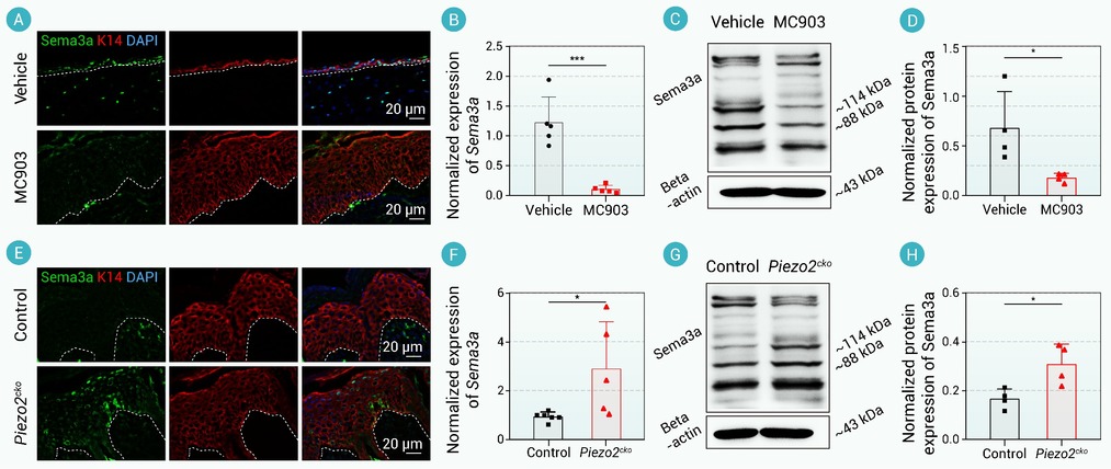
Figure 4.
PIEZO2 is required for suppressing Sema3a expression in the context of AD
-
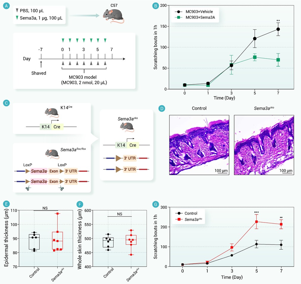
Figure 5.
Keratinocyte Sema3a is essential for AD-associated chronic itch
-
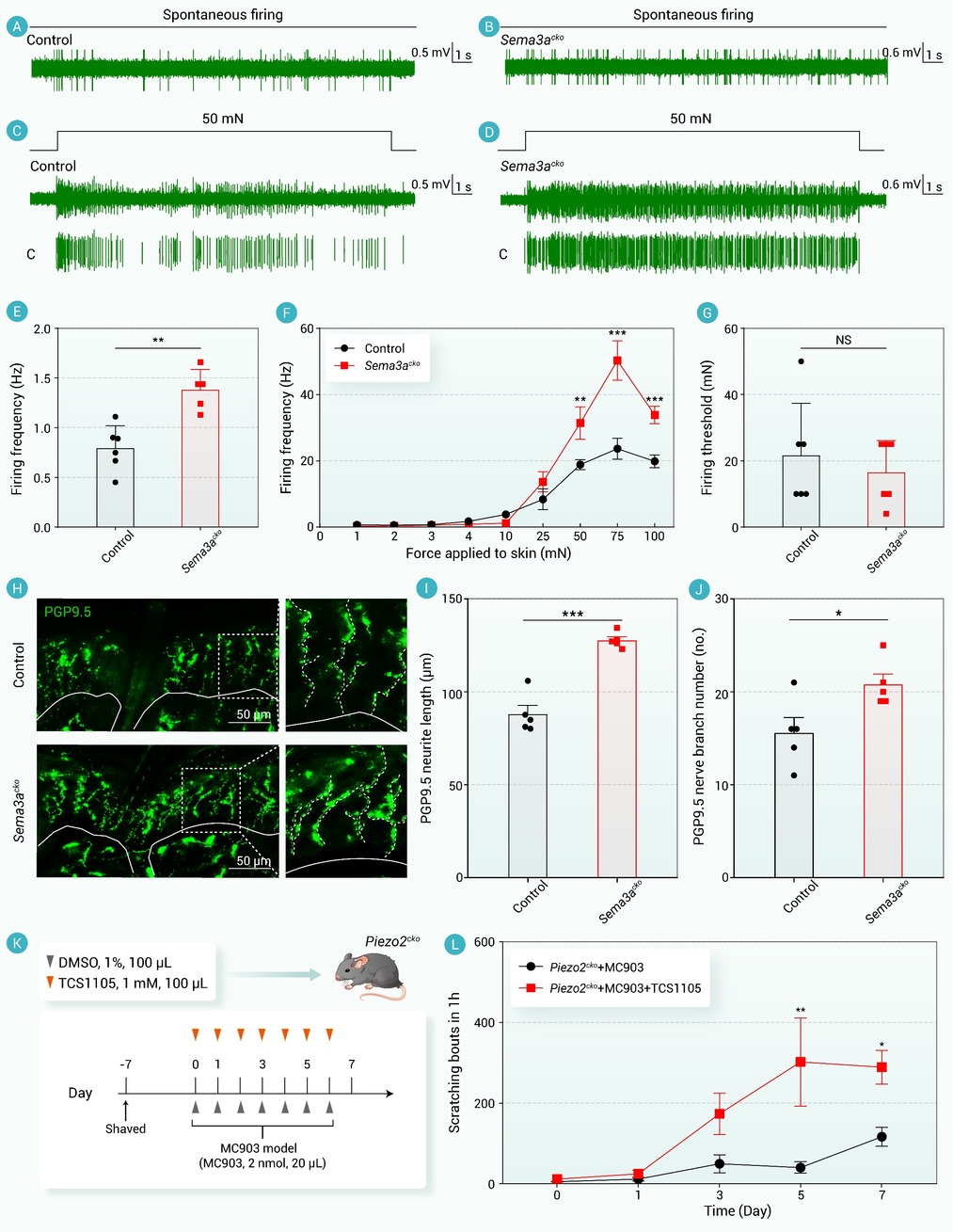
Figure 6.
Piezo2-Sema3a signaling is essential for AD-associated chronic itch
