
NMN-SIRT1 signaling ameliorates IUGR via oxidative stress-mediated mitophagy to promote placental angiogenesis
-
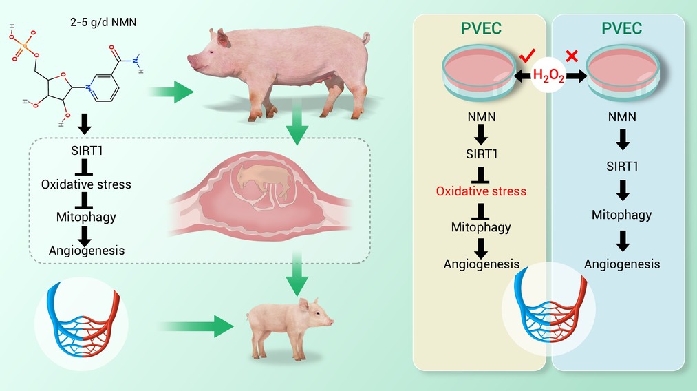
GRAPHICAL ABSTRACT
 DownLoad:
Full size image
DownLoad:
Full size image
-
PUBLIC SUMMARY
-
Dietary NMN (2-5g/d) enhances placental angiogenesis and reduces IUGR incidence in sows.
NMN upregulates placental SIRT1, alleviating oxidative stress in sows/offspring.
NMN-SIRT1 axis inhibits excessive mitophagy and promotes angiogenesis in placenta.
NMN-SIRT1 activation restores PVEC migration via oxidative stress/mitophagy regulation.
Baseline NMN-SIRT1 signaling stimulates mitophagy to sustain PVEC angiogenic capacity.
-
-
ABSTRACT
Nicotinamide mononucleotide (NMN) is a natural anti-aging agent. However, its effects on intrauterine growth restriction (IUGR) remain insufficiently explored. This study found that daily intake of 2-5 g/d of NMN significantly enhanced placental angiogenesis and reduced the incidence of IUGR by using a pregnant sow model (Landrace × Yorkshire breed). Mechanistically, NMN upregulated SIRT1 expression in the placenta, alleviated oxidative stress in sows, placentas, and their offspring, inhibited excessive mitophagy in the placenta, and promoted angiogenesis. Furthermore, activation of the NMN-SIRT1 signaling pathway was observed to mitigate oxidative stress and mitophagy, thereby restoring the migratory capacity of porcine vascular endothelial cells (PVECs) and improving endothelial function. However, in the absence of oxidative stress, NMN-SIRT1 acts as a potent mitophagy-promoting signal, positively regulating PVEC angiogenesis. In conclusion, our study demonstrates that the activation of NMN-SIRT1 signaling is crucial for reducing adverse pregnancy outcomes by ameliorating placental dysfunction induced by perinatal oxidative stress.-
Keywords:
- IUGR /
- NMN /
- Oxidative stress /
- Mitophagy /
- Angiogenesis
-

-
REFERENCES
[1] Huang Z., Huang S., Song T., et al. (2021). Placental angiogenesis in mammals: A review of the regulatory effects of signaling pathways and functional nutrients. Adv. Nutr. 12:2415−2434. DOI:10.1093/advances/nmab070 [2] Wang J., Chen L., Li D., et al. (2008). Intrauterine growth restriction affects the proteomes of the small intestine, liver, and skeletal muscle in newborn pigs. J. Nutr. 138:60−66. DOI:10.1093/jn/138.1.60 [3] ACOG Practice Bulletin No. 227: Fetal growth restriction: Correction. (2021). Obstet. Gynecol. 137:754. DOI:10.1097/aog.0000000000004350 [4] Kesavan K. and Devaskar S. U. (2019). Intrauterine growth restriction: Postnatal monitoring and outcomes. Pediatr. Clin. North Am. 6:403−423. DOI:10.1016/j.pcl.2018.12.009 [5] Wei Y., Tang W., Mao P., et al. (2024). Sexually dimorphic response to hepatic Injury in newborn suffering from intrauterine growth restriction. Adv. Sci. (Weinh) 11:e2403095. DOI:10.1002/advs.202403095 [6] Dessi A., Marincola F. C. and Fanos V. (2015). Metabolomics and the great obstetrical syndromes--GDM, PET, and IUGR. Best. Pract. Res. Clin. Obstet. Gynaecol. 29:156-164. DOI:10.1016/j.bpobgyn.2014.04.023 [7] Hunter D. S., Hazel S. J., Kind K. L., et al. (2016). Programming the brain: Common outcomes and gaps in knowledge from animal studies of IUGR. Physiol. Behav. 164:233−248. DOI:10.1016/j.physbeh.2016.06.005 [8] Wu Z., Xiang Q., Feng L., et al. (2023). Adenosine-ADORA2A promotes ang-induced angiogenesis in intrauterine growth restriction placenta via the Stat3/Akt pathway. Arterioscler. Thromb. Vasc. Biol. 43:e190−e209. DOI:10.1161/ATVBAHA.122.318948 [9] Nardozza L. M., Caetano A. C., Zamarian A. C., et al. (2017). Fetal growth restriction: Current knowledge. Arch. Gynecol. Obstet. 295:1061−1077. DOI:10.1007/s00404-017-4341-9 [10] Lai T. (2019). Effect of exogenous spermine on angiogenes for procine umbilical vein endothelial cells and its mechanism. Master's Dissertation (Jiangxi Agricultural University). DOI:10.27177/d.cnki.gjxnu.2019.000073 [11] Peng J., Xiong J., Cui C., et al. (2019). Maternal eicosapentaenoic acid feeding decreases placental lipid deposition and improves the homeostasis of oxidative stress through a Sirtuin-1 (SIRT1) independent manner. Mol. Nutr. Food Res. 63:e1900343. DOI:10.1002/mnfr.201900343 [12] Peng J., Zhou Y., Hong Z., et al. (2019). Maternal eicosapentaenoic acid feeding promotes placental angiogenesis through a Sirtuin-1 independent inflammatory pathway. Biochim. Biophys. Acta Mol. Cell Biol. Lipids 1864:147−157. DOI:10.1016/j.bbalip.2018.11.003 [13] Pereira R. D., De Long N. E., Wang R. C., et al. (2015). Angiogenesis in the placenta: The role of reactive oxygen species signaling. Biomed Res. Int. 2015:814543. DOI:10.1155/2015/814543 [14] Wu D., Nie J., Zhou S., et al. (2025). Iron homeostasis: Effects of different levels of protein iron on placental iron handling in sows. J. Nutr. Biochem. 148:110141. DOI:10.1016/j.jnutbio.2025.110141 [15] Chen C. P., Bajoria R. and Aplin J. D. (2002). Decreased vascularization and cell proliferation in placentas of intrauterine growth-restricted fetuses with abnormal umbilical artery flow velocity waveforms. Am. J. Obstet. Gynecol. 187:764−769. DOI:10.1067/mob.2002.125243 [16] Jackson M. R., Walsh A. J., Morrow R. J., et al. (1995). Reduced placental villous tree elaboration in small-for-gestational-age pregnancies: relationship with umbilical artery doppler waveforms. Am. J. Obstet. Gynecol. 172:518−525. DOI:10.1016/0002-9378(95)90566-9 [17] Krebs C., Macara L. M., Leiser R., et al. (1996). Intrauterine growth restriction with absent end-diastolic flow velocity in the umbilical artery is associated with maldevelopment of the placental terminal villous tree. Am. J. Obstet. Gynecol. 175:1534−1542. DOI:10.1016/s0002-9378(96)70103-5 [18] Amdi C., Krogh U., Flummer C., et al. (2013). Intrauterine growth restricted piglets defined by their head shape ingest insufficient amounts of colostrum. J. Anim. Sci. 91:5605−5613. DOI:10.2527/jas.2013-6824 [19] Wang J., Feng C., Liu T., et al. (2017). Physiological alterations associated with intrauterine growth restriction in fetal pigs: Causes and insights for nutritional optimization. Mol. Reprod. Dev. 84:897−904. DOI:10.1002/mrd.22842 [20] Hu C., Wu Z., Huang Z., et al. (2021). Nox2 impairs VEGF-A-induced angiogenesis in placenta via mitochondrial ROS-STAT3 pathway. Redox Biol. 45:102051. DOI:10.1016/j.redox.2021.102051 [21] Xia M., Pan Y., Guo L., et al. (2019). Effect of gestation dietary methionine/lysine ratio on placental angiogenesis and reproductive performance of sows. J. Anim. Sci. 97:3487−3497. DOI:10.1093/jas/skz175 [22] Abdullah N., Lau C.C. and Ismail S. (2016). Potential use of Lentinus squarrosulus mushroom as fermenting agent and source of natural antioxidant additive in livestock feed. J. Sci. Food Agric. 96:1459−1466. DOI:10.1002/jsfa.7242 [23] Huang S., Zhang L., Luo J., et al. (2023). Cysteamine and N-Acetyl-cysteine alleviate placental oxidative stress and barrier function damage induced by deoxynivalenol. J. Agric. Food Chem. 71:6846−6858. DOI:10.1021/acs.jafc.3c00399 [24] Peng J., Zhang Y., Liu Q., et al. (2024). Allicin in pregnancy diets modulates steroid metabolism in pregnant sows and placental sulphate metabolism promoting placental angiogenesis and foetal development. Animal 18:101224. DOI:10.1016/j.animal.2024.101224 [25] Wu G., Bazer F. W., Johnson G. A., et al. (2017). Functional amino acids in the development of the pig placenta. Mol. Reprod. Dev. 84:870−882. DOI:10.1002/mrd.22809 [26] Al-Gubory K. H., Fowler P. A. and Garrel C. (2010). The roles of cellular reactive oxygen species, oxidative stress and antioxidants in pregnancy outcomes. Int. J. Biochem. Cell Biol. 42:1634−1650. DOI:10.1016/j.biocel.2010.06.001 [27] Li Z., Zhang W., Yang M., et al. (2025). Fermented Lentinus edodes improves insulin sensitivity in dam and offspring mice via activation of the hepatic PI3K/AKT signaling pathway. Br. J. Nutr. 134:16−27. DOI:10.1017/S0007114525103796 [28] Wang C., Tang Y., Zhang Y., et al. (2025). Pentadecanoic acid (C15:0) promotes placental angiogenesis by activating the placental PI3K-AKT signaling. Food Funct. 16:9344−9358. DOI:10.1039/d5fo03684d [29] Wang J., Zheng S., Li Z., et al. (2024). Pentadecanoic acid (C15:0, PA) induces mild maternal glucose intolerance and promotes the growth of the offspring partly through up-regulating liver PPARα and MAPK signaling pathways. Food Funct. 15:11400−11414. DOI:10.1039/d4fo03970j [30] Duhig K., Chappell L. C. and Shennan A. H. (2016). Oxidative stress in pregnancy and reproduction. Obstet. Med. 9:113−116. DOI:10.1177/1753495x16648495 [31] Hussain T., Murtaza G., Metwally E., et al. (2021). The role of xxidative stress and antioxidant balance in pregnancy. Mediators Inflamm. 2021:9962860. DOI:10.1155/2021/9962860 [32] Xia M., Peng J., Cui C., et al. (2021). Effect of gestation dietary methionine-to-lysine ratio on methionine metabolism and antioxidant ability of high-prolific sows. Anim. Nutr. 7:849−858. DOI:10.1016/j.aninu.2021.02.006 [33] Peng X., Cai X., Li J., et al. (2021). Effects of melatonin supplementation during pregnancy on reproductive performance, maternal-placental-fetal redox status, and placental mitochondrial function in a sow model. Antioxidants (Basel) 10:1867. DOI:10.3390/antiox10121867 [34] Li Q., Yang S., Chen F., et al. (2022). Nutritional strategies to alleviate oxidative stress in sows. Anim. Nutr. 9:60−73. DOI:10.1016/j.aninu.2021.10.006 [35] Zhou Y., Xu T., Cai A., et al. (2018). Excessive backfat of sows at 109 d of gestation induces lipotoxic placental environment and is associated with declining reproductive performance. J. Anim. Sci. 96:250−257. DOI:10.1093/jas/skx041 [36] Marcu R., Zheng Y. and Hawkins B. J. (2017). Mitochondria and angiogenesis. Adv. Exp. Med. Biol. 982:371−406. DOI:10.1007/978-3-319-55330-6_21 [37] Covarrubias A. J., Perrone R., Grozio A., et al. (2021). NAD(+) metabolism and its roles in cellular processes during ageing. Nat. Rev. Mol. Cell Biol. 22:119−141. DOI:10.1038/s41580-020-00313-x [38] Grozio A., Mills K. F., Yoshino J., et al. (2019). Slc12a8 is a Nicotinamide mononucleotide transporter. Nat. Metab. 1:47−57. DOI:10.1038/s42255-018-0009-4 [39] Ma R. Y., Li L., Yang H., et al. (2024). Therapeutic effect of Nicotinamide mononucleotide on Alzheimer's disease through activating autophagy and anti-oxidative stress. Biomed. Pharmacother. 178:117199. DOI:10.1016/j.biopha.2024.117199 [40] Mills K., Yoshida S., Stein L., et al. (2016). Long-Term administration of Nicotinamide mononucleotide mitigates age-associated physiological decline in mice. Cell metab. 24:795−806. DOI:10.1016/j.cmet.2016.09.013 [41] Nadeeshani H., Li J., Ying T., et al. (2022). Nicotinamide mononucleotide (NMN) as an anti-aging health product - promises and safety concerns. J. Adv. Res. 37:267−278. DOI:10.1016/j.jare.2021.08.003 [42] Yoshino M., Yoshino J., Kayser B. D., et al. (2021). Nicotinamide mononucleotide increases muscle insulin sensitivity in prediabetic women. Science 372:1224−1229. DOI:10.1126/science.abe9985 [43] Hao Z., Xu G., Yuan M., et al. (2022). Leucine supplementation in middle-aged male mice improved aging-induced vascular remodeling and dysfunction via activating the Sirt1-Foxo1 axis. Nutrients 14. DOI:10.3390/nu14183856 [44] Fang E. F., Scheibye-Knudsen M., Brace L. E., et al. (2014). Defective mitophagy in XPA via PARP-1 hyperactivation and NAD(+)/SIRT1 reduction. Cell 157:882−896. DOI:10.1016/j.cell.2014.03.026 [45] Nunnari J. and Suomalainen A. (2012). Mitochondria: In sickness and in health. Cell 148:1145−1159. DOI:10.1016/j.cell.2012.02.035 [46] Shadel G. S. and Horvath T. L. (2015). Mitochondrial ROS signaling in organismal homeostasis. Cell 163:560−569. DOI:10.1016/j.cell.2015.10.001 [47] Holland O., Dekker -Nitert M., Gallo L. A., et al. (2017). Review: Placental mitochondrial function and structure in gestational disorders. Placenta 54:2−9. DOI:10.1016/j.placenta.2016.12.012 [48] Poidatz D., Dos -Santos E., Duval F., et al. (2015). Involvement of estrogen-related receptor-γ and mitochondrial content in intrauterine growth restriction and preeclampsia. Fertil. Steril. 104:483−490. DOI:10.1016/j.fertnstert.2015.05.005 [49] Nair A. B. and Jacob S. (2016). A simple practice guide for dose conversion between animals and human. J. Basic Clin. Pharm. 7:27−31. DOI:10.4103/0976-0105.177703 [50] Braidy N., Berg J., Clement J., et al. (2019). Role of Nicotinamide adenine dinucleotide and related precursors as therapeutic targets for age-related degenerative diseases: Rationale, biochemistry, pharmacokinetics, and outcomes. Antioxid. Redox Signal. 30:251−294. DOI:10.1089/ars.2017.7269 [51] Song Q., Zhou X., Xu K., et al. (2023). The safety and antiaging effects of Nicotinamide mononucleotide in human clinical rrials: An update. Adv. Nutr. 14:1416−1435. DOI:10.1016/j.advnut.2023.08.008 [52] Huang S., Wu Z., Huang Z., et al. (2021). Maternal supply of cysteamine alleviates oxidative stress and enhances angiogenesis in porcine placenta. J. Anim. Sci. Biotechnol. 12:91. DOI:10.1186/s40104-021-00609-8 [53] Kasikova L., Rakova J., Hensler M., et al. (2024). Tertiary lymphoid structures and B cells determine clinically relevant T cell phenotypes in ovarian cancer. Nat. Commun. 15:2528. DOI:10.1038/s41467-024-46873-w [54] Livak K. J. and Schmittgen T. D. (2001). Analysis of relative gene expression data using real-time quantitative PCR and the 2(-Delta Delta C(T)) Method. Methods 25:402−408. DOI:10.1006/meth.2001.1262 [55] Liu L., Jin X., Hu C., et al. (2017). Exosomes derived from mesenchymal stem cells rescue myocardial ischaemia/reperfusion injury by inducing cardiomyocyte autophagy via AMPK and Akt pathways. Cell Physiol. Biochem. 43:52−68. DOI:10.1159/000480317 [56] Qi H., Xiong L. and Tong C. (2022). Aging of the placenta. Aging (Albany NY) 14:5294−5295. DOI:10.18632/aging.204175 [57] Chen Z., Xiong L., Jin H., et al. (2021). Advanced maternal age causes premature placental senescence and malformation via dysregulated α-Klotho expression in trophoblasts. Aging Cell 20:e13417. DOI:10.1111/acel.13417 [58] Marbrey M. W., Douglas E. S., Goodwin E. R., et al. (2023). Coumestrol induces oxidative stress and impairs migration and embryonic growth. Reproduction 166:1−11. DOI:10.1530/rep-23-0017 [59] Hong W., Mo F., Zhang Z., et al. (2020). Nicotinamide mononucleotide: A promising molecule for therapy of diverse diseases by targeting NAD+ metabolism. Front. Cell Dev. Biol. 8:246. DOI:10.3389/fcell.2020.00246 [60] Marinescu G. C., Popescu R. G. and Dinischiotu A. (2018). Size exclusion chromatography method for purification of Nicotinamide mononucleotide (NMN) from bacterial cells. Sci. Rep. 8:4433. DOI:10.1038/s41598-018-22806-8 [61] Hou Y., Lautrup S., Cordonnier S., et al. (2018). NAD(+) supplementation normalizes key Alzheimer's features and DNA damage responses in a new AD mouse model with introduced DNA repair deficiency. Proc. Natl. Acad. Sci. USA 115:E1876−E1885. DOI:10.1073/pnas.1718819115 [62] Irie J., Inagaki E., Fujita M., et al. (2020). Effect of oral administration of Nicotinamide mononucleotide on clinical parameters and Nicotinamide metabolite levels in healthy Japanese men. Endocr. J. 67:153−160. DOI:10.1507/endocrj.EJ19-0313 [63] Luo Z., Yao J., Wang Z., et al. (2023). Mitochondria in endothelial cells angiogenesis and function: Current understanding and future perspectives. J. Transl. Med. 21:441. DOI:10.1186/s12967-023-04286-1 [64] Onishi M., Yamano K., Sato M., et al. (2021). Molecular mechanisms and physiological functions of mitophagy. EMBO. J. 40:e104705. DOI:10.15252/embj.2020104705 [65] Wong H. S., Dighe P. A., Mezera V., et al. (2017). Production of superoxide and hydrogen peroxide from specific mitochondrial sites under different bioenergetic conditions. J. Biol. Chem. 292:16804−16809. DOI:10.1074/jbc.R117.789271 [66] Pickles S., Vigié P. and Youle R. J. (2018). Mitophagy and quality control mechanisms in mitochondrial maintenance. Curr. Biol. 28:R170−r185. DOI:10.1016/j.cub.2018.01.004 [67] Xiao B., Goh J. Y., Xiao L., et al. (2017). Reactive oxygen species trigger Parkin/PINK1 pathway-dependent mitophagy by inducing mitochondrial recruitment of Parkin. J. Biol. Chem. 292:16697−16708. DOI:10.1074/jbc.M117.787739 [68] Sansanwal P., Yen B., Gahl W. A., et al. (2010). Mitochondrial autophagy promotes cellular injury in nephropathic cystinosis. J. Am. Soc. Nephrol. 21:272−283. DOI:10.1681/ASN.2009040383 [69] Sharma A., Ahmad S., Ahmad T., et al. (2021). Mitochondrial dynamics and mitophagy in lung disorders. Life Sci. 284:119876. DOI:10.1016/j.lfs.2021.119876 [70] Zhong Y., Xia S., Wang G., et al. (2024). The interplay between mitophagy and mitochondrial ROS in acute lung injury. Mitochondrion 78:101920. DOI:10.1016/j.mito.2024.101920 [71] Basit F., van Oppen L. M., Schockel L., et al. (2017). Mitochondrial complex I inhibition triggers a mitophagy-dependent ROS increase leading to necroptosis and ferroptosis in melanoma cells. Cell Death Dis. 8:e2716. DOI:10.1038/cddis.2017.133 [72] Zhou T., Wang X., Wang K., et al. (2023). Activation of aldehyde dehydrogenase-2 improves ischemic random skin flap survival in rats. Front. Immunol. 14:1127610. DOI:10.3389/fimmu.2023.1127610 [73] Bai G., Jiang X., Qin J., et al. (2022). Perinatal exposure to glyphosate-based herbicides impairs progeny health and placental angiogenesis by disturbing mitochondrial function. Environ. Int. 170:107579. DOI:10.1016/j.envint.2022.107579 [74] Lu Z., Jiang L., Lesani P., et al. (2023). Nicotinamide mononucleotide alleviates osteoblast senescence induction and promotes bone healing in osteoporotic mice. J. Gerontol. A. Biol. Sci. Med. Sci. 78:186−194. DOI:10.1093/gerona/glac175 [75] Forte M., Bianchi F., Cotugno M., et al. (2020). Pharmacological restoration of autophagy reduces hypertension-related stroke occurrence. Autophagy 16:1468−1481. DOI:10.1080/15548627.2019.1687215 [76] Imai S. and Yoshino J. (2013). The importance of NAMPT/NAD/SIRT1 in the systemic regulation of metabolism and ageing. Diabetes Obes. Metab. 15:26−33. DOI:10.1111/dom.12171 [77] Gomes A. P., Price N. L., Ling A. J., et al. (2013). Declining NAD(+) induces a pseudohypoxic state disrupting nuclear-mitochondrial communication during aging. Cell 155:1624−1638. DOI:10.1016/j.cell.2013.11.037 [78] Kiss T., Balasubramanian P., Valcarcel-Ares M. N., et al. (2019). Nicotinamide mononucleotide (NMN) treatment attenuates oxidative stress and rescues angiogenic capacity in aged cerebromicrovascular endothelial cells: A potential mechanism for the prevention of vascular cognitive impairment. GeroScience 41:619−630. DOI:10.1007/s11357-019-00074-2 -
ABOUT THIS ARTICLE
Cite this article:
Peng J., Zhang Y., Gao K., et al. (2026). NMN-SIRT1 signaling ameliorates IUGR via oxidative stress-mediated mitophagy to promote placental angiogenesis. The Innovation Life 4:100201. https://doi.org/10.59717/j.xinn-life.2026.100201
Welcome!
To request copyright permission to republish or share portions of our works, please visit Copyright Clearance Center's (CCC) Marketplace website at marketplace.copyright.com.
Article Metrics
Relative Articles
Cited by
Export File
Citation
Peng J., Zhang Y., Gao K., et al. (2026). NMN-SIRT1 signaling ameliorates IUGR via oxidative stress-mediated mitophagy to promote placental angiogenesis. The Innovation Life 4:100201. https://doi.org/10.59717/j.xinn-life.2026.100201

Format
Content
-
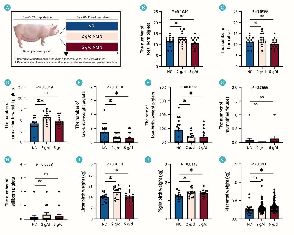
Figure 1.
Effects of NMN supplementation on reproductive performance of sows during pregnancy
-
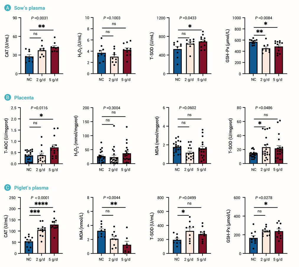
Figure 2.
Effects of NMN supplementation on oxidative stress in sows, placentas and piglets
-
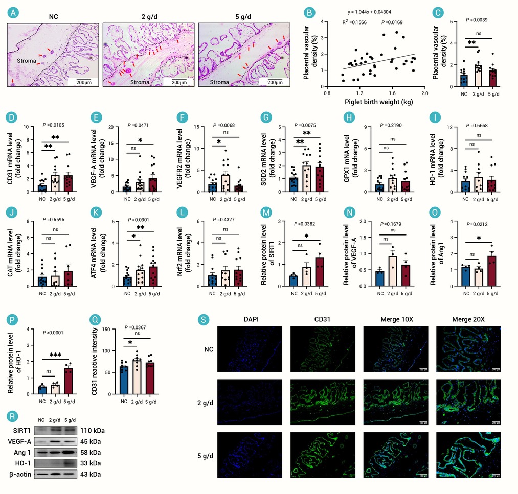
Figure 3.
Effects of adding NMN to pregnancy diets on placental angiogenesis and oxidative stress status
-
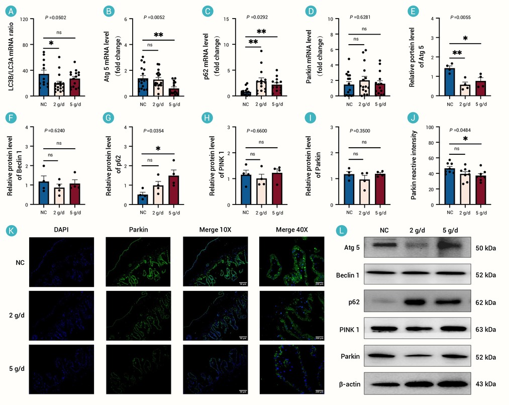
Figure 4.
The effect of adding NMN to pregnancy diet on placental mitophagy
-
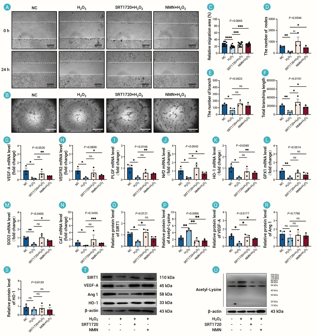
Figure 5.
Effects of adding NMN on angiogenesis and oxidative stress status of PVECs under oxidative stress
-
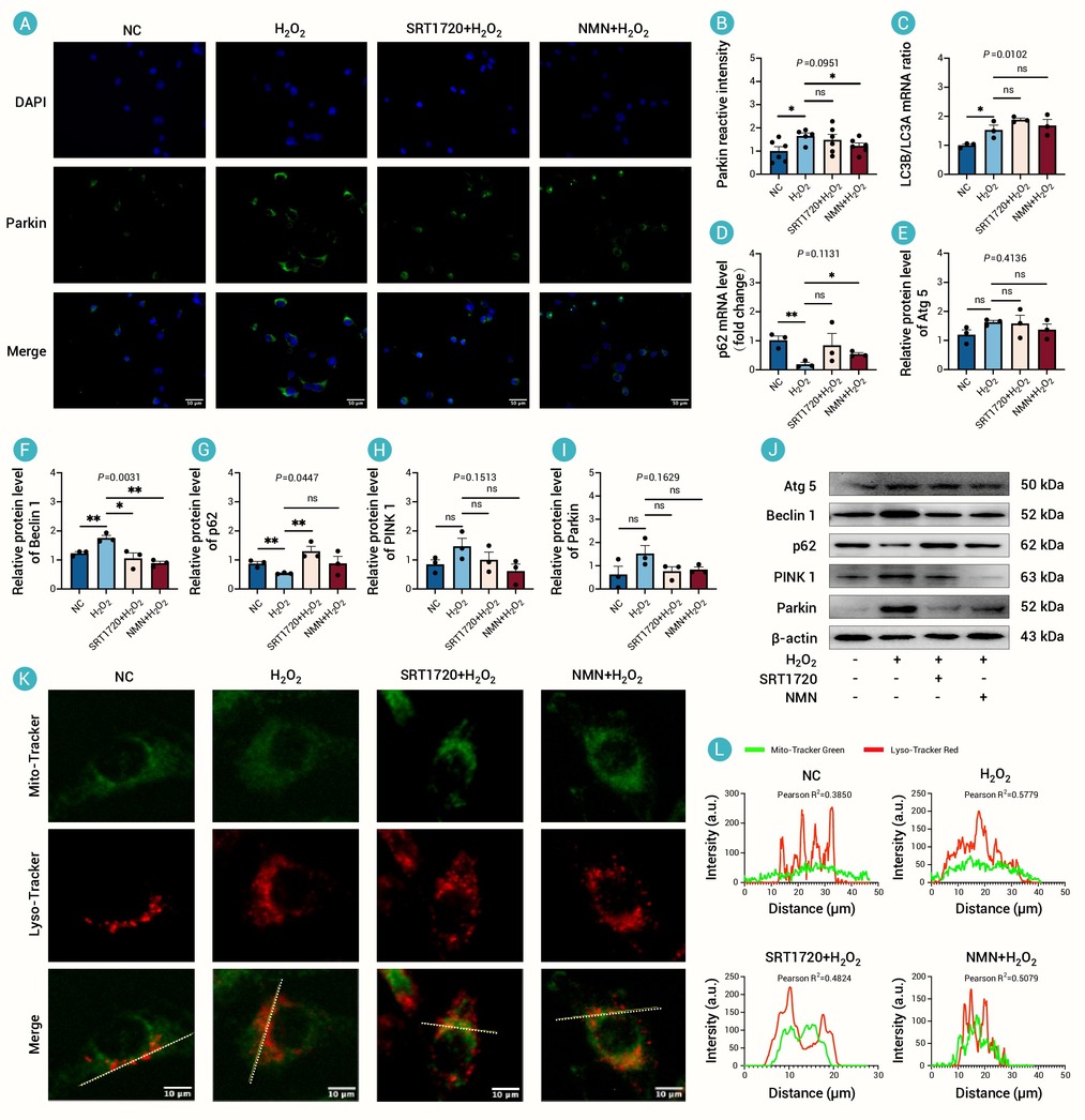
Figure 6.
The effect of adding NMN on mitophagy in PVECs under oxidative stress
-
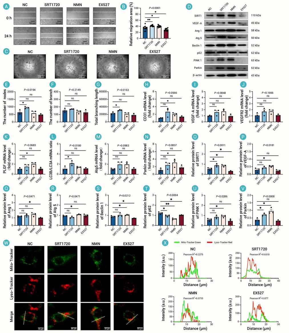
Figure 7.
Effects of activation or inhibition of NMN-SIRT1 on angiogenesis and mitophagy in PVECs
-
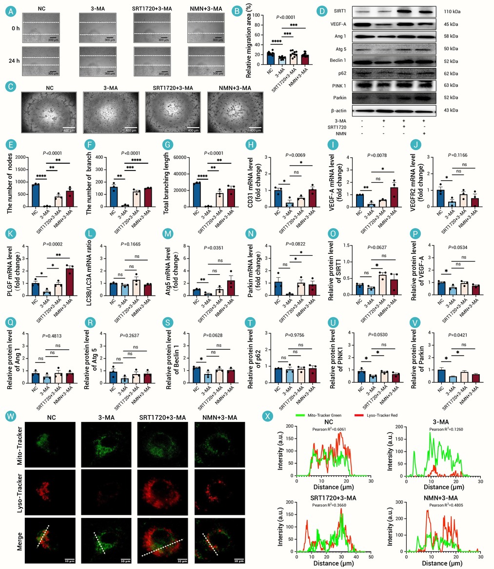
Figure 8.
Effects of activating NMN-SIRT1 after inhibiting mitophagy on angiogenesis and mitophagy in PVECs
