
Advances of fluorescent DNA nanostructures in biomedical applications
-
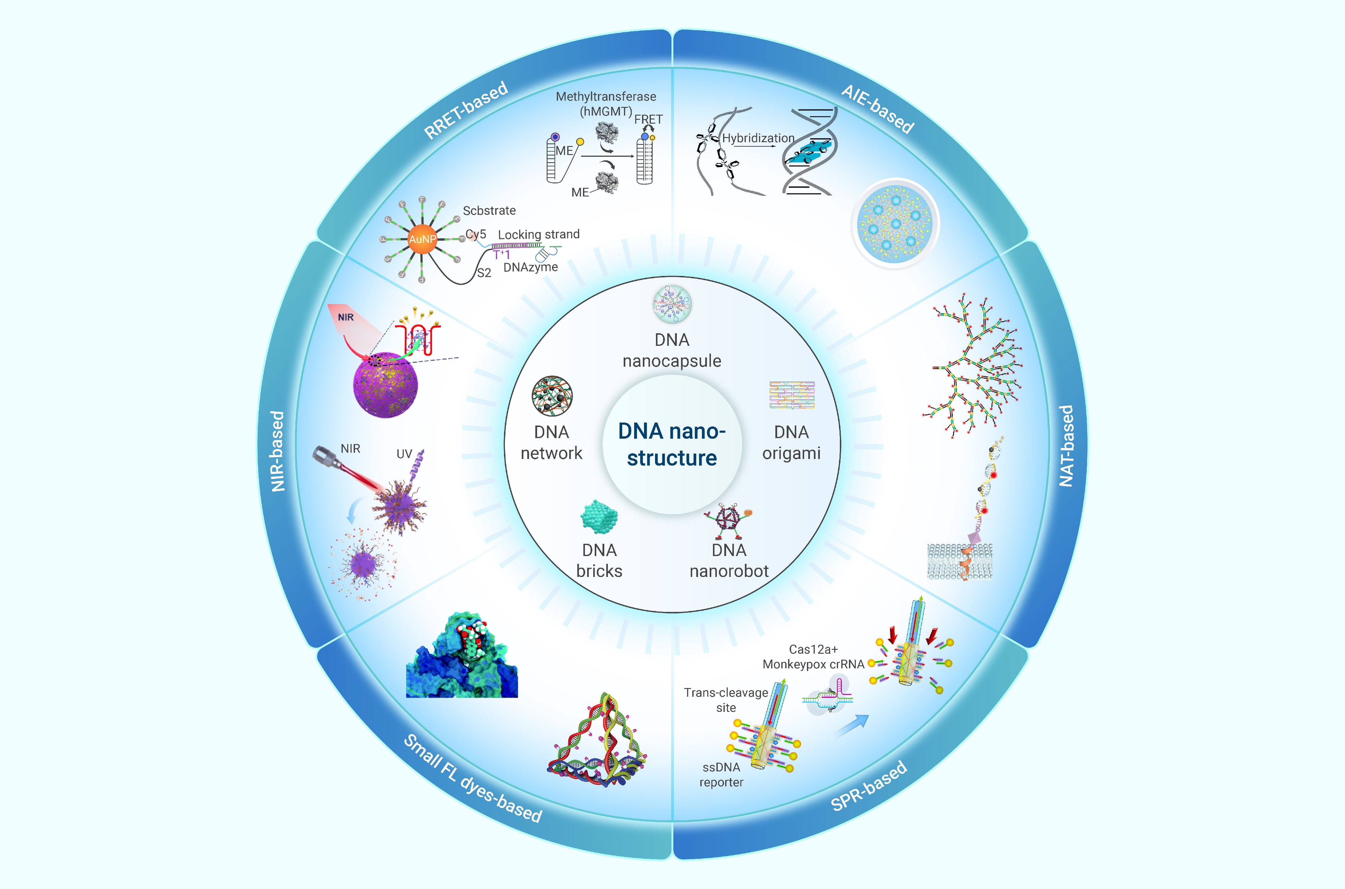
GRAPHICAL ABSTRACT
 DownLoad:
Full size image
DownLoad:
Full size image
-
PUBLIC SUMMARY
-
Advances of fluorescent DNA nanostructures (FDNs) in biomedical applications are reviewed.
Various DNA nanostructures are summarized.
Remaining challenges and key research directions for enlarging biological applications of FDNs are proposed.
-
-
ABSTRACT
With the rapid development of DNA nanotechnology, the emergence of fluorescent DNA nanostructures (FDNs) has enlarged the biological applications. FDNs have great advantages of precise localization and real-time tracing in bioimaging. In this review, the recent biomedical developments of FDNs have been reviewed, including the design of FDNs, and the corresponding applications on biomarker sensing, bioimaging, cancer diagnosis and therapy. Firstly, the development of DNA nanostructures and the corresponding DNA-based nanomaterials were briefly introduced. Simultaneously, to make a better demonstration, the background and theory of the fluorescence detections were briefly introduced. Thereafter, the synthetic strategies of DNA nanostructure were summarized and classified, which facilitated the multiple functionalizations for sensing and bioimaging. Subsequently, the biomedical applications of FDNs are comprehensively summarized based on different detection strategies, including fluorescence resonance energy transfer (FRET), nucleic acid amplification (NAT), aggregation-induced emission (AIE), near-infrared (NIR)-photoactivation, small fluorescent dyes loading, and surface plasmon resonance (SPR) technologies. Finally, an insight into the challenges and future perspectives is provided. As reviewed, FDNs are important tools in precision medicine, showing great potential in both in vivo and in vitro cancer diagnosis and treatments. Undoubtedly, FDN-based technology is a promising strategy for constructing versatile nanodevices in biological applications and will excel in human healthcare. -

-
REFERENCES
[1] Gao, Q., Zeng, Q., Wang, Z., et al. (2022). Circulating cell-free DNA for cancer early detection. The Innovation 3: 100259. DOI: 10.1016/j.xinn.2022.100259. [2] Seeman, N.C., and Sleiman, H.F. (2017). DNA nanotechnology. Nature Reviews Materials 3: 17068. DOI: 10.1038/natrevmats.2017.68. [3] Awaja, F., Wakelin, E.A., Sage, J., et al. (2015). Description of DNA molecular motion for nanotechnology applications. Progress in Materials Science 74: 308−331. DOI: 10.1016/j.pmatsci.2015.03.001. [4] Zhang, F., Nangreave, J., Liu, Y., et al. (2014). Structural DNA nanotechnology: State of the art and future perspective. Journal of the American Chemical Society 136: 11198−11211. DOI: 10.1021/ja505101a. [5] Aldaye, F.A., Palmer, A.L., and Sleiman, H.F. (2008). Assembling Materials with DNA as the Guide. Science 321: 1795−1799. DOI: 10.1126/science.1154533. [6] Julin, S., Nummelin, S., Kostiainen, M.A., et al. (2018). DNA nanostructure-directed assembly of metal nanoparticle superlattices. Journal of Nanoparticle Research 20: 119. DOI: 10.1007/s11051-018-4225-3. [7] Mirkin, C.A., Letsinger, R.L., Mucic, R.C., et al. (1996). A DNA-based method for rationally assembling nanoparticles into macroscopic materials. Nature 382: 607−609. DOI: 10.1038/382607a0. [8] Alivisatos, A.P., Johnsson, K.P., Peng, X., et al. (1996). Organization of 'nanocrystal molecules' using DNA. Nature 382: 609−611. DOI: 10.1038/382609a0. [9] Fu, J., Liu, M., Liu, Y., et al. (2012). Spatially-Interactive biomolecular networks organized by nucleic acid nanostructures. Accounts of Chemical Research 45: 1215−1226. DOI: 10.1021/ar200295q. [10] Wang, X., Chandrasekaran, A.R., Shen, Z., et al. (2019). Paranemic crossover DNA: There and back again. Chemical Reviews 119: 6273−6289. DOI: 10.1021/acs.chemrev.8b00207. [11] Loescher, S., Groeer, S., and Walther, A. (2018). 3D DNA origami nanoparticles: From basic design principles to emerging applications in soft matter and (bio-) nanosciences. Angewandte Chemie International Edition 57: 10436−10448. DOI: 10.1002/anie.201801700. [12] Tørring, T., Voigt, N.V., Nangreave, J., et al. (2011). DNA origami: a quantum leap for self-assembly of complex structures. Chemical Society Reviews 40: 5636−5646. DOI: 10.1039/C1CS15057J. [13] Hong, F., Zhang, F., Liu, Y., et al. (2017). DNA origami: Scaffolds for creating higher order structures. Chemical Reviews 117: 12584−12640. DOI: 10.1021/acs.chemrev.6b00825. [14] Rothemund, P.W.K. (2006). Folding DNA to create nanoscale shapes and patterns. Nature 440: 297−302. DOI: 10.1038/nature04586. [15] Kopperger, E., List, J., Madhira, S., et al. (2018). A self-assembled nanoscale robotic arm controlled by electric fields. Science 359: 296−301. DOI: 10.1126/science.aao4284. [16] Simmel, F.C., Yurke, B., and Singh, H.R. (2019). Principles and applications of nucleic acid strand displacement reactions. Chemical Reviews 119: 6326−6369. DOI: 10.1021/acs.chemrev.8b00580. [17] Wang, S.S., and Ellington, A.D. (2019). Pattern generation with nucleic acid chemical reaction networks. Chemical Reviews 119: 6370−6383. DOI: 10.1021/acs.chemrev.8b00625. [18] Petersen, P., Tikhomirov, G., and Qian, L. (2018). Information-based autonomous reconfiguration in systems of interacting DNA nanostructures. Nature Communications 9: 5362. DOI: 10.1038/s41467-018-07805-7. [19] Chen, J., Chen, Z., Meng, C., et al. (2023). CRISPR-powered optothermal nanotweezers: Diverse bio-nanoparticle manipulation and single nucleotide identification. Light: Science & Applications 12: 273. DOI: 10.1038/s41377-023-01326-9. [20] Chen, Z., Wu, C., Yuan, Y., et al. (2023). CRISPR-Cas13a-powered electrochemical biosensor for the detection of the L452R mutation in clinical samples of SARS-CoV-2 variants. Journal of Nanobiotechnology 21: 141. DOI: 10.1186/s12951-023-01903-5. [21] Shen, J., Chen, Z., Xie, R., et al. (2023). CRISPR/Cas12a-assisted isothermal amplification for rapid and specific diagnosis of respiratory virus on an microfluidic platform. Biosensors and Bioelectronics 237: 115523. DOI: 10.1016/j.bios.2023.115523. [22] Yang, D., Hartman, M.R., Derrien, T.L., et al. (2014). DNA materials: bridging nanotechnology and biotechnology. Accounts of Chemical Research 47: 1902−1911. DOI: 10.1021/ar5001082. [23] Li, J., Green, A.A., Yan, H., et al. (2017). Engineering nucleic acid structures for programmable molecular circuitry and intracellular biocomputation. Nature Chemistry 9: 1056−1067. DOI: 10.1038/nchem.2852. [24] Kohman, R.E., Kunjapur, A.M., Hysolli, E., et al. (2018). From designing the molecules of life to designing life: Future applications derived from advances in DNA technologies. Angewandte Chemie International Edition 57: 4313−4328. DOI: 10.1002/anie.201707976. [25] Hu, Q., Li, H., Wang, L., et al. (2019). DNA nanotechnology-enabled drug delivery systems. Chemical Reviews 119: 6459−6506. DOI: 10.1021/acs.chemrev.7b00663. [26] Walter, J.A., Mikhail, Y.B., Hyeran, L., et al. (2010). Biological applications of fluorescence lifetime imaging beyond microscopy. Reporters, Markers, Dyes, Nanoparticles, and Molecular Probes for Biomedical Applications II. SPIE 7576: 214−222. DOI: 10.1117/12.849453. [27] Yao, J., Yang, M., and Duan, Y. (2014). Chemistry, biology, and medicine of fluorescent nanomaterials and related systems: New insights into biosensing, bioimaging, genomics, diagnostics, and therapy. Chemical Reviews 114: 6130−6178. DOI: 10.1021/cr200359p. [28] Xing, L., Todd, N.W., Yu, L., et al. (2010). Early detection of squamous cell lung cancer in sputum by a panel of microRNA markers. Modern Pathology 23: 1157−1164. DOI: 10.1038/modpathol.2010.111. [29] Choi, Y.-E., Kwak, J.-W., and Park, J.W. (2010). Nanotechnology for early cancer detection. Sensors 10: 428−455. DOI: 10.3390/s100100428. [30] Lu, H., Wang, Y., and Yu, R. (2023). Immune cell membrane-coated nanoparticles for targeted myocardial ischemia/reperfusion injury therapy. The Innovation Medicine 1: 100015. DOI: 10.59717/j.xinn-med.2023.100015. [31] Cao, C., and Cao, X. (2023). Nanowire-based smart windows achieving dynamic solar radiation regulation. The Innovation Materials 1: 100024. DOI: 10.59717/j.xinn-mater.2023.100024. [32] Huang, Z., Zeng, S., Wen, S., et al. (2023). Fluorescence-guided visualization of pancreatic neuroendocrine tumor borders. The Innovation Medicine 1: 100025. DOI: 10.59717/j.xinn-med.2023.100025. [33] Chinen, A.B., Guan, C.M., Ferrer, J.R., et al. (2015). Nanoparticle probes for the detection of cancer biomarkers, cells, and tissues by fluorescence. Chemical Reviews 115: 10530−10574. DOI: 10.1021/acs.chemrev.5b00321. [34] Berezin, M.Y., and Achilefu, S. (2010). Fluorescence lifetime measurements and biological imaging. Chemical Reviews 110: 2641−2684. DOI: 10.1021/cr900343z. [35] Hötzer, B., Medintz, I.L., and Hildebrandt, N. (2012). Fluorescence in nanobiotechnology: sophisticated fluorophores for novel applications. Small 8: 2297−2326. DOI: 10.1002/smll.201200109. [36] Seeman, N.C. (1982). Nucleic acid junctions and lattices. Journal of Theoretical Biology 99: 237−247. DOI: 10.1016/0022-5193(82)90002-9. [37] Ma, R.-I., Kallenbach, N.R., Sheardy, R.D., et al. (1986). Three-arm nucleic acid junctions are flexible. Nucleic Acids Research 14: 9745−9753. DOI: 10.1093/nar/14.24.9745. [38] Baig, M.M.F.A., Gao, X., Khan, M.A., et al. (2022). Nanoscale packing of DNA tiles into DNA macromolecular lattices. International Journal of Biological Macromolecules 220: 520−527. DOI: 10.1016/j.ijbiomac.2022.08.107. [39] Han, D., Qi, X., Myhrvold, C., et al. (2017). Single-stranded DNA and RNA origami. Science 358: eaao2648. DOI: 10.1126/science.aao2648. [40] Liu, D., Wang, M., Deng, Z., et al. (2004). Tensegrity: Construction of rigid DNA triangles with flexible four-arm DNA junctions. Journal of the American Chemical Society 126: 2324−2325. DOI: 10.1021/ja031754r. [41] Liu, D., Park, S.H., Reif, J.H., et al. (2004). DNA nanotubes self-assembled from triple-crossover tiles as templates for conductive nanowires. Proceedings of the National Academy of Sciences 101: 717−722. DOI: 10.1073/pnas.0305860101. [42] Goodman, R.P., Berry, R.M., and Turberfield, A.J. (2004). The single-step synthesis of a DNA tetrahedron. Chemical Communications(12):1372-1373. DOI: 10.1039/B402293A. [43] Shih, W.M., Quispe, J.D., and Joyce, G.F. (2004). A 1.7-kilobase single-stranded DNA that folds into a nanoscale octahedron. Nature 427 (6975):618-621. DOI: 10.1038/nature02307. [44] Wang, H., Luo, D., Wang, H., et al. (2021). Construction of smart stimuli-responsive DNA nanostructures for biomedical applications. Chemistry – A European Journal 27: 3929−3943. DOI: 10.1002/chem.202003145. [45] Tang, J., Yao, C., Gu, Z., et al. (2020). Super-soft and super-elastic DNA robot with magnetically driven navigational locomotion for cell delivery in confined space. Angewandte Chemie International Edition 59: 2490−2495. DOI: 10.1002/anie.201913549. [46] Gong, X., Li, R., Wang, J., et al. (2020). A smart theranostic nanocapsule for spatiotemporally programmable photo-gene therapy. Angewandte Chemie International Edition 59: 21648−21655. DOI: 10.1002/anie.202008413. [47] Pogu, S.V., Dehariya, D., Yadav, D.N., et al. (2023). A review on fabrication, actuation, and application of magnetic force driven, light driven and DNA nano/microrobots in modern theranostics. Molecular Systems Design & Engineering 8: 416−430. DOI: 10.1039/D2ME00247G. [48] Wei, B., Dai, M., and Yin, P. (2012). Complex shapes self-assembled from single-stranded DNA tiles. Nature 485: 623−626. DOI: 10.1038/nature11075. [49] Zhang, Y., and Seeman, N.C. (1994). Construction of a DNA-truncated octahedron. Journal of the American Chemical Society 116: 1661−1669. DOI: 10.1021/ja00084a006. [50] Zhou, Y., Dong, J., Zhou, C., et al. (2022). Finite assembly of three-dimensional DNA hierarchical nanoarchitectures through orthogonal and directional bonding. Angewandte Chemie International Edition 61: e202116416. DOI: 10.1002/anie.202116416. [51] Han, D., Pal, S., Nangreave, J., et al. (2011). DNA origami with complex curvatures in three-dimensional space. Science 332: 342−346. DOI: 10.1126/science.1202998. [52] Yurke, B., Turberfield, A.J., Mills, A.P., et al. (2000). A DNA-fuelled molecular machine made of DNA. Nature 406: 605−608. DOI: 10.1038/35020524. [53] Yan, H., Zhang, X., Shen, Z., et al. (2002). A robust DNA mechanical device controlled by hybridization topology. Nature 415: 62−65. DOI: 10.1038/415062a. [54] Omabegho, T., Sha, R., and Seeman, N.C. (2009). A bipedal DNA brownian motor with coordinated legs. Science 324: 67−71. DOI: 10.1126/science.1170336. [55] Yan, J., Hu, C., Wang, P., et al. (2015). Growth and origami folding of DNA on nanoparticles for high-efficiency molecular transport in cellular imaging and drug delivery. Angewandte Chemie International Edition 54: 2431−2435. DOI: 10.1002/anie.201408247. [56] Shen, X., Jiang, Q., Wang, J., et al. (2012). Visualization of the intracellular location and stability of DNA origami with a label-free fluorescent probe. Chemical Communications 48: 11301−11303. DOI: 10.1039/C2CC36185J. [57] Edwardson, T.G.W., Lau, K.L., Bousmail, D., et al. (2016). Transfer of molecular recognition information from DNA nanostructures to gold nanoparticles. Nature Chemistry 8: 162−170. DOI: 10.1038/nchem.2420. [58] Li, J., Wei, J., Gao, Y., et al. (2023). Peptide-assembled siRNA nanomicelles confine MnOx-loaded silicages for synergistic chemical and gene-regulated cancer therapy. Chinese Chemical Letters 34: 107662. DOI: 10.1016/j.cclet.2022.07.005. [59] Rosenzweig, B.A., Ross, N.T., Tagore, D.M., et al. (2009). Multivalent protein binding and precipitation by self-assembling molecules on a DNA pentaplex scaffold. Journal of the American Chemical Society 131: 5020−5021. DOI: 10.1021/ja809219p. [60] Lan, X., Su, Z., Zhou, Y., et al. (2017). Programmable supra-assembly of a DNA surface adapter for tunable chiral directional self-assembly of gold nanorods. Angewandte Chemie International Edition 56: 14632−14636. DOI: 10.1002/anie.201709775. [61] Shen, X., Wang, Y., Zhang, Y., et al. (2018). Unique SiO2 nanourchins enable amplification in living cells for in situ imaging of mRNAs. Advanced Functional Materials 28: 1803286. DOI: 10.1002/adfm.201803286. [62] Wang, Y., Lu, X., Wu, X., et al. (2022). Chemically modified DNA nanostructures for drug delivery. The Innovation 3: 100217. DOI: 10.1016/j.xinn.2022.100217. [63] Förster, T. (1948). Zwischenmolekulare energiewanderung und fluoreszenz. Annalen der Physik 437(1-2): 55−75. DOI: 10.1002/andp.19484370105. [64] Piston, D.W., and Kremers, G.-J. (2007). Fluorescent protein FRET: the good, the bad and the ugly. Trends in Biochemical Sciences 32: 407−414. DOI: 10.1016/j.tibs.2007.08.003. [65] Shen, X., Xu, W., Ouyang, J., et al. (2022). Fluorescence resonance energy transfer-based nanomaterials for the sensing in biological systems. Chinese Chemical Letters 33: 4505−4516. DOI: 10.1016/j.cclet.2021.12.061. [66] Yang, L., Liu, Y., Ma, C., et al. (2015). Naphthalene-fused BODIPY with large Stokes shift as saturated-red fluorescent dye for living cell imaging. Dyes and Pigments 122: 1−5. DOI: 10.1016/j.dyepig.2015.06.006. [67] Guo, B., Nie, H., Yang, W., et al. (2016). A highly sensitive and rapidly responding fluorescent probe with a large Stokes shift for imaging intracellular hypochlorite. Sensors and Actuators B: Chemical 236: 459−465. DOI: 10.1016/j.snb.2016.06.004. [68] Gao, Z., Hao, Y., Zheng, M., et al. (2017). A fluorescent dye with large Stokes shift and high stability: synthesis and application to live cell imaging. RSC Advances 7: 7604−7609. DOI: 10.1039/C6RA27547H. [69] Zhang, D., Wen, Y., Xiao, Y., et al. (2008). Bulky 4-tritylphenylethynyl substituted boradiazaindacene: pure red emission, relatively large Stokes shift and inhibition of self-quenching. Chemical Communications (39):4777-4779. DOI: 10.1039/B808681H. [70] Sednev, M.V., Belov, V.N., and Hell, S.W. (2015). Fluorescent dyes with large Stokes shifts for super-resolution optical microscopy of biological objects: A review. Methods and Applications in Fluorescence 3: 042004. DOI: 10.1088/2050-6120/3/4/042004. [71] Lin, W., Yuan, L., Cao, Z., et al. (2010). Through-bond energy transfer cassettes with minimal spectral overlap between the donor emission and acceptor absorption: Coumarin–rhodamine dyads with large pseudo-stokes shifts and emission shifts. Angewandte Chemie International Edition 49: 375−379. DOI: 10.1002/anie.200904515. [72] Zhang, H., Li, H., Cao, Z., et al. (2020). Investigation of the in vivo integrity of polymeric micelles via large Stokes shift fluorophore-based FRET. Journal of Controlled Release 324: 47−54. DOI: 10.1016/j.jconrel.2020.04.046. [73] Jungmann, R., Avendaño, M.S., Woehrstein, J.B., et al. (2014). Multiplexed 3D cellular super-resolution imaging with DNA-PAINT and exchange-PAINT. Nature Methods 11: 313−318. DOI: 10.1038/nmeth.2835. [74] Ma, Y., Song, M., Li, L., et al. (2024). Attomolar-level detection of respiratory virus long-chain oligonucleotides based on FRET biosensor with upconversion nanoparticles and Au–Au dimer. Biosensors and Bioelectronics 243: 115778. DOI: 10.1016/j.bios.2023.115778. [75] Farag, N., Mattossovich, R., Merlo, R., et al. (2021). Folding-upon-repair DNA nanoswitches for monitoring the activity of DNA repair enzymes. Angewandte Chemie International Edition 60: 7283−7289. DOI: 10.1002/anie.202016223. [76] Li, L., Wang, J., Jiang, H., et al. (2023). DNA tetrahedron-based split aptamer probes for reliable imaging of ATP in living cells. Chinese Chemical Letters 34: 107506. DOI: 10.1016/j.cclet.2022.05.020. [77] Huizenga, D.E., and Szostak, J.W. (1995). A DNA aptamer that binds adenosine and ATP. Biochemistry 34: 656−665. DOI: 10.1021/bi00002a033. [78] Nutiu, R., and Li, Y. (2003). Structure-switching signaling aptamers. Journal of the American Chemical Society 125: 4771−4778. DOI: 10.1021/ja028962o. [79] Di, Z., Zhao, J., Chu, H., et al. (2019). An Acidic-microenvironment-driven DNA nanomachine enables specific ATP imaging in the extracellular milieu of tumor. Advanced Materials 31: 1901885. DOI: 10.1002/adma.201901885. [80] Carrasquilla, C., Kapteyn, E., Li, Y., et al. (2017). Sol–gel-derived biohybrid materials incorporating long-chain DNA aptamers. Angewandte Chemie International Edition 56: 10686−10690. DOI: 10.1002/anie.201702859. [81] Zheng, J., Wang, Q., Shi, L., et al. (2021). Logic-gated proximity aptasensing for cell-surface real-time monitoring of apoptosis. Angewandte Chemie International Edition 60: 20858−20864. DOI: 10.1002/anie.202106651. [82] Xiang, Z., Zhao, J., Qu, J., et al. (2022). A multivariate-gated DNA nanodevice for spatioselective imaging of pro-metastatic targets in extracellular microenvironment. Angewandte Chemie International Edition 61: e202111836. DOI: 10.1002/anie.202111836. [83] Zhou, X., Zhao, M., Duan, X., et al. (2017). Collapse of DNA tetrahedron nanostructure for “off–on” fluorescence detection of DNA methyltransferase activity. ACS Applied Materials & Interfaces 9: 40087−40093. DOI: 10.1021/acsami.7b13551. [84] Zhu, G., Zhang, S., Song, E., et al. (2013). Building fluorescent DNA nanodevices on target living cell surfaces. Angewandte Chemie International Edition 52: 5490−5496. DOI: 10.1002/anie.201301439. [85] He, Y., and Liu, D.R. (2010). Autonomous multistep organic synthesis in a single isothermal solution mediated by a DNA walker. Nature Nanotechnology 5: 778−782. DOI: 10.1038/nnano.2010.190. [86] Xu, Z., Dong, Y., Li, J., et al. (2015). A ferrocene-switched electrochemiluminescence “off–on” strategy for the sensitive detection of cardiac troponin I based on target transduction and a DNA walking machine. Chemical Communications 51: 14369−14372. DOI: 10.1039/C5CC04745E. [87] Wang, L., Deng, R., and Li, J. (2015). Target-fueled DNA walker for highly selective miRNA detection. Chemical Science 6: 6777−6782. DOI: 10.1039/C5SC02784E. [88] Wickham, S.F.J., Endo, M., Katsuda, Y., et al. (2011). Direct observation of stepwise movement of a synthetic molecular transporter. Nature Nanotechnology 6: 166−169. DOI: 10.1038/nnano.2010.284. [89] Tomov, T.E., Tsukanov, R., Glick, Y., et al. (2017). DNA bipedal motor achieves a large number of steps due to operation using microfluidics-based interface. ACS Nano 11: 4002−4008. DOI: 10.1021/acsnano.7b00547. [90] Qu, X., Zhu, D., Yao, G., et al. (2017). An exonuclease III-powered, on-particle stochastic DNA walker. Angewandte Chemie International Edition 56: 1855−1858. DOI: 10.1002/anie.201611777. [91] Yang, X., Tang, Y., Mason, S.D., et al. (2016). Enzyme-powered three-dimensional DNA nanomachine for DNA walking, payload release, and biosensing. ACS Nano 10: 2324−2330. DOI: 10.1021/acsnano.5b07102. [92] Xu, Z.-Q., Zhang, P., Chai, Y.-Q., et al. (2018). A biosensor based on a 3D-DNA walking machine network and distance-controlled electrochemiluminescence energy transfer for ultrasensitive detection of tenascin C and lead ions. Chemical Communications 54: 8741−8744. DOI: 10.1039/C8CC04953J. [93] Li, D., Zhou, W., Yuan, R., et al. (2017). A DNA-fueled and catalytic molecule machine lights up trace under-expressed microRNAs in living cells. Analytical Chemistry 89: 9934−9940. DOI: 10.1021/acs.analchem.7b02247. [94] Ye, S., Li, X., Wang, M., et al. (2017). Fluorescence and SERS imaging for the simultaneous absolute quantification of multiple miRNAs in living cells. Analytical Chemistry 89: 5124−5130. DOI: 10.1021/acs.analchem.7b00697. [95] Zhang, H., Lai, M., Zuehlke, A., et al. (2015). Binding-induced DNA nanomachines triggered by proteins and nucleic acids. Angewandte Chemie International Edition 54: 14326−14330. DOI: 10.1002/anie.201506312. [96] Luan, M., Li, N., Pan, W., et al. (2017). Simultaneous detection of multiple targets involved in the PI3K/AKT pathway for investigating cellular migration and invasion with a multicolor fluorescent nanoprobe. Chemical Communications 53: 356−359. DOI: 10.1039/C6CC07605J. [97] Chen, J., Zuehlke, A., Deng, B., et al. (2017). A target-triggered DNAzyme motor enabling homogeneous, amplified detection of proteins. Analytical Chemistry 89: 12888−12895. DOI: 10.1021/acs.analchem.7b03529. [98] Liang, C.-P., Ma, P.-Q., Liu, H., et al. (2017). Rational engineering of a dynamic, entropy-driven DNA nanomachine for intracellular microRNA imaging. Angewandte Chemie International Edition 56: 9077−9081. DOI: 10.1002/anie.201704147. [99] Ma, P.-Q., Liang, C.-P., Zhang, H.-H., et al. (2018). A highly integrated DNA nanomachine operating in living cells powered by an endogenous stimulus. Chemical Science 9: 3299−3304. DOI: 10.1039/C8SC00049B. [100] Mason, S.D., Wang, G.A., Yang, P., et al. (2019). Probing and controlling dynamic interactions at biomolecule–nanoparticle interfaces using stochastic DNA walkers. ACS Nano 13: 8106−8113. DOI: 10.1021/acsnano.9b03053. [101] Kong, L., Kou, B., Zhang, X., et al. (2021). A core–brush 3D DNA nanostructure: the next generation of DNA nanomachine for ultrasensitive sensing and imaging of intracellular microRNA with rapid kinetics. Chemical Science 12: 15953−15959. DOI: 10.1039/D1SC04571G. [102] Liu, J., and Lu, Y. (2002). FRET study of a trifluorophore-labeled DNAzyme. Journal of the American Chemical Society 124: 15208−15216. DOI: 10.1021/ja027647z. [103] Deng, R., Yang, H., Dong, Y., et al. (2018). Temperature-robust DNAzyme biosensors confirming ultralow background detection. ACS Sensors 3: 2660−2666. DOI: 10.1021/acssensors.8b01122. [104] Wu, Z., Fan, H., Satyavolu, N.S.R., et al. (2017). Imaging endogenous metal ions in living cells using a DNAzyme–catalytic hairpin assembly probe. Angewandte Chemie International Edition 56: 8721−8725. DOI: 10.1002/anie.201703540. [105] Peng, H., Li, X.-F., Zhang, H., et al. (2017). A microRNA-initiated DNAzyme motor operating in living cells. Nature Communications 8: 14378. DOI: 10.1038/ncomms14378. [106] Wu, M., Zhang, M., Fan, Z., et al. (2021). Ultrasensitive DNA methyltransferase activity sensing and inhibitor evaluation with highly photostable upconversion nanoparticle transducer. Microchimica Acta 188: 169. DOI: 10.1007/s00604-021-04831-z. [107] Cha, T.-G., Pan, J., Chen, H., et al. (2015). Design principles of DNA enzyme-based walkers: Translocation kinetics and photoregulation. Journal of the American Chemical Society 137: 9429−9437. DOI: 10.1021/jacs.5b05522. [108] Cha, T.-G., Pan, J., Chen, H., et al. (2014). A synthetic DNA motor that transports nanoparticles along carbon nanotubes. Nature Nanotechnology 9: 39−43. DOI: 10.1038/nnano.2013.257. [109] Yang, K., Wang, H., Ma, N., et al. (2018). Programmable target-initiated DNAzyme walker walking along a spatially isolated and highly hybridizable substrate track on a nanoparticle surface. ACS Applied Materials & Interfaces 10: 44546−44553. DOI: 10.1021/acsami.8b16408. [110] Liu, C., Hu, Y., Pan, Q., et al. (2019). A microRNA-triggered self-powered DNAzyme walker operating in living cells. Biosensors and Bioelectronics 136: 31−37. DOI: 10.1016/j.bios.2019.04.031. [111] Liu, C., Hu, Y., Pan, Q., et al. (2020). A photocontrolled and self-powered bipedal DNA walking machine for intracellular microRNA imaging. Chemical Communications 56: 3496−3499. DOI: 10.1039/D0CC00017E. [112] Dyla, M., Andersen, J.L., Kjaergaard, M., et al. (2016). Engineering a prototypic P-type ATPase listeria monocytogenes Ca2+-ATPase 1 for single-molecule FRET studies. Bioconjugate Chemistry 27: 2176−2187. DOI: 10.1021/acs.bioconjchem.6b00387. [113] Fijen, C., Montón Silva, A., Hochkoeppler, A., et al. (2017). A single-molecule FRET sensor for monitoring DNA synthesis in real time. Physical Chemistry Chemical Physics 19: 4222−4230. DOI: 10.1039/C6CP05919H. [114] Koo, H., Park, I., Lee, Y., et al. (2016). Visualization and quantification of microRNA in a single cell using atomic force microscopy. Journal of the American Chemical Society 138: 11664−11671. DOI: 10.1021/jacs.6b05048. [115] Wei, J., Gong, X., Wang, Q., et al. (2018). Construction of an autonomously concatenated hybridization chain reaction for signal amplification and intracellular imaging. Chemical Science 9: 52−61. DOI: 10.1039/C7SC03939E. [116] Gao, R., Hao, C., Xu, L., et al. (2018). Spiny nanorod and upconversion nanoparticle satellite assemblies for ultrasensitive detection of messenger RNA in living cells. Analytical Chemistry 90: 5414−5421. DOI: 10.1021/acs.analchem.8b00617. [117] Xu, L., Gao, Y., Kuang, H., et al. (2018). MicroRNA-directed intracellular self-assembly of chiral nanorod dimers. Angewandte Chemie International Edition 57: 10544−10548. DOI: 10.1002/anie.201805640. [118] Ma, W., Sun, M., Fu, P., et al. (2017). A chiral-nanoassemblies-enabled strategy for simultaneously profiling surface glycoprotein and microRNA in living cells. Advanced Materials 29: 1703410. DOI: 10.1002/adma.201703410. [119] Dai, W., Dong, H., Guo, K., et al. (2018). Near-infrared triggered strand displacement amplification for MicroRNA quantitative detection in single living cells. Chemical Science 9: 1753−1759. DOI: 10.1039/c7sc04243d. [120] Meng, X., Dai, W., Zhang, K., et al. (2018). Imaging multiple microRNAs in living cells using ATP self-powered strand-displacement cascade amplification. Chemical Science 9: 1184−1190. DOI: 10.1039/C7SC04725H. [121] Meng, X., Zhang, K., Dai, W., et al. (2018). Multiplex microRNA imaging in living cells using DNA-capped-Au assembled hydrogels. Chemical Science 9: 7419−7425. DOI: 10.1039/C8SC02858C. [122] Shen, X., Zhang, Y., Sun, J., et al. (2019). Biodegradable nanosyringes for intracellular amplification-based dual-diagnosis and gene therapy in single living cells††Electronic supplementary information (ESI) available: Experimental details, additional data and graphs. Chemical Science 10 (24): 6113-6119. DOI: 10.1039/c9sc01894h. [123] Liu, Y., Chen, Q., Liu, J., et al. (2017). Design of a modular DNA triangular-prism sensor enabling ratiometric and multiplexed biomolecule detection on a single microbead. Analytical Chemistry 89: 3590−3596. DOI: 10.1021/acs.analchem.6b04918. [124] Yin, P., Choi, H.M.T., Calvert, C.R., et al. (2008). Programming biomolecular self-assembly pathways. Nature 451: 318−322. DOI: 10.1038/nature06451. [125] Chandran, H., Rangnekar, A., Shetty, G., et al. (2013). An autonomously self-assembling dendritic DNA nanostructure for target DNA detection. Biotechnology Journal 8: 221−227. DOI: 10.1002/biot.201100499. [126] Xuan, F., and Hsing, I.M. (2014). Triggering hairpin-free chain-branching growth of fluorescent DNA dendrimers for nonlinear hybridization chain reaction. Journal of the American Chemical Society 136: 9810−9813. DOI: 10.1021/ja502904s. [127] Di, Z., Lu, X., Zhao, J., et al. (2022). Mild acidosis-directed signal amplification in tumor microenvironment via spatioselective recruitment of DNA amplifiers. Angewandte Chemie International Edition 61: e202205436. DOI: 10.1002/anie.202205436. [128] Peng, Y., Pang, H., Gao, Z., et al. (2023). Kinetics-accelerated one-step detection of microRNA through spatially localized reactions based on DNA tile self-assembly. Biosensors and Bioelectronics 222: 114932. DOI: 10.1016/j.bios.2022.114932. [129] Peng, Y., Gao, Z., Qiao, B., et al. (2023). Size-controlled DNA tile self-assembly nanostructures through caveolae-mediated endocytosis for signal-amplified imaging of microRNAs in living cells. Advanced Science 10: 2300614. DOI: 10.1002/advs.202300614. [130] Zhou, Z., Lin, N., Ouyang, Y., et al. (2023). Cascaded, feedback-driven, and spatially localized emergence of constitutional dynamic networks driven by enzyme-free catalytic DNA circuits. Journal of the American Chemical Society 145: 12617−12629. DOI: 10.1021/jacs.3c02083. [131] Zhou, X.-M., Zhuo, Y., Yuan, R., et al. (2023). Target-mediated self-assembly of DNA networks for sensitive detection and intracellular imaging of APE1 in living cells. Chemical Science 14: 2318−2324. DOI: 10.1039/D2SC06968G. [132] Li, J., Wang, J., Liu, S., et al. (2020). Amplified FRET nanoflares: An endogenous mRNA-powered nanomachine for intracellular microRNA imaging. Angewandte Chemie International Edition 59: 20104−20111. DOI: 10.1002/anie.202008245. [133] Chen, Y., Dai, W., Wang, D., et al. (2022). A cancer cell membrane vesicle-packaged DNA nanomachine for intracellular microRNA imaging. Chemical Communications 58: 9488−9491. DOI: 10.1039/D2CC03068C. [134] Lu, H., Yang, F., Liu, B., et al. (2019). Intracellular low-abundance microRNA imaging by a NIR-assisted entropy-driven DNA system. Nanoscale Horizons 4: 472−479. DOI: 10.1039/C8NH00330K. [135] Liu, F., Li, N., Shang, Y., et al. (2022). A DNA-based plasmonic nanodevice for cascade signal amplification. Angewandte Chemie International Edition 61: e202114706. DOI: 10.1002/anie.202114706. [136] Wang, C., Xie, Y., Song, X., et al. (2023). A NIR programmable in vivo miRNA magnifier for NIR-II imaging of early stage cancer. Angewandte Chemie International Edition 62: e202312665. DOI: 10.1002/anie.202312665. [137] Shustova, N.B., Ong, T.-C., Cozzolino, A.F., et al. (2012). Phenyl ring dynamics in a tetraphenylethylene-bridged metal–organic framework: Implications for the mechanism of aggregation-induced emission. Journal of the American Chemical Society 134: 15061−15070. DOI: 10.1021/ja306042w. [138] Ding, D., Li, K., Liu, B., et al. (2013). Bioprobes based on AIE fluorogens. Accounts of Chemical Research 46: 2441−2453. DOI: 10.1021/ar3003464. [139] Mei, J., Huang, Y., and Tian, H. (2018). Progress and trends in AIE-based bioprobes: A brief overview. ACS Applied Materials & Interfaces 10: 12217−12261. DOI: 10.1021/acsami.7b14343. [140] Mei, J., Leung, N.L.C., Kwok, R.T.K., et al. (2015). Aggregation-induced emission: Together we shine, united we soar! Chemical Reviews 115 (21):11718-11940. DOI: 10.1021/acs.chemrev.5b00263. [141] Jia, Y., Zuo, X., Lou, X., et al. (2015). Rational designed bipolar, conjugated polymer-DNA composite beacon for the sensitive detection of proteins and ions. Analytical Chemistry 87: 3890−3894. DOI: 10.1021/ac504690y. [142] Zhang, M., Saha, M.L., Wang, M., et al. (2017). Multicomponent platinum(II) cages with tunable emission and amino acid sensing. Journal of the American Chemical Society 139: 5067−5074. DOI: 10.1021/jacs.6b12536. [143] Liu, Y., Deng, C., Tang, L., et al. (2011). Specific detection of d-glucose by a tetraphenylethene-based fluorescent sensor. Journal of the American Chemical Society 133: 660−663. DOI: 10.1021/ja107086y. [144] Li, Y., Kwok, R.T.K., Tang, B.Z., et al. (2013). Specific nucleic acid detection based on fluorescent light-up probe from fluorogens with aggregation-induced emission characteristics. RSC Advances 3: 10135−10138. DOI: 10.1039/C3RA41983E. [145] Ou, X., Hong, F., Zhang, Z., et al. (2017). A highly sensitive and facile graphene oxide-based nucleic acid probe: Label-free detection of telomerase activity in cancer patient's urine using AIEgens. Biosensors and Bioelectronics 89: 417−421. DOI: 10.1016/j.bios.2016.05.035. [146] Hong, Y., Lam, J.W.Y., and Tang, B.Z. (2011). Aggregation-induced emission. Chemical Society Reviews 40: 5361−5388. DOI: 10.1039/C1CS15113D. [147] Hong, Y., Xiong, H., Lam, J.W.Y., et al. (2010). Fluorescent bioprobes: Structural matching in the docking processes of aggregation-induced emission fluorogens on DNA surfaces. Chemistry – A European Journal 16: 1232−1245. DOI: 10.1002/chem.200900778. [148] Li, W., Zhang, Y., Wang, Y., et al. (2021). Nucleic acids induced peptide-based AIE nanoparticles for fast cell imaging. Chinese Chemical Letters 32: 1571−1574. DOI: 10.1016/j.cclet.2020.09.054. [149] Tong, H., Hong, Y., Dong, Y., et al. (2007). Protein detection and quantitation by tetraphenylethene-based fluorescent probes with aggregation-induced emission characteristics. The Journal of Physical Chemistry B 111: 11817−11823. DOI: 10.1021/jp073147m. [150] Wang, M., Zhang, D., Zhang, G., et al. (2008). The convenient fluorescence turn-on detection of heparin with a silole derivative featuring an ammonium group. Chemical Communications 37 :4469-4471. DOI: 10.1039/B808877B. [151] Yuan, Y., Zhang, C.-J., and Liu, B. (2015). A photoactivatable AIE polymer for light-controlled gene delivery: Concurrent endo/lysosomal escape and DNA unpacking. Angewandte Chemie International Edition 54: 11419−11423. DOI: 10.1002/anie.201503640. [152] Zhang, C., Hao, L., Calabrese, C.M., et al. (2015). Biodegradable DNA-brush block copolymer spherical nucleic acids enable transfection agent-free intracellular gene regulation. Small 11: 5360−5368. DOI: 10.1002/smll.201501573. [153] Würthner, F. (2020). Aggregation-induced emission (AIE): A historical perspective. Angewandte Chemie International Edition 59: 14192−14196. DOI: 10.1002/anie.202007525. [154] Yu, Y., Liu, J., Zhao, Z., et al. (2012). Facile preparation of non-self-quenching fluorescent DNA strands with the degree of labeling up to the theoretic limit. Chemical Communications 48: 6360−6362. DOI: 10.1039/C2CC32038J. [155] Li, S., Langenegger, S.M., and Häner, R. (2013). Control of aggregation-induced emission by DNA hybridization. Chemical Communications 49: 5835−5837. DOI: 10.1039/C3CC42706D. [156] Ucar, H., and Wagenknecht, H.-A. (2021). DNA-templated control of chirality and efficient energy transport in supramolecular DNA architectures with aggregation-induced emission. Chemical Science 12: 10048−10053. DOI: 10.1039/D1SC02351A. [157] Jiang, M., Gu, X., Lam, J.W.Y., et al. (2017). Two-photon AIE bio-probe with large Stokes shift for specific imaging of lipid droplets. Chemical Science 8: 5440−5446. DOI: 10.1039/C7SC01400G. [158] Gao, Y., He, Z., He, X., et al. (2019). Dual-color emissive AIEgen for specific and label-free double-stranded DNA recognition and single-nucleotide polymorphisms detection. Journal of the American Chemical Society 141: 20097−20106. DOI: 10.1021/jacs.9b09239. [159] Nobe, S., Yamamoto, K., Miyakawa, H., et al. (2023). Sulfonium-cation-containing aggregation-induced emission block copolymers: self-assembly, multicolor emission, and detection for DNA polyplexes. European Polymer Journal 197: 112359. DOI: 10.1016/j.eurpolymj.2023.112359. [160] Chen, Z., Wei, Z., Xiao, F., et al. (2022). The hydrophobicity of AIE dye facilitates DNA condensation for carrier-free gene therapy. Advanced Functional Materials 32: 2207845. DOI: 10.1002/adfm.202207845. [161] Tang, X., Zhu, Y., Guan, W., et al. (2023). Assembling aggregation-induced emission with natural DNA to maximize donor/acceptor ratio for efficient light-harvesting antennae. Aggregate 4: e348. DOI: 10.1002/agt2.348. [162] Li, M., Cai, Y.-N., Peng, C.-F., et al. (2021). DNA dendrimer–templated copper nanoparticles: self-assembly, aggregation-induced emission enhancement and sensing of lead ions. Microchimica Acta 188: 346. DOI: 10.1007/s00604-021-04967-y. [163] Liu, H., Zhang, Y., Xiong, W., et al. (2023). Aggregation-induced enhancement of peroxidase-mimetic activity of DNAzyme-gold nanoparticles for ultrasensitive detection of lead ions. Analytical Methods 15: 4972−4979. DOI: 10.1039/D3AY00399J. [164] Liu, Z., Liu, Y., Chang, Y., et al. (2016). Nanoscale optomechanical actuators for controlling mechanotransduction in living cells. Nature Methods 13: 143−146. DOI: 10.1038/nmeth.3689. [165] Hwang, K., Wu, P., Kim, T., et al. (2014). Photocaged DNAzymes as a general method for sensing metal ions in living cells. Angewandte Chemie International Edition 53: 13798−13802. DOI: 10.1002/anie.201408333. [166] Wang, W., Satyavolu, N.S.R., Wu, Z., et al. (2017). Near-infrared photothermally activated DNAzyme–gold nanoshells for imaging metal ions in living cells. Angewandte Chemie International Edition 56: 6798−6802. DOI: 10.1002/anie.201701325. [167] Gao, W., Fan, X., Bi, Y., et al. (2022). Preparation of NIR-responsive gold nanocages as efficient carrier for controlling release of EGCG in anticancer application. Frontiers in Chemistry 10 . DOI: 10.3389/fchem.2022.926002. [168] Ye, M., Kong, Y., Zhang, C., et al. (2021). Near-infrared light controllable DNA walker driven by endogenous adenosine triphosphate for in situ spatiotemporal imaging of intracellular microRNA. ACS Nano 15: 14253−14262. DOI: 10.1021/acsnano.1c02229. [169] Li, Y., Xie, Y., Zhang, Y., et al. (2022). DNA nanomachine activation and Zn2+ imaging in living cells with single NIR irradiation. Analytica Chimica Acta 1221: 340149. DOI: 10.1016/j.aca.2022.340149. [170] Jiang, D., Sun, Y., Li, J., et al. (2016). Multiple-armed tetrahedral DNA nanostructures for tumor-targeting, dual-modality in vivo imaging. ACS Applied Materials & Interfaces 8: 4378−4384. DOI: 10.1021/acsami.5b10792. [171] Ding, F., Zhang, S., Liu, S., et al. (2022). Molecular visualization of early-stage acute kidney injury with a DNA framework nanodevice. Advanced Science 9: 2105947. DOI: 10.1002/advs.202105947. [172] Xiao, F., Lin, L., Chao, Z., et al. (2020). Organic spherical nucleic acids for the transport of a NIR-II-emitting dye across the blood–brain barrier. Angewandte Chemie International Edition 59: 9702−9710. DOI: 10.1002/anie.202002312. [173] Zhang, W., Wang, F., Wang, Y., et al. (2016). PH and near-infrared light dual-stimuli responsive drug delivery using DNA-conjugated gold nanorods for effective treatment of multidrug resistant cancer cells. Journal of Controlled Release 232: 9−19. DOI: 10.1016/j.jconrel.2016.04.001. [174] Zeng, L., Ma, G., Lin, J., et al. (2018). Photoacoustic probes for molecular detection: Recent advances and perspectives. Small 14: 1800782. DOI: 10.1002/smll.201800782. [175] Zeng, Y., Chang, P., Ma, J., et al. (2022). DNA origami–anthraquinone hybrid nanostructures for in vivo quantitative monitoring of the progression of tumor hypoxia affected by chemotherapy. ACS Applied Materials & Interfaces 14: 6387−6403. DOI: 10.1021/acsami.1c22620. [176] Liu, J.-X., Xin, M.-K., Sun, X., et al. (2023). NIR photocontrolled fluorescent nanosensor under a six-branched DNA nanowheel-Induced nucleic acid confinement effect for high-performance bioimaging. ACS Applied Materials & Interfaces 15: 10529−10540. DOI: 10.1021/acsami.2c23165. [177] Zhang, P., Guo, R., Zhang, H., et al. (2023). Fluoropolymer coated DNA nanoclews for volumetric visualization of oligonucleotides delivery and near infrared light activated anti-angiogenic oncotherapy. Advanced Science 10: 2304633. DOI: 10.1002/advs.202304633. [178] Yang, L., Zhu, S., He, Z., et al. (2022). Aqueous-phase synthesis of upconversion metal-organic frameworks for ATP-responsive in situ imaging and targeted combinational cancer therapy. Chinese Chemical Letters 33: 314−319. DOI: 10.1016/j.cclet.2021.07.007. [179] Chen, B., Mei, L., Fan, R., et al. (2021). Facile construction of targeted pH-responsive DNA-conjugated gold nanoparticles for synergistic photothermal-chemotherapy. Chinese Chemical Letters 32: 1775−1779. DOI: 10.1016/j.cclet.2020.12.058. [180] Pang, L., Tang, X., Yao, L., et al. (2023). Smart down/upconversion nanomachines integrated with “AND” logic computation and enzyme-free amplification for NIR-II fluorescence-assisted precise and enhanced photodynamic therapy. Chemical Science 14: 3070−3075. DOI: 10.1039/D2SC06601G. [181] Zhao, J., Chu, H., Zhao, Y., et al. (2019). A NIR light gated DNA nanodevice for spatiotemporally controlled imaging of microRNA in cells and animals. Journal of the American Chemical Society 141: 7056−7062. DOI: 10.1021/jacs.9b01931. [182] Zhao, J., Gao, J., Xue, W., et al. (2018). Upconversion luminescence-activated DNA nanodevice for ATP sensing in living cells. Journal of the American Chemical Society 140: 578−581. DOI: 10.1021/jacs.7b11161. [183] Qu, A., Wu, X., Li, S., et al. (2020). An NIR-responsive DNA-mediated nanotetrahedron enhances the clearance of senescent cells. Advanced Materials 32: 2000184. DOI: 10.1002/adma.202000184. [184] Jin, Y., Wang, H., Li, X., et al. (2020). Multifunctional DNA polymer-assisted upconversion therapeutic nanoplatform for enhanced photodynamic therapy. ACS Applied Materials & Interfaces 12: 26832−26841. DOI: 10.1021/acsami.0c03274. [185] Liu, Q., Cheng, H.-B., Ma, R., et al. (2023). A DNA-based nanodevice for near-infrared light-controlled drug release and bioimaging. Nano Today 48: 101747. DOI: 10.1016/j.nantod.2022.101747. [186] Li, M., Yang, G., Zheng, Y., et al. (2023). NIR/pH-triggered aptamer-functionalized DNA origami nanovehicle for imaging-guided chemo-phototherapy. Journal of Nanobiotechnology 21: 186. DOI: 10.1186/s12951-023-01953-9. [187] Hu, R., Zhang, X., Zhao, Z., et al. (2014). DNA nanoflowers for multiplexed cellular imaging and traceable targeted drug delivery. Angewandte Chemie International Edition 53: 5821−5826. DOI: 10.1002/anie.201400323. [188] Szukalski, A., Moffa, M., Camposeo, A., et al. (2019). All-optical switching in dye-doped DNA nanofibers. Journal of Materials Chemistry C 7: 170−176. DOI: 10.1039/C8TC04677H. [189] Pasternack, R.F., Giannetto, A., Pagano, P., et al. (1991). Self-assembly of porphyrins on nucleic acids and polypeptides. Journal of the American Chemical Society 113: 7799−7800. DOI: 10.1021/ja00020a072. [190] Hannah, K.C., and Armitage, B.A. (2004). DNA-templated assembly of helical cyanine dye aggregates: A supramolecular chain polymerization. Accounts of Chemical Research 37: 845−853. DOI: 10.1021/ar030257c. [191] Ahrens, M.J., Sinks, L.E., Rybtchinski, B., et al. (2004). Self-assembly of supramolecular light-harvesting arrays from covalent multi-chromophore perylene-3,4:9,10-bis(dicarboximide) building blocks. Journal of the American Chemical Society 126: 8284−8294. DOI: 10.1021/ja039820c. [192] Benvin, A.L., Creeger, Y., Fisher, G.W., et al. (2007). Fluorescent DNA nanotags: supramolecular fluorescent labels based on intercalating dye arrays assembled on nanostructured DNA templates. Journal of the American Chemical Society 129: 2025−2034. DOI: 10.1021/ja066354t. [193] Xu, Y., Huang, S.-w., Ma, Y.-q., et al. (2022). Loading of DOX into a tetrahedral DNA nanostructure: the corner does matter. Nanoscale Advances 4: 754−760. DOI: 10.1039/D1NA00753J. [194] Li, J., Fan, C., Pei, H., et al. (2013). Smart drug delivery nanocarriers with self-assembled DNA nanostructures. Advanced Materials 25: 4386−4396. DOI: 10.1002/adma.201300875. [195] Zhang, J., Guo, Y., Ding, F., et al. (2019). A camptothecin-grafted DNA tetrahedron as a precise nanomedicine to inhibit tumor growth. Angewandte Chemie International Edition 58: 13794−13798. DOI: 10.1002/anie.201907380. [196] Tacar, O., Sriamornsak, P., and Dass, C.R. (2013). Doxorubicin: an update on anticancer molecular action, toxicity and novel drug delivery systems. Journal of Pharmacy and Pharmacology 65: 157−170. DOI: 10.1111/j.2042-7158.2012.01567.x. [197] Zhang, Q., Jiang, Q., Li, N., et al. (2014). DNA origami as an in vivo drug delivery vehicle for cancer therapy. ACS Nano 8: 6633−6643. DOI: 10.1021/nn502058j. [198] Xin, H., Sim, W.J., Namgung, B., et al. (2019). Quantum biological tunnel junction for electron transfer imaging in live cells. Nature Communications 10: 3245. DOI: 10.1038/s41467-019-11212-x. [199] Xin, H., Namgung, B., and Lee, L.P. (2018). Nanoplasmonic optical antennas for life sciences and medicine. Nature Reviews Materials 3: 228−243. DOI: 10.1038/s41578-018-0033-8. [200] Gong, P., Lee, C.-Y., Gamble, L.J., et al. (2006). Hybridization behavior of mixed DNA/Alkylthiol monolayers on gold: Characterization by surface plasmon resonance and 32P radiometric assay. Analytical Chemistry 78: 3326−3334. DOI: 10.1021/ac052138b. [201] Ivanov, A.N., Kuzin, Y.I., and Evtugyn, G.A. (2019). SPR sensor based on polyelectrolyte complexes with DNA inclusion. Sensors and Actuators B: Chemical 281: 574−581. DOI: 10.1016/j.snb.2018.10.156. [202] Chen, Z., Li, J., Li, T., et al. (2022). A CRISPR/Cas12a-empowered surface plasmon resonance platform for rapid and specific diagnosis of the Omicron variant of SARS-CoV-2. National Science Review 9: nwac104. DOI: 10.1093/nsr/nwac104. [203] Chen, Y., Chen, Z., Li, T., et al. (2023). Ultrasensitive and specific clustered regularly interspaced short palindromic repeats empowered a plasmonic fiber tip system for amplification-free monkeypox virus detection and genotyping. ACS Nano 17: 12903−12914. DOI: 10.1021/acsnano.3c05007. [204] Bi, R., Li, Y., Xu, M., et al. (2022). Direct evidence of CRISPR-Cas9-mediated mitochondrial genome editing. The Innovation 3: 100329. DOI: 10.1016/j.xinn.2022.100329. [205] Tang, D., Jia, T., Luo, Y., et al. (2023). DnaQ mediates directional spacer acquisition in the CRISPR-Cas system by a time-dependent mechanism. The Innovation 4: 100495. DOI: 10.1016/j.xinn.2023.100495. [206] Zheng, F., Chen, Z., Li, J., et al. (2022). A highly sensitive CRISPR-empowered surface plasmon resonance sensor for diagnosis of inherited diseases with femtomolar-level real-time quantification. Advanced Science 9: 2105231. DOI: 10.1002/advs.202105231. -
ABOUT THIS ARTICLE
Cite this article:
Shen X., Cao M., Xing X., et al., (2024). Advances of fluorescent DNA nanostructures in biomedical applications. The Innovation Materials 2(2): 100064. https://doi.org/10.59717/j.xinn-mater.2024.100064
Welcome!
To request copyright permission to republish or share portions of our works, please visit Copyright Clearance Center's (CCC) Marketplace website at marketplace.copyright.com.

Export File
Citation
Shen X., Cao M., Xing X., et al., (2024). Advances of fluorescent DNA nanostructures in biomedical applications. The Innovation Materials 2(2): 100064. https://doi.org/10.59717/j.xinn-mater.2024.100064

Format
Content


-
Figure 1.
Principle of fluorescence detection
-
Figure 2.
The design and classification of DNA nanostructures
-
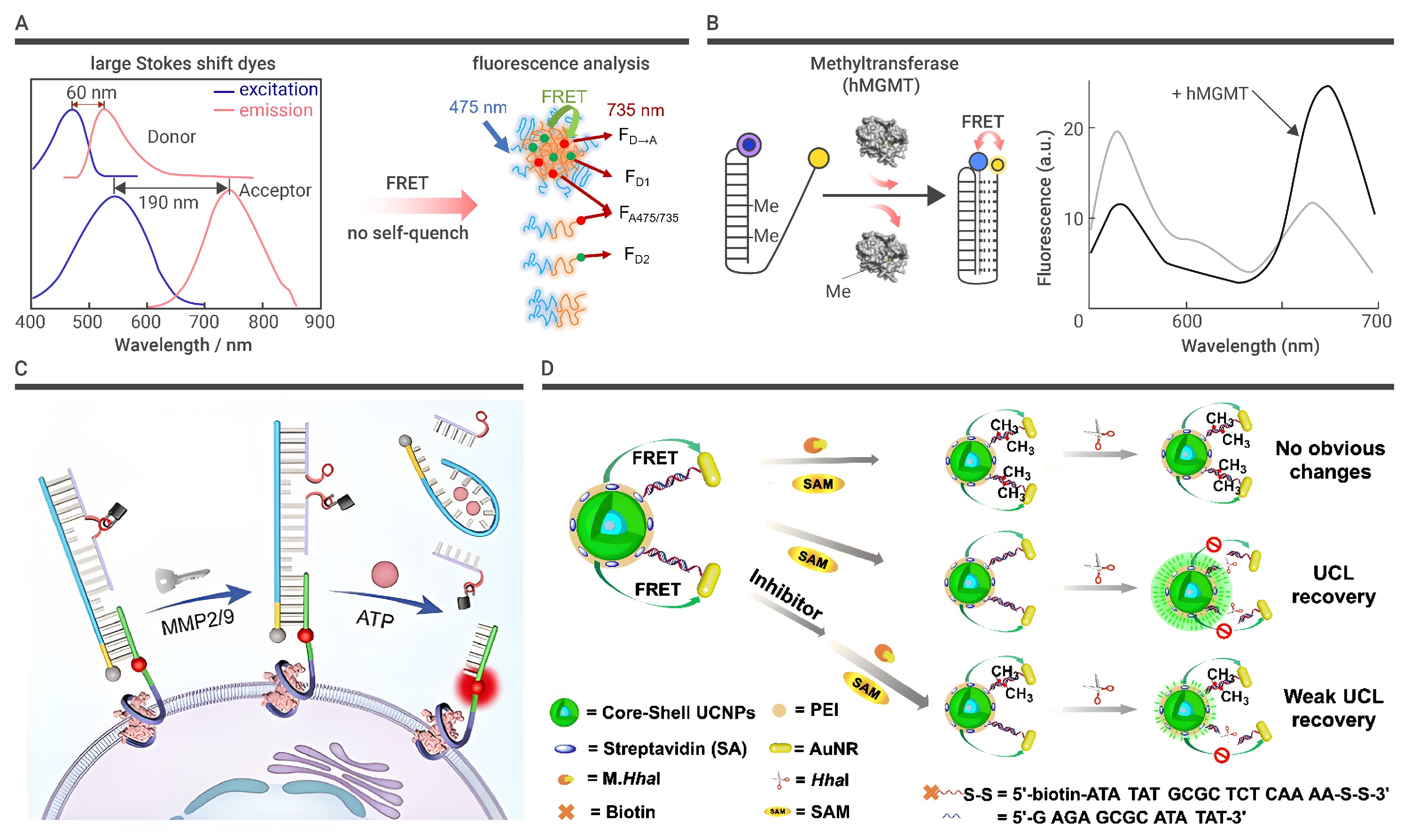
Figure 3.
FRET-based DNA nanostructures for biomedical application
-
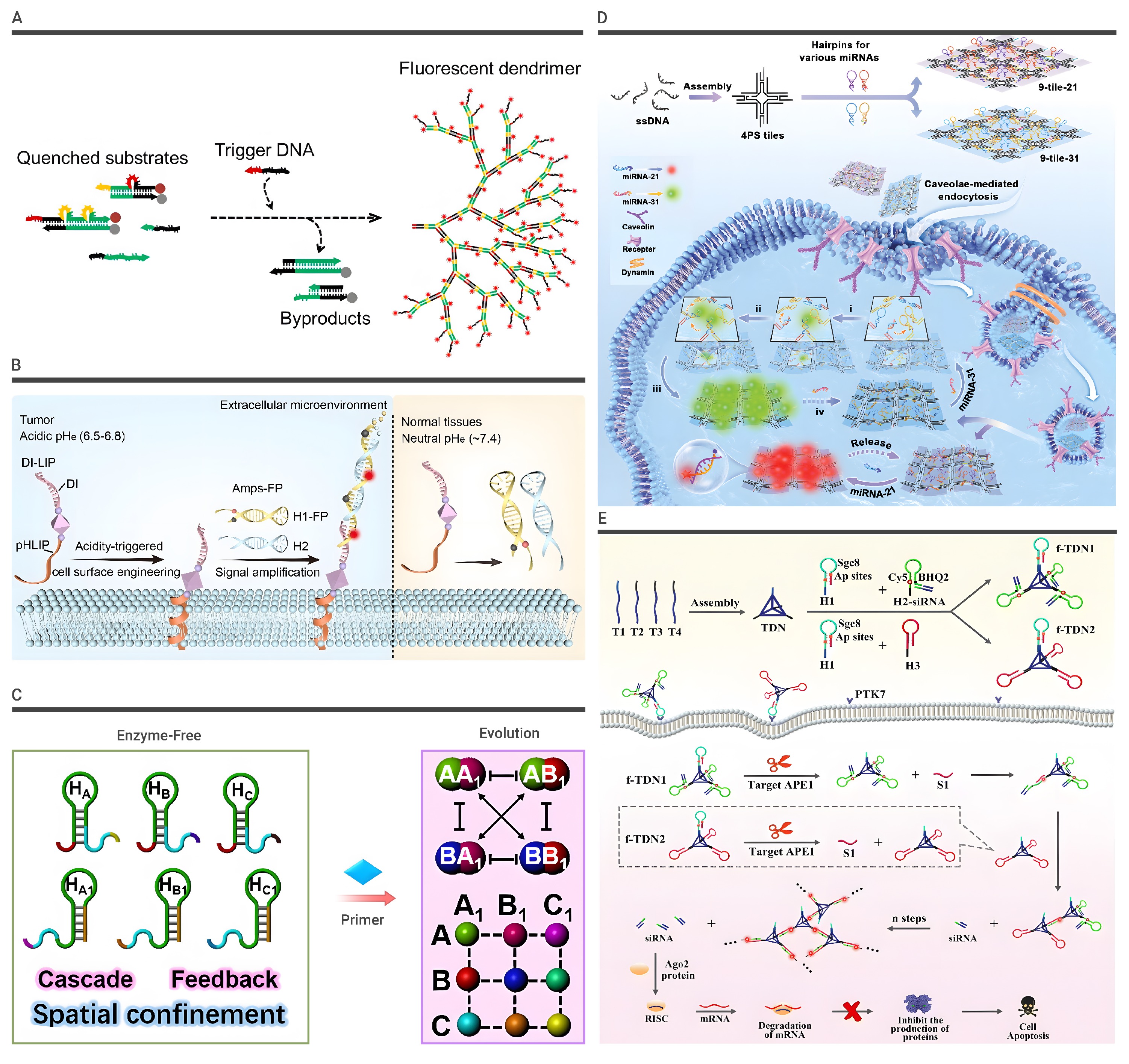
Figure 4.
Nucleic acid amplification-based DNA nanostructures
-
Figure 5.
AIE properties and AIE-based DNA nanostructures’ applications
-
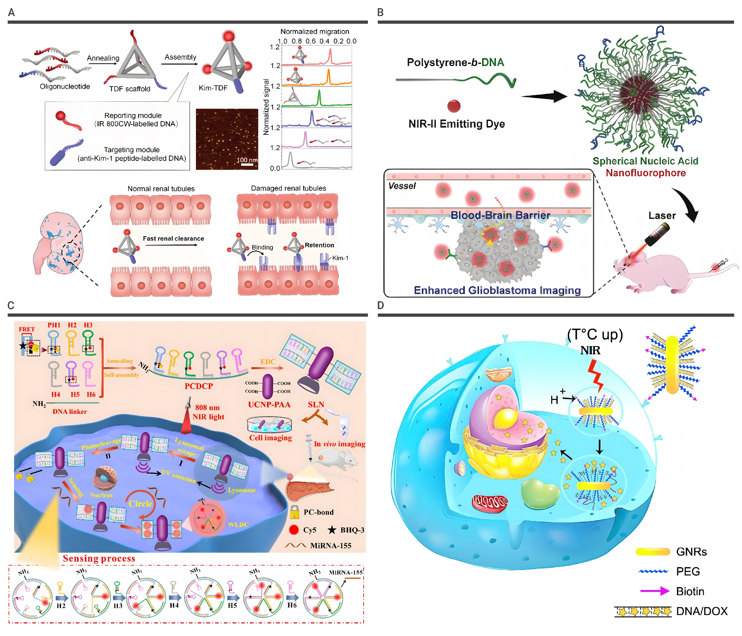
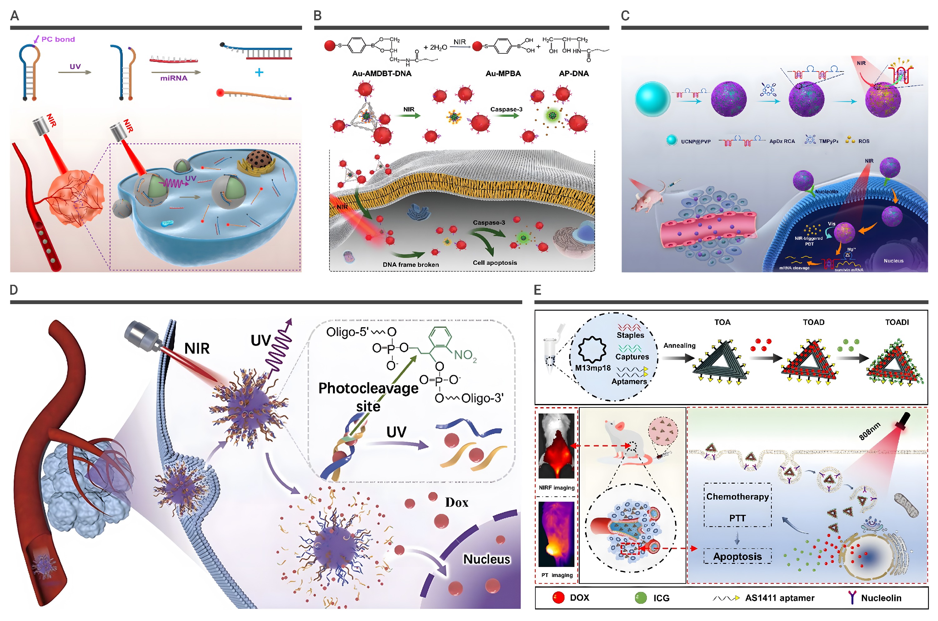
Figure 6.
The application of NIR-based DNA nanostructures
-
Figure 7.
The application of UCNPs-based DNA nanostructures in cancer treatments
-
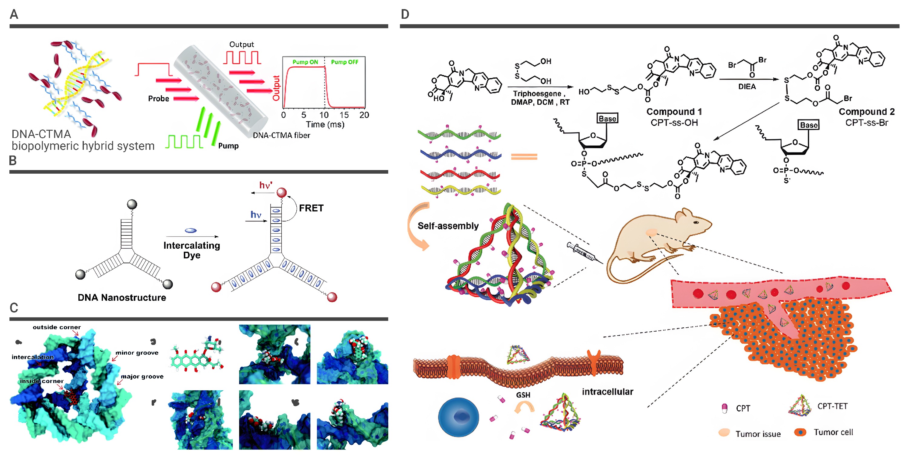
Figure 8.
Small fluorescent dye-intercalated DNA complexes
-
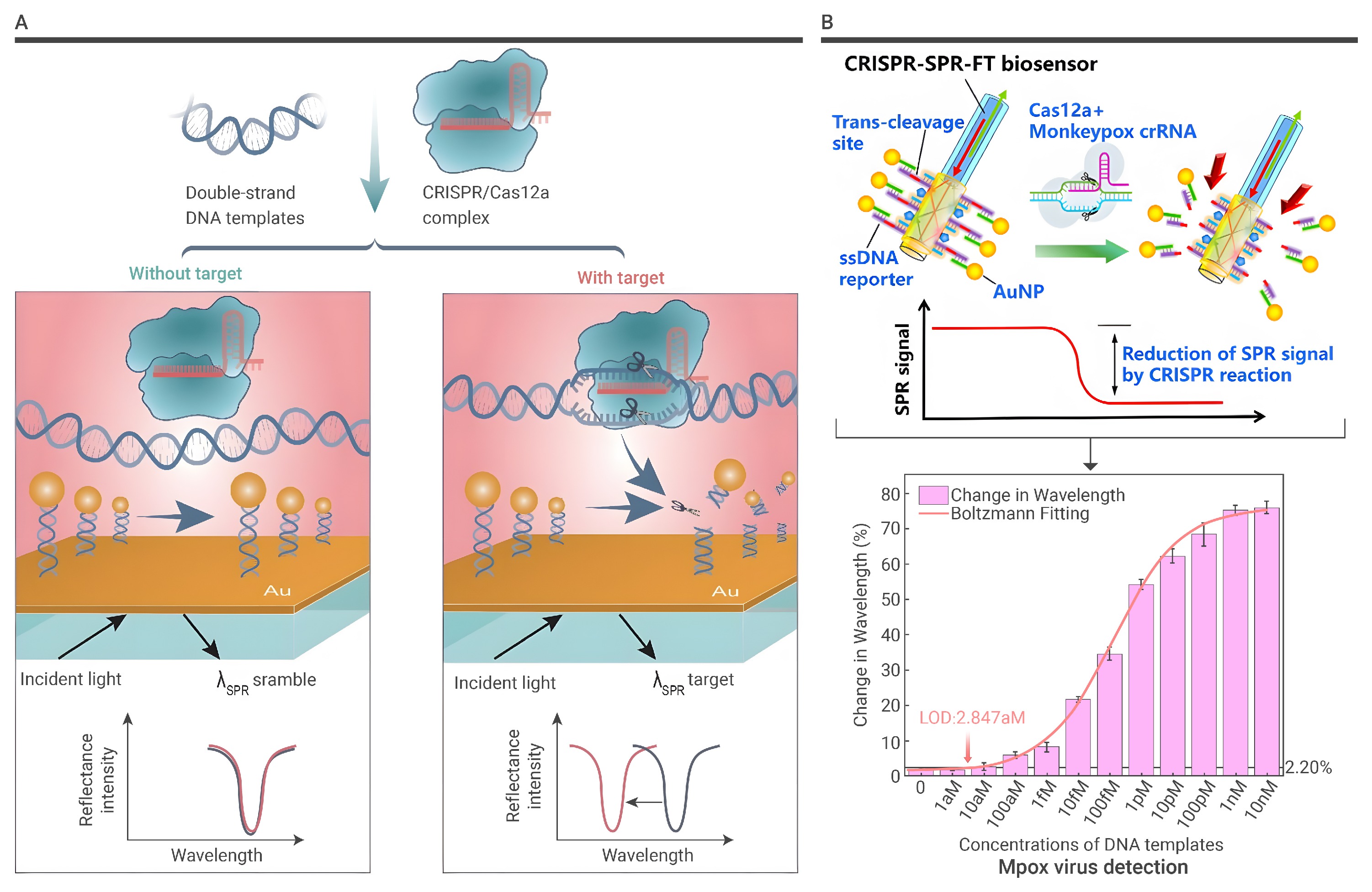
Figure 9.
The detection methods with SPR-based DNA nanostructures
