
Preparation, microstructure, and mechanical properties of enamel-like nanocomposites for dental repair application
-
GRAPHICAL ABSTRACT

 DownLoad:
Full size image
DownLoad:
Full size image
-
PUBLIC SUMMARY
-
The structure-mechanical property-function relationship of natural human tooth enamel was discussed.
The synthesis, microstructure and mechanical property of biomimetic tooth enamel nanocomposites were reviewed.
The biosafety and application of nano-enamel composite in dental prosthesis were discussed.
-
-
ABSTRACT
Tooth enamel, as the hardest and the most resilient bioceramic material (~95.5 wt% apatite minerals) in human body, forms complex, highly ordered, hierarchical hetero-phase array structure over millions of years of evolution. This multiscale complex structure endows tooth enamel with excellent mechanical stability (especially the resistance to fracture, wear, and impact), high chop efficiency, and superb durability. However, in the complex oral environment, several factors such as oral bacteria, acidic foods, and mechanical collisions, can cause the dissolution of apatite crystals and even the damage of the enamel, resulting in a series of lesions such as dental caries that severely affects human health and life quality. Therefore, the urgent need for restoring to the normal function of natural teeth by repairing enamel has motivated researchers to develop advanced synthetic strategies for constructing artificial enamels. In this review, based on the understanding of the hierarchical heterogeneous structure-mechanical property-function relationship of natural human tooth enamel, we firstly introduced several synthetic strategies of biomimetic enamel nanocomposites such as cell-based tissue engineering, organic matrix-guided crystal growth, microgel-based microenvironment mineralization, amorphous precursor mineralization, and physicochemical methods, as well as presenting their microstructures and mechanical properties published in recent years. Finally, we discussed the biological safety of these artificial enamel nanocomposites and their dental repair applications. -

-
REFERENCES
[1] Wegst U. G., Bai H., Saiz E., et al. (2015). Bioinspired structural materials. Nat. Mater. 14:23−36. DOI:10.1038/nmat4089 [2] Sansom I. J., Smith M. P., Armstrong H. A., et al. (1992). Presence of the earliest vertebrate hard tissue in conodonts. Science 256:1308−1311. DOI:10.1126/science.1598573 [3] Sadier A., Jackman W. R., Laudet V., et al. (2019). The vertebrate tooth row: is it initiated by a single organizing tooth. BioEssays. 42:1900229. DOI:10.1002/bies.201900229 [4] Chen H., Tang Z., Liu J., et al. (2006). Acellular synthesis of a human enamel-like microstructure. Adv. Mater. 18:1846−1851. DOI:10.1002/adma.200502401 [5] Angelova Volponi A., Zaugg L. K., Neves V., et al. (2018). Tooth repair and regeneration. Curr. Oral. Health. Rep. 5:295−303. DOI:10.1007/s40496-018-0196-9 [6] Lacruz R. S., Habelitz S., Wright J. T., et al. (2016). Dental enamel formation and implications for oral health and disease. Physiol. Rev. 97:939−993. DOI:10.1152/physrev.00030.2016 [7] Arnold S., Plate U., Wiesmann H. P., et al. (2001). Quantitative analyses of the biomineralization of different hard tissues. J. Microsc. 202:488−494. DOI:10.1046/j.1365-2818.2001.00831.x [8] Hannig M. and Hannig C. (2010). Nanomaterials in preventive dentistry. Nat. Nanotechnol. 5:565−569. DOI:10.1038/nnano.2010.83 [9] Wei Y., Liu S., Xiao Z., et al. (2020). Enamel repair with amorphous ceramics. Adv. Mater. 32:e1907067. DOI:10.1002/adma.201907067 [10] Peres M. A., Macpherson L. M., Weyant R. J., et al. (2019). Oral diseases: A global public health challenge. Lancet. 394:249−260. DOI:10.1016/s0140-6736(19)31146-8 [11] Koulourides T., Feagin F. and Pigman W. (1965). Remineralization of dental enamel by saliva in vitro. Ann. N. Y. Acad. Sci. 131:751−757. DOI:10.1111/j.1749-6632.1965.tb34839.x [12] Iafisco M., Degli Esposti L., Ramirez-Rodriguez G. B., et al. (2018). Fluoride-doped amorphous calcium phosphate nanoparticles as a promising biomimetic material for dental remineralization. Sci. Rep. 8:17016. DOI:10.1038/s41598-018-35258-x [13] Abou Neel E. A., Strange A., Ibrahim S., et al. (2016). Demineralization–remineralization dynamics in teeth and bone. Int. J. Nanomed. 11:4743−4763. DOI:10.2147/ijn.s107624 [14] Palmer L., Newcomb C., Kaltz S., et al. (2008). Biomimetic systems for hydroxyapatite mineralization inspired by bone and enamel. Chem. Rev. 108:4754−4783. DOI:10.1021/cr8004422 [15] Chatzistavrou X., Papagerakis S., Ma P. X., et al. (2012). Innovative approaches to regenerate enamel and dentin. Int. J. Dent. 2012:856470. DOI:10.1155/2012/856470 [16] Cui F.-Z., and Ge J. (2007). New observations of the hierarchical structure of human enamel, from nanoscale to microscale. J. Tissue Eng. Regen. Med. 1:185−191. DOI:10.1002/term.21 [17] Koenigswald W. V. and Clemens W. A. (1992). Levels of complexity in the microstructure of mammalian enamel and their application in studies of systematics. Scanning Microsc. 6 :195–217. https://digitalcommons.usu.edu/microscopy/vol6/iss1/16. [18] Wilmers J. and Bargmann S. (2020). Nature’s design solutions in dental enamel: uniting high strength and extreme damage resistance. Acta Biomater. 107:1−24. DOI:10.1016/j.actbio.2020.02.019 [19] Zhang N., Wang X., Xiang W., et al. (2019). Hierarchy structure and fracture mechanisms of the wild wolf tusk's enamel. Biomater. Adv. 106:110277. DOI:10.1016/j.msec.2019.110277 [20] Xu H., Smith D. T., Jahanmir S., et al. (1998). Indentation damage and mechanical properties of human enamel and dentin. J. Dent. Res. 77:472−480. DOI:10.1177/00220345980770030601 [21] Habelitz S., Marshall S. J., Marshall G. W., et al. (2001). Mechanical properties of human dental enamel on the nanometre scale. Arch. Oral Biol. 46:173−183. DOI:10.1016/s0003-9969(00)00089-3 [22] Mahoney E., Holt A., Swain M., et al. (2000). The hardness and modulus of elasticity of primary molar teeth: an ultra-micro-indentation study. J. Dent. 28:589−594. DOI:10.1016/s0300-5712(00)00043-9 [23] Lawn B. R., Lee J. W. and Chai, H. (2010). Teeth: Among nature's most durable biocomposites. Annu. Rev. Mater. Res. 40:55−75. DOI:10.1146/annurev-matsci-070909-104537 [24] Bajaj D. and Arola D. (2009). Role of prism decussation on fatigue crack growth and fracture of human enamel. Acta Biomater. 5:3045−3056. DOI:10.1016/j.actbio.2009.04.013 [25] Thompson V. P. (2019). The tooth: An analogue for biomimetic materials design and processing. Dent. Mater. 36:25−42. DOI:10.1016/j.dental.2019.08.106 [26] Risnes S. and Li C. (2017). Aspects of the final phase of enamel formation as evidenced by observations of superficial enamel of human third molars using scanning electron microscopy. Arch. Oral Biol. 86:72−79. DOI:10.1016/j.archoralbio.2017.11.008 [27] Reyes-Gasga J. and Brès E. F. (2021). High resolution STEM images of the human tooth enamel crystals. Appl. Sci. 11:7477. DOI:10.3390/app11167477 [28] Batina N., Renugopalakrishnan V., Casillas Lavín P. N., et al. (2004). Ultrastructure of dental enamel afflicted with hypoplasia: an atomic force microscopic study. Calcif. Tissue Int. 74:294−301. DOI:10.1007/s00223-002-1045-2 [29] Alwadai G. S., Roberts G., Ungar P. S., et al. (2020). Monitoring of simulated occlusal tooth wear by objective outcome measures. J. Dent. 102:103467. DOI:10.1016/j.jdent.2020.103467 [30] Reyes-Gasga J., Martínez-Piñeiro E. L., Rodríguez-Álvarez G., et al. (2013). XRD and FTIR crystallinity indices in sound human tooth enamel and synthetic hydroxyapatite. Biomater. Adv. 33:4568−4574. DOI:10.1016/j.msec.2013.07.014 [31] Leroy G., Penel G., Leroy N., et al. (2002). Human tooth enamel: A raman polarized approach. Appl. Spectrosc. 56:103−1034. DOI:10.1366/000370202760249765 [32] Lopes C. D., Limirio P. H., Novais V. R., et al. (2018). Fourier transform infrared spectroscopy (FTIR) application chemical characterization of enamel, dentin and bone. Appl. Spectrosc. Rev. 53:747−769. DOI:10.1080/05704928.2018.1431923 [33] Holcomb D. W. and Young, R. A. (1980). Thermal decomposition of human tooth enamel. Calcif. Tissue Int. 31:189. DOI:10.1007/bf02407181 [34] Zheng J., Zeng Y., Wen J., et al. (2016). Impact wear behavior of human tooth enamel under simulated chewing conditions. J. Mech. Behav. Biomed. Mater. 62:119−127. DOI:10.1016/j.jmbbm.2016.04.039 [35] Gao S., An B. B., Yahyazadehfar M., et al. (2016). Contact fatigue of human enamel: Experiments, mechanisms and modeling. J. Mech. Behav. Biomed. Mater. 60:438−450. DOI:10.1016/j.jmbbm.2016.02.030 [36] Beniash E., Stifler C., Sun C., et al. (2019). The hidden structure of human enamel. Nat. Commun. 10:1−13. DOI:10.1038/s41467-019-12185-7 [37] Thiagarajan G., Vizcarra B., Bodapudi V., et al. (2017). Stress analysis of irradiated human tooth enamel using finite element methods. Comput. Method Biomech. Biomed. Eng. 20:1533−1542. DOI:10.1080/10255842.2017.1383401 [38] O'Keefe T. J. and Christie, R. D. (1977). Electroless and chemical metal deposition on human tooth enamel. J. Dent. Res. 56:595−602. DOI:10.1177/00220345770560060701 [39] Gautam C., Joyner J., Gautam A., et al. (2016). Zirconia based dental ceramics: Structure, mechanical properties, biocompatibility and applications. Dalton Trans. 45:19194−19215. DOI:10.1039/c6dt03484e [40] Xu X., He L., Zhu B., et al. (2017). Advances in polymeric materials for dental applications. Polym. Chem. 8:807−823. DOI:10.1039/c6py01957a [41] Li L., Mao C., Wang J., et al. (2011). Bio-inspired enamel repair via glu-directed assembly of apatite nanoparticles: an approach to biomaterials with optimal characteristics. Adv. Mater. 23:4695−4701. DOI:10.1002/adma.201102773 [42] Zhou Y., Zhou Y., Gao L., et al. (2018). Synthesis of artificial dental enamel by an elastin-like polypeptide assisted biomimetic approach. J. Mater. Chem. B. 6:844−853. DOI:10.1039/c7tb02576a [43] Pitts NB., Zero DT. and Marsh PD. (2017). Dental caries. Nat. Rev. Dis. Primers. 3:17030. DOI:10.1038/nrdp.2017.30 [44] Wang D., Deng J., Deng X., et al. (2020). Controlling enamel remineralization by amyloid-like amelogenin mimics. Adv. Mater. 32:e2002080. DOI:10.1002/adma.202002080 [45] Honda M., Shinmura Y. and Shinohara Y., (2008). Enamel tissue engineering using subcultured enamel organ epithelial cells in combination with dental pulp cells. Cells Tissues Organs. 189 :261–267. DOI:10.1159/000151743. [46] Yeom B., Sain T., Lacevic N., et al. (2017). Abiotic tooth enamel. Nature. 543:95−98. DOI:10.1038/nature21410 [47] Sezen M., Ow-Yang C., Karahan Ö., et al. (2018). Micro and nanostructural analysis of a human tooth using correlated focused ion beam (FIB) and transmission electron microscopy (TEM) investigations. Micron 115:17−24. DOI:10.1016/j.micron.2018.08.004 [48] Jágr M., Ergang P., Pataridis S., et al. (2019). Proteomic analysis of dentin-enamel junction and adjacent protein-containing enamel matrix layer of healthy human molar teeth. Eur. J. Oral Sci. 127:112−121. DOI:10.1111/eos.12594 [49] Castiblanco G. A., Rutishauser D., Ilag L. L., et al. (2015). Identification of proteins from human permanent erupted enamel. Eur. J. Oral Sci. 123:390−395. DOI:10.1111/eos.12214 [50] Açil Y., Mobasseri A. E., Warnke P. H., et al. (2005). Detection of mature collagen in human dental enamel. Calcif. Tissue Int. 76:121−126. DOI:10.1007/s00223-004-0122-0 [51] Weidmann S. M. and Eyre D. R. (1967). Amino acid composition of enamel protein in the fully developed human tooth. Caries Res. 1:349−355. DOI:10.1159/000259535 [52] Vymetal J., Slabý I., Spahr A., et al. (2008). Bioinformatic analysis and molecular modelling of human ameloblastin suggest a two-domain intrinsically unstructured calcium-binding protein. Eur. J. Oral Sci. 116:124−134. DOI:10.1111/j.1600-0722.2008.00526.x [53] Fasano M., Curry S., Terreno E., et al. (2005). The extraordinary ligand binding properties of human serum albumin. IUBMB Life. 57:787−796. DOI:10.1080/15216540500404093 [54] Daubert D. M., Kelley J. L., Udod Y. G., et al. (2016). Human enamel thickness and ENAM polymorphism. Int. J. Oral Sci. 8:93−97. DOI:10.1038/ijos.2016.1 [55] He L. and Swain M. V. (2008). Understanding the mechanical behaviour of human enamel from its structural and compositional characteristics. J. Mech. Behav. Biomed. Mater. 1:18−29. DOI:10.1016/j.jmbbm.2007.05.001 [56] Poole D. F. G. and Brooks A. W. (1961). The arrangement of crystallites in enamel prisms. Arch. Oral Biol. 5:14−26. DOI:10.1016/0003-9969(61)90110-8 [57] Wood C. B. and Stern D. N. (1997). The earliest prisms in mammalian and reptilian enamel. Koenigswald, W., Sander, P M. Tooth Enamel Microstructure. CRC Press. pp. 63–83. DOI:10.1201/9781003077930-4. [58] Meckel A. H., Griebstein W. J. and Neal, R. J. (1965). Structure of mature human dental enamel as observed by electron microscopy. Arch. Oral Biol. 10:775−783. DOI:10.1016/0003-9969(65)90131-7 [59] Mao F., Chuan-Kui L. I., Meng J., et al. (2017). Introduction to terminologies of tooth enamel microstructures and a proposal for their standard Chinese translations. Vertebrata Palasiatica. 55:347−366. DOI:10.19615/j.cnki.1000-3118.170923 [60] Duverger O., Beniash E. and Morasso M. I., (2016). Keratins as components of the enamel organic matrix. Matrix Biol. 52-54 :260–265. DOI:10.1016/j.matbio.2015.12.007 [61] Wang C., Fang Y., Zhang L., et al. (2021). Enamel microstructural features of bovine and human incisors: A comparative study. Ann. Anat. 235:151700. DOI:10.1016/j.aanat.2021.151700 [62] Osborn J. W. (1990). A 3-dimensional model to describe the relation between prism directions, parazones and diazones, and the Hunter-Schreger bands in human tooth enamel. Arch. Oral Biol. 35:869−878. DOI:10.1016/0003-9969(90)90065-i [63] Lynch C. D., O’Sullivan V. R., Dockery P., et al. (2010). Hunter-Schreger band patterns in human tooth enamel. J. Anat. 217:106−115. DOI:10.1111/j.1469-7580.2010.01255.x [64] Shin N. Y., Yamazaki H., Beniash E., et al. (2020). Amelogenin phosphorylation regulates tooth enamel formation by stabilizing a transient amorphous mineral precursor. J. Biol. Chem. 295:1943−1959. DOI:10.1074/jbc.ra119.010506 [65] Popowics T. E., Rensberger J. M. and Herring S. W. (2004). Enamel microstructure and microstrain in the fracture of human and pig molar cusps. Arch. Oral Biol. 49:595−605. DOI:10.1016/j.archoralbio.2004.01.016 [66] He L., Yin Z., Vuuren L. J., et al. (2013). A natural functionally graded biocomposite coating-human enamel. Acta Biomater. 9:6330−6337. DOI:10.1016/j.actbio.2012.12.029 [67] Cuy J. L., Mann A. B., Livi K. J., et al. (2002). Nanoindentation mapping of the mechanical properties of human molar tooth enamel. Arch. Oral Biol. 47:281−291. DOI:10.1016/s0003-9969(02)00006-7 [68] Bajaj D. and Arola D. D. (2009). On the R-curve behavior of human tooth enamel. Biomaterials. 30:4037−4046. DOI:10.1016/j.biomaterials.2009.04.017 [69] Craig R. G. and Peyton F. A., (1958). The microhardness of enamel and dentin. J. Dent. Res. 37 :661–668. DOI:10.1177/00220345580370041301. [70] Craig R. G., Peyton F. A. and Johnson D. W., (1961). Compressive properties of enamel, dental cements, and gold. J. Dent. Res. 40 :936–945. DOI:10.1177/00220345610400051901 [71] Stanford J. W., Weigel K. V., Paffenbarger G. C., et al. (1960). Compressive properties of hard tooth tissues and some restorative materials. J. Am. Dental Assoc. 60:746−756. DOI:10.14219/jada.archive.1960.0258 [72] He L. H. and Swain M. V., (2007). Influence of environment on the mechanical behaviour of mature human enamel. Biomaterials. 28 :4512–4520. DOI:10.1016/j.biomaterials.2007.06.020. [73] Xie Z., Swain M. V., Swadener G., et al. (2009). Effect of microstructure upon elastic behaviour of human tooth enamel. J. Biomech. 42:1075−1080. DOI:10.1016/j.jbiomech.2009.02.004 [74] Park S., Wang D., Zhang D., et al. (2008). Mechanical properties of human enamel as a function of age and location in the tooth. J. Mater. Sci. Mater. Med. 19:2317−2324. DOI:10.1007/s10856-007-3340-y [75] Shen L., Sousa F. B., Tay N., et al. (2020). Deformation behavior of normal human enamel: a study by nanoindentation. J. Mech. Behav. Biomed. Mater. 108:103799. DOI:10.1016/j.jmbbm.2020.103799 [76] An B., Wang R., Arola D., et al. (2012). The role of property gradients on the mechanical behavior of human enamel. J. Mech. Behav. Biomed. Mater. 9:63−72. DOI:10.1016/j.jmbbm.2012.01.009 [77] Sutrakar V. K., Javvaji B. and Budarapu P. R. (2021). Fracture strength and fracture toughness of graphene: MD simulations. Appl. Phys. A. 127:949. DOI:10.1007/s00339-021-05047-x [78] Chan Y., Ngan A. H. and King N. M. (2009). Use of focused ion beam milling for investigating the mechanical properties of biological tissues: a study of human primary molars. J. Mech. Behav. Biomed. Mater. 2:375−383. DOI:10.1016/j.jmbbm.2009.01.006 [79] Lewis R. and Dwyer-Joyce R. S. (2005). Wear of human teeth: A tribological perspective. Proc. Inst. Mech. Eng. Part J J. Eng. Tribol. 219:1−18. DOI:10.1243/1350650053295394 [80] Kaidonis J. A., Richards L. C., Townsend G. C., et al. (1998). Wear of human enamel: A quantitative in vitro assessment. J. Dent. Res. 77:1983−1990. DOI:10.1177/00220345980770120601 [81] Zheng J. and Zhou Z. (2007). Study of in vitro wear of human tooth enamel. Tribol. Lett. 26:181−189. DOI:10.1007/s11249-006-9192-7 [82] Lee J. J., Kwon J. Y., Chai H., et al. (2009). Fracture modes in human teeth. J. Dent. Res. 88:224−228. DOI:10.1177/0022034508330055 [83] Viswanath B., Raghavan R., Ramamurty U., et al. (2007). Mechanical properties and anisotropy in hydroxyapatite single crystals. Scr. Mater. 57:361−364. DOI:10.1016/j.scriptamat.2007.04.027 [84] Xia J., Tian Z., Hua L., et al. (2017). Enamel crystallite strength and wear: nanoscale responses of teeth to chewing loads. J. Royal Soc. Interface. 14:20170456. DOI:10.1098/rsif.2017.0456 [85] White S. N., Luo W., Paine M. L., et al. (2001). Biological organization of hydroxyapatite crystallites into a fibrous continuum toughens and controls anisotropy in human enamel. J. Dent. Res. 80:321−326. DOI:10.1177/00220345010800010501 [86] Yahyazadehfar M., Bajaj D. and Arola D. D. (2012). Hidden contributions of the enamel rods on the fracture resistance of human teeth. Acta Biomater. 9:4806−4814. DOI:10.1016/j.actbio.2012.09.020 [87] Padmanabhan S. K., Balakrishnan A., Chu M. C., et al. (2009). Micro-indentation fracture behavior of human enamel. Dent. Mater. 26:100−104. DOI:10.1016/j.dental.2009.07.015 [88] Yahyazadehfar M. and Arola D. (2015). The role of organic proteins on the crack growth resistance of human enamel. Acta Biomater. 19:33−45. DOI:10.1016/j.actbio.2015.03.011 [89] Jia Y. and Xuan F. (2012). Anisotropic wear behavior of human enamel at the rod level in terms of nanoscratching. Wear 290-291:124−132. DOI:10.1016/j.wear.2012.04.020 [90] Bajaj D., Park S., Quinn G. D., et al. (2010). Fracture processes and mechanisms of crack growth resistance in human enamel. JOM 62:76−82. DOI:10.1007/s11837-010-0113-8 [91] Bajaj D., Nazari A., Eidelman, N., et al. (2008). A comparison of fatigue crack growth in human enamel and hydroxyapatite. Biomaterials. 29:4847−4854. DOI:10.1016/j.biomaterials.2008.08.019 [92] Moradian-Oldak, J., (2012). Protein-mediated enamel mineralization. Front Biosci. 17 :1996–2023. DOI:10.2741/4034. [93] Basheer N., Madhubala M. and Sekar M. (2020). Future perspectives of biomimetics in restorative dentistry. J. Int. Pharm. Res. 32:P19−28. DOI:10.9734/jpri/2020/v32i2530820 [94] Zivkovic P., Petrovic V., Najman S., et al. (2010). Stem cell-based dental tissue engineering. Sci. World J. 10:901−916. DOI:10.1100/tsw.2010.81 [95] Ahmed G., Abou-Auf E., Abubakr N., et al. (2020). Tissue engineering approaches for enamel, dentin, and pulp regeneration: an update. Stem Cells Int. 2020:1−15. DOI:10.1155/2020/5734539 [96] Galler K. M. and D'Souza R. N. (2011). Tissue engineering approaches for regenerative dentistry. Regenerative Medicine. 6:111−124. DOI:10.2217/rme.10.86 [97] Thesleff I. (2003). Epithelial-mesenchymal signalling regulating tooth morphogenesis. J. Cell Sci. 116:1647−1648. DOI:10.1242/jcs.00410 [98] Abdel Meguid E., Ke Y., Ji J., et al. (2018). Stem cells applications in bone and tooth repair and regeneration: new insights, tools, and hopes. J. Cell. Physiol. 233:1825−1835. DOI:10.1002/jcp.25940 [99] Bazina F., Brouxhon S. M., Graham U. M., et al. (2021). Serotonin contributes to the in vitro production of a biomimetic enamel-like material from reprogrammed oral epithelial keratinocytes. Orthod. Craniofac. Res. 24:494−501. DOI:10.1111/ocr.12475 [100] Ono M., Oshima M., Ogawa M., et al. (2017). Practical whole-tooth restoration utilizing autologous bioengineered tooth germ transplantation in a postnatal canine model. Sci. Rep. 7:44522. DOI:10.1038/srep44522 [101] Liu Y., Jiang M., Hao W., et al. (2013). Skin epithelial cells as possible substitutes for ameloblasts during tooth regeneration. J. Tissue Eng. Regen. Med. 7:934−943. DOI:10.1002/term.1485 [102] Zhang W., Vázquez B. and Yelick P. C. (2014). Bioengineered post-natal recombinant tooth bud models. J. Tissue Eng. Regen. Med. 11:658−668. DOI:10.1002/term.1962 [103] Li Q., Zhang S., Sui Y., et al. (2019). Sequential stimulation with different concentrations of BMP4 promotes the differentiation of human embryonic stem cells into dental epithelium with potential for tooth formation. Stem Cell Res. Ther. 10:276. DOI:10.1186/s13287-019-1378-7 [104] Zhang W., Vazquez B., Oreadi D., et al. (2017). Decellularized tooth bud scaffolds for tooth regeneration. J. Dent. Res. 96:516−523. DOI:10.1177/0022034516689082 [105] Angelova Volponi A., Kawasaki M. and Sharpe P. T. (2013). Adult human gingival epithelial cells as a source for whole-tooth bioengineering. J. Dent. Res. 92:329−334. DOI:10.1177/0022034513481041 [106] Yang K., Wang C., Chang H., et al. (2011). Fibrin glue mixed with platelet-rich fibrin as a scaffold seeded with dental bud cells for tooth regeneration. J. Tissue Eng. Regen. Med. 6:777−785. DOI:10.1002/term.483 [107] Hu X., Lee J. W., Zheng X., et al. (2018). Efficient induction of functional ameloblasts from human keratinocyte stem cells. Stem Cell Res. Ther. 9:126. DOI:10.1186/s13287-018-0822-4 [108] Zhang Y., Li Y., Shi R., et al. (2017). Generation of tooth–periodontium complex structures using high-odontogenic potential dental epithelium derived from mouse embryonic stem cells. Stem Cell Res. Ther. 8:141. DOI:10.1186/s13287-017-0583-5 [109] El Gezawi M., Wölfle U. C., Haridy R., et al. (2019). Remineralization, regeneration, and repair of natural tooth structure: Influences on the future of restorative dentistry practice. ACS Biomater. Sci. Eng. 5:4899−4919. DOI:10.1021/acsbiomaterials.9b00591 [110] Grawish M. E., Grawish L. M., Grawish H. M., et al. (2020). Challenges of engineering biomimetic dental and paradental tissues. Tissue Eng. Regen. Med. 17:403−421. DOI:10.1007/s13770-020-00269-1 [111] Duailibi S. E., Duailibi M. T., Zhang W., et al. (2008). Bioengineered dental tissues grown in the rat jaw. J. Dent. Res. 87:745−750. DOI:10.1177/154405910808700811 [112] Shinmura Y., Tsuchiya S., Hata K., et al. (2008). Quiescent epithelial cell rests of Malassez can differentiate into ameloblast-like cells. J. Cell. Physiol. 217:728−738. DOI:10.1002/jcp.21546 [113] Honda M. J., Shinohara Y., Hata K. I., et al. (2007). Subcultured odontogenic epithelial cells in combination with dental mesenchymal cells produce enamel-dentin-like complex structures. Cell Transplant. 16:833−847. DOI:10.3727/000000007783465208 [114] Young C. S., Terada S., Vacanti J. P., et al. (2002). Tissue engineering of complex tooth structures on biodegradable polymer scaffolds. J. Dent. Res. 81:695−700. DOI:10.1177/154405910208101008 [115] Yao S., Jin B., Liu Z., et al. (2017). Biomineralization: From material tactics to biological strategy. Adv. Mater. 29:1605903. DOI:10.1002/adma.201605903 [116] Pandya M. and Diekwisch T. G. (2021). Amelogenesis: Transformation of a protein-mineral matrix into tooth enamel. J. Struct. Biol. 213:107809. DOI:10.1016/j.jsb.2021.107809 [117] Vaissier Welborn V. (2020). Enamel synthesis explained. Proc. Natl. Acad. Sci. USA 117 :21847–21848. DOI:10.1073/pnas.2014394117. [118] Ravindranath R. M., Basilrose R. M., Ravindranath N. H., et al. (2003). Amelogenin interacts with cytokeratin-5 in ameloblasts during enamel growth. J. Biol. Chem. 278:20293−20302. DOI:10.1074/jbc.m211184200 [119] Margolis H. C., Beniash E. and Fowler C. E. (2006). Role of macromolecular assembly of enamel matrix proteins in enamel formation. J. Dent. Res. 85:775−793. DOI:10.1177/154405910608500902 [120] Bai Y., Bonde J., Carneiro K. M., et al. (2021). A brief history of the discovery of amelogenin nanoribbons in vitro and in vivo. J. Dent. Res. 100:1429−1433. DOI:10.1177/00220345211043463 [121] Chang D., Fermani G., Fermani S., et al. (2005). Supramolecular assembly of amelogenin nanospheres into birefringent microribbons. Science. 307:1450−1454. DOI:10.1126/science.1105675 [122] Carneiro K. M., Zhai H., Zhu L., et al. (2016). Amyloid-like ribbons of amelogenins in enamel mineralization. Sci. Rep. 6:23105. DOI:10.1038/srep23105 [123] Martinez-Avila O., Wu S., Kim S. J., et al. (2012). Self-assembly of filamentous amelogenin requires calcium and phosphate: From dimers via nanoribbons to fibrils. Biomacromolecules 13:3494−3502. DOI:10.1021/bm300942c [124] Bai Y., Yu Z., Ackerman L., et al. (2020). Protein nanoribbons template enamel mineralization. Proc. Natl. Acad. Sci. USA 117:19201−19208. DOI:10.1073/pnas.2007838117 [125] Farooq I. and Bugshan A. (2020). The role of salivary contents and modern technologies in the remineralization of dental enamel: A narrative review. F1000Research. 9 :171. DOI:10.12688/f1000research.22499.2. [126] Elsharkawy S., Al-Jawad M., Pantano M. F., et al. (2018). Protein disorder-order interplay to guide the growth of hierarchical mineralized structures. Nat. Commun. 9:2145. DOI:10.1038/s41467-018-04319-0 [127] Mukherjee K., Ruan Q., Nutt S., et al. (2018). Peptide-based bioinspired approach to regrowing multilayered aprismatic enamel. ACS Omega. 3:2546−2557. DOI:10.1021/acsomega.7b02004 [128] Wang H., Xiao Z., Yang J., et al. (2017). Oriented and ordered biomimetic remineralization of the surface of demineralized dental enamel using HAP@ACP nanoparticles guided by glycine. Sci. Rep. 7:40701. DOI:10.1038/srep40701 [129] Bartlett J. D. and Simmer J. P. (1999). Proteinases in developing dental enamel. Crit. rev. oral biol. med. 10:425−441. DOI:10.1177/10454411990100040101 [130] Zhong X., Lai T., Chen L., et al. (2021). Self-assembly and mineralization of full-length human amelogenin and its functional fragments in vitro. West China J. Stoma. 39:419−424. DOI:10.7518/hxkq.2021.04.007 [131] Athanasiadou D., Danesi A. L., Umbrio L., et al. (2021). Hydroxyapatite growth on amelogenin-amelotin recombinamers. ChemNanoMat. 7:851−857. DOI:10.1002/cnma.202100064 [132] Li X., Pan D., Lin S., et al. (2013). Facile in vitro hydroxyapatite remineralization of human enamel with remarkable hardness. CrystEngComm. 15:4351−4356. DOI:10.1039/c3ce26947g [133] Wen Z., Chen J., Wang H., et al. (2016). Abalone water-soluble matrix for self-healing biomineralization of tooth defects. Biomater. Adv. 67:182−187. DOI:10.1016/j.msec.2016.05.015 [134] Featherstone J. D. (1999). Prevention and reversal of dental caries: Role of low level fluoride. Community Dent. Oral. 27:31−40. DOI:10.1111/j.1600-0528.1999.tb01989.x [135] Pearce E. I., Coote G. E. and Larsen M. J. (1995). The distribution of fluoride in carious human enamel. J. Dent. Res. 74:1775−1782. DOI:10.1177/00220345950740111001 [136] Wang Z., Ma G., Liu X., et al. (2009). Will fluoride toughen or weaken our teeth. Understandings based on nucleation, morphology, and structural assembly. J. Phys. Chem. B. 113:16393−16399. DOI:10.1021/jp905846p [137] Aulestia F. J., Groeling J., Bomfim G. H., et al. (2020). Fluoride exposure alters Ca2+ signaling and mitochondrial function in enamel cells. Sci. Signal. 13 :eaay0086. DOI:10.1126/scisignal.aay0086. [138] Levin A., Hakala T. A., Schnaider L., et al. (2020). Biomimetic peptide self-assembly for functional materials. Nat. Rev. Chem. 4:615−634. DOI:10.1038/s41570-020-0215-y [139] Dogan S., Fong H., Yucesoy D. T., et al. (2018). Biomimetic tooth repair: amelogenin-derived peptide enables in vitro remineralization of human enamel. ACS Biomater. Sci. Eng. 4:1788−1796. DOI:10.1021/acsbiomaterials.7b00959 [140] Yang X., Yang B., He L., et al. (2017). Bioinspired peptide-decorated tannic acid for in situ remineralization of tooth enamel: In vitro and in vivo evaluation. Sci. Eng. 3:3553−3562. DOI:10.1021/acsbiomaterials.7b00623 [141] Mohamed N. A. and Abd El-Ghany N. A. (2012). Synthesis and antimicrobial activity of some novel terephthaloyl thiourea cross-linked carboxymethyl chitosan hydrogels. Cellulose. 19:1879−1891. DOI:10.1007/s10570-012-9789-y [142] Xiao Z., Que K., Wang H., et al. (2017). Rapid biomimetic remineralization of the demineralized enamel surface using nano-particles of amorphous calcium phosphate guided by chimaeric peptides. Dent. Mater. 33 :1217–1228. DOI:10.1016/j.dental.2017.07.015 [143] Zhou L., Wong, H. Zhang Y., et al. (2020). Constructing an antibiofouling and mineralizing bioactive tooth surface to protect against decay and promote self-healing. ACS Appl. Mater. Interfaces. 12:3021−3031. DOI:10.1021/acsami.9b19745 [144] Aita K., Oaki Y., Ohtsuki C., et al. (2015). Fabrication of self-standing films consisting of enamel-like oriented nanorods using artificial peptide. Cryst. Eng. Comm. 17:5551−5555. DOI:10.1039/c5ce00598a [145] Hsu C.C., Chung H., Yang J., et al. (2011). Influence of 8DSS peptide on nano-mechanical behavior of human enamel. J. Dent. Res. 90:88−92. DOI:10.1177/0022034510381904 [146] Liu Y., Ding C., He L., et al. (2018). Bioinspired heptapeptides as functionalized mineralization inducers with enhanced hydroxyapatite affinity. J. Mater. Chem. B. 6:1984−1994. DOI:10.1039/c7tb03067c [147] Wu D., Yang J., Li J., et al. (2013). Hydroxyapatite-anchored dendrimer for in situ remineralization of human tooth enamel. Biomaterials. 34:5036−5047. DOI:10.1016/j.biomaterials.2013.03.053 [148] Chen M., Yang J., Li J., et al. (2014). Modulated regeneration of acid-etched human tooth enamel by a functionalized dendrimer that is an analog of amelogenin. Acta Biomater. 10:4437−4446. DOI:10.1016/j.actbio.2014.05.016 [149] Jaymand M., Lotfi M. and Lotfi R. (2016). Functional dendritic compounds: Potential prospective candidates for dental restorative materials and in situ re-mineralization of human tooth enamel. RSC Adv. 6:43127−43146. DOI:10.1039/c6ra05722e [150] Chen L., Liang K., Li J., et al. (2013). Regeneration of biomimetic hydroxyapatite on etched human enamel by anionic PAMAM template in vitro. Arch. Oral Biol. 58:975−980. DOI:10.1016/j.archoralbio.2013.03.008 [151] Jia L., Tao S., Yang J., et al. (2020). Adhesion of streptococcus mutans on remineralized enamel surface induced by poly(amido amine) dendrimers. Colloids Surf. B Biointerfaces. 197:111409. DOI:10.1016/j.colsurfb.2020.111409 [152] Cao Y., Mei M., Li Q., et al. (2013). Agarose hydrogel biomimetic mineralization model for the regeneration of enamel prismlike tissue. ACS Appl. Mater. Interfaces. 6:410−420. DOI:10.1021/am4044823 [153] Wu X., Cao Y., Mei M., et al. (2014). An electrophoresis-aided biomineralization system for regenerating dentin-and enamel-like microstructures for the self-healing of tooth defects. Cryst. Growth Des. 14:5537−5548. DOI:10.1021/cg5007633 [154] Chen Z., Miao Z., Zhang P., et al. (2019). Bioinspired enamel-like oriented minerals on general surfaces: towards improved mechanical properties. J. Mater. Chem. B. 7:5237−5244. DOI:10.1039/c9tb00676a [155] Fan Y., Wen Z., Liao S., et al. (2012). Novel amelogenin-releasing hydrogel for remineralization of enamel artificial caries. J. Bioact. Compat. Polym. 27:585−603. DOI:10.1177/0883911512458050 [156] Ruan Q., Zhang Y., Yang X., et al. (2013). An amelogenin-chitosan matrix promotes assembly of an enamel-like layer with a dense interface. Acta Biomater. 9:7289−7297. DOI:10.1016/j.actbio.2013.04.004 [157] Ling Z., He Y., Huang H., et al. (2019). Effects of oligopeptide simulating DMP-1/mineral trioxide aggregate/agarose hydrogel biomimetic mineralisation model for the treatment of dentine hypersensitivity. J. Mater. Chem. B. 7:5825−5833. DOI:10.1039/c9tb01684h [158] Cao C. and Li Q. (2016). Scanning electron microscopic analysis of using agarose hydrogel microenvironment to create enamel prism-like tissue on dentine surface. J. Dent. 55:54−60. DOI:10.1016/j.jdent.2016.09.015 [159] Fletcher J., Walsh D., Fowler C. E., et al. (2011). Electrospun mats of PVP/ACP nanofibres for remineralization of enamel tooth surfaces. CrystEngComm. 13:3692−3697. DOI:10.1039/c0ce00806k [160] Simeonov M., Gussiyska A., Mironova J., et al. (2019). Novel hybrid chitosan/calcium phosphates microgels for remineralization of demineralized enamel-A model study. Eur. Polym. J. 119:14−21. DOI:10.1016/j.eurpolymj.2019.07.005 [161] Guentsch A., Busch S., Seidler K., et al. (2010). Biomimetic mineralization: effects on human enamel in vivo. Adv. Eng. Mater. 12:B571−B576. DOI:10.1002/adem.201080008 [162] Han M., Li Q. L., Cao Y., et al. (2017). In vivo remineralization of dentin using an agarose hydrogel biomimetic mineralization system. Sci. Rep. 7:41955. DOI:10.1038/srep41955 [163] Cao Y., Mei M., Li Q., et al. (2014). Enamel prism-like tissue regeneration using enamel matrix derivative. J. Dent. 42:1535−1542. DOI:10.1016/j.jdent.2014.08.014 [164] Prajapati S., Ruan Q., Mukherjee K., et al. (2018). The presence of MMP-20 reinforces biomimetic enamel regrowth. J. Dent. Res. 97:84−90. DOI:10.1177/0022034517728504 [165] Fang Z., Guo M., Zhou Q., et al. (2021). Enamel-like tissue regeneration by using biomimetic enamel matrix proteins. Int. J. Biol. Macromol. 183:2131−2141. DOI:10.1016/j.ijbiomac.2021.06.028 [166] Li Q., Ning T., Cao Y., et al. (2014). A novel self-assembled oligopeptide amphiphile for biomimetic mineralization of enamel. BMC Biotechnology 14:32. DOI:10.1186/1472-6750-14-32 [167] Beniash E., Metzler R. A., Lam R. S., (2009). Transient amorphous calcium phosphate in forming enamel. J. Struct. Biol. 166 :133–143. DOI:10.1016/j.jsb.2009.02.001. [168] Onuma K. (2006). Recent research on pseudobiological hydroxyapatite crystal growth and phase transition mechanisms. Prog. Cryst. Growth Ch. Mater. 52:223−245. DOI:10.1016/j.pcrysgrow.2006.06.003 [169] Dey A., Bomans P. H., Müller F. A., et al. (2010). The role of prenucleation clusters in surface-induced calcium phosphate crystallization. Nat. Mater. 9:1010−1014. DOI:10.1038/nmat2900 [170] Onuma K. and Ito A. (1998). Cluster growth model for hydroxyapatite. Chem. Mater. 10:3346−3351. DOI:10.1021/cm980062c [171] Gelli R., Ridi F. and Baglioni P. (2019). The importance of being amorphous: calcium and magnesium phosphates in the human body. Adv. Colloid Interface Sci. 269:219−235. DOI:10.1016/j.cis.2019.04.011 [172] Jin B., Liu Z., Shao C., et al. (2021). Phase transformation mechanism of amorphous calcium phosphate to hydroxyapatite investigated by liquid-cell transmission electron microscopy. Cryst. Growth Des. 21:5126−5134. DOI:10.1021/acs.cgd.1c00503 [173] Kim S., Ryu H. S., Shin H., et al. (2005). In situ observation of hydroxyapatite nanocrystal formation from amorphous calcium phosphate in calcium-rich solutions. Mater. Chem. Phys. 91:500−506. DOI:10.1016/j.matchemphys.2004.12.016 [174] Puech J., Heughebaert J. C. and Montel G. (1982). A new mode of growing apatite crystallites. J. Cryst. Growth. 56:20−25. DOI:10.1016/0022-0248(82)90008-2 [175] Pan H., Liu X., Tang R., et al. (2010). Mystery of the transformation from amorphous calcium phosphate to hydroxyapatite. Chem. Commun. 46:7415−7417. DOI:10.1039/c0cc00971g [176] Jiang S., Pan H., Chen Y., et al. (2015). Amorphous calcium phosphate phase-mediated crystal nucleation kinetics and pathway. Faraday Discuss. 179:451−461. DOI:10.1039/c4fd00212a [177] Wang C., Liao J., Gou B., et al. (2009). Crystallization at multiple sites inside particles of amorphous calcium phosphate. Cryst. Growth Des. 9:2620−2626. DOI:10.1021/cg801069t [178] Dorozhkin S. V. (2010). Amorphous calcium (ortho)phosphates. Acta Biomater. 6:4457−4475. DOI:10.1016/j.actbio.2010.06.031 [179] Combes C. and Rey C. (2010). Amorphous calcium phosphates: Synthesis, properties and uses in biomaterials. Acta Biomater. 6:3362−3378. DOI:10.1016/j.actbio.2010.02.017 [180] Iijima M. and Onuma K. (2018). Roles of fluoride on octacalcium phosphate and apatite formation on amorphous calcium phosphate substrate. Cryst. Growth Des. 18:2279−2288. DOI:10.1021/acs.cgd.7b01717 [181] Shao C., Jin B., Mu Z., et al. (2019). Repair of tooth enamel by a biomimetic mineralization frontier ensuring epitaxial growth. Sci. Adv. 5:1−9. DOI:10.1126/sciadv.aaw9569 [182] Hua F., Yan J., Zhao S., et al. (2019). In vitro remineralization of enamel white spot lesions with a carrier-based amorphous calcium phosphate delivery system. Clin. Oral Investig. 24:2079−2089. DOI:10.1007/s00784-019-03073-x [183] Onuma K. and Iijima M. (2017). Artificial enamel induced by phase transformation of amorphous nanoparticles. Sci. Rep. 7:2711. DOI:10.1038/s41598-017-02949-w [184] Fontaine A. L., Zavgorodniy A., Liu H., et al. (2016). Atomic-scale compositional mapping reveals Mg-rich amorphous calcium phosphate in human dental enamel. Sci. Adv. 2:e1601145. DOI:10.1126/sciadv.1601145 [185] Ding H., Pan H., Xu X., et al. (2014). Toward a detailed understanding of magnesium Ions on hydroxyapatite crystallization inhibition. Cryst. Growth Des. 14:763−769. DOI:10.1021/cg401619s [186] Li Y., Ping H., Wei J., et al. (2021). Bioprocess-inspired room-temperature synthesis of enamel-like fluorapatite/polymer nanocomposites controlled by magnesium Ions. ACS Appl. Mater. Interfaces. 13:25260−25269. DOI:10.1021/acsami.1c04575 [187] Song J., Li T., Gao J., et al. (2021). Building an aprismatic enamel-like layer on a demineralized enamel surface by using carboxymethyl chitosan and lysozyme-encapsulated amorphous calcium phosphate nanogels. J. Dent. 107:103599. DOI:10.1016/j.jdent.2021.103599 [188] Fernando J. R., Shen P., Sim C. P., et al. (2019). Self-assembly of dental surface nanofilaments and remineralisation by SnF2 and CPP-ACP nanocomplexes. Sci. Rep. 9:1285. DOI:10.1038/s41598-018-37580-w [189] Ren F., Ding Y., Ge X., et al. (2019). Growth of one-dimensional single-crystalline hydroxyapatite nanorods. J. Cryst. Growth. 349:75−82. DOI:10.1016/j.jcrysgro.2012.04.003 [190] Manafi S. and Rahimipour M. R. (2011). Synthesis of nanocrystalline hydroxyapatite nanorods via hydrothermal conditions. Chem. Eng. Technol. 34:972−976. DOI:10.1002/ceat.201000393 [191] Jin X., Zhuang J., Zhang Z., et al. (2014). Hydrothermal synthesis of hydroxyapatite nanorods in the presence of sodium citrate and its aqueous colloidal stability evaluation in neutral pH. Chem. Eng. Technol. 443:125−130. DOI:10.1016/j.jcis.2014.12.010 [192] Cao H., Zhang L., Zheng H., et al. (2010). Hydroxyapatite nanocrystals for biomedical applications. J. Phys. Chem. C. 114:18352−18357. DOI:10.1021/jp106078b [193] Wang X., Sun Y. and Lin K. (2015). Facile synthesis of dental enamel-like hydroxyapatite nanorod arrays via hydrothermal transformation of hillebrandite nanobelts. J. Mater. Chem. B. 3:7334−7339. DOI:10.1039/c5tb01506e [194] Zhang J., Jiang D., Zhang J., et al. (2010). Synthesis of dental enamel-like hydroxyapatite through solution mediated solid-state conversion. Langmuir. 26:2989−2994. DOI:10.1021/la9043649 [195] Li K., Kou H., Rao J., et al. (2021). Fabrication of enamel-like structure on polymer-infiltrated zirconia ceramics. Dent. Mater. 37:e245−e255. DOI:10.1016/j.dental.2021.01.002 [196] Wang S., Zhang L., Chen W., et al. (2020). Rapid regeneration of enamel-like-oriented inorganic crystals by using rotary evaporation. Mater. Sci. Eng. C 115:111141. DOI:10.1016/j.msec.2020.111141 [197] Sun M., Wu N. and Chen H. (2017). Laser-assisted rapid mineralization of human tooth enamel. Sci. Rep. 7:9611. DOI:10.1038/s41598-017-10082-x [198] Zhao M., Yang D., Fan S., et al. (2021). 3D-printed strong dental crown with multi-scale ordered architecture, high-precision, and bioactivity. Adv. Sci. 9:2104001. DOI:10.1002/advs.202104001 [199] Yin Y., Yun S., Fang J., et al. (2009). Chemical regeneration of human tooth enamel under near-physiological conditions. Chem. Commun. 39:5892−5894. DOI:10.1039/b911407f [200] Xia W., Lausmaa J., Thomsen P., et al. (2012). Highly packed and aligned fluoride substituted hydroxyapatite via a surfactant-free process. J. Biomed. Mater. Res. Part B Appl. Biomater. 100B:75−81. DOI:10.1002/jbm.b.31924 [201] Zhuang Z., Yoshimura H. and Aizawa M. (2013). Synthesis and ultrastructure of plate-like apatite single crystals as a model for tooth enamel. Biomater. Adv. 33:2534−2540. DOI:10.1016/j.msec.2013.02.035 [202] Xu Y., Ma G., Wang X., et al. (2012). Solution–air interface synthesis and growth mechanism of tooth enamel-like hydroxyapatite/chondroitin sulfate films. Cryst. Growth Des. 12:3362−3368. DOI:10.1021/cg3001653 [203] Wang J., Cheng Q. and Tang Z. (2011). Layered nanocomposites inspired by the structure and mechanical properties of nacre. Chem. Soc. Rev. 41:1111−1129. DOI:10.1039/c1cs15106a [204] Qi X., Zhang D., Ma Z., et al. (2018). An epidermis-like hierarchical smart coating with a hardness of tooth enamel. ACS Nano. 12:1062−1073. DOI:10.1021/acsnano.7b05478 [205] Wei J., Ping H., Xie J., et al. (2019). Bioprocess-inspired microscale additive manufacturing of multilayered TiO2/polymer composites with enamel-like structures and high mechanical properties. Adv. Funct. Mater. 30:1904880. DOI:10.1002/adfm.201904880 [206] Wong H., Zhang Y. and Li Q. (2021). An enamel-inspired bioactive material with multiscale structure and antibacterial adhesion property. Bioact. Mater. 7:491−503. DOI:10.1016/j.bioactmat.2021.05.035 [207] Chen L., Hontsu S., Komasa S., et al. (2021). Hydroxyapatite film coating by er: YAG pulsed laser deposition method for the repair of enamel defects. Materials 14:7475. DOI:10.3390/ma14237475 [208] Ye W. and Wang X. (2007). Ribbon-like and rod-like hydroxyapatite crystals deposited on titanium surface with electrochemical method. Mater. Lett. 61:4062−4065. DOI:10.1016/j.matlet.2007.01.040 [209] Liao Y., Feng Z. and Li S. (2008). Preparation and characterization of hydroxyapatite coatings on human enamel by electrodeposition. Thin Solid Films. 516:6145−6150. DOI:10.1016/j.tsf.2007.11.022 [210] Zhang R., Metoki N., Sharabani-Yosef O., et al. (2016). Hydroxyapatite/mesoporous graphene/single-walled carbon nanotubes freestanding flexible hybrid membranes for regenerative medicine. Adv. Funct. Mater. 26:7965−7974. DOI:10.1002/adfm.201670287 [211] Shi Y. and Wang W. (2020). 3D inkjet printing of the zirconia ceramic implanted teeth. Mater. Lett. 261:127131. DOI:10.1016/j.matlet.2019.127131 [212] Chung Y., Park J. M., Kim T. H., et al. (2021). 3D printing of resin material for denture artificial teeth: Chipping and indirect tensile fracture resistance. Materials 11:1798. DOI:10.3390/ma11101798 [213] Sheela U. B., Usha P. G., Joseph M. M., (2021). 3D printing in dental implants. Thomas, D.; Singh, D. In 3D printing in medicine and surgery. Woodhead Publishing. pp83–104. DOI:10.1016/B978-0-08-102542-0.00007-5 [214] Zheng B., Luo Y., Liao H., et al. (2017). Investigation of the crystallinity of suspension plasma sprayed hydroxyapatite coatings. J. Eur. Ceram. Soc. 37:5017−5021. DOI:10.1016/j.jeurceramsoc.2017.07.007 [215] Zarif M. E., Yehia S. A., Biță B., et al. (2021). Atmospheric pressure plasma activation of hydroxyapatite to improve fluoride incorporation and modulate bacterial biofilm. Int. J. Mol. Sci. 22:13103. DOI:10.3390/ijms222313103 [216] Fathollah S., Abbasi H., Akhoundi S., et al. (2022). Cold plasma enamel surface treatment to increase fluoride varnish uptake. Sci. Rep. 12:4657. DOI:10.1038/s41598-022-08069-4 [217] El-Wassefy N. A. (2017). Remineralizing effect of cold plasma and/or bioglass on demineralized enamel. Dent. Mater. J. 36:157−167. DOI:10.4012/dmj.2016-219 [218] Zhao H., Liu S., Wei Y., et al. (2022). Multiscale engineered artificial tooth enamel. Science. 375:551−556. DOI:10.1126/science.abj3343 [219] Yue H., Yao Y., Li Y., et al. (2023). Thermally resistant, mechanically robust, enamel-inspired hydroxyapatite/polyethylene nanocomposite battery separator. Adv. Funct. Mater. 34:2308039. DOI:10.1002/adfm.202308039 [220] O’Hagan-Wong K., Enax J., Meyer F., et al. (2022). The use of hydroxyapatite toothpaste to prevent dental caries. Odontology 110:223−230. DOI:10.1007/s10266-021-00675-4 [221] Bossù M., Saccucci M., Salucci A., et al. (2019). Enamel remineralization and repair results of biomimetic hydroxyapatite toothpaste on deciduous teeth: an effective option to fluoride toothpaste. J. Nanobiotechnol. 17:17. DOI:10.1186/s12951-019-0454-6 [222] Amaechi B. T., Alshareif D. O., Azees P. A., et al. (2021). Anti-caries evaluation of a nano-hydroxyapatite dental lotion for use after toothbrushing: An in situ study. J. Dent. 115:103863. DOI:10.1016/j.jdent.2021.103863 [223] Shang R. and Kunzelmann K. (2021). Biomimetic tooth-whitening effect of hydroxyapatite-containing mouthrinses after long-term simulated oral rinsing. Am. J. Dent. 34 :307–312. https://europepmc.org/article/med/35051317 [224] Chen K., Ding J., Li L., et al. (2019). Amorphous alumina nanosheets/polylactic acid artificial nacre. Matter 1:1385−1398. DOI:10.1016/j.matt.2019.09.012 -
ABOUT THIS ARTICLE
Cite this article:
Chen K., Huang J., Ding L., et al. (2025). Preparation, microstructure, and mechanical properties of enamel-like nanocomposites for dental repair application. The Innovation Materials 3:100111. https://doi.org/10.59717/j.xinn-mater.2024.100111
Welcome!
To request copyright permission to republish or share portions of our works, please visit Copyright Clearance Center's (CCC) Marketplace website at marketplace.copyright.com.
Article Metrics
Relative Articles
Cited by
Export File
Citation
Chen K., Huang J., Ding L., et al. (2025). Preparation, microstructure, and mechanical properties of enamel-like nanocomposites for dental repair application. The Innovation Materials 3:100111. https://doi.org/10.59717/j.xinn-mater.2024.100111

Format
Content


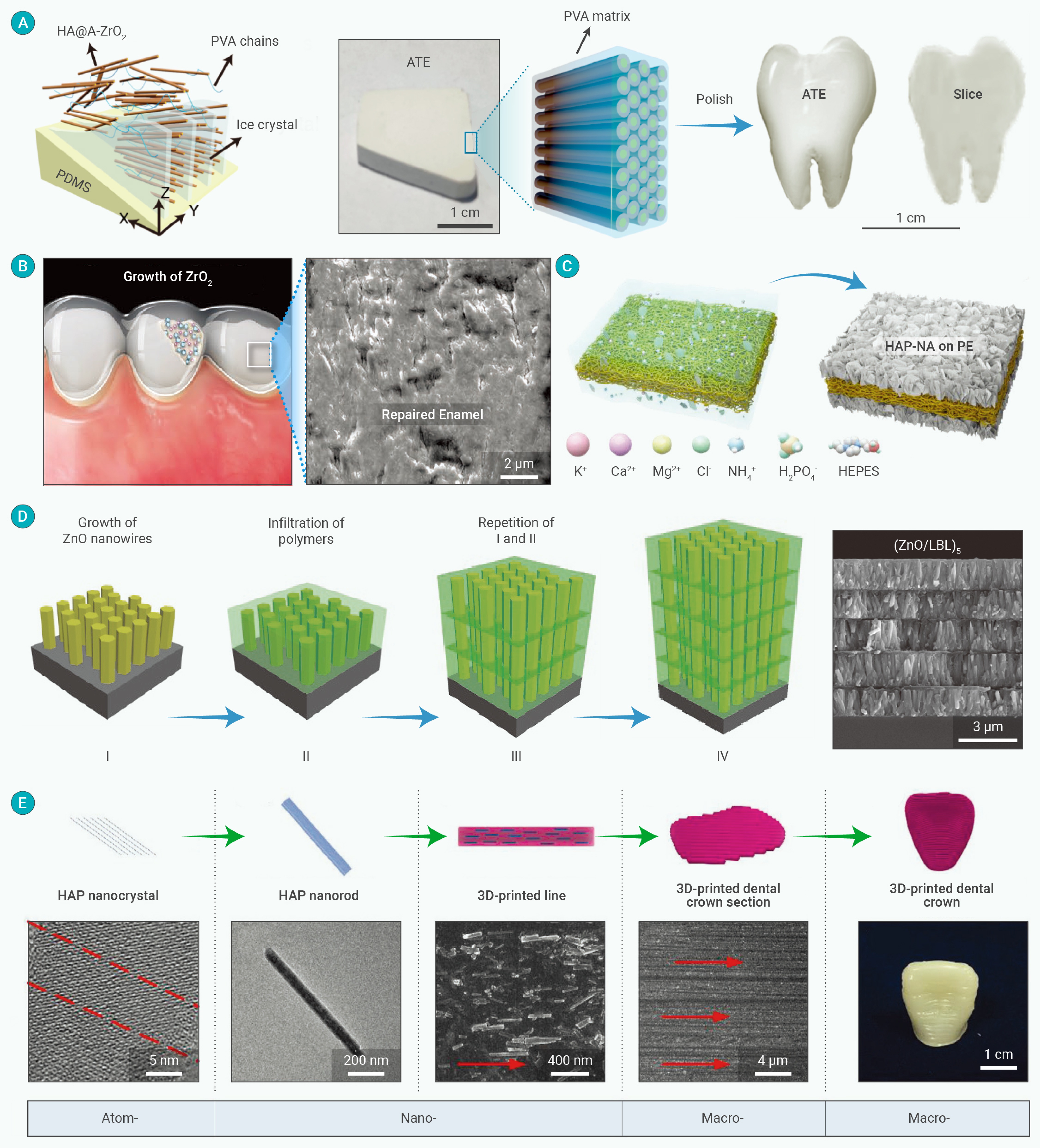

-
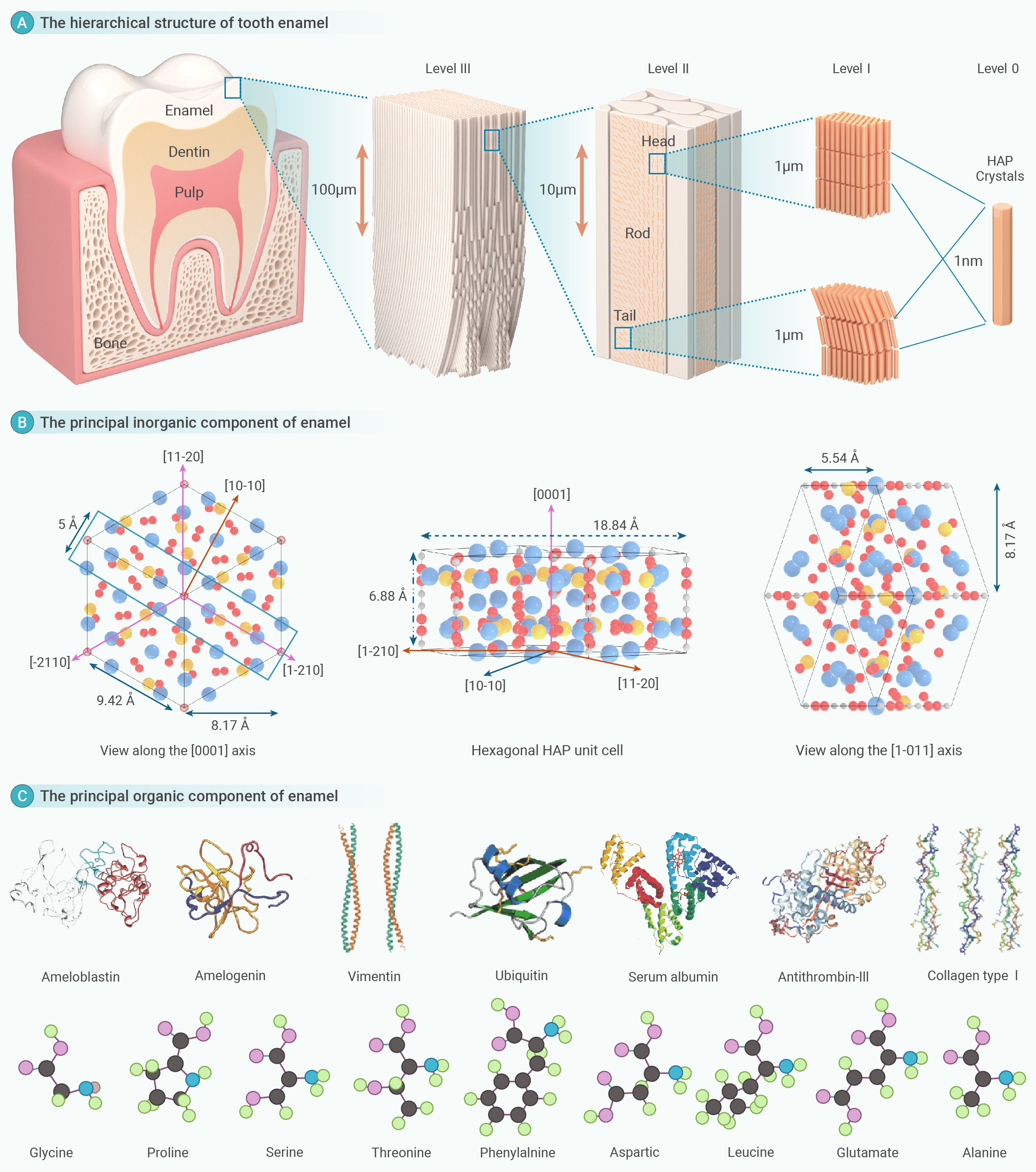
Figure 1.
The hierarchical structure and composition of human tooth enamel
-
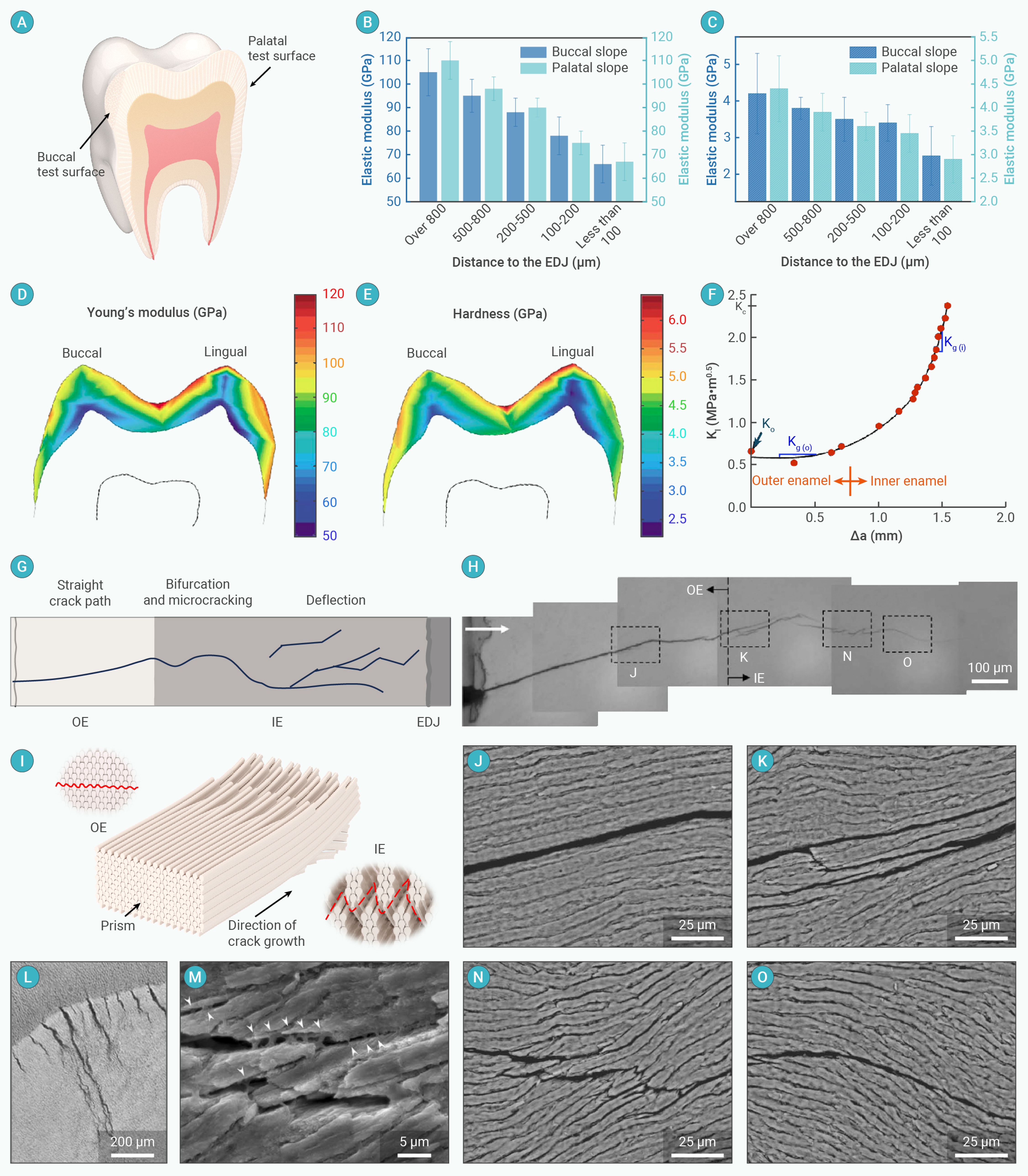
Figure 2.
Mechanical properties and strengthening and toughening mechanisms of human tooth enamel
-
Figure 3.
Tooth formation and regeneration process by tissue engineering
-
Figure 4.
Schematic diagram and SEM images of regenerated enamel of biomimetic mineralization guided by protein matrix
-
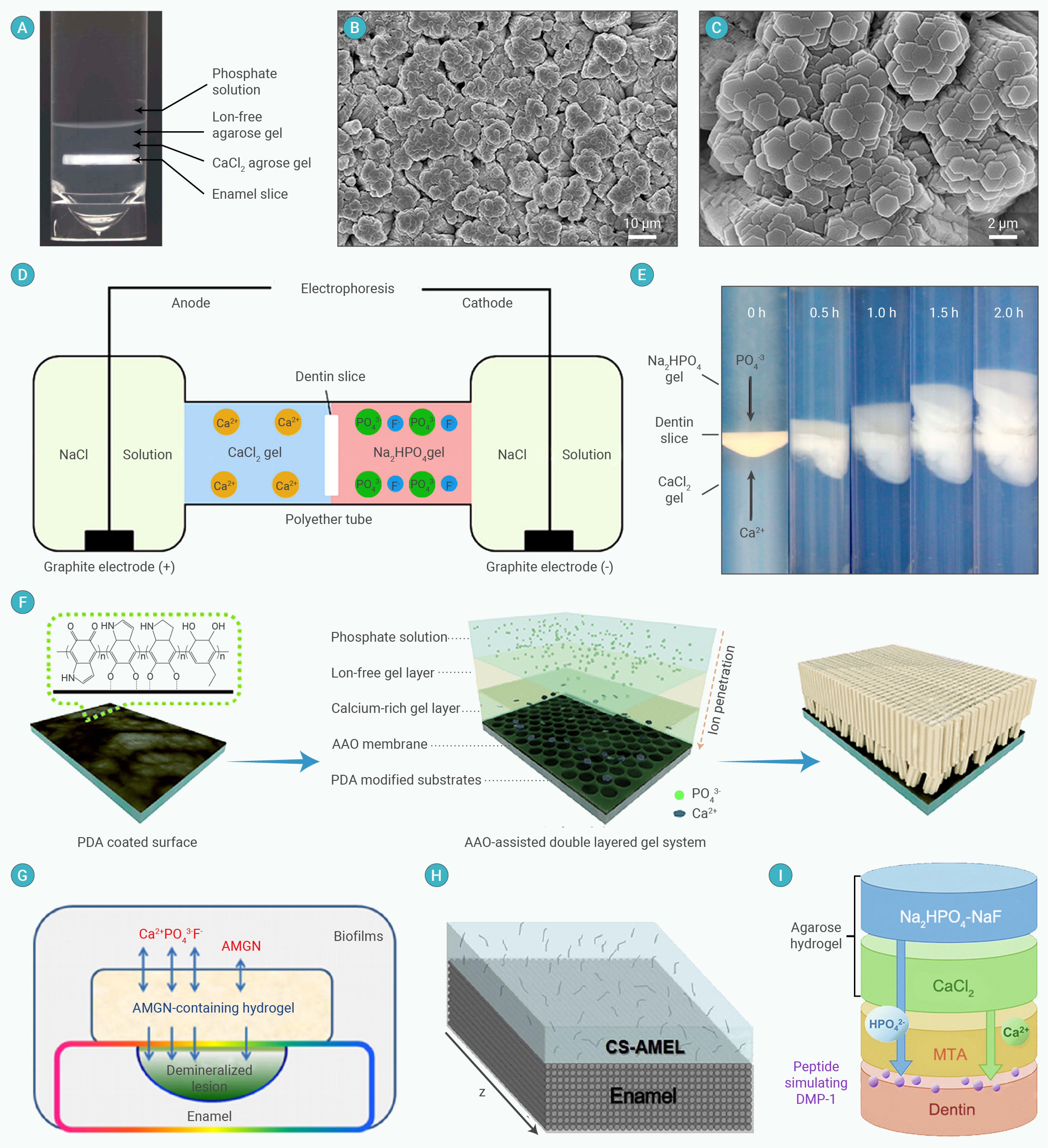
Figure 5.
Schematic and electron microscope images of enamel regeneration by a gel microenvironment mimicking a biomineralization process
-
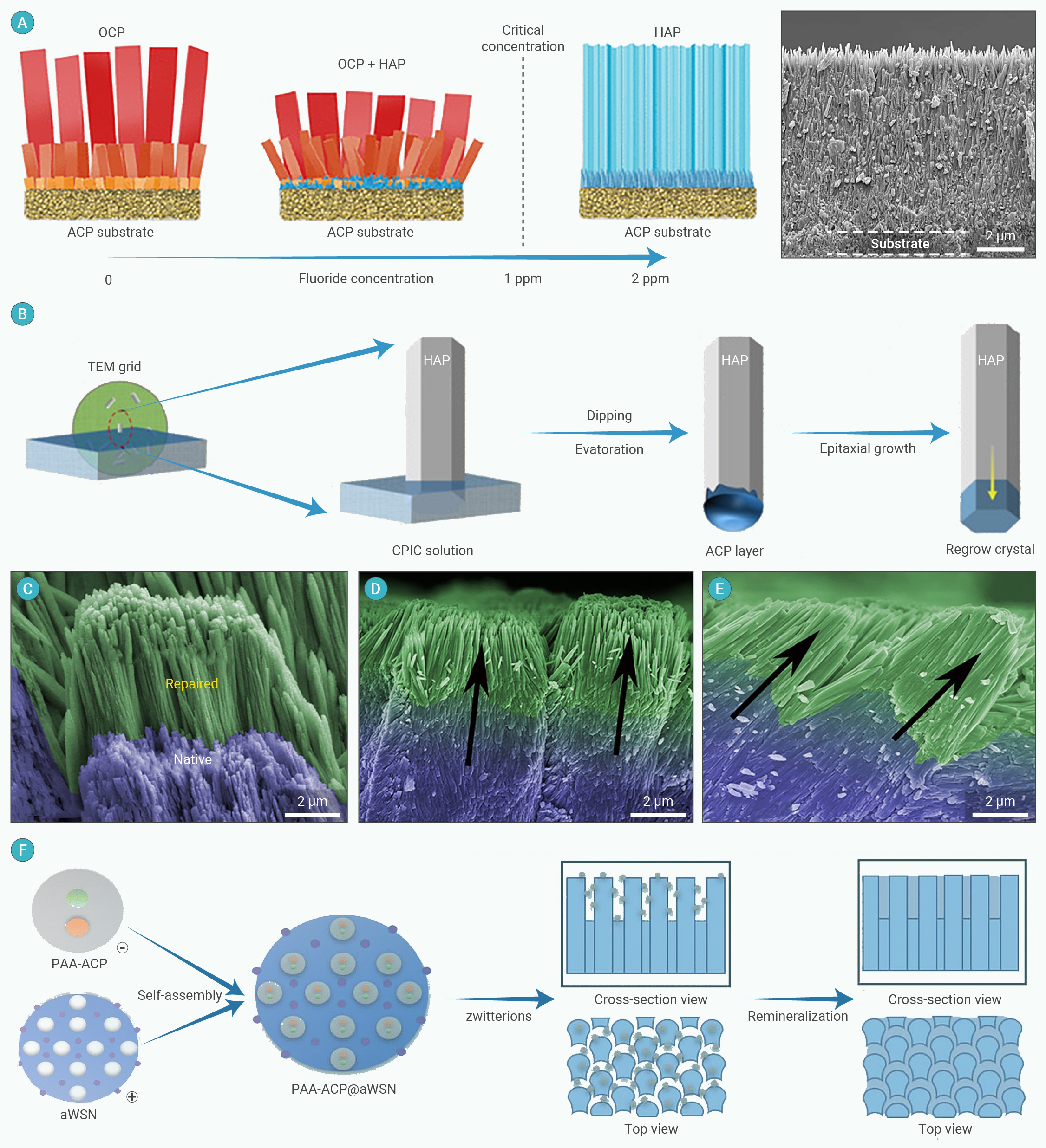
Figure 6.
Repair of enamel due to amorphous-crystalline phase transition
-
Figure 7.
Schematic diagram and electron microscope images of enamel regeneration by physicochemical methods
-
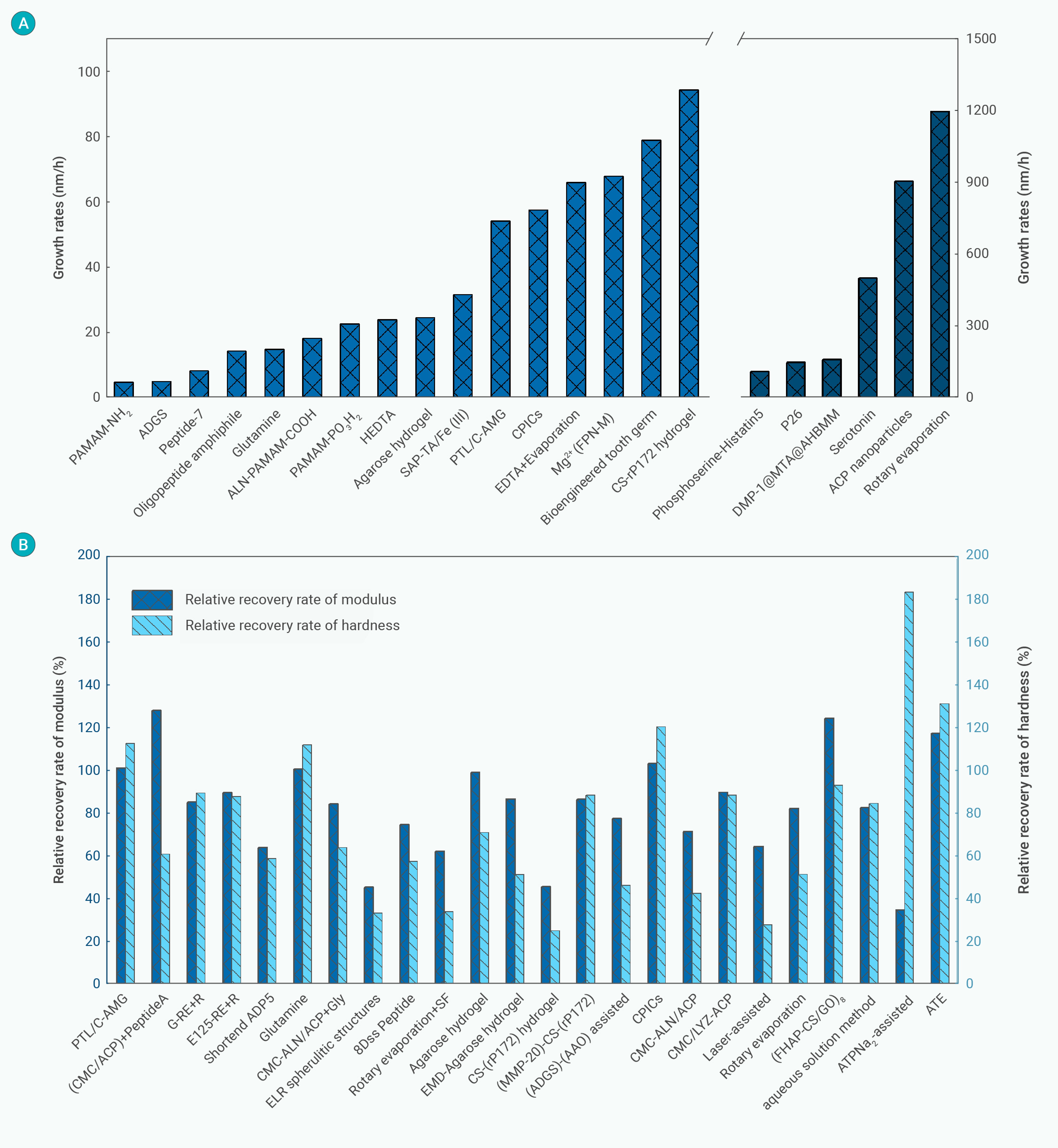
Figure 8.
The growth rates as well as relative recovery rates of modulus and hardness in various biomimetic enamel materials
-
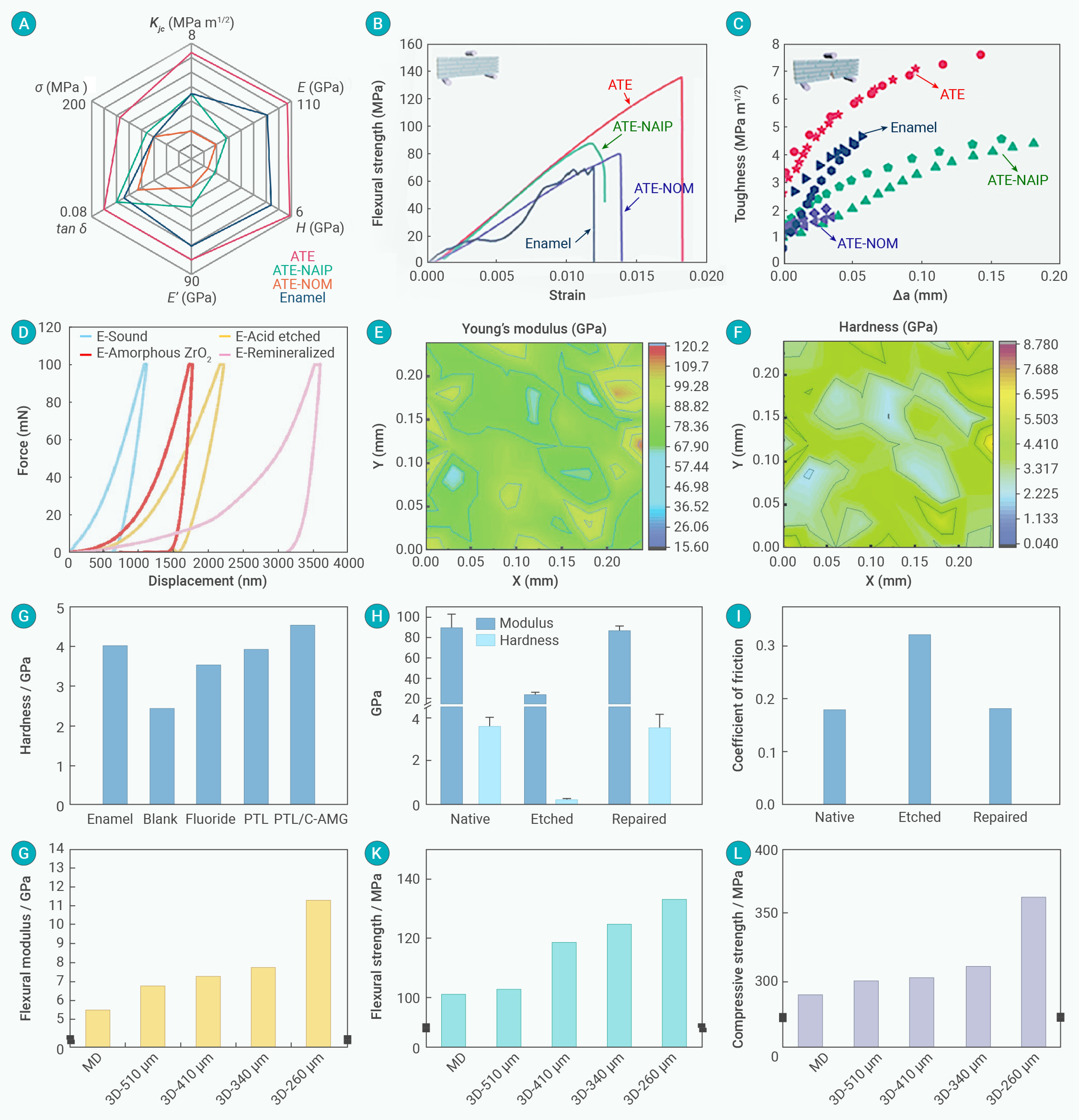
Figure 9.
Mechanical performances of the regenerated enamel-like nanocomposites
-
Figure 10.
In vivo mineralization test and cytotoxicity test
