
Prediction of angle closure after laser peripheral iridotomy: The fourteen-year Zhongshan Angle Closure Prevention trial
-
GRAPHICAL ABSTRACT
 DownLoad:
Full size image
DownLoad:
Full size image
-
PUBLIC SUMMARY
-
Anterior chamber angle can continue to narrow after laser peripheral iridotomy.
Long-term risk of progression after laser peripheral iridotomy can be predicted.
The narrowing of anterior chamber angle is an important risk factor.
Dynamic changes of intraocular pressure and lens vault are additional predictors.
-
-
ABSTRACT
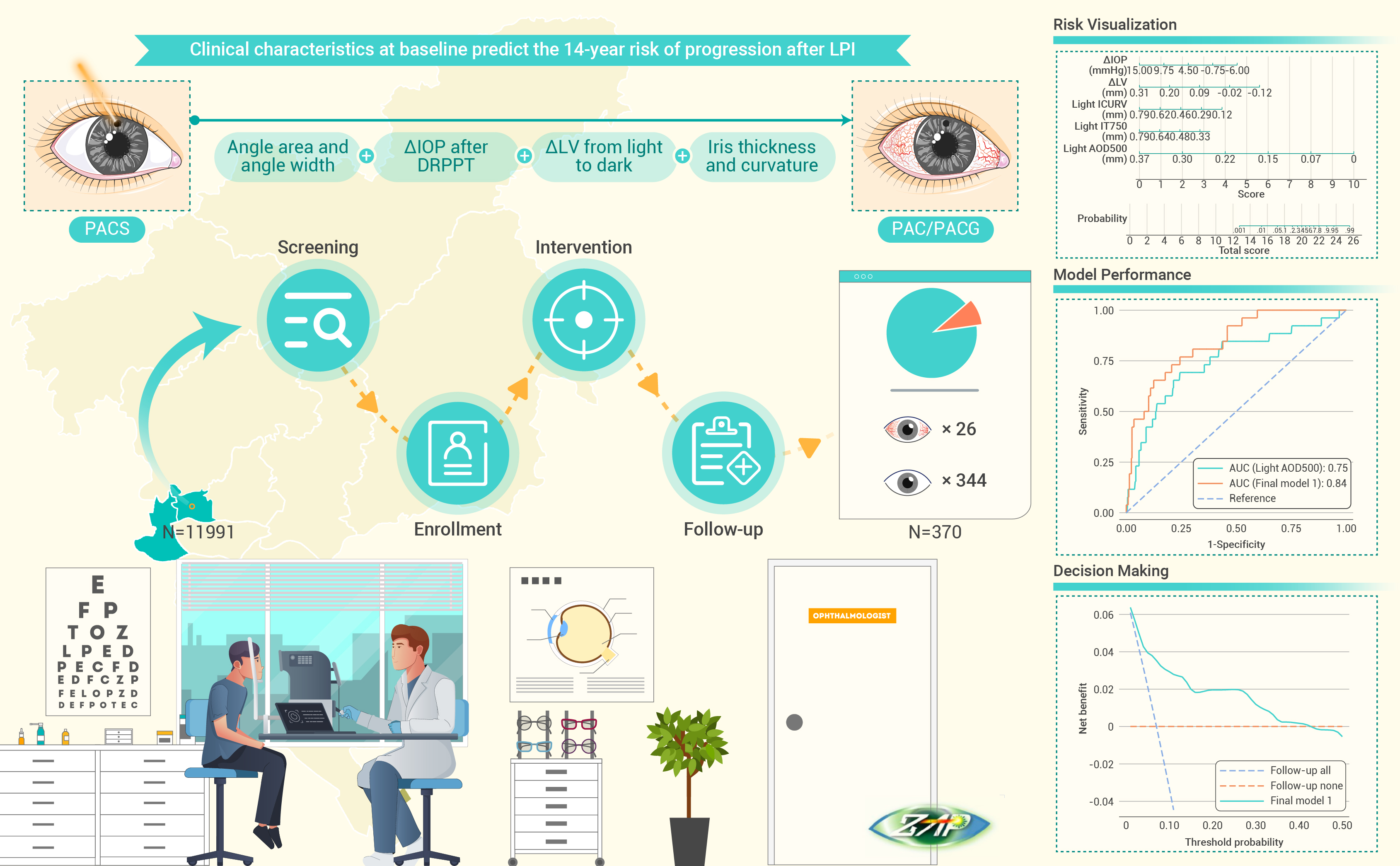
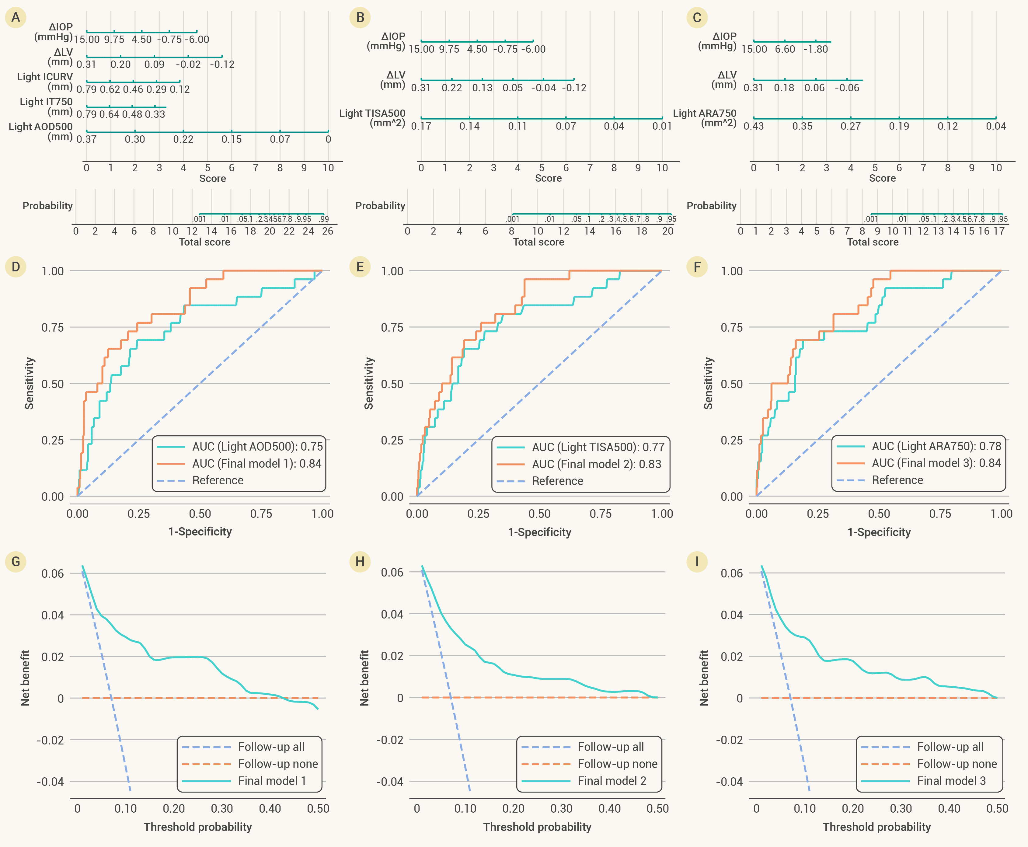
Anterior chamber angles in primary angle closure suspects (PACS) can continue to narrow after laser peripheral iridotomy (LPI). The aim of this study is to identify risk factors and develop prediction models for the progression in LPI-treated eyes during a 14-year follow-up. From 2008 to 2010, 889 Chinese participants aged 50-70 years with bilateral PACS were enrolled in the Zhongshan Angle Closure Prevention (ZAP) trial and received LPI in one randomly selected eye. Examinations before LPI included Goldmann tonometry, ultrasound A-scan biometry, both light-room and dark-room anterior-segment optical coherence tomography (AS-OCT). Logistic regression models were built to predict the 14-year risk of progression in PACS eyes after LPI (peripheral anterior synechiae, intraocular pressure [IOP] > 24 mmHg, or acute angle closure). Within 370 eligible PACS eyes, 26 progressed to PAC during 14 years after LPI. For both light-room and dark-room AS-OCT metrics before LPI, the narrowing of anterior chamber angle was identified as risk factor for the 14-year risk of progression in LPI-treated PACS eyes. In addition, change in IOP after dark-room prone provocative test and change in lens vault from light to dark before LPI were found to be negatively associated with the risk of progression during 14 years after LPI. Based on aforementioned predictors, multivariable logistic models provided good performance in the prediction for long-term risk of progression after LPI (area under the curve = 0.80-0.84). This study suggested that closer monitoring is still required for PACS eyes at high risk of progression even after prophylactic LPI. -

-
REFERENCES
[1] Prum, B.E., Herndon, L.W., Moroi, S.E., et al. (2016). Primary angle closure preferred practice pattern guidelines. Ophthalmology 123: P1−P40. DOI: 10.1016/j.ophtha.2015.12.023. [2] Tham, Y.C., Li, X., Wong, T.Y., et al. (2014). Global prevalence of glaucoma and projections of glaucoma burden through 2040: A systematic review and meta-analysis. Ophthalmology 121: 2081−2090. DOI: 10.1016/j.ophtha.2014.05.013. [3] Quigley, H.A., and Broman, A.T. (2006). The number of people with glaucoma worldwide in 2010 and 2020. Br. J. Ophthalmol. 90: 262−267. DOI: 10.1136/bjo.2005.081224. [4] Radhakrishnan, S., Chen, P.P., Junk, A.K., et al. (2018). Laser peripheral iridotomy in primary angle closure: A report by the American Academy of Ophthalmology. Ophthalmology 125: 1110−1120. DOI: 10.1016/j.ophtha.2018.01.015. [5] He, M., Jiang, Y., Huang, S., et al. (2019). Laser peripheral iridotomy for the prevention of angle closure: A single-centre, randomised controlled trial. Lancet 393: 1609−1618. DOI: 10.1016/S0140-6736(18)32607-2. [6] Baskaran, M., Kumar, R.S., Friedman, D.S., et al. (2022). The Singapore Asymptomatic Narrow Angles Laser Iridotomy Study: Five-year results of a randomized controlled trial. Ophthalmology 129: 147−158. DOI: 10.1016/j.ophtha.2021.08.017. [7] Jiang, Y., Chang, D.S., Zhu, H., et al. (2014). Longitudinal changes of angle configuration in primary angle-closure suspects: The Zhongshan Angle-Closure Prevention Trial. Ophthalmology 121: 1699−1705. DOI: 10.1016/j.ophtha.2014.03.039. [8] Yan, W., and He, M. (2017). Predictive medicine in ophthalmology. Ophthalmology 124: 420−421. DOI: 10.1016/j.ophtha.2016.12.018. [9] Bao, Y.K., Xu, B.Y., Friedman, D.S., et al. (2023). Biometric risk factors for angle closure progression after laser peripheral iridotomy. JAMA Ophthalmol. 141: 516−524. DOI: 10.1001/jamaophthalmol.2023.0937. [10] Jiang, Y., Friedman, D.S., He, M., et al. (2010). Design and methodology of a randomized controlled trial of laser iridotomy for the prevention of angle closure in southern China: The Zhongshan Angle Closure Prevention Trial. Ophthalmic. Epidemiol. 17: 321−332. DOI: 10.3109/09286586.2010.508353. [11] Yuan, Y., Wang, W., Xiong, R., et al. (2023). Fourteen-year outcome of angle-closure prevention with laser iridotomy in the Zhongshan Angle-Closure Prevention Study: extended follow-up of a randomized controlled trial. Ophthalmology 130: 786−794. DOI: 10.1016/j.ophtha.2023.03.024. [12] Console, J.W., Sakata, L.M., Aung, T., et al. (2008). Quantitative analysis of anterior segment optical coherence tomography images: The Zhongshan Angle Assessment Program. Br. J. Ophthalmol. 92: 1612−1616. DOI: 10.1136/bjo.2007.129932. [13] Kattan, M.W., Eastham, J.A., Stapleton, A.M., et al. (1998). A preoperative nomogram for disease recurrence following radical prostatectomy for prostate cancer. J. Natl. Cancer Inst. 90: 766−771. DOI: 10.1093/jnci/90.10.766. [14] Vickers, A.J., and Elkin, E.B. (2006). Decision curve analysis: A novel method for evaluating prediction models. Med. Decis. Making 26: 565−574. DOI: 10.1177/0272989X06295361. [15] Hong, C., Park, K.H., Hyung, S.M., et al. (1996). Evaluation of pupillary block component in angle-closure glaucoma. Jpn. J. Ophthalmol. 40: 239−243. [16] Sihota, R., Rishi, K., Srinivasan, G., et al. (2016). Functional evaluation of an iridotomy in primary angle closure eyes. Graefes. Arch. Clin. Exp. Ophthalmol. 254: 1141−1149. DOI: 10.1007/s00417-016-3298-x. [17] Kumar, R.S., Baskaran, M., Chew, P.T., et al. (2008). Prevalence of plateau iris in primary angle closure suspects an ultrasound biomicroscopy study. Ophthalmology 115: 430−434. DOI: 10.1016/j.ophtha.2007.07.026. [18] Wang, N., Ouyang, J., Zhou, W., et al. (2000). Multiple patterns of angle closure mechanisms in primary angle closure glaucoma in Chinese. Zhonghua Yan Ke Za Zhi 36: 46−51. [19] Quigley, H.A. (2009). Angle-closure glaucoma-simpler answers to complex mechanisms: LXVI Edward Jackson Memorial Lecture. Am. J. Ophthalmol. 148: 657−669. DOI: 10.1016/j.ajo.2009.08.009. [20] Baskaran, M., Yang, E., Trikha, S., et al. (2017). Residual angle closure one year after laser peripheral iridotomy in primary angle closure suspects. Am. J. Ophthalmol. 183: 111−117. DOI: 10.1016/j.ajo.2017.08.016. [21] Koh, V., Keshtkaran, M.R., Hernstadt, D., et al. (2019). Predicting the outcome of laser peripheral iridotomy for primary angle closure suspect eyes using anterior segment optical coherence tomography. Acta Ophthalmol. 97: e57−e63. DOI: 10.1111/aos.13822. [22] Lee, R.Y., Kasuga, T., Cui, Q.N., et al. (2013). Association between baseline angle width and induced angle opening following prophylactic laser peripheral iridotomy. Invest. Ophthalmol. Vis. Sci. 54: 3763−3770. DOI: 10.1167/iovs.13-11597. [23] Sung, K.R., Lee, K.S., and Hong, J.W. (2015). Baseline anterior segment parameters associated with the long-term outcome of laser peripheral iridotomy. Curr. Eye Res. 40: 1128−1133. DOI: 10.3109/02713683.2014.986334. [24] Masoodi, H., Jafarzadehpur, E., Esmaeili, A., et al. (2014). Evaluation of anterior chamber angle under dark and light conditions in angle closure glaucoma: An anterior segment OCT study. Cont. Lens Anterior Eye. 37: 300−304. DOI: 10.1016/j.clae.2014.04.002. [25] Nolan, W.P., See, J.L., Chew, P.T., et al. (2007). Detection of primary angle closure using anterior segment optical coherence tomography in Asian eyes. Ophthalmology 114: 33−39. DOI: 10.1016/j.ophtha.2006.05.073. [26] Jiang, Y., Wang, W., Wang, L., et al. (2021). Association of anterior segment parameters and 5-year incident narrow angles: findings from an older Chinese population. Br. J. Ophthalmol. 105: 970−976. DOI: 10.1136/bjophthalmol-2020-315852. [27] Wilensky, J.T., Kaufman, P.L., Frohlichstein, D, et al. (1993). Follow-up of angle-closure glaucoma suspects. Am. J. Ophthalmol. 115: 338−346. DOI: 10.1016/S0002-9394(14)73585-8. [28] Friedman, D.S., Chang, D.S., Jiang, Y., et al. (2019). Darkroom prone provocative testing in primary angle closure suspects and those with open angles. Br. J. Ophthalmol. 103: 1834−1839. DOI: 10.1136/bjophthalmol-2018-313362. [29] Harris, L.S., and Galin, M.A. (1972). Prone provocative testing for narrow angle glaucoma. Arch. Ophthalmol. 87: 493−496. DOI: 10.1001/archopht.1972.01000020495001. [30] Ozaki, M., Nongpiur, M.E., Aung, T., et al. (2012). Increased lens vault as a risk factor for angle closure: Confirmation in a Japanese population. Graefes. Arch. Clin. Exp. Ophthalmol. 250: 1863−1868. DOI: 10.1007/s00417-012-2011-y. [31] Moghimi, S., Vahedian, Z., Zandvakil, N., et al. (2014). Role of lens vault in subtypes of angle closure in Iranian subjects. Eye 28: 337−343. DOI: 10.1038/eye.2013.296. [32] Wang, Z., Chung, C., Lin, J, et al. (2016). Quantitative measurements of the ciliary body in eyes with acute primary-angle closure. Invest. Ophthalmol. Vis. Sci. 57: 3299−3305. DOI: 10.1167/iovs.16-19558. [33] Tan, G.S., He, M., Zhao, W., et al. (2012). Determinants of lens vault and association with narrow angles in patients from Singapore. Am. J. Ophthalmol. 154: 39−46. DOI: 10.1016/j.ajo.2012.01.015. [34] Ang, M., Baskaran, M., Werkmeister, R.M., et al. (2018). Anterior segment optical coherence tomography. Prog. Retin. Eye Res. 66: 132−156. DOI: 10.1016/j.preteyeres.2018.04.002. [35] Sakata, L.M., Lavanya, R., Friedman, D.S., et al. (2008). Assessment of the scleral spur in anterior segment optical coherence tomography images. Arch. Ophthalmol. 126: 181−185. DOI: 10.1001/archophthalmol.2007.46. [36] Wanichwecharungruang, B., Pattanapongpaiboon, W., Kongsomboon, K., et al. (2022). Diagnostic performance of anterior segment optical coherence tomography in detecting plateau iris. BMJ Open Ophthalmol. 7: e000931. DOI: 10.1136/bmjophth-2021-000931. [37] Peng, P.H., Nguyen, H., Lin, H.S., et al. (2011). Long-term outcomes of laser iridotomy in Vietnamese patients with primary angle closure. Br. J. Ophthalmol. 95: 1207−1211. DOI: 10.1136/bjo.2010.181016. -
ABOUT THIS ARTICLE
Cite this article:
Yuan Y., Yang S., Wang W., et al., (2023). Prediction of angle closure after laser peripheral iridotomy: The fourteen-year Zhongshan Angle Closure Prevention trial. The Innovation Medicine 1(3), 100033. https://doi.org/10.59717/j.xinn-med.2023.100033
Welcome!
To request copyright permission to republish or share portions of our works, please visit Copyright Clearance Center's (CCC) Marketplace website at marketplace.copyright.com.

Article Metrics
Relative Articles
Cited by
Export File
Citation
Yuan Y., Yang S., Wang W., et al., (2023). Prediction of angle closure after laser peripheral iridotomy: The fourteen-year Zhongshan Angle Closure Prevention trial. The Innovation Medicine 1(3), 100033. https://doi.org/10.59717/j.xinn-med.2023.100033

Format
Content
-
Figure 1.
Anterior segment optical coherence tomography metrics determined by the Zhongshan Angle Assessment Program
-
Figure 2.
Prediction models for the 14-year risk of progression from PACS to PAC after LPI based on light-room AS-OCT metrics
