
Stem cell niches functionalized strategies for organ regeneration and manufacturing
-
GRAPHICAL ABSTRACT

 DownLoad:
Full size image
DownLoad:
Full size image
-
PUBLIC SUMMARY
-
Stem cell niches determine stem cells fate and homeostasis.
Stem cell niches provide opportunities for organ regeneration.
Stem cell niches-functionalized strategies have been emerging.
-
-
ABSTRACT
Organ regeneration and manufacturing are promising new research directions in the life sciences. Stem cells and their niches, which exist in most adult organs, play a central role in organ development, homeostasis, and regeneration. Although considerable advances have been made in stem cell-mediated organ regeneration and manufacturing in recent decades, their clinical effectiveness remains unsatisfactory. Stem cell niches comprise a dynamic microenvironment that supports stem cells throughout their lifetime and are critical to stem cell fate. Based on recent research on stem cell niches, tremendous progress has been made in organ regeneration and manufacturing in vivo and in vitro. In this review, we discuss recent advances in the composition and function of stem cell niches during regeneration. We also discuss stem cell niche remodeling using cell-cell interaction, extracellular matrix (ECM) reconstruction, cell-ECM interaction, and key signaling-based niche strategies to promote endogenous tooth, gastrointestinal tract, and liver regeneration and its application in organoids and organoid-on-chip construction. -

-
REFERENCES
[1] Yamanaka, S. (2020). Pluripotent stem cell-based cell therapy-promise and challenges. Cell Stem Cell 27: 523−531. DOI: 10.1016/j.stem.2020.09.014. [2] Watt, F.M., and Driskell, R.R. (2010). The therapeutic potential of stem cells. Philos. Trans. R. Soc. Lond. B Biol. Sci. 365: 155−163. DOI: 10.1098/rstb.2009.0149. [3] Daley, G.Q. (2012). The promise and perils of stem cell therapeutics. Cell Stem Cell 10: 740−749. DOI: 10.1016/j.stem.2012.05.010. [4] Galipeau, J., and Sensébé, L. (2018). Mesenchymal stromal cells: clinical challenges and therapeutic opportunities. Cell Stem Cell 22: 824−833. DOI: 10.1016/j.stem.2018.05.004. [5] Hofer, M., and Lutolf, M.P. (2021). Engineering organoids. Nat. Rev. Mater. 6: 402−420. DOI: 10.1038/s41578-021-00279-y. [6] Rossi, G., Manfrin, A., and Lutolf, M.P. (2018). Progress and potential in organoid research. Nat. Rev. Genet. 19: 671−687. DOI: 10.1038/s41576-018-0051-9. [7] Kim, W., Gwon, Y., Park, S., et al. (2023). Therapeutic strategies of three-dimensional stem cell spheroids and organoids for tissue repair and regeneration. Bioact. Mater. 19: 50−74. DOI: 10.1016/j.bioactmat.2022.03.039. [8] Lane, S.W., Williams, D.A., and Watt, F.M. (2014). Modulating the stem cell niche for tissue regeneration. Nat. Biotechnol. 32: 795−803. DOI: 10.1038/nbt.2978. [9] Chacón-Martínez, C.A., Koester, J., and Wickström, S.A. (2018). Signaling in the stem cell niche: regulating cell fate, function and plasticity. Development 145 : dev165399. DOI: 10.1242/dev.165399. [10] Hicks, M.R., and Pyle, A.D. (2023). The emergence of the stem cell niche. Trends Cell Biol. 33: 112−123. DOI: 10.1016/j.tcb.2022.07.003. [11] Xie, T., and Spradling, A.C. (2000). A niche maintaining germ line stem cells in the Drosophila ovary. Science 290: 328−330. DOI: 10.1126/science.290.5490.328. [12] Morrison, S.J., and Spradling, A.C. (2008). Stem cells and niches: mechanisms that promote stem cell maintenance throughout life. Cell 132: 598−611. DOI: 10.1016/j.cell.2008.01.038. [13] Schmidt, M., Schüler, S.C., Hüttner, S.S., et al. (2019). Adult stem cells at work: regenerating skeletal muscle. Cell. Mol. Life Sci. 76: 2559−2570. DOI: 10.1007/s00018-019-03093-6. [14] Brittan, M., and Wright, N.A. (2002). Gastrointestinal stem cells. J. Pathol. 197: 492−509. DOI: 10.1002/path.1155. [15] Shaker, A., and Rubin, D.C. (2010). Intestinal stem cells and epithelial-mesenchymal interactions in the crypt and stem cell niche. Transl. Res. 156: 180−187. DOI: 10.1016/j.trsl.2010.06.003. [16] Zhang, L., Theise, N., Chua, M., et al. (2008). The stem cell niche of human livers: Symmetry between development and regeneration. Hepatology 48: 1598−1607. DOI: 10.1002/hep.22516. [17] Gonzales, K.A.U., and Fuchs, E. (2017). Skin and its regenerative powers: An alliance between stem cells and their niche. Dev. Cell 43: 387−401. DOI: 10.1016/j.devcel.2017.10.001. [18] Ort, C., Dayekh, K., Xing, M., et al. (2018). Emerging strategies for stem cell lineage commitment in tissue engineering and regenerative medicine. ACS Biomater. Sci. Eng. 4: 3644−3657. DOI: 10.1021/acsbiomaterials.8b00532. [19] Wattrus, S.J., and Zon, L.I. (2018). Stem cell safe harbor: The hematopoietic stem cell niche in zebrafish. Blood Adv. 2: 3063−3069. DOI: 10.1182/bloodadvances.2018021725. [20] Diotel, N., Lübke, L., Strähle, U., et al. (2020). Common and distinct features of adult neurogenesis and regeneration in the telencephalon of zebrafish and mammals. Front. Neurosci. 14: 568930. DOI: 10.3389/fnins.2020.568930. [21] Schwarzer, S., Rekhade, D.R., Machate, A., et al. (2022). Reactivation of the neurogenic niche in the adult zebrafish statoacoustic ganglion following a mechanical lesion. Front. Cell Dev. Biol. 10: 850624. DOI: 10.3389/fcell.2022.850624. [22] Rajan, V., Melong, N., Hing Wong, W., et al. (2020). Humanized zebrafish enhance human hematopoietic stem cell survival and promote acute myeloid leukemia clonal diversity. Haematologica 105: 2391−2399. DOI: 10.3324/haematol.2019.223040. [23] Ishizuya-Oka, A., and Hasebe, T. (2013). Establishment of intestinal stem cell niche during amphibian metamorphosis. Curr. Top. Dev. Biol. 103: 305−327. DOI: 10.1016/B978-0-12-385979-2.00011-3. [24] Ghaddar, B., Lübke, L., Couret, D., et al. (2021). Cellular mechanisms participating in brain repair of adult zebrafish and mammals after injury. Cell 10 : 391. DOI: 10.3390/cells10020391. [25] Ceci, M., Mariano, V., and Romano, N. (2018). Zebrafish as a translational regeneration model to study the activation of neural stem cells and role of their environment. Rev. Neurosci. 30: 45−66. DOI: 10.1515/revneuro-2018-0020. [26] Xia, J., Kang, Z., Xue, Y., et al. (2021). A single-cell resolution developmental atlas of hematopoietic stem and progenitor cell expansion in zebrafish. Proc. Natl. Acad. Sci. U. S. A. 118 : e2015748118. DOI: 10.1073/pnas.2015748118. [27] Hu, H., Duan, Y., Wang, K., et al. (2022). Dental niche cells directly contribute to tooth reconstitution and morphogenesis. Cell Rep. 41: 111737. DOI: 10.1016/j.celrep.2022.111737. [28] Santos, A.J.M., Lo, Y.H., Mah, A.T., et al. (2018). The intestinal stem cell niche: Homeostasis and adaptations. Trends Cell Biol. 28: 1062−1078. DOI: 10.1016/j.tcb.2018.08.001. [29] Ratnayake, D., Nguyen, P.D., Rossello, F.J., et al. (2021). Macrophages provide a transient muscle stem cell niche via NAMPT secretion. Nature 591: 281−287. DOI: 10.1038/s41586-021-03199-7. [30] Li, J., Tan, J., Martino, M.M., et al. (2018). Regulatory T-cells: Potential regulator of tissue repair and regeneration. Front. Immunol. 9: 585. DOI: 10.3389/fimmu.2018.00585. [31] Schüler, S.C., Liu, Y., Dumontier, S., et al. (2022). Extracellular matrix: Brick and mortar in the skeletal muscle stem cell niche. Front. Cell Dev. Biol. 10: 1056523. DOI: 10.3389/fcell.2022.1056523. [32] Lee-Thedieck, C., Schertl, P., and Klein, G. (2022). The extracellular matrix of hematopoietic stem cell niches. Adv. Drug Deliv. Rev. 181: 114069. DOI: 10.1016/j.addr.2021.114069. [33] Arulmoli, J., Pathak, M.M., McDonnell, L.P., et al. (2015). Static stretch affects neural stem cell differentiation in an extracellular matrix-dependent manner. Sci. Rep. 5: 8499. DOI: 10.1038/srep08499. [34] Sun, Y., Xu, S., Jiang, M., et al. (2021). Role of the extracellular matrix in Alzheimer’s disease. Front. Aging Neurosci. 13: 707466. DOI: 10.3389/fnagi.2021.707466. [35] Arranz, A.M., Perkins, K.L., Irie, F., et al. (2014). Hyaluronan deficiency due to Has3 knock-out causes altered neuronal activity and seizures via reduction in brain extracellular space. J. Neurosci. 34: 6164−6176. DOI: 10.1523/JNEUROSCI.3458-13.2014. [36] Yeo, G.C., and Weiss, A.S. (2019). Soluble matrix protein is a potent modulator of mesenchymal stem cell performance. Proc. Natl. Acad. Sci. U. S. A. 116: 2042−2051. DOI: 10.1073/pnas.1812951116. [37] Saw, S., Weiss, A., Khokha, R., et al. (2019). Metalloproteases: On the watch in the hematopoietic niche. Trends Immunol. 40: 1053−1070. DOI: 10.1016/j.it.2019.09.006. [38] Martín-Alonso, M., Iqbal, S., Vornewald, P.M., et al. (2021). Smooth muscle-specific MMP17 (MT4-MMP) regulates the intestinal stem cell niche and regeneration after damage. Nat. Commun. 12: 6741. DOI: 10.1038/s41467-021-26904-6. [39] Nusse, R., and Clevers, H. (2017). Wnt/beta-catenin signaling, disease, and emerging therapeutic modalities. Cell 169: 985−999. DOI: 10.1016/j.cell.2017.05.016. [40] Chakrabarti, R., Celià-Terrassa, T., Kumar, S., et al. (2018). Notch ligand Dll1 mediates cross-talk between mammary stem cells and the macrophageal niche. Science 360 : eaan4153. DOI: 10.1126/science.aan4153. [41] Chung, M.I., Bujnis, M., Barkauskas, C.E., et al. (2018). Niche-mediated BMP/SMAD signaling regulates lung alveolar stem cell proliferation and differentiation. Development 145 : dev163014. DOI: 10.1242/dev.163014. [42] Chen, G., Xu, H., Yao, Y., et al. (2020). BMP signaling in the development and regeneration of cranium bones and maintenance of calvarial stem cells. Front. Cell Dev. Biol. 8: 135. DOI: 10.3389/fcell.2020.00135. [43] Wang, Y., Kim, R., Hinman, S.S., et al. (2018). Bioengineered systems and designer matrices that recapitulate the intestinal stem cell niche. Cell. Mol. Gastroenterol. Hepatol. 5: 440−453.e441. DOI: 10.1016/j.jcmgh.2018.01.008. [44] Parfenova, O.K., Kukes, V.G., and Grishin, D.V. (2021). Follistatin-like proteins: Structure, functions and biomedical importance. Biomedicines 9 : 999. DOI: 10.3390/biomedicines9080999. [45] Nakamura, T., Jimenez-Rojo, L., Koyama, E., et al. (2017). Epiprofin regulates enamel formation and tooth morphogenesis by controlling epithelial-mesenchymal interactions during tooth development. J. Bone Miner. Res. 32: 601−610. DOI: 10.1002/jbmr.3024. [46] Fahmy-Garcia, S., Farrell, E., Witte-Bouma, J., et al. (2019). Follistatin effects in migration, vascularization, and osteogenesis in vitro and bone repair in vivo. Front. Bioeng. Biotechnol. 7: 38. DOI: 10.3389/fbioe.2019.00038. [47] Elbediwy, A., Vincent-Mistiaen, Z.I., and Thompson, B.J. (2016). YAP and TAZ in epithelial stem cells: A sensor for cell polarity, mechanical forces and tissue damage. Bioessays 38: 644−653. DOI: 10.1002/bies.201600037. [48] van Soldt, B.J., and Cardoso, W.V. (2019). Hippo-Yap/Taz signaling: Complex network interactions and impact in epithelial cell behavior. Wiley Interdiscip. Rev. Dev. Biol. 9: e371. DOI: 10.1002/wdev.371. [49] Piccolo, S., Dupont, S., and Cordenonsi, M. (2014). The biology of YAP/TAZ: Hippo signaling and beyond. Physiol. Rev. 94: 1287−1312. DOI: 10.1152/physrev.00005.2014. [50] Moya, I.M., and Halder, G. (2019). Hippo-YAP/TAZ signalling in organ regeneration and regenerative medicine. Nat. Rev. Mol. Cell Biol. 20: 211−226. DOI: 10.1038/s41580-018-0086-y. [51] Park, H.W., Kim, Y.C., Yu, B., et al. (2015). Alternative Wnt signaling activates YAP/TAZ. Cell 162: 780−794. DOI: 10.1016/j.cell.2015.07.013. [52] Adebayo, A.K., and Nakshatri, H. (2022). Modeling preclinical cancer studies under physioxia to enhance clinical translation. Cancer Res. 82: 4313−4321. DOI: 10.1158/0008-5472.CAN-22-2311. [53] Li, C., Wu, B., Li, Y., et al. (2022). Loss of sphingosine kinase 2 promotes the expansion of hematopoietic stem cells by improving their metabolic fitness. Blood 140: 1686−1701. DOI: 10.1182/blood.2022016112. [54] Liu, J., He, J., Ge, L., et al. (2021). Hypoxic preconditioning rejuvenates mesenchymal stem cells and enhances neuroprotection following intracerebral hemorrhage via the miR-326-mediated autophagy. Stem Cell. Res. Ther. 12: 413. DOI: 10.1186/s13287-021-02480-w. [55] Mu, J., Li, L., Wu, J., et al. (2022). Hypoxia-stimulated mesenchymal stem cell-derived exosomes loaded by adhesive hydrogel for effective angiogenic treatment of spinal cord injury. Biomater Sci 10: 1803−1811. DOI: 10.1039/D1BM01722E. [56] Hu, C., Fan, L., Cen, P., et al. (2016). Energy metabolism plays a critical role in stem cell maintenance and differentiation. Int. J. Mol. Sci. 17: 253. DOI: 10.3390/ijms17020253. [57] Folmes, C.D., Dzeja, P.P., Nelson, T.J., et al. (2012). Metabolic plasticity in stem cell homeostasis and differentiation. Cell Stem Cell 11: 596−606. DOI: 10.1016/j.stem.2012.10.002. [58] Morimoto, H., Yamamoto, T., Miyazaki, T., et al. (2021). An interplay of NOX1-derived ROS and oxygen determines the spermatogonial stem cell self-renewal efficiency under hypoxia. Genes Dev. 35: 250−260. DOI: 10.1101/gad.339903.120. [59] He, R., Wang, Z., Cui, M., et al. (2021). HIF1A Alleviates compression-induced apoptosis of nucleus pulposus derived stem cells via upregulating autophagy. Autophagy 17: 3338−3360. DOI: 10.1080/15548627.2021.1872227. [60] Choi, W., Kwon, S.J., Jin, H.J., et al. (2017). Optimization of culture conditions for rapid clinical-scale expansion of human umbilical cord blood-derived mesenchymal stem cells. Clin. Transl. Med. 6: 38. DOI: 10.1186/s40169-017-0168-z. [61] Nakamura, N., Shi, X., Darabi, R., et al. (2021). Hypoxia in cell reprogramming and the epigenetic regulations. Front. Cell Dev. Biol. 9: 609984. DOI: 10.3389/fcell.2021.609984. [62] Wang, W., Hu, C.K., Zeng, A., et al. (2020). Changes in regeneration-responsive enhancers shape regenerative capacities in vertebrates. Science 369 : eaaz3090. DOI: 10.1126/science.aaz3090. [63] Lumelsky, N., O'Hayre, M., Chander, P., et al. (2018). Autotherapies: Enhancing endogenous healing and regeneration. Trends Mol. Med. 24: 919−930. DOI: 10.1016/j.molmed.2018.08.004. [64] Dong, J., Hu, Y., Fan, X., et al. (2018). Single-cell RNA-seq analysis unveils a prevalent epithelial/mesenchymal hybrid state during mouse organogenesis. Genome Biol. 19: 31. DOI: 10.1186/s13059-018-1416-2. [65] Ribatti, D., and Santoiemma, M. (2014). Epithelial-mesenchymal interactions: A fundamental Developmental Biology mechanism. Int. J. Dev. Biol. 58: 303−306. DOI: 10.1387/ijdb.140143dr. [66] Nakao, K., Morita, R., Saji, Y., et al. (2007). The development of a bioengineered organ germ method. Nat. Methods 4: 227−230. DOI: 10.1038/nmeth1012. [67] Wang, Z., Kapadia, W., Li, C., et al. (2021). Tissue-specific engineering: 3D bioprinting in regenerative medicine. J. Control. Release 329: 237−256. DOI: 10.1016/j.jconrel.2020.11.044. [68] Stevens, A.J., Harris, A.R., Gerdts, J., et al. (2023). Programming multicellular assembly with synthetic cell adhesion molecules. Nature 614: 144−152. DOI: 10.1038/s41586-022-05622-z. [69] Song, J.J., and Ott, H.C. (2011). Organ engineering based on decellularized matrix scaffolds. Trends Mol. Med. 17: 424−432. DOI: 10.1016/j.molmed.2011.03.005. [70] Rajab, T.K., O'Malley, T.J., and Tchantchaleishvili, V. (2020). Decellularized scaffolds for tissue engineering: Current status and future perspective. Artif. Organs 44: 1031−1043. DOI: 10.1111/aor.13701. [71] Orlando, G., Booth, C., Wang, Z., et al. (2013). Discarded human kidneys as a source of ECM scaffold for kidney regeneration technologies. Biomaterials 34: 5915−5925. DOI: 10.1016/j.biomaterials.2013.04.033. [72] Nichols, J.E., Niles, J., Riddle, M., et al. (2013). Production and assessment of decellularized pig and human lung scaffolds. Tissue Eng. Part A 19: 2045−2062. DOI: 10.1089/ten.tea.2012.0250. [73] Sánchez, P.L., Fernández-Santos, M.E., Costanza, S., et al. (2015). Acellular human heart matrix: A critical step toward whole heart grafts. Biomaterials 61: 279−289. DOI: 10.1016/j.biomaterials.2015.04.056. [74] Mazza, G., Rombouts, K., Rennie Hall, A., et al. (2015). Decellularized human liver as a natural 3D-scaffold for liver bioengineering and transplantation. Sci. Rep. 5: 13079. DOI: 10.1038/srep13079. [75] Zhang, W., Vazquez, B., Oreadi, D., et al. (2017). Decellularized tooth bud scaffolds for tooth regeneration. J. Dent. Res. 96: 516−523. DOI: 10.1177/0022034516689082. [76] Choudhury, D., Tun, H.W., Wang, T., et al. (2018). Organ-derived decellularized extracellular matrix: A game changer for bioink manufacturing. Trends Biotechnol. 36: 787−805. DOI: 10.1016/j.tibtech.2018.03.003. [77] Xing, H., Lee, H., Luo, L., et al. (2020). Extracellular matrix-derived biomaterials in engineering cell function. Biotechnol. Adv. 42: 107421. DOI: 10.1016/j.biotechadv.2019.107421. [78] Xing, Y., and Naik, S. (2020). Under pressure: Stem cell-niche interactions coordinate tissue adaptation to inflammation. Curr. Opin. Cell Biol. 67: 64−70. DOI: 10.1016/j.ceb.2020.08.009. [79] Chaudhuri, O., Cooper-White, J., Janmey, P.A., et al. (2020). Effects of extracellular matrix viscoelasticity on cellular behaviour. Nature 584: 535−546. DOI: 10.1038/s41586-020-2612-2. [80] Xie, W., Wei, X., Kang, H., et al. (2023). Static and dynamic: Evolving biomaterial mechanical properties to control cellular mechanotransduction. Adv. Sci. 10: e2204594. DOI: 10.1002/advs.202204594. [81] Ikeda, E., Morita, R., Nakao, K., et al. (2009). Fully functional bioengineered tooth replacement as an organ replacement therapy. Proc. Natl. Acad. Sci. U. S. A. 106: 13475−13480. DOI: 10.1073/pnas.0902944106. [82] Zhang, W., and Yelick, P.C. (2021). Tooth repair and regeneration: potential of dental stem cells. Trends Mol. Med. 27: 501−511. DOI: 10.1016/j.molmed.2021.02.005. [83] Wang, F., Xiao, J., Cong, W., et al. (2014). Morphology and chronology of diphyodont dentition in miniature pigs, Sus Scrofa. Oral Dis. 20: 367−379. DOI: 10.1111/odi.12126. [84] Li, Y., Li, G., Wang, F., et al. (2019). Integrated analysis of LncRNA-mRNA coexpression in the extracellular matrix of developing deciduous teeth in miniature pigs. Biomed. Res. Int. 2019: 6159490. DOI: 10.1155/2019/6159490. [85] Li, Y., Gong, Y., Wu, X., et al. (2018). Quantitative proteomic analysis of deciduous molars during cap to bell transition in miniature pig. J. Proteomics 172: 57−67. DOI: 10.1016/j.jprot.2017.10.013. [86] Wang, F., Li, Y., Wu, X., et al. (2017). Transcriptome analysis of coding and long non-coding RNAs highlights the regulatory network of cascade initiation of permanent molars in miniature pigs. BMC Genomics 18: 148. DOI: 10.1186/s12864-017-3546-4. [87] Li, Y., Wang, X., Ren, J., et al. (2018). Mandible exosomal ssc-mir-133b regulates tooth development in miniature swine via endogenous apoptosis. Bone Res. 6: 28. DOI: 10.1038/s41413-018-0028-5. [88] Wu, X., Hu, J., Li, G., et al. (2020). Biomechanical stress regulates mammalian tooth replacement via the integrin β1-RUNX2-Wnt pathway. EMBO J. 39: e102374. DOI: 10.15252/embj.2019102374. [89] Wang, F., Wu, Z., Fan, Z., et al. (2018). The cell re-association-based whole-tooth regeneration strategies in large animal, Sus scrofa. Cell Prolif. 51: e12479. DOI: 10.1111/cpr.12479. [90] Wu, Z., Wang, F., Fan, Z., et al. (2019). Whole-tooth regeneration by allogeneic cell reassociation in pig jawbone. Tissue Eng. Part A 25: 1202−1212. DOI: 10.1089/ten.tea.2018.0243. [91] Gao, Z.H., Hu, L., Liu, G.L., et al. (2016). Bio-root and implant-based restoration as a tooth replacement alternative. J. Dent. Res. 95: 642−649. DOI: 10.1177/0022034516639260. [92] Sonoyama, W., Liu, Y., Fang, D., et al. (2006). Mesenchymal stem cell-mediated functional tooth regeneration in swine. PLoS ONE 1: e79. DOI: 10.1371/journal.pone.0000079. [93] Wei, F., Song, T., Ding, G., et al. (2013). Functional tooth restoration by allogeneic mesenchymal stem cell-based bio-root regeneration in swine. Stem Cells Dev. 22: 1752−1762. DOI: 10.1089/scd.2012.0688. [94] He, L., Zhou, J., Chen, M., et al. (2019). Parenchymal and stromal tissue regeneration of tooth organ by pivotal signals reinstated in decellularized matrix. Nat. Mater. 18: 627−637. DOI: 10.1038/s41563-019-0368-6. [95] Yang, X., Ma, Y., Guo, W., et al. (2019). Stem cells from human exfoliated deciduous teeth as an alternative cell source in bio-root regeneration. Theranostics 9: 2694−2711. DOI: 10.7150/thno.31801. [96] Sui, B.D., Zheng, C.X., Zhao, W.M., et al. (2023). Mesenchymal condensation in tooth development and regeneration: A focus on translational aspects of organogenesis. Physiol. Rev. 103: 1899−1964. DOI: 10.1152/physrev.00019.2022. [97] Yen, T.H., and Wright, N.A. (2006). The gastrointestinal tract stem cell niche. Stem Cell Rev. 2: 203−212. DOI: 10.1007/s12015-006-0048-1. [98] Tullie, L., Jones, B.C., De Coppi, P., et al. (2022). Building gut from scratch - progress and update of intestinal tissue engineering. Nat. Rev. Gastroenterol. Hepatol. 19: 417−431. DOI: 10.1038/s41575-022-00586-x. [99] Raghavan, S., Gilmont, R.R., Miyasaka, E.A., et al. (2011). Successful implantation of bioengineered, intrinsically innervated, human internal anal sphincter. Gastroenterology 141: 310−319. DOI: 10.1053/j.gastro.2011.03.056. [100] Speer, A.L., Ren, X., McNeill, E.P., et al. (2021). Bioengineering of the digestive tract: Approaching the clinic. Cytotherapy 23: 381−389. DOI: 10.1016/j.jcyt.2021.02.006. [101] Orlando, G., Domínguez-Bendala, J., Shupe, T., et al. (2013). Cell and organ bioengineering technology as applied to gastrointestinal diseases. Gut 62: 774−786. DOI: 10.1136/gutjnl-2011-301111. [102] Pentinmikko, N., Iqbal, S., Mana, M., et al. (2019). Notum produced by Paneth cells attenuates regeneration of aged intestinal epithelium. Nature 571: 398−402. DOI: 10.1038/s41586-019-1383-0. [103] Fan, Y., Huo, X., Guo, B., et al. (2022). Cullin 4b-RING ubiquitin ligase targets IRGM1 to regulate Wnt signaling and intestinal homeostasis. Cell Death Differ. 29: 1673−1688. DOI: 10.1038/s41418-022-00954-9. [104] Richmond, C.A., Rickner, H., Shah, M.S., et al. (2018). JAK/STAT-1 signaling is required for reserve intestinal stem cell activation during intestinal regeneration following acute inflammation. Stem Cell Rep. 10: 17−26. DOI: 10.1016/j.stemcr.2017.11.015. [105] Zheng, Y., Song, Y., Han, Q., et al. (2018). Intestinal epithelial cell-specific IGF1 promotes the expansion of intestinal stem cells during epithelial regeneration and functions on the intestinal immune homeostasis. Am. J. Physiol. Endocrinol. Metab. 315: E638−E649. DOI: 10.1152/ajpendo.00022.2018. [106] Campana, L., Esser, H., Huch, M., et al. (2021). Liver regeneration and inflammation: From fundamental science to clinical applications. Nat. Rev. Mol. Cell Biol. 22: 608−624. DOI: 10.1038/s41580-021-00373-7. [107] Zhang, L., Ma, X.J., Fei, Y.Y., et al. (2022). Stem cell therapy in liver regeneration: Focus on mesenchymal stem cells and induced pluripotent stem cells. Pharmacol. Ther. 232: 108004. DOI: 10.1016/j.pharmthera.2021.108004. [108] Stevens, K.R., Scull, M.A., Ramanan, V., et al. (2017). In situ expansion of engineered human liver tissue in a mouse model of chronic liver disease. Sci. Transl. Med. 9 : eaah5505. DOI: 10.1126/scitranslmed.aah5505. [109] Asadi, M., Khalili, M., Lotfi, H., et al. (2021). Liver bioengineering: Recent trends/advances in decellularization and cell sheet technologies towards translation into the clinic. Life Sci. 276: 119373. DOI: 10.1016/j.lfs.2021.119373. [110] Alwahsh, S.M., Rashidi, H., and Hay, D.C. (2018). Liver cell therapy: is this the end of the beginning. Cell. Mol. Life Sci. 75: 1307−1324. DOI: 10.1007/s00018-017-2713-8. [111] Tatsumi, K., and Okano, T. (2017). Hepatocyte transplantation: Cell sheet technology for liver cell transplantation. Curr. Trans. Rep. 4: 184−192. DOI: 10.1007/s40472-017-0156-7. [112] Fujii, M., Yamanouchi, K., Sakai, Y., et al. (2018). In vivo construction of liver tissue by implantation of a hepatic non-parenchymal/adipose-derived stem cell sheet. J. Tissue Eng. Regen. Med. 12: e287−e295. DOI: 10.1002/term.2424. [113] Tang, X.Y., Wu, S., Wang, D., et al. (2022). Human organoids in basic research and clinical applications. Signal Transduct. Target. Ther. 7: 168. DOI: 10.1038/s41392-022-01024-9. [114] Miao, Y., Ha, A., de Lau, W., et al. (2020). Next-generation surrogate Wnts support organoid growth and deconvolute frizzled pleiotropy in vivo. Cell Stem Cell 27: 840−851.e846. DOI: 10.1016/j.stem.2020.07.020. [115] Nguyen, D.T., Althage, M., Magnone, M.C., et al. (2018). Translational strategy: Humanized mini-organs. Drug Discov. Today 23: 1812−1817. DOI: 10.1016/j.drudis.2018.05.039. [116] Mullard, A. (2023). Mini-organs attract big pharma. Nat. Rev. Drug Discov. 22: 175−176. DOI: 10.1038/d41573-023-00030-y. [117] Corrò, C., Novellasdemunt, L., and Li, V.S.W. (2020). A brief history of organoids. Am. J. Physiol. Cell Physiol. 319: C151−c165. DOI: 10.1152/ajpcell.00120.2020. [118] Madl, C.M., and Heilshorn, S.C. (2018). Engineering hydrogel microenvironments to recapitulate the stem cell niche. Annu. Rev. Biomed. Eng. 20: 21−47. DOI: 10.1146/annurev-bioeng-062117-120954. [119] Yan, K.S., Janda, C.Y., Chang, J., et al. (2017). Non-equivalence of Wnt and R-spondin ligands during Lgr5(+) intestinal stem-cell self-renewal. Nature 545: 238−242. DOI: 10.1038/nature22313. [120] Puschhof, J., Pleguezuelos-Manzano, C., Martinez-Silgado, A., et al. (2021). Intestinal organoid cocultures with microbes. Nat. Protoc. 16: 4633−4649. DOI: 10.1038/s41596-021-00589-z. [121] Han, X., Mslati, M.A., Davies, E., et al. (2021). Creating a more perfect union: modeling intestinal bacteria-epithelial interactions using organoids. Cell. Mol. Gastroenterol. Hepatol. 12: 769−782. DOI: 10.1016/j.jcmgh.2021.04.010. [122] Tallapragada, N.P., Cambra, H.M., Wald, T., et al. (2021). Inflation-collapse dynamics drive patterning and morphogenesis in intestinal organoids. Cell Stem Cell 28: 1516−1532.e1514. DOI: 10.1016/j.stem.2021.04.002. [123] Meng, F., Shen, C., Yang, L., et al. (2022). Mechanical stretching boosts expansion and regeneration of intestinal organoids through fueling stem cell self-renewal. Cell Regen. 11: 39. DOI: 10.1186/s13619-022-00137-4. [124] Zhang, W., Huang, G., and Xu, F. (2020). Engineering biomaterials and approaches for mechanical stretching of cells in three dimensions. Front. Bioeng. Biotechnol. 8: 589590. DOI: 10.3389/fbioe.2020.589590. [125] He, L., Si, G., Huang, J., et al. (2018). Mechanical regulation of stem-cell differentiation by the stretch-activated Piezo channel. Nature 555: 103−106. DOI: 10.1038/nature25744. [126] Argentiere, S., Siciliano, P.A., and Blasi, L. (2021). How microgels can improve the impact of organ-on-chip and microfluidic devices for 3d culture: Compartmentalization, single cell encapsulation and control on cell fate. Polymers 13 : 3216. DOI: 10.3390/polym13193216. [127] Rivera, K.R., Pozdin, V.A., Young, A.T., et al. (2019). Integrated phosphorescence-based photonic biosensor (iPOB) for monitoring oxygen levels in 3D cell culture systems. Biosens. Bioelectron. 123: 131−140. DOI: 10.1016/j.bios.2018.07.035. [128] Park, S.E., Georgescu, A., and Huh, D. (2019). Organoids-on-a-chip. Science 364: 960−965. DOI: 10.1126/science.aaw7894. [129] Zhang, S., Wan, Z., and Kamm, R.D. (2021). Vascularized organoids on a chip: Strategies for engineering organoids with functional vasculature. Lab Chip 21: 473−488. DOI: 10.1039/D0LC01186J. [130] Shirure, V.S., Hughes, C.C.W., and George, S.C. (2021). Engineering vascularized organoid-on-a-chip models. Annu. Rev. Biomed. Eng. 23: 141−167. DOI: 10.1146/annurev-bioeng-090120-094330. [131] Not list. (2022). Organoids and organs on a chip. Nat. Biotechnol. 40: 472. DOI: 10.1038/s41587-022-01287-1. [132] Baptista, L.S., Porrini, C., Kronemberger, G.S., et al. (2022). 3D organ-on-a-chip: The convergence of microphysiological systems and organoids. Front. Cell Dev. Biol. 10: 1043117. DOI: 10.3389/fcell.2022.1043117. [133] Wang, Y., Gao, Y., Pan, Y., et al. (2023). Emerging trends in organ-on-a-chip systems for drug screening. Acta Pharm. Sin. B 13: 2483−2509. DOI: 10.1016/j.apsb.2023.02.006. [134] Skardal, A., Aleman, J., Forsythe, S., et al. (2020). Drug compound screening in single and integrated multi-organoid body-on-a-chip systems. Biofabrication 12: 025017. DOI: 10.1088/1758-5090/ab6d36. [135] Mittal, R., Woo, F.W., Castro, C.S., et al. (2019). Organ-on-chip models: Implications in drug discovery and clinical applications. J. Cell. Physiol. 234: 8352−8380. DOI: 10.1002/jcp.27729. [136] Rajan, S.A.P., Aleman, J., Wan, M., et al. (2020). Probing prodrug metabolism and reciprocal toxicity with an integrated and humanized multi-tissue organ-on-a-chip platform. Acta Biomater. 106: 124−135. DOI: 10.1016/j.actbio.2020.02.015. [137] Ouchi, R., and Koike, H. (2023). Modeling human liver organ development and diseases with pluripotent stem cell-derived organoids. Front. Cell Dev. Biol. 11: 1133534. DOI: 10.3389/fcell.2023.1133534. [138] Maldonado, M., Luu, R.J., Ico, G., et al. (2017). Lineage- and developmental stage-specific mechanomodulation of induced pluripotent stem cell differentiation. Stem Cell. Res. Ther. 8: 216. DOI: 10.1186/s13287-017-0667-2. [139] Mashinchian, O., De Franceschi, F., Nassiri, S., et al. (2022). An engineered multicellular stem cell niche for the 3D derivation of human myogenic progenitors from iPSCs. EMBO J. 41: e110655. DOI: 10.15252/embj.2022110655. -
ABOUT THIS ARTICLE
Cite this article:
Liu H, Hu L, Zhang D, et al., (2023). Stem cell niches functionalized strategies for organ regeneration and manufacturing. The Innovation Medicine 1(3), 100037. https://doi.org/10.59717/j.xinn-med.2023.100037
Welcome!
To request copyright permission to republish or share portions of our works, please visit Copyright Clearance Center's (CCC) Marketplace website at marketplace.copyright.com.

Export File
Citation
Liu H, Hu L, Zhang D, et al., (2023). Stem cell niches functionalized strategies for organ regeneration and manufacturing. The Innovation Medicine 1(3), 100037. https://doi.org/10.59717/j.xinn-med.2023.100037

Format
Content


-
Figure 1.
Composition of stem cell niches.
-
Figure 2.
Function of stem cell niches.
-
Figure 3.
Stem cell niche-functionalized strategies for gastrointestinal tract, tooth and liver regeneration.
-
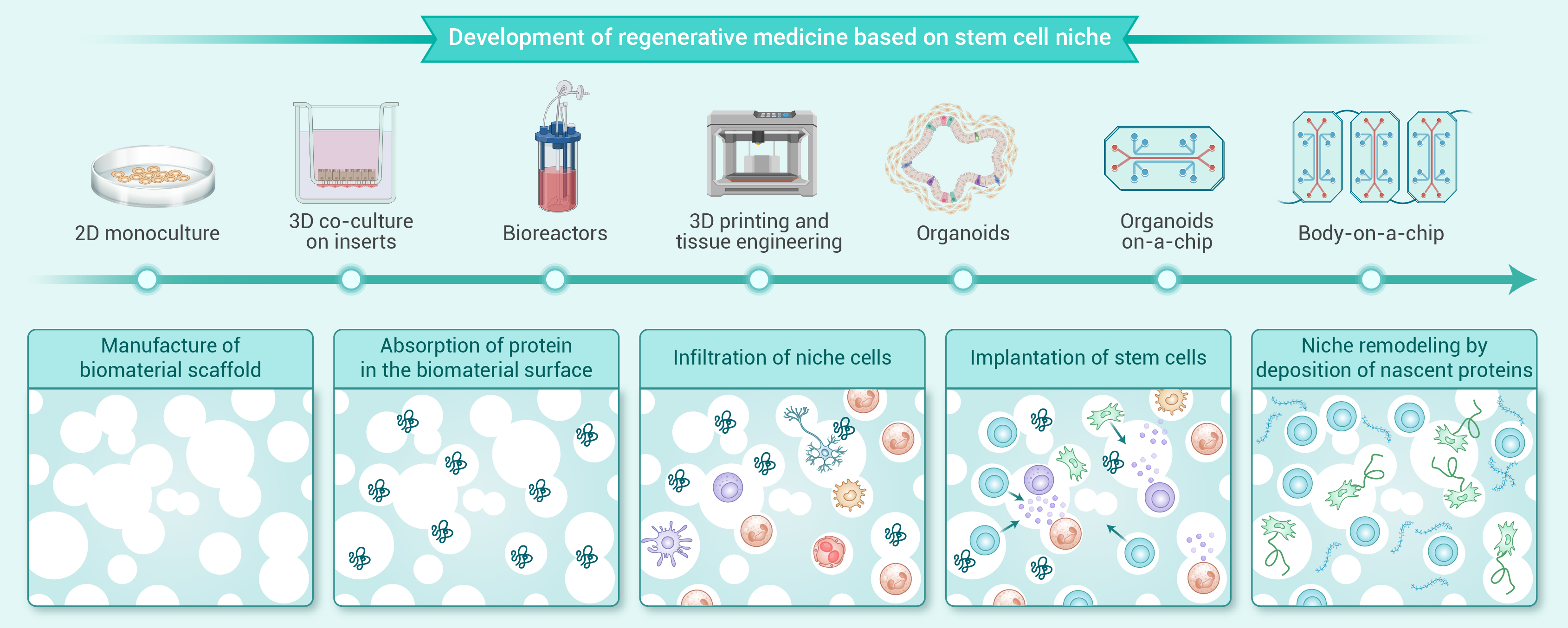
Figure 4.
Development of regenerative medicine based on stem cell niche.
-
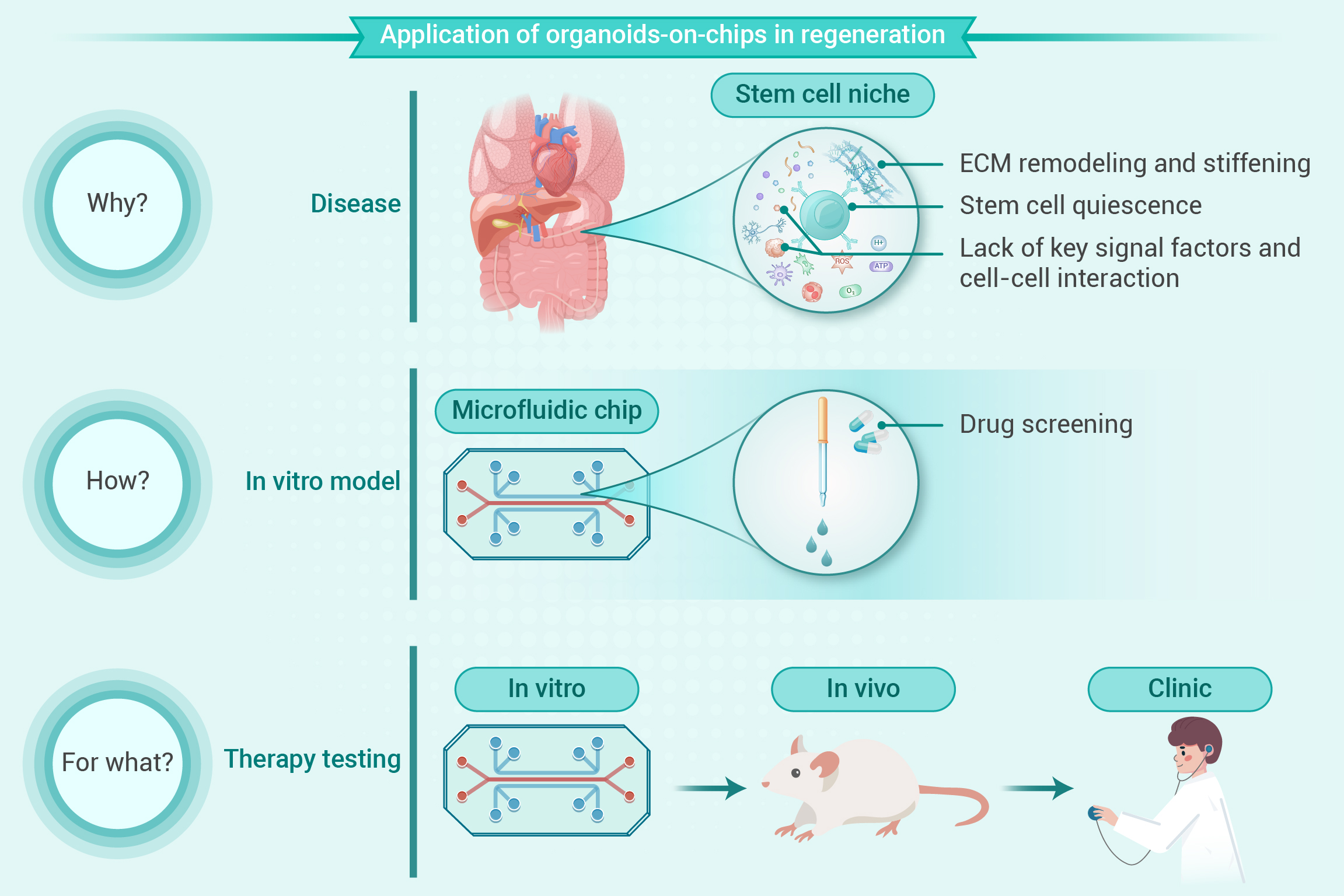
Figure 5.
Application of organoids-on-chips in regeneration.
