
Into the microscale: Low-input sequencing technologies and applications in medicine
-
GRAPHICAL ABSTRACT
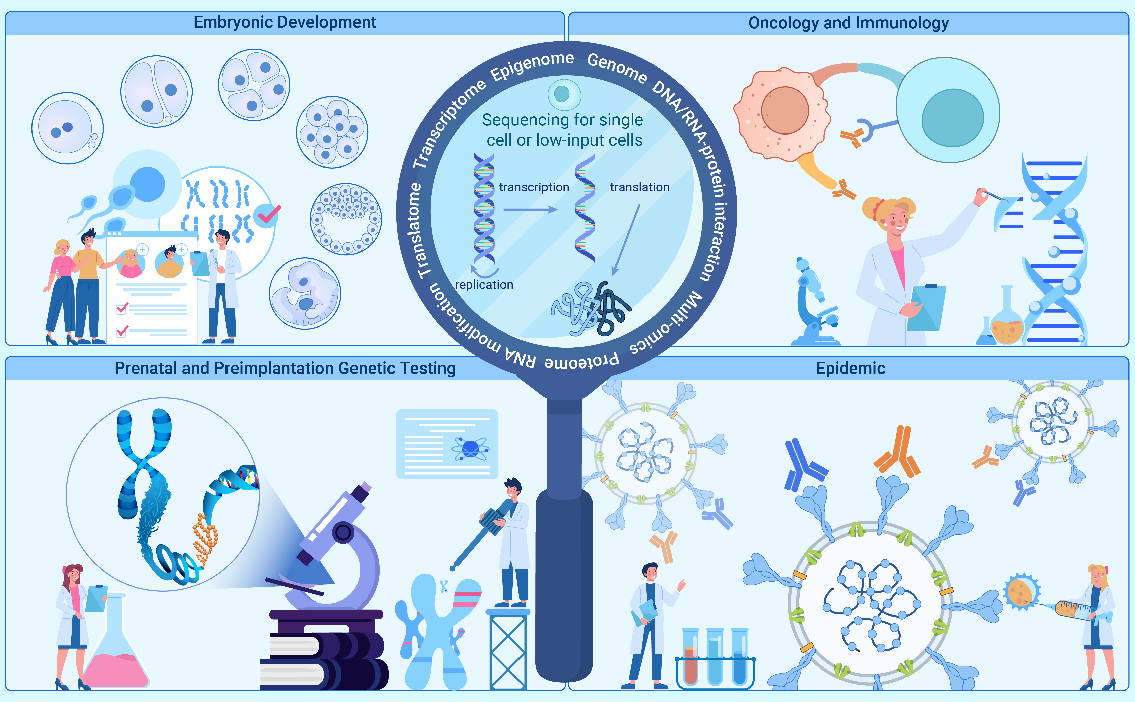
 DownLoad:
Full size image
DownLoad:
Full size image
-
PUBLIC SUMMARY
-
Low-input and single-cell sequencing technologies are revolutionizing biological and medical research.
Low-input and single-cell sequencing technologies investigate biological functions at multiple levels.
Low-input and single-cell sequencing technologies facilitate precision medicine and targeted therapy.
-
-
ABSTRACT
Sequencing technology has undergone major breakthroughs over time and has become indispensable in biological and medical research. Advances in protocols and analysis algorithms allow the analysis of small sample inputs, enabling the characterization of complex networks that govern biological processes in physiology and pathology, driving the development of precision medicine and targeted therapy. In this review, we comprehensively summarize low-input sequencing technologies that include methods for profiling the genome, epigenome, transcriptome, translatome, RNA modification, RNA-RNA interaction, RNA-protein interaction, and multi-omics. The key steps and innovations in different protocols are highlighted, and their advantages, limitations, and scope of application are described. With a focus on the impact of low-input sequencing technologies in biomedical fields, including embryonic development, prenatal and preimplantation genetic testing, oncology and immunology, and pandemic research, we discuss their potential to improve embryo implantation rates, prevent birth defects, develop prospective therapies, and predict prognosis. Lastly, we discuss current limitations and future prospects, providing new insights for medical research. -

-
REFERENCES
[1] Heather, J.M., and Chain, B. (2016). The sequence of sequencers: The history of sequencing DNA. Genomics. 107: 1−8. DOI: 10.1016/j.ygeno.2015.11.003. [2] Goodwin, S., McPherson, J.D., and McCombie, W.R. (2016). Coming of age: Ten years of next-generation sequencing technologies. Nat. Rev. Genet. 17: 333−351. DOI: 10.1038/nrg.2016.49. [3] Preissl, S., Gaulton, K.J., and Ren, B. (2023). Characterizing cis-regulatory elements using single-cell epigenomics. Nat. Rev. Genet. 24: 21−43. DOI: 10.1038/s41576-022-00509-1. [4] Barrett, M.T., Reid, B.J., and Joslyn, G. (1995). Genotypic analysis of multiple loci in somatic cells by whole genome amplification. Nucleic Acids Res. 23: 3488−3492. DOI: 10.1093/nar/23.17.3488. [5] Zhang, L., Cui, X., Schmitt, K., et al. (1992). Whole genome amplification from a single cell: implications for genetic analysis. Proc Natl Acad Sci U S A. 89: 5847−5851. DOI: 10.1073/pnas.89.13.5847. [6] Telenius, H., Carter, N.P., Bebb, C.E., et al. (1992). Degenerate oligonucleotide-primed PCR: general amplification of target DNA by a single degenerate primer. Genomics. 13: 718−725. DOI: 10.1016/0888-7543(92)90147-K. [7] Huang, L., Ma, F., Chapman, A., et al. (2015). Single-Cell Whole-Genome Amplification and Sequencing: Methodology and Applications. Annu Rev Genomics Hum Genet. 16: 79−102. DOI: 10.1146/annurev-genom-090413-025352. [8] Dietmaier, W., Hartmann, A., Wallinger, S., et al. (1999). Multiple mutation analyses in single tumor cells with improved whole genome amplification. Am J Pathol. 154: 83−95. DOI: 10.1016/S0002-9440(10)65254-6. [9] Buchanan, A.V., Risch, G.M., Robichaux, M., et al. (2000). Long DOP-PCR of rare archival anthropological samples. Hum Biol. 72: 911−925. [10] Zheng, S., Ma, X., Buffler, P.A., et al. (2001). Whole genome amplification increases the efficiency and validity of buccal cell genotyping in pediatric populations. Cancer Epidemiol Biomarkers Prev. 10: 697−700. [11] Cheung, V.G., and Nelson, S.F. (1996). Whole genome amplification using a degenerate oligonucleotide primer allows hundreds of genotypes to be performed on less than one nanogram of genomic DNA. Proc Natl Acad Sci U S A. 93: 14676−14679. DOI: 10.1073/pnas.93.25.14676. [12] Paunio, T., Reima, I., and Syvanen, A.C. (1996). Preimplantation diagnosis by whole-genome amplification, PCR amplification, and solid-phase minisequencing of blastomere DNA. Clin Chem. 42: 1382−1390. DOI: 10.1093/clinchem/42.9.1382. [13] Dean, F.B., Hosono, S., Fang, L., et al. (2002). Comprehensive human genome amplification using multiple displacement amplification. Proc Natl Acad Sci U S A. 99: 5261−5266. DOI: 10.1073/pnas.082089499. [14] Barker, D.L., Hansen, M.S., Faruqi, A.F., et al. (2004). Two methods of whole-genome amplification enable accurate genotyping across a 2320-SNP linkage panel. Genome Res. 14: 901−907. DOI: 10.1101/gr.1949704. [15] Lizardi, P.M., Huang, X., Zhu, Z., et al. (1998). Mutation detection and single-molecule counting using isothermal rolling-circle amplification. Nat Genet. 19: 225−232. DOI: 10.1038/898. [16] Dean, F.B., Nelson, J.R., Giesler, T.L., and Lasken, R.S. (2001). Rapid amplification of plasmid and phage DNA using Phi 29 DNA polymerase and multiply-primed rolling circle amplification. Genome Res. 11: 1095−1099. DOI: 10.1101/gr.180501. [17] Lasken, R.S. (2007). Single-cell genomic sequencing using Multiple Displacement Amplification. Curr Opin Microbiol. 10: 510−516. DOI: 10.1016/j.mib.2007.08.005. [18] Zong, C., Lu, S., Chapman, A.R., and Xie, X.S. (2012). Genome-wide detection of single-nucleotide and copy-number variations of a single human cell. Science. 338: 1622−1626. DOI: 10.1126/science.1229164. [19] Chen, C., Xing, D., Tan, L., et al. (2017). Single-cell whole-genome analyses by Linear Amplification via Transposon Insertion (LIANTI). Science. 356: 189−194. DOI: 10.1126/science.aak9787. [20] Yin, Y., Jiang, Y., Lam, K.G., et al. (2019). High-Throughput Single-Cell Sequencing with Linear Amplification. Mol Cell. 76 : 676-690 e610. [21] Xing, D., Tan, L., Chang, C.H., et al. (2021). Accurate SNV detection in single cells by transposon-based whole-genome amplification of complementary strands. Proc Natl Acad Sci U S A. 118 : 10.1073/pnas.2013106118. [22] Booth, M.J., Ost, T.W., Beraldi, D., et al. (2013). Oxidative bisulfite sequencing of 5-methylcytosine and 5-hydroxymethylcytosine. Nat Protoc. 8: 1841−1851. DOI: 10.1038/nprot.2013.115. [23] Smallwood, S.A., Lee, H.J., Angermueller, C., et al. (2014). Single-cell genome-wide bisulfite sequencing for assessing epigenetic heterogeneity. Nat Methods. 11: 817−820. DOI: 10.1038/nmeth.3035. [24] Farlik, M., Sheffield, N.C., Nuzzo, A., et al. (2015). Single-cell DNA methylome sequencing and bioinformatic inference of epigenomic cell-state dynamics. Cell Rep. 10: 1386−1397. DOI: 10.1016/j.celrep.2015.02.001. [25] Olova, N., Krueger, F., Andrews, S., et al. (2018). Comparison of whole-genome bisulfite sequencing library preparation strategies identifies sources of biases affecting DNA methylation data. Genome Biol. 19: 33. DOI: 10.1186/s13059-018-1408-2. [26] Gu, C., Liu, S., Wu, Q., et al. (2019). Integrative single-cell analysis of transcriptome, DNA methylome and chromatin accessibility in mouse oocytes. Cell Res. 29: 110−123. DOI: 10.1038/s41422-018-0125-4. [27] Wu, P., Gao, Y., Guo, W., and Zhu, P. (2019). Using local alignment to enhance single-cell bisulfite sequencing data efficiency. Bioinformatics. 35: 3273−3278. DOI: 10.1093/bioinformatics/btz125. [28] Luo, C., Keown, C.L., Kurihara, L., et al. (2017). Single-cell methylomes identify neuronal subtypes and regulatory elements in mammalian cortex. Science. 357: 600−604. DOI: 10.1126/science.aan3351. [29] Luo, C., Rivkin, A., Zhou, J., et al. (2018). Robust single-cell DNA methylome profiling with snmC-seq2. Nat Commun. 9: 3824. DOI: 10.1038/s41467-018-06355-2. [30] Mulqueen, R.M., Pokholok, D., Norberg, S.J., et al. (2018). Highly scalable generation of DNA methylation profiles in single cells. Nat Biotechnol. 36 : 428-431, 10.1038/nbt.4112. [31] Tanaka, K., and Okamoto, A. (2007). Degradation of DNA by bisulfite treatment. Bioorg Med Chem Lett. 17: 1912−1915. DOI: 10.1016/j.bmcl.2007.01.040. [32] Sakamoto, Y., Zaha, S., Nagasawa, S., et al. (2021). Long-read whole-genome methylation patterning using enzymatic base conversion and nanopore sequencing. Nucleic Acids Res. 49: e81. DOI: 10.1093/nar/gkab397. [33] Liu, Y., Siejka-Zielinska, P., Velikova, G., et al. (2019). Bisulfite-free direct detection of 5-methylcytosine and 5-hydroxymethylcytosine at base resolution. Nat Biotechnol. 37: 424−429. DOI: 10.1038/s41587-019-0041-2. [34] Liu, Y., Cheng, J., Siejka-Zielinska, P., et al. (2020). Accurate targeted long-read DNA methylation and hydroxymethylation sequencing with TAPS. Genome Biol. 21: 54. DOI: 10.1186/s13059-020-01969-6. [35] Guo, H., Zhu, P., Wu, X., et al. (2013). Single-cell methylome landscapes of mouse embryonic stem cells and early embryos analyzed using reduced representation bisulfite sequencing. Genome Res. 23: 2126−2135. DOI: 10.1101/gr.161679.113. [36] Shareef, S.J., Bevill, S.M., Raman, A.T., et al. (2021). Extended-representation bisulfite sequencing of gene regulatory elements in multiplexed samples and single cells. Nat Biotechnol. 39: 1086−1094. DOI: 10.1038/s41587-021-00910-x. [37] Booth, M.J., Branco, M.R., Ficz, G., et al. (2012). Quantitative sequencing of 5-methylcytosine and 5-hydroxymethylcytosine at single-base resolution. Science. 336: 934−937. DOI: 10.1126/science.1220671. [38] Yu, M., Hon, G.C., Szulwach, K.E., et al. (2012). Base-resolution analysis of 5-hydroxymethylcytosine in the mammalian genome. Cell. 149 : 1368-1380, 10.1016/j.cell.2012.04.027. [39] Sun, Z., Terragni, J., Borgaro, J.G., et al. (2013). High-resolution enzymatic mapping of genomic 5-hydroxymethylcytosine in mouse embryonic stem cells. Cell Rep. 3: 567−576. DOI: 10.1016/j.celrep.2013.01.001. [40] Tan, L., Xiong, L., Xu, W., et al. (2013). Genome-wide comparison of DNA hydroxymethylation in mouse embryonic stem cells and neural progenitor cells by a new comparative hMeDIP-seq method. Nucleic Acids Res. 41: e84. DOI: 10.1093/nar/gkt091. [41] Wu, X., Inoue, A., Suzuki, T., and Zhang, Y. (2017). Simultaneous mapping of active DNA demethylation and sister chromatid exchange in single cells. Genes Dev. 31: 511−523. DOI: 10.1101/gad.294843.116. [42] Zhu, C., Gao, Y., Guo, H., et al. (2017). Single-Cell 5-Formylcytosine Landscapes of Mammalian Early Embryos and ESCs at Single-Base Resolution. Cell Stem Cell. 20 : 720-731 e725. [43] Gao, W., Ku, W.L., Pan, L., et al. (2021). Multiplex indexing approach for the detection of DNase I hypersensitive sites in single cells. Nucleic Acids Res. 49: e56. DOI: 10.1093/nar/gkab102. [44] Jin, W., Tang, Q., Wan, M., et al. (2015). Genome-wide detection of DNase I hypersensitive sites in single cells and FFPE tissue samples. Nature. 528: 142−146. DOI: 10.1038/nature15740. [45] Lu, F., Liu, Y., Inoue, A., et al. (2016). Establishing Chromatin Regulatory Landscape during Mouse Preimplantation Development. Cell. 165: 1375−1388. DOI: 10.1016/j.cell.2016.05.050. [46] Chen, X., Miragaia, R.J., Natarajan, K.N., and Teichmann, S.A. (2018). A rapid and robust method for single cell chromatin accessibility profiling. Nat Commun. 9: 5345. DOI: 10.1038/s41467-018-07771-0. [47] Mezger, A., Klemm, S., Mann, I., et al. (2018). High-throughput chromatin accessibility profiling at single-cell resolution. Nat Commun. 9: 3647. DOI: 10.1038/s41467-018-05887-x. [48] Buenrostro, J.D., Wu, B., Litzenburger, U.M., et al. (2015). Single-cell chromatin accessibility reveals principles of regulatory variation. Nature. 523: 486−490. DOI: 10.1038/nature14590. [49] Cusanovich, D.A., Daza, R., Adey, A., et al. (2015). Multiplex single cell profiling of chromatin accessibility by combinatorial cellular indexing. Science. 348: 910−914. DOI: 10.1126/science.aab1601. [50] Meyer, C.A., and Liu, X.S. (2014). Identifying and mitigating bias in next-generation sequencing methods for chromatin biology. Nat Rev Genet. 15: 709−721. [51] Karabacak Calviello, A., Hirsekorn, A., Wurmus, R., et al. (2019). Reproducible inference of transcription factor footprints in ATAC-seq and DNase-seq datasets using protocol-specific bias modeling. Genome Biol. 20: 42. DOI: 10.1186/s13059-019-1654-y. [52] Hu, Y., Jiang, Z., Chen, K., et al. (2023). scNanoATAC-seq: a long-read single-cell ATAC sequencing method to detect chromatin accessibility and genetic variants simultaneously within an individual cell. Cell Res. 33: 83−86. [53] Kelly, T.K., Liu, Y., Lay, F.D., et al. (2012). Genome-wide mapping of nucleosome positioning and DNA methylation within individual DNA molecules. Genome Res. 22: 2497−2506. DOI: 10.1101/gr.143008.112. [54] Pott, S. (2017). Simultaneous measurement of chromatin accessibility, DNA methylation, and nucleosome phasing in single cells. Elife. 6 : 10.7554/eLife.23203. [55] Barski, A., Cuddapah, S., Cui, K., et al. (2007). High-resolution profiling of histone methylations in the human genome. Cell. 129: 823−837. DOI: 10.1016/j.cell.2007.05.009. [56] Johnson, D.S., Mortazavi, A., Myers, R.M., and Wold, B. (2007). Genome-wide mapping of in vivo protein-DNA interactions. Science. 316: 1497−1502. DOI: 10.1126/science.1141319. [57] Park, P.J. (2009). ChIP-seq: advantages and challenges of a maturing technology. Nat Rev Genet. 10: 669−680. [58] Mikkelsen, T.S., Ku, M., Jaffe, D.B., et al. (2007). Genome-wide maps of chromatin state in pluripotent and lineage-committed cells. Nature. 448: 553−560. DOI: 10.1038/nature06008. [59] Shankaranarayanan, P., Mendoza-Parra, M.A., Walia, M., et al. (2011). Single-tube linear DNA amplification (LinDA) for robust ChIP-seq. Nat Methods. 8: 565−567. DOI: 10.1038/nmeth.1626. [60] Cao, Z., Chen, C., He, B., et al. (2015). A microfluidic device for epigenomic profiling using 100 cells. Nat Methods. 12: 959−962. DOI: 10.1038/nmeth.3488. [61] Brind'Amour, J., Liu, S., Hudson, M., et al. (2015). An ultra-low-input native ChIP-seq protocol for genome-wide profiling of rare cell populations. Nat Commun. 6: 6033. DOI: 10.1038/ncomms7033. [62] Rotem, A., Ram, O., Shoresh, N., et al. (2015). Single-cell ChIP-seq reveals cell subpopulations defined by chromatin state. Nat Biotechnol. 33: 1165−1172. DOI: 10.1038/nbt.3383. [63] Ai, S., Xiong, H., Li, C.C., et al. (2019). Profiling chromatin states using single-cell itChIP-seq. Nat Cell Biol. 21: 1164−1172. DOI: 10.1038/s41556-019-0383-5. [64] van Steensel, B., Delrow, J., and Henikoff, S. (2001). Chromatin profiling using targeted DNA adenine methyltransferase. Nat Genet. 27: 304−308. DOI: 10.1038/85871. [65] Zentner, G.E., Kasinathan, S., Xin, B., et al. (2015). ChEC-seq kinetics discriminates transcription factor binding sites by DNA sequence and shape in vivo. Nat Commun. 6: 8733. DOI: 10.1038/ncomms9733. [66] Skene, P.J., and Henikoff, S. (2017). An efficient targeted nuclease strategy for high-resolution mapping of DNA binding sites. Elife. 6 : 10.7554/eLife.21856. [67] Harada, A., Maehara, K., Handa, T., et al. (2019). A chromatin integration labelling method enables epigenomic profiling with lower input. Nat Cell Biol. 21: 287−296. DOI: 10.1038/s41556-018-0248-3. [68] Wang, Q., Xiong, H., Ai, S., et al. (2019). CoBATCH for High-Throughput Single-Cell Epigenomic Profiling. Mol Cell. 76 : 206-216 e207. [69] Carter, B., Ku, W.L., Kang, J.Y., et al. (2019). Mapping histone modifications in low cell number and single cells using antibody-guided chromatin tagmentation (ACT-seq). Nat Commun. 10: 3747. DOI: 10.1038/s41467-019-11559-1. [70] Liu, B., Xu, Q., Wang, Q., et al. (2020). The landscape of RNA Pol II binding reveals a stepwise transition during ZGA. Nature. 587: 139−144. DOI: 10.1038/s41586-020-2847-y. [71] Tang, F., Barbacioru, C., Wang, Y., et al. (2009). mRNA-Seq whole-transcriptome analysis of a single cell. Nat Methods. 6: 377−382. DOI: 10.1038/nmeth.1315. [72] Ziegenhain, C., Vieth, B., Parekh, S., et al. (2017). Comparative Analysis of Single-Cell RNA Sequencing Methods. Mol Cell. 65 : 631-643 e634. [73] Mereu, E., Lafzi, A., Moutinho, C., et al. (2020). Benchmarking single-cell RNA-sequencing protocols for cell atlas projects. Nat Biotechnol. 38: 747−755. DOI: 10.1038/s41587-020-0469-4. [74] Luecken, M.D., and Theis, F.J. (2019). Current best practices in single-cell RNA-seq analysis: a tutorial. Mol Syst Biol. 15: e8746. DOI: 10.15252/msb.20188746. [75] Bolger, A.M., Lohse, M., and Usadel, B. (2014). Trimmomatic: a flexible trimmer for Illumina sequence data. Bioinformatics. 30: 2114−2120. DOI: 10.1093/bioinformatics/btu170. [76] Martin, M. (2011). Cutadapt removes adapter sequences from high-throughput sequencing reads. EMBnet.journal. 17: 10−12. [77] Dobin, A., Davis, C.A., Schlesinger, F., et al. (2013). STAR: ultrafast universal RNA-seq aligner. Bioinformatics. 29: 15−21. DOI: 10.1093/bioinformatics/bts635. [78] Kim, D., Paggi, J.M., Park, C., et al. (2019). Graph-based genome alignment and genotyping with HISAT2 and HISAT-genotype. Nat Biotechnol. 37: 907−915. DOI: 10.1038/s41587-019-0201-4. [79] Zheng, G.X., Terry, J.M., Belgrader, P., et al. (2017). Massively parallel digital transcriptional profiling of single cells. Nat Commun. 8: 14049. DOI: 10.1038/ncomms14049. [80] Butler, A., Hoffman, P., Smibert, P., et al. (2018). Integrating single-cell transcriptomic data across different conditions, technologies, and species. Nat Biotechnol. 36: 411−420. DOI: 10.1038/nbt.4096. [81] McCarthy, D.J., Campbell, K.R., Lun, A.T., and Wills, Q.F. (2017). Scater: pre-processing, quality control, normalization and visualization of single-cell RNA-seq data in R. Bioinformatics. 33: 1179−1186. DOI: 10.1093/bioinformatics/btw777. [82] Wolf, F.A., Angerer, P., and Theis, F.J. (2018). SCANPY: large-scale single-cell gene expression data analysis. Genome Biol. 19: 15. DOI: 10.1186/s13059-017-1382-0. [83] Love, M.I., Huber, W., and Anders, S. (2014). Moderated estimation of fold change and dispersion for RNA-seq data with DESeq2. Genome Biol. 15: 550. DOI: 10.1186/s13059-014-0550-8. [84] Robinson, M.D., McCarthy, D.J., and Smyth, G.K. (2010). edgeR: a Bioconductor package for differential expression analysis of digital gene expression data. Bioinformatics. 26: 139−140. DOI: 10.1093/bioinformatics/btp616. [85] Ritchie, M.E., Phipson, B., Wu, D., et al. (2015). limma powers differential expression analyses for RNA-sequencing and microarray studies. Nucleic Acids Res. 43: e47. DOI: 10.1093/nar/gkv007. [86] Cao, J., Spielmann, M., Qiu, X., et al. (2019). The single-cell transcriptional landscape of mammalian organogenesis. Nature. 566: 496−502. DOI: 10.1038/s41586-019-0969-x. [87] Wolf, F.A., Hamey, F.K., Plass, M., et al. (2019). PAGA: graph abstraction reconciles clustering with trajectory inference through a topology preserving map of single cells. Genome Biol. 20: 59. DOI: 10.1186/s13059-019-1663-x. [88] Efremova, M., Vento-Tormo, M., Teichmann, S.A., and Vento-Tormo, R. (2020). CellPhoneDB: inferring cell-cell communication from combined expression of multi-subunit ligand-receptor complexes. Nat Protoc. 15: 1484−1506. DOI: 10.1038/s41596-020-0292-x. [89] Ren, X., Zhong, G., Zhang, Q., et al. (2020). Reconstruction of cell spatial organization from single-cell RNA sequencing data based on ligand-receptor mediated self-assembly. Cell Res. 30: 763−778. DOI: 10.1038/s41422-020-0353-2. [90] Jin, S., Guerrero-Juarez, C.F., Zhang, L., et al. (2021). Inference and analysis of cell-cell communication using CellChat. Nat Commun. 12: 1088. DOI: 10.1038/s41467-021-21246-9. [91] Fan, X., Tang, D., Liao, Y., et al. (2020). Single-cell RNA-seq analysis of mouse preimplantation embryos by third-generation sequencing. PLoS Biol. 18: e3001017. DOI: 10.1371/journal.pbio.3001017. [92] Liao, Y., Liu, Z., Zhang, Y., et al. (2023). High-throughput and high-sensitivity full-length single-cell RNA-seq analysis on third-generation sequencing platform. Cell Discov. 9: 5. [93] Volden, R., Palmer, T., Byrne, A., et al. (2018). Improving nanopore read accuracy with the R2C2 method enables the sequencing of highly multiplexed full-length single-cell cDNA. Proc Natl Acad Sci U S A. 115: 9726−9731. DOI: 10.1073/pnas.1806447115. [94] Gupta, I., Collier, P.G., Haase, B., et al. (2018). Single-cell isoform RNA sequencing characterizes isoforms in thousands of cerebellar cells. Nat Biotechnol 10.1038/nbt.4259. [95] Lebrigand, K., Magnone, V., Barbry, P., and Waldmann, R. (2020). High throughput error corrected Nanopore single cell transcriptome sequencing. Nat Commun. 11: 4025. DOI: 10.1038/s41467-020-17800-6. [96] Singh, M., Al-Eryani, G., Carswell, S., et al. (2019). High-throughput targeted long-read single cell sequencing reveals the clonal and transcriptional landscape of lymphocytes. Nat Commun. 10: 3120. DOI: 10.1038/s41467-019-11049-4. [97] Liu, Y., Nie, H., Liu, H., and Lu, F. (2019). Poly(A) inclusive RNA isoform sequencing (PAIso-seq) reveals wide-spread non-adenosine residues within RNA poly(A) tails. Nat Commun. 10: 5292. DOI: 10.1038/s41467-019-13228-9. [98] Shi, Z.X., Chen, Z.C., Zhong, J.Y., et al. (2023). High-throughput and high-accuracy single-cell RNA isoform analysis using PacBio circular consensus sequencing. Nat Commun. 14: 2631. DOI: 10.1038/s41467-023-38324-9. [99] Zheng, Y.-F., Chen, Z.-C., Shi, Z.-X., et al. (2020). HIT-scISOseq: High-throughput and High-accuracy Single-cell Full-length Isoform Sequencing for Corneal Epithelium. bioRxiv 2020.2007.2027.222349, 10.1101/2020.07.27.222349. [100] Al'Khafaji, A.M., Smith, J.T., Garimella, K.V., et al. (2023). High-throughput RNA isoform sequencing using programmed cDNA concatenation. Nat Biotechnol 10.1038/s41587-023-01815-7. [101] Legnini, I., Alles, J., Karaiskos, N., et al. (2019). FLAM-seq: full-length mRNA sequencing reveals principles of poly(A) tail length control. Nat Methods. 16: 879−886. DOI: 10.1038/s41592-019-0503-y. [102] Liu, D., Chen, Y., Ren, Y., et al. (2022). Primary specification of blastocyst trophectoderm by scRNA-seq: New insights into embryo implantation. Sci Adv. 8: eabj3725. DOI: 10.1126/sciadv.abj3725. [103] Liu, Y., Zhao, H., Shao, F., et al. (2023). Remodeling of maternal mRNA through poly(A) tail orchestrates human oocyte-to-embryo transition. Nat Struct Mol Biol. 30: 200−215. DOI: 10.1038/s41594-022-00908-2. [104] Williams, C.G., Lee, H.J., Asatsuma, T., et al. (2022). An introduction to spatial transcriptomics for biomedical research. Genome Med. 14: 68. DOI: 10.1186/s13073-022-01075-1. [105] Lubeck, E., Coskun, A.F., Zhiyentayev, T., et al. (2014). Single-cell in situ RNA profiling by sequential hybridization. Nat Methods. 11: 360−361. DOI: 10.1038/nmeth.2892. [106] Chen, K.H., Boettiger, A.N., Moffitt, J.R., et al. (2015). RNA imaging. Spatially resolved, highly multiplexed RNA profiling in single cells. Science. 348: aaa6090. [107] Wang, X., Allen, W.E., Wright, M.A., et al. (2018). Three-dimensional intact-tissue sequencing of single-cell transcriptional states. Science. 361 : 10.1126/science.aat5691. [108] Rodriques, S.G., Stickels, R.R., Goeva, A., et al. (2019). Slide-seq: A scalable technology for measuring genome-wide expression at high spatial resolution. Science. 363: 1463−1467. DOI: 10.1126/science.aaw1219. [109] Stickels, R.R., Murray, E., Kumar, P., et al. (2021). Highly sensitive spatial transcriptomics at near-cellular resolution with Slide-seqV2. Nat Biotechnol. 39: 313−319. DOI: 10.1038/s41587-020-0739-1. [110] Vickovic, S., Eraslan, G., Salmen, F., et al. (2019). High-definition spatial transcriptomics for in situ tissue profiling. Nat Methods. 16: 987−990. DOI: 10.1038/s41592-019-0548-y. [111] Cho, C.S., Xi, J., Si, Y., et al. (2021). Microscopic examination of spatial transcriptome using Seq-Scope. Cell. 184 : 3559-3572 e3522. [112] Xia, K., Sun, H.-X., Li, J., et al. (2021). Single-cell Stereo-seq enables cell type-specific spatial transcriptome characterization in <em>Arabidopsis</em> leaves. bioRxiv 2021.2010.2020.465066, 10.1101/2021.10.20.465066. [113] Fu, X., Sun, L., Chen, J.Y., et al. (2021). Continuous Polony Gels for Tissue Mapping with High Resolution and RNA Capture Efficiency. bioRxiv 2021.2003.2017.435795, 10.1101/2021.03.17.435795. [114] Liu, Y., Yang, M., Deng, Y., et al. (2020). High-Spatial-Resolution Multi-Omics Sequencing via Deterministic Barcoding in Tissue. Cell. 183 : 1665-1681 e1618. [115] Zhao, J., Qin, B., Nikolay, R., et al. (2019). Translatomics: The Global View of Translation. Int J Mol Sci. 20 : 10.3390/ijms20010212. [116] VanInsberghe, M., van den Berg, J., Andersson-Rolf, A., et al. (2021). Single-cell Ribo-seq reveals cell cycle-dependent translational pausing. Nature. 597: 561−565. DOI: 10.1038/s41586-021-03887-4. [117] Xiong, Z., Xu, K., Lin, Z., et al. (2022). Ultrasensitive Ribo-seq reveals translational landscapes during mammalian oocyte-to-embryo transition and pre-implantation development. Nat Cell Biol. 24: 968−980. DOI: 10.1038/s41556-022-00928-6. [118] Zhang, C., Wang, M., Li, Y., and Zhang, Y. (2022). Profiling and functional characterization of maternal mRNA translation during mouse maternal-to-zygotic transition. Sci Adv. 8: eabj3967. DOI: 10.1126/sciadv.abj3967. [119] Hu, W., Zeng, H., Shi, Y., et al. (2022). Single-cell transcriptome and translatome dual-omics reveals potential mechanisms of human oocyte maturation. Nat Commun. 13: 5114. DOI: 10.1038/s41467-022-32791-2. [120] Dominissini, D., Moshitch-Moshkovitz, S., Schwartz, S., et al. (2012). Topology of the human and mouse m6A RNA methylomes revealed by m6A-seq. Nature. 485: 201−206. DOI: 10.1038/nature11112. [121] Wu, Y., Xu, X., Qi, M., et al. (2022). N(6)-methyladenosine regulates maternal RNA maintenance in oocytes and timely RNA decay during mouse maternal-to-zygotic transition. Nat Cell Biol. 24: 917−927. DOI: 10.1038/s41556-022-00915-x. [122] Chen, K., Luo, G.Z., and He, C. (2015). High-Resolution Mapping of N(6)-Methyladenosine in Transcriptome and Genome Using a Photo-Crosslinking-Assisted Strategy. Methods Enzymol. 560: 161−185. [123] Linder, B., Grozhik, A.V., Olarerin-George, A.O., et al. (2015). Single-nucleotide-resolution mapping of m6A and m6Am throughout the transcriptome. Nat Methods. 12: 767−772. DOI: 10.1038/nmeth.3453. [124] Molinie, B., Wang, J., Lim, K.S., et al. (2016). m(6)A-LAIC-seq reveals the census and complexity of the m(6)A epitranscriptome. Nat Methods. 13: 692−698. DOI: 10.1038/nmeth.3898. [125] Zhang, Z., Chen, L.Q., Zhao, Y.L., et al. (2019). Single-base mapping of m(6)A by an antibody-independent method. Sci Adv. 5: eaax0250. DOI: 10.1126/sciadv.aax0250. [126] Garcia-Campos, M.A., Edelheit, S., Toth, U., et al. (2019). Deciphering the "m(6)A Code" via Antibody-Independent Quantitative Profiling. Cell. 178 : 731-747 e716. [127] Meyer, K.D. (2019). DART-seq: an antibody-free method for global m(6)A detection. Nat Methods. 16: 1275−1280. DOI: 10.1038/s41592-019-0570-0. [128] Wang, Y., Xiao, Y., Dong, S., et al. (2020). Antibody-free enzyme-assisted chemical approach for detection of N(6)-methyladenosine. Nat Chem Biol. 16: 896−903. DOI: 10.1038/s41589-020-0525-x. [129] Shu, X., Cao, J., Cheng, M., et al. (2020). A metabolic labeling method detects m(6)A transcriptome-wide at single base resolution. Nat Chem Biol. 16: 887−895. DOI: 10.1038/s41589-020-0526-9. [130] Hu, L., Liu, S., Peng, Y., et al. (2022). m(6)A RNA modifications are measured at single-base resolution across the mammalian transcriptome. Nat Biotechnol. 40: 1210−1219. DOI: 10.1038/s41587-022-01243-z. [131] Xia, Q., Li, S., Ding, T., et al. (2023). Nanopore sequencing for detecting reciprocal translocation carrier status in preimplantation genetic testing. BMC Genomics. 24: 1. [132] Xiao, Y.L., Liu, S., Ge, R., et al. (2023). Transcriptome-wide profiling and quantification of N(6)-methyladenosine by enzyme-assisted adenosine deamination. Nat Biotechnol 10.1038/s41587-022-01587-6. [133] Liu, C., Sun, H., Yi, Y., et al. (2023). Absolute quantification of single-base m(6)A methylation in the mammalian transcriptome using GLORI. Nat Biotechnol. 41: 355−366. DOI: 10.1038/s41587-022-01487-9. [134] Davis, F.F., and Allen, F.W. (1957). Ribonucleic acids from yeast which contain a fifth nucleotide. J Biol Chem. 227: 907−915. DOI: 10.1016/S0021-9258(18)70770-9. [135] Carlile, T.M., Rojas-Duran, M.F., Zinshteyn, B., et al. (2014). Pseudouridine profiling reveals regulated mRNA pseudouridylation in yeast and human cells. Nature. 515: 143−146. DOI: 10.1038/nature13802. [136] Lovejoy, A.F., Riordan, D.P., and Brown, P.O. (2014). Transcriptome-wide mapping of pseudouridines: pseudouridine synthases modify specific mRNAs in S. cerevisiae. PLoS One. 9: e110799. DOI: 10.1371/journal.pone.0110799. [137] Schwartz, S., Bernstein, D.A., Mumbach, M.R., et al. (2014). Transcriptome-wide mapping reveals widespread dynamic-regulated pseudouridylation of ncRNA and mRNA. Cell. 159: 148−162. DOI: 10.1016/j.cell.2014.08.028. [138] Bakin, A., and Ofengand, J. (1993). Four newly located pseudouridylate residues in Escherichia coli 23S ribosomal RNA are all at the peptidyltransferase center: analysis by the application of a new sequencing technique. Biochemistry. 32: 9754−9762. DOI: 10.1021/bi00088a030. [139] Li, X., Zhu, P., Ma, S., et al. (2015). Chemical pulldown reveals dynamic pseudouridylation of the mammalian transcriptome. Nat Chem Biol. 11: 592−597. DOI: 10.1038/nchembio.1836. [140] Khoddami, V., Yerra, A., Mosbruger, T.L., et al. (2019). Transcriptome-wide profiling of multiple RNA modifications simultaneously at single-base resolution. Proc Natl Acad Sci U S A. 116: 6784−6789. DOI: 10.1073/pnas.1817334116. [141] Fleming, A.M., Alenko, A., Kitt, J.P., et al. (2019). Structural Elucidation of Bisulfite Adducts to Pseudouridine That Result in Deletion Signatures during Reverse Transcription of RNA. J Am Chem Soc. 141: 16450−16460. DOI: 10.1021/jacs.9b08630. [142] Dai, Q., Zhang, L.S., Sun, H.L., et al. (2023). Quantitative sequencing using BID-seq uncovers abundant pseudouridines in mammalian mRNA at base resolution. Nat Biotechnol. 41: 344−354. DOI: 10.1038/s41587-022-01505-w. [143] Torres, A.G., Batlle, E., and Ribas de Pouplana, L. (2014). Role of tRNA modifications in human diseases. Trends Mol Med. 20: 306−314. DOI: 10.1016/j.molmed.2014.01.008. [144] Zheng, G., Qin, Y., Clark, W.C., et al. (2015). Efficient and quantitative high-throughput tRNA sequencing. Nat Methods. 12: 835−837. DOI: 10.1038/nmeth.3478. [145] Cozen, A.E., Quartley, E., Holmes, A.D., et al. (2015). ARM-seq: AlkB-facilitated RNA methylation sequencing reveals a complex landscape of modified tRNA fragments. Nat Methods. 12: 879−884. DOI: 10.1038/nmeth.3508. [146] Lucas, M.C., Pryszcz, L.P., Medina, R., et al. (2023). Quantitative analysis of tRNA abundance and modifications by nanopore RNA sequencing. Nat Biotechnol 10.1038/s41587-023-01743-6. [147] Butcher, S.E., and Pyle, A.M. (2011). The molecular interactions that stabilize RNA tertiary structure: RNA motifs, patterns, and networks. Acc Chem Res. 44: 1302−1311. DOI: 10.1021/ar200098t. [148] Helwak, A., Kudla, G., Dudnakova, T., and Tollervey, D. (2013). Mapping the human miRNA interactome by CLASH reveals frequent noncanonical binding. Cell. 153: 654−665. DOI: 10.1016/j.cell.2013.03.043. [149] Nguyen, T.C., Cao, X., Yu, P., et al. (2016). Mapping RNA-RNA interactome and RNA structure in vivo by MARIO. Nat Commun. 7: 12023. DOI: 10.1038/ncomms12023. [150] Lu, Z., Zhang, Q.C., Lee, B., et al. (2016). RNA Duplex Map in Living Cells Reveals Higher-Order Transcriptome Structure. Cell. 165: 1267−1279. DOI: 10.1016/j.cell.2016.04.028. [151] Aw, J.G., Shen, Y., Wilm, A., et al. (2016). In Vivo Mapping of Eukaryotic RNA Interactomes Reveals Principles of Higher-Order Organization and Regulation. Mol Cell. 62: 603−617. DOI: 10.1016/j.molcel.2016.04.028. [152] Sharma, E., Sterne-Weiler, T., O'Hanlon, D., and Blencowe, B.J. (2016). Global Mapping of Human RNA-RNA Interactions. Mol Cell. 62: 618−626. DOI: 10.1016/j.molcel.2016.04.030. [153] Cai, Z., Cao, C., Ji, L., et al. (2020). RIC-seq for global in situ profiling of RNA-RNA spatial interactions. Nature. 582: 432−437. DOI: 10.1038/s41586-020-2249-1. [154] Cao, C., Cai, Z., Ye, R., et al. (2021). Global in situ profiling of RNA-RNA spatial interactions with RIC-seq. Nat Protoc. 16: 2916−2946. DOI: 10.1038/s41596-021-00524-2. [155] Tang, L. (2020). Mapping RNA–RNA interactions. Nature Methods. 17: 760−760. DOI: 10.1038/s41592-020-0922-9. [156] Re, A., Joshi, T., Kulberkyte, E., et al. (2014). RNA-protein interactions: an overview. Methods Mol Biol. 1097: 491−521. [157] Zhao, J., Ohsumi, T.K., Kung, J.T., et al. (2010). Genome-wide identification of polycomb-associated RNAs by RIP-seq. Mol Cell. 40: 939−953. DOI: 10.1016/j.molcel.2010.12.011. [158] Ule, J., Jensen, K., Mele, A., and Darnell, R.B. (2005). CLIP: a method for identifying protein-RNA interaction sites in living cells. Methods. 37: 376−386. DOI: 10.1016/j.ymeth.2005.07.018. [159] Licatalosi, D.D., Mele, A., Fak, J.J., et al. (2008). HITS-CLIP yields genome-wide insights into brain alternative RNA processing. Nature. 456: 464−469. DOI: 10.1038/nature07488. [160] Hafner, M., Landthaler, M., Burger, L., et al. (2010). Transcriptome-wide identification of RNA-binding protein and microRNA target sites by PAR-CLIP. Cell. 141: 129−141. DOI: 10.1016/j.cell.2010.03.009. [161] Konig, J., Zarnack, K., Rot, G., et al. (2010). iCLIP reveals the function of hnRNP particles in splicing at individual nucleotide resolution. Nat Struct Mol Biol. 17: 909−915. DOI: 10.1038/nsmb.1838. [162] Van Nostrand, E.L., Pratt, G.A., Shishkin, A.A., et al. (2016). Robust transcriptome-wide discovery of RNA-binding protein binding sites with enhanced CLIP (eCLIP). Nat Methods. 13: 508−514. DOI: 10.1038/nmeth.3810. [163] Zarnegar, B.J., Flynn, R.A., Shen, Y., et al. (2016). irCLIP platform for efficient characterization of protein-RNA interactions. Nat Methods. 13: 489−492. DOI: 10.1038/nmeth.3840. [164] McMahon, A.C., Rahman, R., Jin, H., et al. (2016). TRIBE: Hijacking an RNA-Editing Enzyme to Identify Cell-Specific Targets of RNA-Binding Proteins. Cell. 165: 742−753. DOI: 10.1016/j.cell.2016.03.007. [165] Su, R., Fan, L.H., Cao, C., et al. (2021). Global profiling of RNA-binding protein target sites by LACE-seq. Nat Cell Biol. 23: 664−675. DOI: 10.1038/s41556-021-00696-9. [166] Levy, E., and Slavov, N. (2018). Single cell protein analysis for systems biology. Essays Biochem. 62: 595−605. DOI: 10.1042/EBC20180014. [167] Gebreyesus, S.T., Siyal, A.A., Kitata, R.B., et al. (2022). Streamlined single-cell proteomics by an integrated microfluidic chip and data-independent acquisition mass spectrometry. Nat Commun. 13: 37. [168] Zhang, L., and Vertes, A. (2018). Single-Cell Mass Spectrometry Approaches to Explore Cellular Heterogeneity. Angew Chem Int Ed Engl. 57: 4466−4477. DOI: 10.1002/anie.201709719. [169] Comi, T.J., Do, T.D., Rubakhin, S.S., and Sweedler, J.V. (2017). Categorizing Cells on the Basis of their Chemical Profiles: Progress in Single-Cell Mass Spectrometry. J Am Chem Soc. 139: 3920−3929. DOI: 10.1021/jacs.6b12822. [170] Labib, M., and Kelley, S.O. (2020). Single-cell analysis targeting the proteome. Nat Rev Chem. 4: 143−158. DOI: 10.1038/s41570-020-0162-7. [171] Lombard-Banek, C., Moody, S.A., and Nemes, P. (2016). Single-Cell Mass Spectrometry for Discovery Proteomics: Quantifying Translational Cell Heterogeneity in the 16-Cell Frog (Xenopus) Embryo. Angew Chem Int Ed Engl. 55: 2454−2458. DOI: 10.1002/anie.201510411. [172] Bendall, S.C., Simonds, E.F., Qiu, P., et al. (2011). Single-cell mass cytometry of differential immune and drug responses across a human hematopoietic continuum. Science. 332: 687−696. DOI: 10.1126/science.1198704. [173] Lombard-Banek, C., Moody, S.A., Manzini, M.C., and Nemes, P. (2019). Microsampling Capillary Electrophoresis Mass Spectrometry Enables Single-Cell Proteomics in Complex Tissues: Developing Cell Clones in Live Xenopus laevis and Zebrafish Embryos. Anal Chem. 91: 4797−4805. DOI: 10.1021/acs.analchem.9b00345. [174] Slavov, N. (2021). Single-cell protein analysis by mass spectrometry. Curr Opin Chem Biol. 60: 1−9. DOI: 10.1016/j.cbpa.2020.04.018. [175] Chen, Q., Yan, G., Gao, M., and Zhang, X. (2015). Ultrasensitive Proteome Profiling for 100 Living Cells by Direct Cell Injection, Online Digestion and Nano-LC-MS/MS Analysis. Anal Chem. 87: 6674−6680. DOI: 10.1021/acs.analchem.5b00808. [176] Chen, W., Wang, S., Adhikari, S., et al. (2016). Simple and Integrated Spintip-Based Technology Applied for Deep Proteome Profiling. Anal Chem. 88: 4864−4871. DOI: 10.1021/acs.analchem.6b00631. [177] Zhu, Y., Piehowski, P.D., Zhao, R., et al. (2018). Nanodroplet processing platform for deep and quantitative proteome profiling of 10-100 mammalian cells. Nat Commun. 9: 882. DOI: 10.1038/s41467-018-03367-w. [178] Zhu, Y., Clair, G., Chrisler, W.B., et al. (2018). Proteomic Analysis of Single Mammalian Cells Enabled by Microfluidic Nanodroplet Sample Preparation and Ultrasensitive NanoLC-MS. Angew Chem Int Ed Engl. 57: 12370−12374. DOI: 10.1002/anie.201802843. [179] Budnik, B., Levy, E., Harmange, G., and Slavov, N. (2018). SCoPE-MS: mass spectrometry of single mammalian cells quantifies proteome heterogeneity during cell differentiation. Genome Biol. 19: 161. DOI: 10.1186/s13059-018-1547-5. [180] Specht, H., Emmott, E., Petelski, A.A., et al. (2021). Single-cell proteomic and transcriptomic analysis of macrophage heterogeneity using SCoPE2. Genome Biol. 22: 50. DOI: 10.1186/s13059-021-02267-5. [181] Mertins, P., Udeshi, N.D., Clauser, K.R., et al. (2012). iTRAQ labeling is superior to mTRAQ for quantitative global proteomics and phosphoproteomics. Mol Cell Proteomics 11 : M111 014423. [182] Treitz, C., Cassidy, L., Hockendorf, A., et al. (2015). Quantitative proteome analysis of Caenorhabditis elegans upon exposure to nematicidal Bacillus thuringiensis. J Proteomics. 113: 337−350. DOI: 10.1016/j.jprot.2014.09.027. [183] Virreira Winter, S., Meier, F., Wichmann, C., et al. (2018). EASI-tag enables accurate multiplexed and interference-free MS2-based proteome quantification. Nat Methods. 15: 527−530. DOI: 10.1038/s41592-018-0037-8. [184] Mnatsakanyan, R., Shema, G., Basik, M., et al. (2018). Detecting post-translational modification signatures as potential biomarkers in clinical mass spectrometry. Expert Rev Proteomics. 15: 515−535. DOI: 10.1080/14789450.2018.1483340. [185] Dey, S.S., Kester, L., Spanjaard, B., et al. (2015). Integrated genome and transcriptome sequencing of the same cell. Nature Biotechnology. 33: 285−289. DOI: 10.1038/nbt.3129. [186] Macaulay, I.C., Haerty, W., Kumar, P., et al. (2015). G&T-seq: parallel sequencing of single-cell genomes and transcriptomes. Nature Methods. 12: 519−522. DOI: 10.1038/nmeth.3370. [187] Zhu, C., Preissl, S., and Ren, B. (2020). Single-cell multimodal omics: the power of many. Nat Methods. 17: 11−14. DOI: 10.1038/s41592-019-0691-5. [188] Angermueller, C., Clark, S.J., Lee, H.J., et al. (2016). Parallel single-cell sequencing links transcriptional and epigenetic heterogeneity. Nat Methods. 13: 229−232. DOI: 10.1038/nmeth.3728. [189] Hu, Y., Huang, K., An, Q., et al. (2016). Simultaneous profiling of transcriptome and DNA methylome from a single cell. Genome Biol. 17: 88. DOI: 10.1186/s13059-016-0950-z. [190] Zhu, C., Yu, M., Huang, H., et al. (2019). An ultra high-throughput method for single-cell joint analysis of open chromatin and transcriptome. Nature Structural & Molecular Biology. 26: 1063−1070. [191] Hou, Y., Guo, H., Cao, C., et al. (2016). Single-cell triple omics sequencing reveals genetic, epigenetic, and transcriptomic heterogeneity in hepatocellular carcinomas. Cell Res. 26: 304−319. DOI: 10.1038/cr.2016.23. [192] Clark, S.J., Argelaguet, R., Kapourani, C.A., et al. (2018). scNMT-seq enables joint profiling of chromatin accessibility DNA methylation and transcription in single cells. Nat Commun. 9: 781. DOI: 10.1038/s41467-018-03149-4. [193] Wang, Y., Yuan, P., Yan, Z., et al. (2021). Single-cell multiomics sequencing reveals the functional regulatory landscape of early embryos. Nature Communications. 12: 1247. DOI: 10.1038/s41467-021-21409-8. [194] Li, G., Liu, Y., Zhang, Y., et al. (2019). Joint profiling of DNA methylation and chromatin architecture in single cells. Nat Methods. 16: 991−993. DOI: 10.1038/s41592-019-0502-z. [195] Cao, J., Cusanovich, D.A., Ramani, V., et al. (2018). Joint profiling of chromatin accessibility and gene expression in thousands of single cells. Science. 361: 1380−1385. DOI: 10.1126/science.aau0730. [196] Chen, S., Lake, B.B., and Zhang, K. (2019). High-throughput sequencing of the transcriptome and chromatin accessibility in the same cell. Nat Biotechnol. 37: 1452−1457. DOI: 10.1038/s41587-019-0290-0. [197] Ma, S., Zhang, B., LaFave, L.M., et al. (2020). Chromatin Potential Identified by Shared Single-Cell Profiling of RNA and Chromatin. Cell. 183 : 1103-1116 e1120. [198] Yan, L., Yang, M., Guo, H., et al. (2013). Single-cell RNA-Seq profiling of human preimplantation embryos and embryonic stem cells. Nat Struct Mol Biol. 20: 1131−1139. DOI: 10.1038/nsmb.2660. [199] Zou, Z., Zhang, C., Wang, Q., et al. (2022). Translatome and transcriptome co-profiling reveals a role of TPRXs in human zygotic genome activation. Science. 378: abo7923. DOI: 10.1126/science.abo7923. [200] Stirparo, G.G., Boroviak, T., Guo, G., et al. (2018). Integrated analysis of single-cell embryo data yields a unified transcriptome signature for the human pre-implantation epiblast. Development. 145 : 10.1242/dev.158501. [201] Meistermann, D., Bruneau, A., Loubersac, S., et al. (2021). Integrated pseudotime analysis of human pre-implantation embryo single-cell transcriptomes reveals the dynamics of lineage specification. Cell Stem Cell. 28 : 1625-1640 e1626. [202] Zhu, P., Guo, H., Ren, Y., et al. (2018). Single-cell DNA methylome sequencing of human preimplantation embryos. Nature genetics. 50: 12−19. DOI: 10.1038/s41588-017-0007-6. [203] Li, L., Guo, F., Gao, Y., et al. (2018). Single-cell multi-omics sequencing of human early embryos. Nat Cell Biol. 20: 847−858. DOI: 10.1038/s41556-018-0123-2. [204] Xia, W., Xu, J., Yu, G., et al. (2019). Resetting histone modifications during human parental-to-zygotic transition. Science. 365: 353−360. DOI: 10.1126/science.aaw5118. [205] Yu, H., Chen, M., Hu, Y., et al. (2022). Dynamic reprogramming of H3K9me3 at hominoid-specific retrotransposons during human preimplantation development. Cell Stem Cell. 29 : 1031-1050 e1012. [206] Xu, R., Li, S., Wu, Q., et al. (2022). Stage-specific H3K9me3 occupancy ensures retrotransposon silencing in human pre-implantation embryos. Cell Stem Cell. 29 : 1051-1066 e1058. [207] Xue, Z., Huang, K., Cai, C., et al. (2013). Genetic programs in human and mouse early embryos revealed by single-cell RNA sequencing. Nature. 500: 593−597. DOI: 10.1038/nature12364. [208] Petropoulos, S., Edsgard, D., Reinius, B., et al. (2016). Single-Cell RNA-Seq Reveals Lineage and X Chromosome Dynamics in Human Preimplantation Embryos. Cell. 165: 1012−1026. DOI: 10.1016/j.cell.2016.03.023. [209] Shahbazi, M.N. (2020). Mechanisms of human embryo development: from cell fate to tissue shape and back. Development. 147 : 10.1242/dev.190629. [210] Rossant, J., and Tam, P.P.L. (2022). Early human embryonic development: Blastocyst formation to gastrulation. Dev Cell. 57: 152−165. DOI: 10.1016/j.devcel.2021.12.022. [211] Weltner, J., and Lanner, F. (2021). Refined transcriptional blueprint of human preimplantation embryos. Cell Stem Cell. 28: 1503−1504. DOI: 10.1016/j.stem.2021.08.011. [212] Guo, H., Zhu, P., Yan, L., et al. (2014). The DNA methylation landscape of human early embryos. Nature. 511: 606−610. DOI: 10.1038/nature13544. [213] Zhou, F., Wang, R., Yuan, P., et al. (2019). Reconstituting the transcriptome and DNA methylome landscapes of human implantation. Nature. 572: 660−664. DOI: 10.1038/s41586-019-1500-0. [214] Gao, L., Wu, K., Liu, Z., et al. (2018). Chromatin Accessibility Landscape in Human Early Embryos and Its Association with Evolution. Cell. 173 : 248-259 e215. [215] Wu, K., Fan, D., Zhao, H., et al. (2023). Dynamics of histone acetylation during human early embryogenesis. Cell Discov. 9: 29. [216] Yan, Z., An, J., Peng, Y., et al. (2021). DevOmics: an integrated multi-omics database of human and mouse early embryo. Brief Bioinform. 22 : 10.1093/bib/bbab208. [217] Huang, X., Tang, X., Bai, X., et al. (2022). dbEmbryo multi-omics database for analyses of synergistic regulation in early mammalian embryo development. Genome Res. 32: 1612−1625. DOI: 10.1101/gr.276744.122. [218] Handyside, A.H., Kontogianni, E.H., Hardy, K., and Winston, R.M. (1990). Pregnancies from biopsied human preimplantation embryos sexed by Y-specific DNA amplification. Nature. 344: 768−770. DOI: 10.1038/344768a0. [219] Zegers-Hochschild, F., Adamson, G.D., Dyer, S., et al. (2017). The International Glossary on Infertility and Fertility Care, 2017. Hum Reprod. 32: 1786−1801. DOI: 10.1093/humrep/dex234. [220] Group, E.P.-M.W., Carvalho, F., Moutou, C., et al. (2020). ESHRE PGT Consortium good practice recommendations for the detection of monogenic disorders. Hum Reprod Open. 2020: hoaa018. DOI: 10.1093/hropen/hoaa018. [221] Group, E.P.-S.P.-A.W., Coonen, E., Rubio, C., et al. (2020). ESHRE PGT Consortium good practice recommendations for the detection of structural and numerical chromosomal aberrations. Hum Reprod Open. 2020: hoaa017. DOI: 10.1093/hropen/hoaa017. [222] Peters, B.A., Kermani, B.G., Alferov, O., et al. (2015). Detection and phasing of single base de novo mutations in biopsies from human in vitro fertilized embryos by advanced whole-genome sequencing. Genome Res. 25: 426−434. DOI: 10.1101/gr.181255.114. [223] Viotti, M. (2020). Preimplantation Genetic Testing for Chromosomal Abnormalities: Aneuploidy, Mosaicism, and Structural Rearrangements. Genes (Basel). 11 : 10.3390/genes11060602. [224] Liu, S., Wang, H., Leigh, D., et al. (2021). Third-generation sequencing: any future opportunities for PGT. J Assist Reprod Genet. 38: 357−364. DOI: 10.1007/s10815-020-02009-9. [225] Wang, Y., Qin, M., Yan, Z., et al. (2020). A strategy using SNP linkage analysis for monogenic diseases PGD combined with HLA typing. Clin Genet. 98: 138−146. DOI: 10.1111/cge.13770. [226] Yan, L., Huang, L., Xu, L., et al. (2015). Live births after simultaneous avoidance of monogenic diseases and chromosome abnormality by next-generation sequencing with linkage analyses. Proc Natl Acad Sci U S A. 112: 15964−15969. DOI: 10.1073/pnas.1523297113. [227] Backenroth, D., Zahdeh, F., Kling, Y., et al. (2019). Haploseek: a 24-hour all-in-one method for preimplantation genetic diagnosis (PGD) of monogenic disease and aneuploidy. Genet Med. 21: 1390−1399. DOI: 10.1038/s41436-018-0351-7. [228] Masset, H., Zamani Esteki, M., Dimitriadou, E., et al. (2019). Multi-centre evaluation of a comprehensive preimplantation genetic test through haplotyping-by-sequencing. Hum Reprod. 34: 1608−1619. DOI: 10.1093/humrep/dez106. [229] Chen, S., Yin, X., Zhang, S., et al. (2021). Comprehensive preimplantation genetic testing by massively parallel sequencing. Hum Reprod. 36: 236−247. [230] Xie, P., Hu, X., Kong, L., et al. (2022). A novel multifunctional haplotyping-based preimplantation genetic testing for different genetic conditions. Hum Reprod. 37: 2546−2559. DOI: 10.1093/humrep/deac190. [231] Zhang, S., Liang, F., Lei, C., et al. (2019). Long-read sequencing and haplotype linkage analysis enabled preimplantation genetic testing for patients carrying pathogenic inversions. J Med Genet. 56: 741−749. DOI: 10.1136/jmedgenet-2018-105976. [232] Wei, S., Weiss, Z.R., Gaur, P., et al. (2018). Rapid preimplantation genetic screening using a handheld, nanopore-based DNA sequencer. Fertil Steril. 110 : 910-916 e912. [233] Chow, J.F.C., Cheng, H.H.Y., Lau, E.Y.L., et al. (2020). Distinguishing between carrier and noncarrier embryos with the use of long-read sequencing in preimplantation genetic testing for reciprocal translocations. Genomics. 112: 494−500. DOI: 10.1016/j.ygeno.2019.04.001. [234] McGranahan, N., and Swanton, C. (2017). Clonal Heterogeneity and Tumor Evolution: Past, Present, and the Future. Cell. 168: 613−628. DOI: 10.1016/j.cell.2017.01.018. [235] Li, P.H., Kong, X.Y., He, Y.Z., et al. (2022). Recent developments in application of single-cell RNA sequencing in the tumour immune microenvironment and cancer therapy. Mil Med Res. 9: 52. [236] Wen, L., Li, G., Huang, T., et al. (2022). Single-cell technologies: From research to application. Innovation (Camb). 3: 100342. [237] Pu, W., Shi, X., Yu, P., et al. (2021). Single-cell transcriptomic analysis of the tumor ecosystems underlying initiation and progression of papillary thyroid carcinoma. Nat Commun. 12: 6058. DOI: 10.1038/s41467-021-26343-3. [238] van Galen, P., Hovestadt, V., Wadsworth Ii, M.H., et al. (2019). Single-Cell RNA-Seq Reveals AML Hierarchies Relevant to Disease Progression and Immunity. Cell. 176 : 1265-1281 e1224. [239] Satpathy, A.T., Saligrama, N., Buenrostro, J.D., et al. (2018). Transcript-indexed ATAC-seq for precision immune profiling. Nat Med. 24: 580−590. DOI: 10.1038/s41591-018-0008-8. [240] Bian, S., Hou, Y., Zhou, X., et al. (2018). Single-cell multiomics sequencing and analyses of human colorectal cancer. Science. 362: 1060−1063. DOI: 10.1126/science.aao3791. [241] Lei, Y., Tang, R., Xu, J., et al. (2021). Applications of single-cell sequencing in cancer research: progress and perspectives. J Hematol Oncol. 14: 91. DOI: 10.1186/s13045-021-01105-2. [242] Lavin, Y., Kobayashi, S., Leader, A., et al. (2017). Innate Immune Landscape in Early Lung Adenocarcinoma by Paired Single-Cell Analyses. Cell. 169 : 750-765 e717. [243] Yuan, D., Tao, Y., Chen, G., and Shi, T. (2019). Systematic expression analysis of ligand-receptor pairs reveals important cell-to-cell interactions inside glioma. Cell Commun Signal. 17: 48. DOI: 10.1186/s12964-019-0363-1. [244] Chen, Y.P., Yin, J.H., Li, W.F., et al. (2020). Single-cell transcriptomics reveals regulators underlying immune cell diversity and immune subtypes associated with prognosis in nasopharyngeal carcinoma. Cell Res. 30: 1024−1042. DOI: 10.1038/s41422-020-0374-x. [245] Xu, J., Liao, K., Yang, X., et al. (2021). Using single-cell sequencing technology to detect circulating tumor cells in solid tumors. Mol Cancer. 20: 104. DOI: 10.1186/s12943-021-01392-w. [246] Cao, Y., Su, B., Guo, X., et al. (2020). Potent Neutralizing Antibodies against SARS-CoV-2 Identified by High-Throughput Single-Cell Sequencing of Convalescent Patients' B Cells. Cell. 182 : 73-84 e16. [247] Marsh, S.E., Walker, A.J., Kamath, T., et al. (2022). Dissection of artifactual and confounding glial signatures by single-cell sequencing of mouse and human brain. Nat Neurosci. 25: 306−316. DOI: 10.1038/s41593-022-01022-8. [248] Armand, E.J., Li, J., Xie, F., et al. (2021). Single-Cell Sequencing of Brain Cell Transcriptomes and Epigenomes. Neuron. 109: 11−26. DOI: 10.1016/j.neuron.2020.12.010. [249] Mathys, H., Davila-Velderrain, J., Peng, Z., et al. (2019). Single-cell transcriptomic analysis of Alzheimer's disease. Nature. 570: 332−337. DOI: 10.1038/s41586-019-1195-2. [250] Pan, H., Xue, C., Auerbach, B.J., et al. (2020). Single-Cell Genomics Reveals a Novel Cell State During Smooth Muscle Cell Phenotypic Switching and Potential Therapeutic Targets for Atherosclerosis in Mouse and Human. Circulation. 142: 2060−2075. DOI: 10.1161/CIRCULATIONAHA.120.048378. [251] Yazar, S., Alquicira-Hernandez, J., Wing, K., et al. (2022). Single-cell eQTL mapping identifies cell type-specific genetic control of autoimmune disease. Science. 376: eabf3041. DOI: 10.1126/science.abf3041. [252] Wang, S., Zhang, Q., Hui, H., et al. (2020). An atlas of immune cell exhaustion in HIV-infected individuals revealed by single-cell transcriptomics. Emerg Microbes Infect. 9: 2333−2347. DOI: 10.1080/22221751.2020.1826361. [253] Xuan, J., Yu, Y., Qing, T., et al. (2013). Next-generation sequencing in the clinic: promises and challenges. Cancer Lett. 340: 284−295. DOI: 10.1016/j.canlet.2012.11.025. [254] Ding, S., Chen, X., and Shen, K. (2020). Single-cell RNA sequencing in breast cancer: Understanding tumor heterogeneity and paving roads to individualized therapy. Cancer Commun (Lond). 40: 329−344. DOI: 10.1002/cac2.12078. [255] Sklavenitis-Pistofidis, R., Getz, G., and Ghobrial, I. (2021). Single-cell RNA sequencing: one step closer to the clinic. Nat Med. 27: 375−376. DOI: 10.1038/s41591-021-01276-y. [256] Cohen, Y.C., Zada, M., Wang, S.Y., et al. (2021). Identification of resistance pathways and therapeutic targets in relapsed multiple myeloma patients through single-cell sequencing. Nat Med. 27: 491−503. DOI: 10.1038/s41591-021-01232-w. [257] Jaitin, D.A., Weiner, A., Yofe, I., et al. (2016). Dissecting Immune Circuits by Linking CRISPR-Pooled Screens with Single-Cell RNA-Seq. Cell. 167 : 1883-1896 e1815. [258] Dixit, A., Parnas, O., Li, B., et al. (2016). Perturb-Seq: Dissecting Molecular Circuits with Scalable Single-Cell RNA Profiling of Pooled Genetic Screens. Cell. 167 : 1853-1866 e1817. [259] Datlinger, P., Rendeiro, A.F., Schmidl, C., et al. (2017). Pooled CRISPR screening with single-cell transcriptome readout. Nat Methods. 14: 297−301. DOI: 10.1038/nmeth.4177. [260] Replogle, J.M., Saunders, R.A., Pogson, A.N., et al. (2022). Mapping information-rich genotype-phenotype landscapes with genome-scale Perturb-seq. Cell. 185 : 2559-2575 e2528. [261] Funk, L., Su, K.C., Ly, J., et al. (2022). The phenotypic landscape of essential human genes. Cell. 185 : 4634-4653 e4622. [262] Lu, S., Zong, C., Fan, W., et al. (2012). Probing meiotic recombination and aneuploidy of single sperm cells by whole-genome sequencing. Science. 338: 1627−1630. DOI: 10.1126/science.1229112. [263] Hou, Y., Fan, W., Yan, L., et al. (2013). Genome analyses of single human oocytes. Cell. 155: 1492−1506. DOI: 10.1016/j.cell.2013.11.040. [264] Pellegrino, M., Sciambi, A., Treusch, S., et al. (2018). High-throughput single-cell DNA sequencing of acute myeloid leukemia tumors with droplet microfluidics. Genome Res. 28: 1345−1352. DOI: 10.1101/gr.232272.117. [265] Zhang, Y., Yan, Z., Qin, Q., et al. (2018). Transcriptome Landscape of Human Folliculogenesis Reveals Oocyte and Granulosa Cell Interactions. Molecular cell. 72 : 1021-1034 e1024. [266] Wang, M., Liu, X., Chang, G., et al. (2018). Single-Cell RNA Sequencing Analysis Reveals Sequential Cell Fate Transition during Human Spermatogenesis. Cell Stem Cell. 23 : 599-614 e594. [267] Zhang, M., Hu, S., Min, M., et al. (2021). Dissecting transcriptional heterogeneity in primary gastric adenocarcinoma by single cell RNA sequencing. Gut. 70: 464−475. DOI: 10.1136/gutjnl-2019-320368. [268] Ren, X., Wen, W., Fan, X., et al. (2021). COVID-19 immune features revealed by a large-scale single-cell transcriptome atlas. Cell. 184 : 1895-1913 e1819. [269] Zeng, Y., Wang, S., Gao, S., et al. (2018). Refined RIP-seq protocol for epitranscriptome analysis with low input materials. PLoS Biol. 16: e2006092. DOI: 10.1371/journal.pbio.2006092. [270] Liu, J., Xu, Y.P., Li, K., et al. (2021). The m(6)A methylome of SARS-CoV-2 in host cells. Cell Res. 31: 404−414. DOI: 10.1038/s41422-020-00465-7. [271] Wu, J., Xu, J., Liu, B., et al. (2018). Chromatin analysis in human early development reveals epigenetic transition during ZGA. Nature. 557: 256−260. DOI: 10.1038/s41586-018-0080-8. [272] You, M., Chen, L., Zhang, D., et al. (2021). Single-cell epigenomic landscape of peripheral immune cells reveals establishment of trained immunity in individuals convalescing from COVID-19. Nat Cell Biol. 23: 620−630. DOI: 10.1038/s41556-021-00690-1. [273] Gao, L., Mathur, V., Tam, S.K.M., et al. (2023). Single-cell analysis reveals transcriptomic and epigenomic impacts on the maternal-fetal interface following SARS-CoV-2 infection. Nat Cell Biol. 25: 1047−1060. DOI: 10.1038/s41556-023-01169-x. [274] Liu, Y., Nie, H., Zhang, Y., et al. (2023). Comprehensive analysis of mRNA poly(A) tails by PAIso-seq2. Sci China Life Sci. 66: 187−190. DOI: 10.1007/s11427-022-2186-8. [275] Han, K.Y., Kim, K.T., Joung, J.G., et al. (2018). SIDR: simultaneous isolation and parallel sequencing of genomic DNA and total RNA from single cells. Genome Res. 28: 75−87. DOI: 10.1101/gr.223263.117. [276] Rodriguez-Meira, A., Buck, G., Clark, S.A., et al. (2019). Unravelling Intratumoral Heterogeneity through High-Sensitivity Single-Cell Mutational Analysis and Parallel RNA Sequencing. Mol Cell. 73 : 1292-1305 e1298. [277] Guo, F., Li, L., Li, J., et al. (2017). Single-cell multi-omics sequencing of mouse early embryos and embryonic stem cells. Cell Res. 27: 967−988. DOI: 10.1038/cr.2017.82. [278] Yan, R., Gu, C., You, D., et al. (2021). Decoding dynamic epigenetic landscapes in human oocytes using single-cell multi-omics sequencing. Cell Stem Cell. 28 : 1641-1656 e1647. [279] Lee, D.S., Luo, C., Zhou, J., et al. (2019). Simultaneous profiling of 3D genome structure and DNA methylation in single human cells. Nat Methods. 16: 999−1006. DOI: 10.1038/s41592-019-0547-z. [280] Zhu, C., Yu, M., Huang, H., et al. (2019). An ultra high-throughput method for single-cell joint analysis of open chromatin and transcriptome. Nat Struct Mol Biol. 26: 1063−1070. DOI: 10.1038/s41594-019-0323-x. [281] Stoeckius, M., Hafemeister, C., Stephenson, W., et al. (2017). Simultaneous epitope and transcriptome measurement in single cells. Nat Methods. 14: 865−868. DOI: 10.1038/nmeth.4380. [282] Peterson, V.M., Zhang, K.X., Kumar, N., et al. (2017). Multiplexed quantification of proteins and transcripts in single cells. Nat Biotechnol. 35: 936−939. DOI: 10.1038/nbt.3973. [283] Wagner, J., Rapsomaniki, M.A., Chevrier, S., et al. (2019). A Single-Cell Atlas of the Tumor and Immune Ecosystem of Human Breast Cancer. Cell. 177 : 1330-1345 e1318. [284] Li, H., van der Leun, A.M., Yofe, I., et al. (2019). Dysfunctional CD8 T Cells Form a Proliferative, Dynamically Regulated Compartment within Human Melanoma. Cell. 176 : 775-789 e718. [285] Peng, J., Sun, B.F., Chen, C.Y., et al. (2019). Single-cell RNA-seq highlights intra-tumoral heterogeneity and malignant progression in pancreatic ductal adenocarcinoma. Cell Res. 29: 725−738. DOI: 10.1038/s41422-019-0195-y. [286] Oh, D.Y., Kwek, S.S., Raju, S.S., et al. (2020). Intratumoral CD4(+) T Cells Mediate Anti-tumor Cytotoxicity in Human Bladder Cancer. Cell. 181 : 1612-1625 e1613. [287] Zhang, L., Li, Z., Skrzypczynska, K.M., et al. (2020). Single-Cell Analyses Inform Mechanisms of Myeloid-Targeted Therapies in Colon Cancer. Cell. 181 : 442-459 e429. [288] Obradovic, A., Chowdhury, N., Haake, S.M., et al. (2021). Single-cell protein activity analysis identifies recurrence-associated renal tumor macrophages. Cell. 184 : 2988-3005 e2916. [289] Mathewson, N.D., Ashenberg, O., Tirosh, I., et al. (2021). Inhibitory CD161 receptor identified in glioma-infiltrating T cells by single-cell analysis. Cell. 184 : 1281-1298 e1226. [290] Sun, Y., Wu, L., Zhong, Y., et al. (2021). Single-cell landscape of the ecosystem in early-relapse hepatocellular carcinoma. Cell. 184 : 404-421 e416. [291] Kfoury, Y., Baryawno, N., Severe, N., et al. (2021). Human prostate cancer bone metastases have an actionable immunosuppressive microenvironment. Cancer Cell. 39 : 1464-1478 e1468. [292] Chan, J.M., Quintanal-Villalonga, A., Gao, V.R., et al. (2021). Signatures of plasticity, metastasis, and immunosuppression in an atlas of human small cell lung cancer. Cancer Cell. 39 : 1479-1496 e1418. [293] Liu, Y., Zhang, Q., Xing, B., et al. (2022). Immune phenotypic linkage between colorectal cancer and liver metastasis. Cancer Cell. 40 : 424-437 e425. [294] Xue, R., Zhang, Q., Cao, Q., et al. (2022). Liver tumour immune microenvironment subtypes and neutrophil heterogeneity. Nature. 612: 141−147. DOI: 10.1038/s41586-022-05400-x. [295] He, J., Xiong, X., Yang, H., et al. (2022). Defined tumor antigen-specific T cells potentiate personalized TCR-T cell therapy and prediction of immunotherapy response. Cell Res. 32: 530−542. DOI: 10.1038/s41422-022-00627-9. -
ABOUT THIS ARTICLE
Cite this article:
LI Y., Xu F., Qiao J., et al., (2023). Into the microscale: Low-input sequencing technologies and applications in medicine. The Innovation Medicine 1(3), 100041. https://doi.org/10.59717/j.xinn-med.2023.100041
Welcome!
To request copyright permission to republish or share portions of our works, please visit Copyright Clearance Center's (CCC) Marketplace website at marketplace.copyright.com.
Article Metrics
Relative Articles
Cited by
Export File
Citation
LI Y., Xu F., Qiao J., et al., (2023). Into the microscale: Low-input sequencing technologies and applications in medicine. The Innovation Medicine 1(3), 100041. https://doi.org/10.59717/j.xinn-med.2023.100041

Format
Content



-
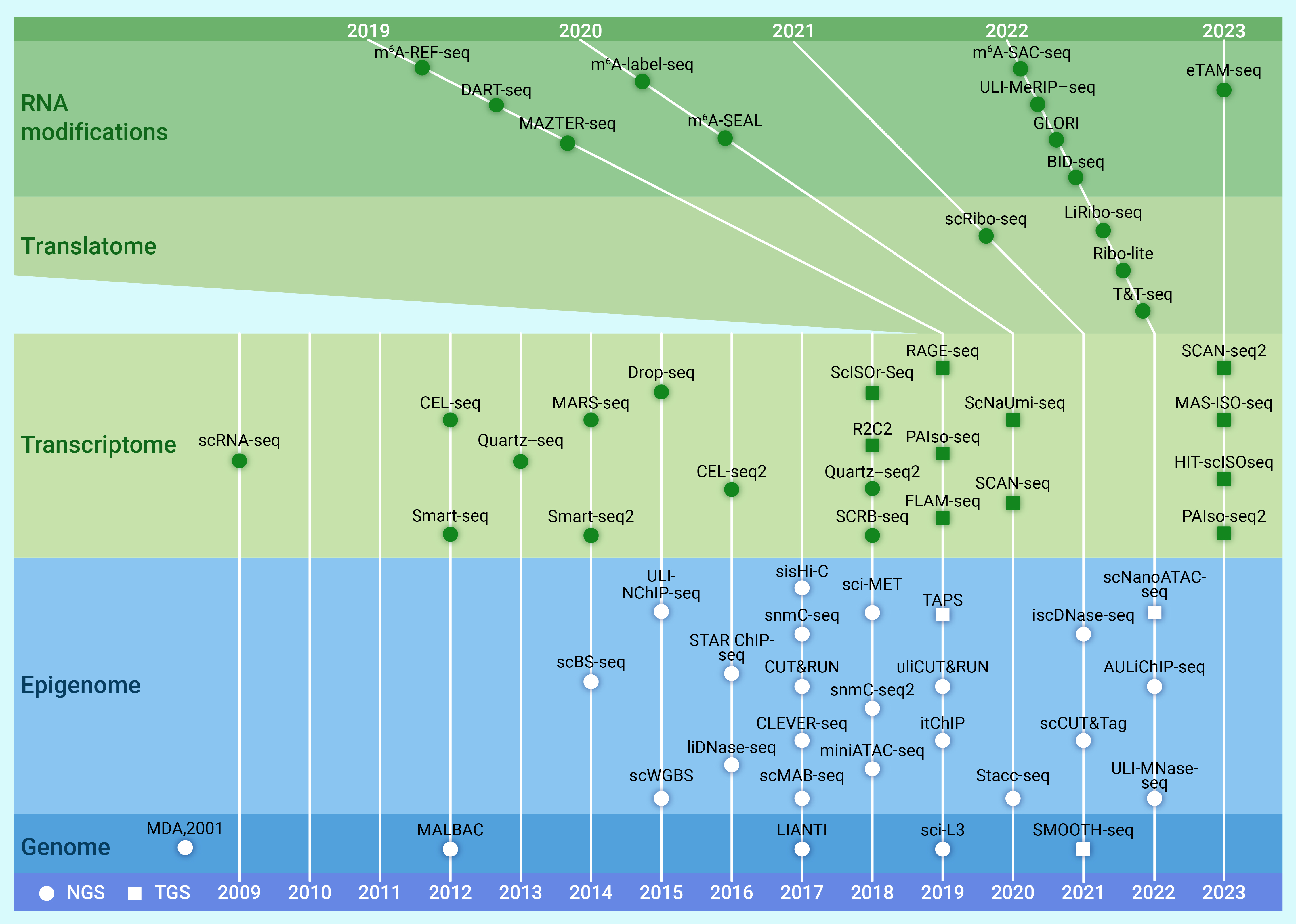
Figure 1.
Representative sequencing technologies for low-input cells and single cells
-
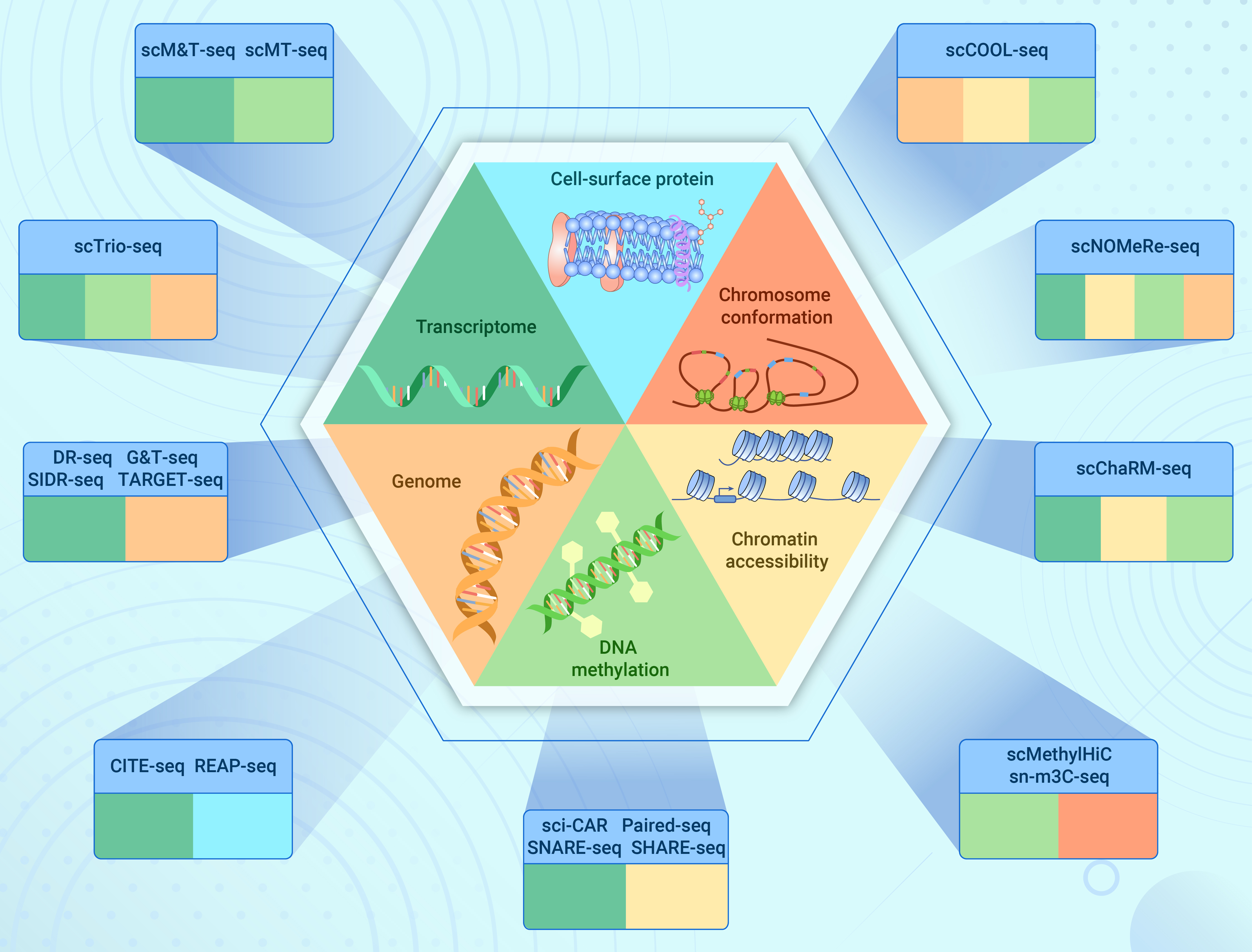
Figure 2.
A brief schematic for representative multi-omics sequencing technologies
-
Figure 3.
Representative advances in human early embryonic development using sequencing technology
-
Figure 4.
Common pipelines for PGT
-
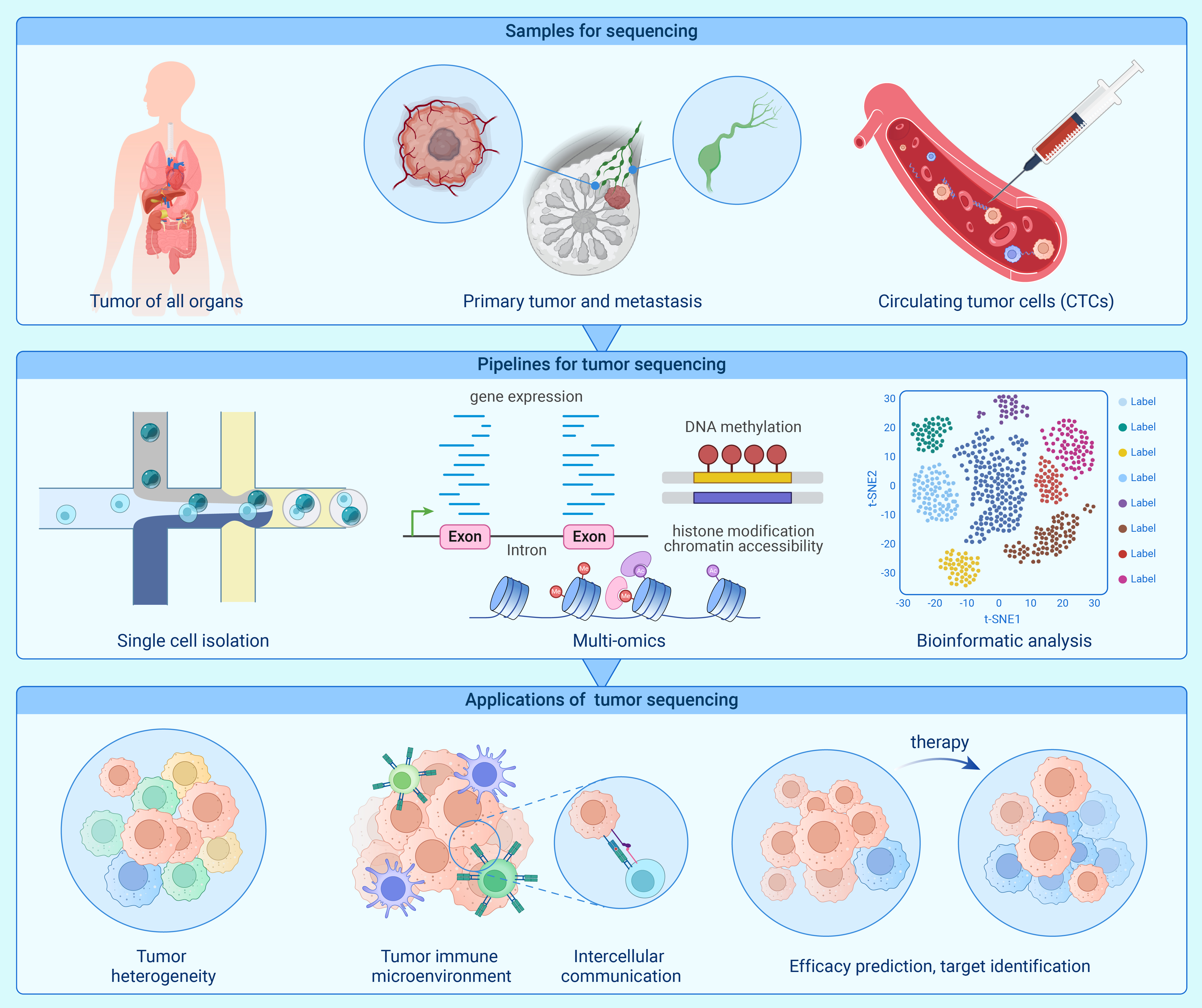
Figure 5.
Low-input sequencing in tumor research
-
Figure 6.
The application of low-input sequencing in other diseases and areas
