
Artificial intelligence- and pediatric CBCT-aided developmental recognition and personalized treatment of periapical diseases
-

-
REFERENCES
[1] Nolla C. M. (1952). The development of permanent teeth. (University of Michigan Ann Arbor [2] Liu Y., Zhang S., Wu X., et al. (2024). Individual graph representation learning for pediatric tooth segmentation from dental CBCT. IEEE Transactions on Medical Imaging. DOI:10.1109/TMI.2024.3501365 [3] Cui Z., Li C. and Wang W. (2019). ToothNet: Automatic tooth instance segmentation and identification from cone beam CT images. IEEE/CVF Conference on Computer Vision and Pattern Recognition 6361-6370. DOI:10.1109/CVPR.2019.00653 [4] Mizutani T., Ohno N. and Nakamura H. (1992). Anatomical study of the root apex in the maxillary anterior teeth. J. Endod. 18:344−347 DOI:10.1016/S0099-2399(06)80486-3 [5] Turosz N., Chęcińska K., Chęciński M., et al. (2024). Artificial intelligence (AI) assessment of pediatric dental panoramic radiographs (DPRs): A clinical study. Pediatr. Rep. 16:794−805 DOI:10.3390/pediatric16030067 [6] Turosz N., Chęcińska K., Chęciński M., et al. (2023). Applications of artificial intelligence in the analysis of dental panoramic radiographs: An overview of systematic reviews. Dentomaxillofac. Radiol. 52:20230284 DOI:10.1259/dmfr.20230284 [7] Mureșanu S., Almășan O., Hedeșiu M., et al. (2023). Artificial intelligence models for clinical usage in dentistry with a focus on dentomaxillofacial CBCT: A systematic review. Oral Radiol. 39:18−40 DOI:10.1007/s11282-022-00660-9 [8] Wu X., Chen H., Huang Y., et al. (2020). Center-sensitive and boundary-aware tooth instance segmentation and classification from cone-beam CT. 2020 IEEE 17th International Symposium on Biomedical Imaging (ISBI) 939-942. DOI:10.1109/ISBI45749.2020.9098542 [9] Cui Z., Fang Y., Mei L., et al. (2022). A fully automatic AI system for tooth and alveolar bone segmentation from cone-beam CT images. Nat. Commun. 13:2096 DOI:10.1038/s41467-022-29637-2 [10] Li P., Liu Y., Cui Z., et al. (2022). Semantic graph attention with explicit anatomical association modeling for tooth segmentation from CBCT images. IEEE Transactions on Medical Imaging 41:3116−3127 DOI:10.1109/TMI.2022.3179128 -
ABOUT THIS ARTICLE
Cite this article:
Zhang S., Liu Y., Li D., et al. (2025). Artificial intelligence- and pediatric CBCT-aided developmental recognition and personalized treatment of periapical diseases. The Innovation Medicine 3:100118. https://doi.org/10.59717/j.xinn-med.2025.100118
Welcome!
To request copyright permission to republish or share portions of our works, please visit Copyright Clearance Center's (CCC) Marketplace website at marketplace.copyright.com.
Export File
Citation
Zhang S., Liu Y., Li D., et al. (2025). Artificial intelligence- and pediatric CBCT-aided developmental recognition and personalized treatment of periapical diseases. The Innovation Medicine 3:100118. https://doi.org/10.59717/j.xinn-med.2025.100118

Format
Content
 DownLoad:
-
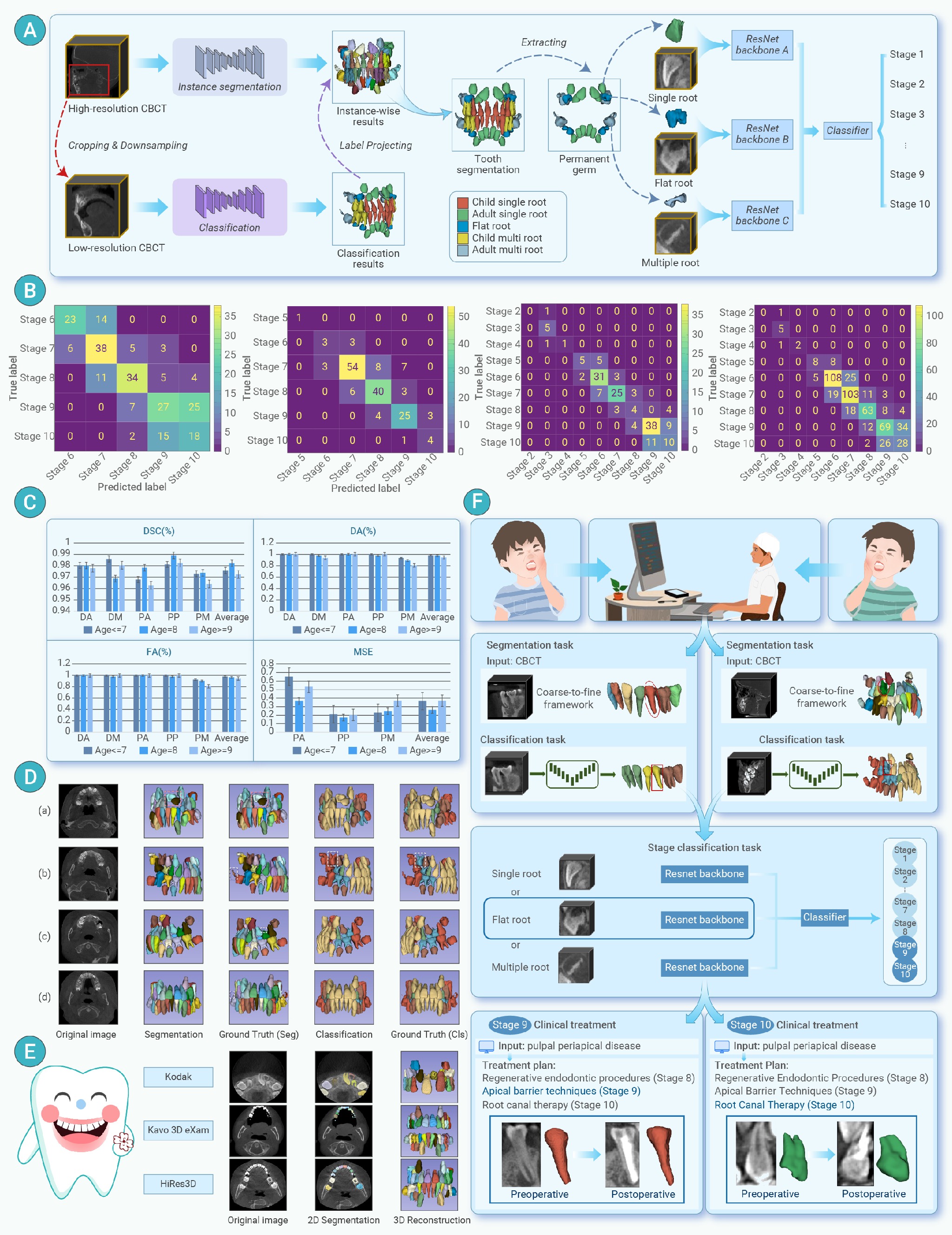
Figure 1.
(A) This illustration presents the architecture of the proposed multi-stage deep learning model. It incorporates a multi-task network specifically engineered for simultaneous tooth segmentation and classification, supplemented by a hierarchical region of interest-based classification network assigned with the task of predicting the staging of tooth germs. (B) Confusion matrices depicting the model's performance for permanent anterior, permanent premolar, permanent molar teeth, and the overall sample set. (C) Performance comparison among different age groups regarding the mean Dice Similarity Coefficient, average symmetric surface distance, detection and identification accuracy for tooth identification, and mean square error for tooth germ staging. (D) Visual representation of the model's segmentation and classification performance in four representative cases. The red box indicates a tooth with an abnormal orientation, the green box indicates a misclassification, and the yellow box indicates a missed detection. (E) Visual demonstration of the model's segmentation and classification performance across various CBCT imaging sources. (F) A flow chart outlining the end-to-end treatment planning process for premolars using the deep learning model. The input comprises of a CBCT image and a periapical disease prompt, with the output encompassing tooth segmentation, classification, and staging results, culminating in a personalized treatment plan.
