
Non-invasive neuromodulation assisted by exogenous stimuli-responsive nanoplatforms for Alzheimer’s disease and Parkinson's disease therapy
-
GRAPHICAL ABSTRACT
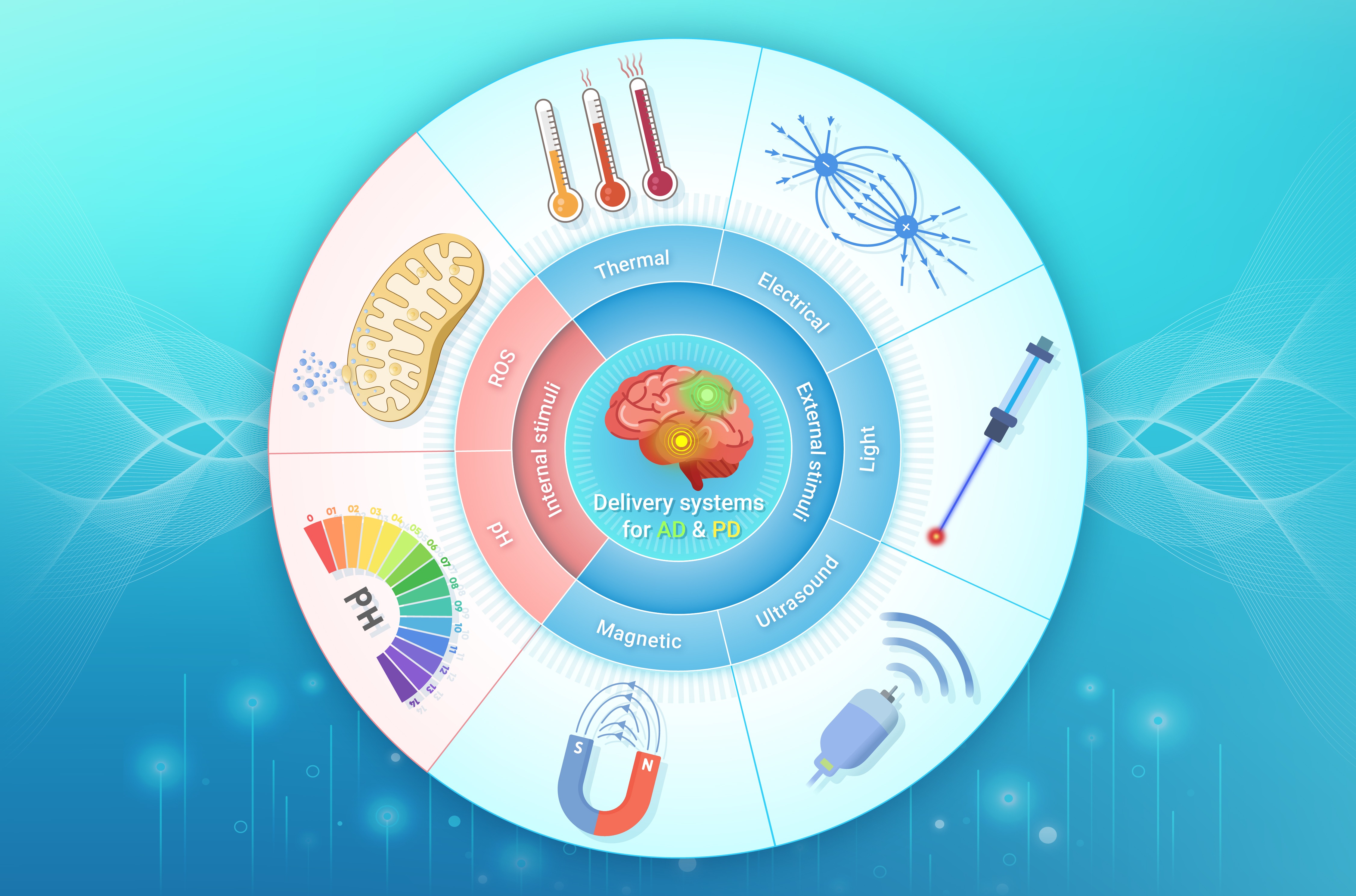
 DownLoad:
Full size image
DownLoad:
Full size image
-
PUBLIC SUMMARY
-
Stimuli-responsive nanoplatforms enable non-invasive neuromodulation for Alzheimer’s and Parkinson’s therapy.
External stimuli enhance drug delivery efficiency in neurodegenerative disease treatment.
Nanoplatforms respond to brain regions or pathologies like amyloid plaques and dopamine deficits.
Challenges in precision, safety, and efficacy are addressed through advanced targeting strategies.
-
-
ABSTRACT
In recent decades, the incidence of neurodegenerative diseases such as Alzheimer’s disease (AD) and Parkinson’s disease (PD) has risen continuously, significantly impairing patients’ quality of life while imposing growing economic and social burdens. Traditional treatments exhibit limited effectiveness in halting disease progression. Non-invasive neuromodulation techniques, utilizing electromagnetic fields, light, or ultrasound, have emerged as promising strategies to modulate neural activity and alleviate symptoms. However, achieving spatially precise and targeted neuromodulation remains challenging. The integration of stimuli-responsive nanoplatforms addresses this limitation. These nanoplatforms, engineered to respond to specific stimuli, can deliver therapeutic agents to the desired brain regions. By enabling controlled and localized drug release, they facilitate precise neuromodulation. Despite their potential, several hurdles must be overcome, including the optimization of nanoplatform design, elucidating complex nanoplatform-brain interactions, and ensuring long-term safety and efficacy. Nonetheless, combining non-invasive neuromodulation with stimuli-responsive nanoplatforms holds revolutionary potential for neurodegenerative disease therapy, offering targeted, personalized treatments that may halt or slow disease progression. This comprehensive review explores the therapeutic potential and applications of non-invasive neuromodulation assisted by exogenous stimuli-responsive nanoplatforms for AD and PD therapy. -

-
REFERENCES
[1] Zhou J., Jangili P., Son S., et al. (2020). Fluorescent diagnostic probes in neurodegenerative diseases. Adv. Mater. 32:e2001945. DOI:10.1002/adma.202001945 [2] Lin M. T. and Beal M. F. (2006). Mitochondrial dysfunction and oxidative stress in neurodegenerative diseases. Nature 443:787−795. DOI:10.1038/nature05292 [3] Tian M., Ma Z. and Yang G.-Z. (2024). Micro/nanosystems for controllable drug delivery to the brain. The Innovation 5:100548. DOI:10.1016/j.xinn.2023.100548 [4] Furtado D., Bjornmalm M., Ayton S., et al. (2018). Overcoming the blood-brain barrier: The role of nanomaterials in treating neurological diseases. Adv. Mater. 30:1801362. DOI:10.1002/adma.201801362 [5] Pandit R., Chen L. and Gotz J. (2020). The blood-brain barrier: Physiology and strategies for drug delivery. Adv. Drug Deliv. Rev. 165-166:1−14. DOI:10.1016/j.addr.2019.11.009 [6] Colombo E., Feyen P., Antognazza M. R., et al. (2016). Nanoparticles: A challenging vehicle for neural stimulation. Front. Neurosci. 10:105. DOI:10.3389/fnins.2016.00105 [7] Yoo S., Mittelstein D. R., Hurt R., et al. (2022). Focused ultrasound excites cortical neurons via mechanosensitive calcium accumulation and ion channel amplification. Nat. Commun. 13:493. DOI:10.1038/s41467-022-28040-1 [8] Tortiglione C., Antognazza M. R., Tino A., et al. (2017). Semiconducting polymers are light nanotransducers in eyeless animals. Sci. Adv. 3:e1601699. DOI:10.1126/sciadv.1601699 [9] Fernandez-Yague M. A., Trotier A., Demir S., et al. (2021). A self-powered piezo-bioelectric device regulates tendon repair-associated signaling pathways through modulation of mechanosensitive ion channels. Adv. Mater. 33:2008788. DOI:10.1002/adma.202008788 [10] Liu H. J. and Xu P. (2022). Strategies to overcome/penetrate the BBB for systemic nanoparticle delivery to the brain/brain tumor. Adv. Drug Deliv. Rev. 191:114619. DOI:10.1016/j.addr.2022.114619 [11] Majumder J. and Minko T. (2021). Multifunctional and stimuli-responsive nanocarriers for targeted therapeutic delivery. Expert Opin. Drug Deliv. 18:205−227. DOI:10.1080/17425247.2021.1828339 [12] Zhang Z., You Y., Ge M., et al. (2023). Functional nanoparticle-enabled non-genetic neuromodulation. J. Nanobiotechnol. 21:319. DOI:10.1186/s12951-023-02084-x [13] Huang H., Delikanli S., Zeng H., et al. (2010). Remote control of ion channels and neurons through magnetic-field heating of nanoparticles. Nat. Nanotechnol. 5:602−606. DOI:10.1038/nnano.2010.125 [14] Graff-Radford J., Yong K. X. X., Apostolova L. G., et al. (2021). New insights into atypical Alzheimer's disease in the era of biomarkers. Lancet Neurol. 20:222−234. DOI:10.1016/s1474-4422(20)30440-3 [15] Kavitha C., Mani V., Srividhya S. R., et al. (2022). Early-stage alzheimer's disease prediction using machine learning models. Front. Public Health 10:853294. DOI:10.3389/fpubh.2022.853294 [16] Busche M. A. and Hyman B. T. (2020). Synergy between amyloid-beta and tau in Alzheimer's disease. Nat. Neurosci. 23:1183−1193. DOI:10.1038/s41593-020-0687-6 [17] Shin W. S., Di J., Cao Q., et al. (2019). Amyloid beta-protein oligomers promote the uptake of tau fibril seeds potentiating intracellular tau aggregation. Alzheimers Res. Ther. 11:86. DOI:10.1186/s13195-019-0541-9 [18] Budimir A. (2011). Metal ions, Alzheimer's disease and chelation therapy. Acta Pharm. 61:1−14. DOI:10.2478/v10007-011-0006-6 [19] Heneka M. T., Carson M. J., El Khoury J., et al. (2015). Neuroinflammation in Alzheimer's disease. Lancet Neurol. 14:388−405. DOI:10.1016/s1474-4422(15)70016-5 [20] Irwin M. R. and Vitiello M. V. (2019). Implications of sleep disturbance and inflammation for Alzheimer's disease dementia. Lancet Neurol. 18:296−306. DOI:10.1016/s1474-4422(18)30450-2 [21] Dubois B., Villain N., Frisoni G. B., et al. (2021). Clinical diagnosis of Alzheimer's disease: Recommendations of the International Working Group. Lancet Neurol. 20:484−496. DOI:10.1016/s1474-4422(21)00066-1 [22] Tao Q.-Q., Cai X., Xue Y.-Y., et al. (2024). Alzheimer's disease early diagnostic and staging biomarkers revealed by large-scale cerebrospinal fluid and serum proteomic profiling. The Innovation 5:100544. DOI:10.1016/j.xinn.2023.100544 [23] Kalia L. V. and Lang A. E. (2015). Parkinson's disease. Lancet 386:896−912. DOI:10.1016/s0140-6736(14)61393-3 [24] McGregor M. M. and Nelson A. B. (2019). Circuit mechanisms of Parkinson's disease. Neuron 101:1042−1056. DOI:10.1016/j.neuron.2019.03.004 [25] Rocha E. M., Keeney M. T., Di Maio R., et al. (2022). LRRK2 and idiopathic Parkinson's disease. Trends Neurosci. 45:224−236. DOI:10.1016/j.tins.2021.12.002 [26] Malpartida A. B., Williamson M., Narendra D. P., et al. (2021). Mitochondrial dysfunction and mitophagy in Parkinson's disease: From mechanism to therapy. Trends Biochem.Sci. 46:329−343. DOI:10.1016/j.tibs.2020.11.007 [27] Weintraub D., Aarsland D., Chaudhuri K. R., et al. (2022). The neuropsychiatry of Parkinson's disease: Advances and challenges. Lancet Neurol. 21:89−102. DOI:10.1016/s1474-4422(21)00330-6 [28] Tolosa E., Garrido A., Scholz S. W., et al. (2021). Challenges in the diagnosis of Parkinson's disease. Lancet Neurol. 20:385−397. DOI:10.1016/s1474-4422(21)00030-2 [29] Mahoney-Sanchez L., Bouchaoui H., Ayton S., et al. (2021). Ferroptosis and its potential role in the physiopathology of Parkinson's disease. Prog. Neurobiol. 196:101890. DOI:10.1016/j.pneurobio.2020.101890 [30] Zhang W., Mehta A., Tong Z., et al. (2021). Development of polymeric nanoparticles for blood-brain barrier transfer-strategies and challenges. Adv. Sci. 8:2003937. DOI:10.1002/advs.202003937 [31] Abbott N. J. and Friedman A. (2012). Overview and introduction: The blood-brain barrier in health and disease. Epilepsia 53:1−6. DOI:10.1111/j.1528-1167.2012.03696.x [32] Bell R. D., Winkler E. A., Sagare A. P., et al. (2010). Pericytes control key neurovascular functions and neuronal phenotype in the adult brain and during brain aging. Neuron 68:409−427. DOI:10.1016/j.neuron.2010.09.043 [33] Ising C. and Heneka M. T. (2018). Functional and structural damage of neurons by innate immune mechanisms during neurodegeneration. Cell Death Dis. 9:120. DOI:10.1038/s41419-017-0153-x [34] Stephenson J., Nutma E., van der Valk P., et al. (2018). Inflammation in CNS neurodegenerative diseases. Immunology 154:204−219. DOI:10.1111/imm.12922 [35] Brighi C., Puttick S., Rose S., et al. (2018). The potential for remodelling the tumour vasculature in glioblastoma. Adv. Drug Deliv. Rev. 136:49−61. DOI:10.1016/j.addr.2018.10.001 [36] Hernandez C. and Shukla S. (2022). Liposome based drug delivery as a potential treatment option for Alzheimer's disease. Neural Regen. Res. 17:1190−1198. DOI:10.4103/1673-5374.327328 [37] Has C. and Sunthar P. (2020). A comprehensive review on recent preparation techniques of liposomes. J. Liposome Res. 30:336−365. DOI:10.1080/08982104.2019.1668010 [38] Elliott R. O. and He M. (2021). Unlocking the power of exosomes for crossing biological barriers in drug delivery. Pharmaceutics 13:122. DOI:10.3390/pharmaceutics13010122 [39] Mardpour S., Hamidieh A. A., Taleahmad S., et al. (2019). Interaction between mesenchymal stromal cell-derived extracellular vesicles and immune cells by distinct protein content. J. Cell. Physiol. 234:8249−8258. DOI:10.1002/jcp.27669 [40] Li Z., Liu F., He X., et al. (2019). Exosomes derived from mesenchymal stem cells attenuate inflammation and demyelination of the central nervous system in EAE rats by regulating the polarization of microglia. Int. Immunopharmacol. 67:268−280. DOI:10.1016/j.intimp.2018.12.001 [41] Armstrong J. P. K., Holme M. N. and Stevens M. M. (2017). Re-engineering extracellular vesicles as smart nanoscale therapeutics. Acs Nano 11:69−83. DOI:10.1021/acsnano.6b07607 [42] Ruan H., Li Y., Zheng D., et al. (2023). Engineered extracellular vesicles for ischemic stroke treatment. The Innovation 4:100394. DOI:10.1016/j.xinn.2023.100394 [43] Teleanu R. I., Preda M. D., Niculescu A.-G., et al. (2022). Current strategies to enhance delivery of drugs across the blood-brain barrier. Pharmaceutics 14:987. DOI:10.3390/pharmaceutics14050987 [44] Schally A. V. and Nagy A. (2004). Chemotherapy targeted to cancers through tumoral hormone receptors. Trends Endocrinol. Metab. 15:300−310. DOI:10.1016/j.tem.2004.07.002 [45] Iqbal I., Saqib F., Mubarak Z., et al. (2024). Alzheimer's disease and drug delivery across the blood-brain barrier: Approaches and challenges. Eur. J. Med. Res. 29:313. DOI:10.1186/s40001-024-01915-3 [46] Huang D., Wang J., Song C., et al. (2023). Ultrasound-responsive matters for biomedical applications. The Innovation 4:100421. DOI:10.1016/j.xinn.2023.100421 [47] Zhang J., Liu H., Du X., et al. (2017). Increasing of blood-brain tumor barrier permeability through transcellular and paracellular pathways by microbubble-enhanced diagnostic ultrasound in a C6 glioma model. Front. Neurosci. 11:86. DOI:10.3389/fnins.2017.00086 [48] Meng Y., Volpini M., Black S., et al. (2017). Focused ultrasound as a novel strategy for Alzheimer disease therapeutics. Ann. Neurol. 81:611−617. DOI:10.1002/ana.24933 [49] Park J., Aryal M., Vykhodtseva N., et al. (2017). Evaluation of permeability, doxorubicin delivery, and drug retention in a rat brain tumor model after ultrasound-induced blood-tumor barrier disruption. J. Control. Release 250:77−85. DOI:10.1016/j.jconrel.2016.10.011 [50] Aryal M., Fischer K., Gentile C., et al. (2017). Effects on p-glycoprotein expression after blood-brain barrier disruption using focused ultrasound and microbubbles. PLoS One 12:e0166061. DOI:10.1371/journal.pone.0166061 [51] Mohapatra A., Uthaman S. and Park I.-K. (2021). External and internal stimuli-responsive metallic nanotherapeutics for enhanced anticancer therapy. Front. Mol. Biosci. 7:597634. DOI:10.3389/fmolb.2020.597634 [52] Rajendrakumar S. K., Cherukula K., Park H. J., et al. (2018). Dual-stimuli-responsive albumin-polyplex nanoassembly for spatially controlled gene release in metastatic breast cancer. J. Control. Release 276:72−83. DOI:10.1016/j.jconrel.2018.02.039 [53] Wu W., Luo L., Wang Y., et al. (2018). Endogenous pH-responsive nanoparticles with programmable size changes for targeted tumor therapy and imaging applications. Theranostics 8:3038−3058. DOI:10.7150/thno.23459 [54] Yao J., Feng J. and Chen J. (2016). External-stimuli responsive systems for cancer theranostic. Asian J. Pharm. Sci. 11:585−595. DOI:10.1016/j.ajps.2016.06.001 [55] Xu Z., Yang D., Long T., et al. (2022). pH-sensitive nanoparticles based on amphiphilic imidazole/cholesterol modified hydroxyethyl starch for tumor chemotherapy. Carbohydr. Polym. 277:118827. DOI:10.1016/j.carbpol.2021.118827 [56] Kanamala M., Wilson W. R., Yang M., et al. (2016). Mechanisms and biomaterials in pH-responsive tumour targeted drug delivery: A review. Biomaterials 85:152−167. DOI:10.1016/j.biomaterials.2016.01.061 [57] Chu S., Shi X., Tian Y., et al. (2022). pH-responsive polymer nanomaterials for tumor therapy. Front. Oncol. 12:855019. DOI:10.3389/fonc.2022.855019 [58] Zhao Y., Liu X., Peng X., et al. (2022). A poloxamer/hyaluronic acid/chitosan-based thermosensitive hydrogel that releases dihydromyricetin to promote wound healing. Int. J. Biol. Macromol. 216:475−486. DOI:10.1016/j.ijbiomac.2022.06.210 [59] Chen Q., Jia C., Xu Y., et al. (2022). Dual-pH responsive chitosan nanoparticles for improving in vivo drugs delivery and chemoresistance in breast cancer. Carbohydr. Polym. 290:119518. DOI:10.1016/j.carbpol.2022.119518 [60] Liu A., Liang C., Liu J., et al. (2022). Reactive oxygen species horizontal line responsive lipid nanoparticles for effective RNAi and corneal neovascularization therapy. ACS Appl. Mater. Interfaces 14:17022−17031. DOI:10.1021/acsami.1c23412 [61] Li Q., Wen Y., You X., et al. (2016). Development of a reactive oxygen species (ROS)-responsive nanoplatform for targeted oral cancer therapy. J. Mat. Chem. B 4:4675−4682. DOI:10.1039/c6tb01016d [62] Xu X., Saw P. E., Tao W., et al. (2017). ROS-responsive polyprodrug nanoparticles for triggered drug delivery and effective cancer therapy. Adv. Mater. 29:1700141. DOI:10.1002/adma.201700141 [63] Saravanakumar G., Kim J. and Kim W. J. (2017). Reactive-oxygen-species-responsive drug delivery systems: Promises and challenges. Adv. Sci. 4:1600124. DOI:10.1002/advs.201600124 [64] Xu Q., He C., Xiao C., et al. (2016). Reactive oxygen species (ROS) responsive polymers for biomedical applications. Macromol. Biosci. 16:635−646. DOI:10.1002/mabi.201500440 [65] Jin L., Zhu Z., Hong L., et al. (2023). ROS-responsive 18beta-glycyrrhetic acid-conjugated polymeric nanoparticles mediate neuroprotection in ischemic stroke through HMGB1 inhibition and microglia polarization regulation. Bioact. Mater. 19:38−49. DOI:10.1016/j.bioactmat.2022.03.040 [66] Zhang R., Liu R., Liu C., et al. (2020). A pH/ROS dual-responsive and targeting nanotherapy for vascular inflammatory diseases. Biomaterials 230:119605. DOI:10.1016/j.biomaterials.2019.119605 [67] Karimi M., Ghasemi A., Zangabad P. S., et al. (2016). Smart micro/nanoparticles in stimulus-responsive drug/gene delivery systems. Chem. Soc. Rev. 45:1457−1501. DOI:10.1039/c5cs00798d [68] Mi P. (2020). Stimuli-responsive nanocarriers for drug delivery, tumor imaging, therapy and theranostics. Theranostics 10:4557−4588. DOI:10.7150/thno.38069 [69] Ovejero J. G., Armenia I., Serantes D., et al. (2021). Selective magnetic nanoheating: Combining iron oxide nanoparticles for multi-hot-spot induction and sequential regulation. Nano Lett. 21:7213−7220. DOI:10.1021/acs.nanolett.1c02178 [70] Wu W., Liu J., Gong P., et al. (2022). Construction of core-shell nanoMOFs@microgel for aqueous lubrication and thermal-responsive drug release. Small 18:e2202510. DOI:10.1002/smll.202202510 [71] Wells C. M., Harris M., Choi L., et al. (2019). Stimuli-responsive drug release from smart polymers. J. Funct. Biomater. 10DOI:10.3390/jfb10030034. [72] Puoci F., Hampel S., Parisi O. I., et al. (2013). Imprinted microspheres doped with carbon nanotubes as novel electroresponsive drug-delivery systems. J. Appl. Polym. Sci. 130:829−834. DOI:10.1002/app.39212 [73] Loh D. M., Nava M. and Nocera D. G. (2022). Polypyrrole-silicon nanowire arrays for controlled intracellular cargo delivery. Nano Lett. 22:366−371. DOI:10.1021/acs.nanolett.1c04033 [74] Wang S., Bai Y., Wang D., et al. (2022). Reversing tumor to “hot”: A NIR light-triggered carrier-free nanoplatform for enhanced tumor penetration and photo-induced immunotherapy. Chem. Eng. J. 442DOI:10.1016/j.cej.2022.136322. [75] Roy B., Mengji R., Roy S., et al. (2022). NIR-responsive lysosomotropic phototrigger: An "AIE plus ESIPT" active naphthalene-based single-component photoresponsive nanocarrier with two-photon uncaging and real-time monitoring ability. ACS Appl. Mater. Interfaces 14:4862−4870. DOI:10.1021/acsami.1c19022 [76] Balakrishnan P. B., Silvestri N., Fernandez-Cabada T., et al. (2020). Exploiting unique alignment of cobalt ferrite nanoparticles, mild hyperthermia, and controlled intrinsic cobalt toxicity for cancer therapy. Adv. Mater. 32:e2003712. DOI:10.1002/adma.202003712 [77] You D. G., Deepagan V. G., Um W., et al. (2016). ROS-generating TiO2 nanoparticles for non-invasive sonodynamic therapy of cancer. Sci. Rep. 6:23200. DOI:10.1038/srep23200 [78] Wang J., Xie L., Shi Y., et al. (2021). Early detection and reversal of cell apoptosis induced by focused ultrasound-mediated blood-brain barrier opening. Acs Nano 15:14509−14521. DOI:10.1021/acsnano.1c04029 [79] Son S., Kim J. H., Wang X., et al. (2020). Multifunctional sonosensitizers in sonodynamic cancer therapy. Chem. Soc. Rev. 49:3244−3261. DOI:10.1039/c9cs00648f [80] Liu T., Zhang N., Wang Z., et al. (2017). Endogenous catalytic generation of O2 bubbles for in situ ultrasound-guided high intensity focused ultrasound ablation. Acs Nano 11:9093−9102. DOI:10.1021/acsnano.7b03772 [81] Peng X., Han Q., Zhou X., et al. (2022). Effect of pH-sensitive nanoparticles on inhibiting oral biofilms. Drug Deliv. 29:561−573. DOI:10.1080/10717544.2022.2037788 [82] Sun Y., Gong L., Yin Y., et al. (2022). A gradient pH-sensitive polymer-based antiviral strategy via viroporin-induced membrane acidification. Adv. Mater. 34:2109580. DOI:10.1002/adma.202109580 [83] Wu H., Xia F., Zhang L., et al. (2022). A ROS-sensitive nanozyme-augmented photoacoustic nanoprobe for early diagnosis and therapy of acute liver failure. Adv. Mater. 34:2108348. DOI:10.1002/adma.202108348 [84] Zhou X., Liu X., Yang X., et al. (2021). Tumor progress intercept by intervening in Caveolin-1 related intercellular communication via ROS-sensitive c-Myc targeting therapy. Biomaterials 275:120958. DOI:10.1016/j.biomaterials.2021.120958 [85] Han L., Zhang X.-Y., Wang Y.-L., et al. (2017). Redox-responsive theranostic nanoplatforms based on inorganic nanomaterials. J. Control. Release 259:40−52. DOI:10.1016/j.jconrel.2017.03.018 [86] Li Y., Feng S., Dai P., et al. (2022). Tailored Trojan horse nanocarriers for enhanced redox-responsive drug delivery. J. Control. Release 342:201−209. DOI:10.1016/j.jconrel.2022.01.006 [87] Riber C. F., Smith A. A. A. and Zelikin A. N. (2015). Self-immolative linkers literally bridge disulfide chemistry and the realm of thiol-free drugs. Adv. Healthc. Mater. 4:1887−1890. DOI:10.1002/adhm.201500344 [88] Lv F., Li C., Ma Y., et al. (2019). Fabrication of step-by-step drug release system both sensitive to magnetic field and temperature based on layered double hydroxides and PNIPAM. Nanotechnology 30:055103. DOI:10.1088/1361-6528/aaf095 [89] Yadav R. and Venkatesu P. (2019). Functionalized carbon nanotubes modulate the phase transition behavior of thermoresponsive polymer via hydrophilic-hydrophobic balance. Polymer 178:121573. DOI:10.1016/j.polymer.2019.121573 [90] Chen T., Yang Y., Peng H., et al. (2021). Cellulose nanocrystals reinforced highly stretchable thermal-sensitive hydrogel with ultra-high drug loading. Carbohydr. Polym. 266:118122. DOI:10.1016/j.carbpol.2021.118122 [91] Bauer S. M., Bauer R. J. and Velazquez O. C. (2005). Angiogenesis, vasculogenesis, and induction of healing in chronic wounds. Vasc. Endovasc. Surg. 39:293−306. DOI:10.1177/153857440503900401 [92] Magaz A., Ashton M. D., Hathout R. M., et al. (2020). Electroresponsive silk-based biohybrid composites for electrochemically controlled growth factor delivery. Pharmaceutics 12:742. DOI:10.3390/pharmaceutics12080742 [93] Ajdary R., Ezazi N. Z., Correia A., et al. (2020). Multifunctional 3D-printed patches for long-term drug release therapies after myocardial infarction. Adv. Funct. Mater. 30:2003440. DOI:10.1002/adfm.202003440 [94] Donahue M. J., Sanchez-Sanchez A., Inal S., et al. (2020). Tailoring PEDOT properties for applications in bioelectronics. Mater. Sci. Eng. R-Rep. 140:100546. DOI:10.1016/j.mser.2020.100546 [95] Enshaei H., Puiggali-Jou A., del Valle L. J., et al. (2021). Nanotheranostic interface based on antibiotic-loaded conducting polymer nanoparticles for real-time monitoring of bacterial growth inhibition. Adv. Healthc. Mater. 10:2001636. DOI:10.1002/adhm.202001636 [96] Sun Q., Hou X., Yang J., et al. (2021). Heparin-coated photosensitive metal-organic frameworks as drug delivery nanoplatforms of autophagy inhibitors for sensitized photodynamic therapy against breast cancer. ACS Appl. Mater. Interfaces 13:55577−55590. DOI:10.1021/acsami.1c18055 [97] Tong X., Gan S., Wu J., et al. (2020). A nano-photosensitizer based on covalent organic framework nanosheets with high loading and therapeutic efficacy. Nanoscale 12:7376−7382. DOI:10.1039/c9nr10787h [98] Xiao F., Cao B., Wen L., et al. (2020). Photosensitizer conjugate-functionalized poly(hexamethylene guanidine) for potentiated broad-spectrum bacterial inhibition and enhanced biocompatibility. Chin. Chem. Lett. 31:2516−2519. DOI:10.1016/j.cclet.2020.06.038 [99] Wang Y., Boero G., Zhang X., et al. (2022). Nanopore generation in biodegradable silk/magnetic nanoparticle membranes by an external magnetic field for implantable drug delivery. ACS Appl. Mater. InterfacesDOI:10.1021/acsami.2c10603. [100] Deatsch A. E. and Evans B. A. (2014). Heating efficiency in magnetic nanoparticle hyperthermia. J. Magn. Magn. Mater. 354:163−172. DOI:10.1016/j.jmmm.2013.11.006 [101] Banobre-Lopez M., Teijeiro A. and Rivas J. (2013). Magnetic nanoparticle-based hyperthermia for cancer treatment. Rep. Pract. Oncol. Radiother. 18:397−400. DOI:10.1016/j.rpor.2013.09.011 [102] Boissenot T., Bordat A., Fattal E., et al. (2016). Ultrasound-triggered drug delivery for cancer treatment using drug delivery systems: From theoretical considerations to practical applications. J. Control. Release 241:144−163. DOI:10.1016/j.jconrel.2016.09.026 [103] Canavese G., Ancona A., Racca L., et al. (2018). Nanoparticle-assisted ultrasound: A special focus on sonodynamic therapy against cancer. Chem. Eng. J. 340:155−172. DOI:10.1016/j.cej.2018.01.060 [104] Cesmeli S. and Avci C. B. (2019). Application of titanium dioxide (TiO2) nanoparticles in cancer therapies. J. Drug Target. 27:762−766. DOI:10.1080/1061186x.2018.1527338 [105] Shanei A. and Sazgarnia A. (2019). An overview of therapeutic applications of ultrasound based on synergetic effects with gold nanoparticles and laser excitation. Iran. J. Basic Med. Sci. 22:848−855. [106] Shanei A. and Akbari-Zadeh H. (2019). Investigating the sonodynamic-radiosensitivty effect of gold nanoparticles on HeLa cervical cancer cells. J. Korean Med. Sci. 34:e243. DOI:10.3346/jkms.2019.34.e243 [107] Heinzelman P., Schoborg J. A. and Jewett M. C. (2015). pH responsive granulocyte colony-stimulating factor variants with implications for treating Alzheimer's disease and other central nervous system disorders. Protein Eng. Des. Sel. 28:481−489. DOI:10.1093/protein/gzv022 [108] Zhang L., Liu X. g., Liu D. q., et al. (2020). A conditionally releasable “do not eat me” CD47 signal facilitates microglia‐targeted drug delivery for the treatment of Alzheimer's disease. Adv. Funct. Mater. 30DOI:10.1002/adfm.201910691. [109] Hu B., Dai F., Fan Z., et al. (2015). Nanotheranostics: Congo red/rutin-MNPs with enhanced magnetic resonance imaging and H2O2-responsive therapy of Alzheimer's disease in APPswe/PS1dE9 transgenic mice. Adv. Mater. 27:5499−5505. DOI:10.1002/adma.201502227 [110] Xu S., Yang P., Qian K., et al. (2022). Modulating autophagic flux via ROS-responsive targeted micelles to restore neuronal proteostasis in Alzheimer's disease. Bioact. Mater. 11:300−316. DOI:10.1016/j.bioactmat.2021.06.017 [111] Ballance W. C., Qin E. C., Chung H. J., et al. (2019). Reactive oxygen species-responsive drug delivery systems for the treatment of neurodegenerative diseases. Biomaterials 217:119292. DOI:10.1016/j.biomaterials.2019.119292 [112] Pettignano A., Grijalvo S., Haering M., et al. (2017). Boronic acid-modified alginate enables direct formation of injectable, self-healing and multistimuli-responsive hydrogels. Chem. Commun. 53:3350−3353. DOI:10.1039/c7cc00765e [113] Yang B., Chen Y. and Shi J. (2019). Reactive oxygen species (ROS)-based nanomedicine. Chem. Rev. 119:4881−4985. DOI:10.1021/acs.chemrev.8b00626 [114] Liu Z., Zhang B., Xia S., et al. (2021). ROS-responsive and multifunctional anti-Alzheimer prodrugs: Tacrine-ibuprofen hybrids via a phenyl boronate linker. Eur. J. Med. Chem. 212:112997. DOI:10.1016/j.ejmech.2020.112997 [115] Zhou H., Gong Y., Liu Y., et al. (2020). Intelligently thermoresponsive flower-like hollow nano-ruthenium system for sustained release of nerve growth factor to inhibit hyperphosphorylation of tau and neuronal damage for the treatment of Alzheimer's disease. Biomaterials 237:119822. DOI:10.1016/j.biomaterials.2020.119822 [116] Georgieva D., Ivanova-Mileva K., Ivanova S., et al. (2020). Thermoresponsive poly(N-isopropylacrylamide) copolymer networks for galantamine hydrobromide delivery. Colloid Polym. Sci. 298:377−384. DOI:10.1007/s00396-020-04621-8 [117] Geng H., Yuan H., Qiu L., et al. (2020). Inhibition and disaggregation of amyloid beta protein fibrils through conjugated polymer-core thermoresponsive micelles. J. Mat. Chem. B 8:10126−10135. DOI:10.1039/d0tb01863e [118] Guimard N. K., Gomez N. and Schmidt C. E. (2007). Conducting polymers in biomedical engineering. Prog. Polym. Sci. 32:876−921. DOI:10.1016/j.progpolymsci.2007.05.012 [119] Xu H., Holzwarth J. M., Yan Y., et al. (2014). Conductive PPY/PDLLA conduit for peripheral nerve regeneration. Biomaterials 35:225−235. DOI:10.1016/j.biomaterials.2013.10.002 [120] Wu Y., Wang L., Guo B., et al. (2016). Electroactive biodegradable polyurethane significantly enhanced Schwann cells myelin gene expression and neurotrophin secretion for peripheral nerve tissue engineering. Biomaterials 87:18−31. DOI:10.1016/j.biomaterials.2016.02.010 [121] Xie M., Wang L., Guo B., et al. (2015). Ductile electroactive biodegradable hyperbranched polylactide copolymers enhancing myoblast differentiation. Biomaterials 71:158−167. DOI:10.1016/j.biomaterials.2015.08.042 [122] Chen J., Dong R., Ge J., et al. (2015). Biocompatible, biodegradable, and electroactive polyurethane-urea elastomers with tunable hydrophilicity for skeletal muscle tissue engineering. ACS Appl. Mater. Interfaces 7:28273−28285. DOI:10.1021/acsami.5b10829 [123] Xie M., Wang L., Ge J., et al. (2015). Strong electroactive biodegradable shape memory polymer networks based on star-shaped polylactide and aniline trimer for bone tissue engineering. ACS Appl. Mater. Interfaces 7:6772−6781. DOI:10.1021/acsami.5b00191 [124] Li W., Luo R., Lin X., et al. (2015). Remote modulation of neural activities via near-infrared triggered release of biomolecules. Biomaterials 65:76−85. DOI:10.1016/j.biomaterials.2015.06.041 [125] Kim D., Kwon H. J. and Hyeon T. (2019). Magnetite/ceria nanoparticle assemblies for extracorporeal cleansing of amyloid-beta in Alzheimer's disease. Adv. Mater. 31:1807965. DOI:10.1002/adma.201807965 [126] Dyne E., Prakash P. S., Li J., et al. (2021). Mild magnetic nanoparticle hyperthermia promotes the disaggregation and microglia-mediated clearance of beta-amyloid plaques. Nanomed.-Nanotechnol. Biol. Med. 34:102397. DOI:10.1016/j.nano.2021.102397 [127] Razzokov J., Yusupov M. and Bogaerts A. (2019). Oxidation destabilizes toxic amyloid beta peptide aggregation. Sci. Rep. 9:5476. DOI:10.1038/s41598-019-41931-6 [128] Kang S.-g., Tien H., Xia Z., et al. (2013). Hydrophobic interaction drives surface-assisted epitaxial assembly of amyloid-like peptides. J. Am. Chem. Soc. 135:3150−3157. DOI:10.1021/ja310989u [129] Karthika V., Sridharan B., Nam J. W., et al. (2024). Neuromodulation by nanozymes and ultrasound during Alzheimer's disease management. J. Nanobiotechnol. 22:139. DOI:10.1186/s12951-024-02406-7 [130] Jiang J., Zhao K., Xiao X., et al. (2012). Synthesis and facet-dependent photoreactivity of BiOCl single-crystalline nanosheets. J. Am. Chem. Soc. 134:4473−4476. DOI:10.1021/ja210484t [131] Jang J., Kim K., Yoon J., et al. (2020). Piezoelectric materials for ultrasound-driven dissociation of Alzheimer's β-amyloid aggregate structure. Biomaterials 255:120165. DOI:10.1016/j.biomaterials.2020.120165 [132] Tzankov B., Tzankova V., Aluani D., et al. (2019). Development of MCM-41 mesoporous silica nanoparticles as a platform for pramipexole delivery. J. Drug Deliv. Sci. Technol. 51:26−35. DOI:10.1016/j.jddst.2019.02.008 [133] Gombotz W. R. and Wee S. F. (1998). Protein release from alginate matrices. Adv. Drug Deliv. Rev. 31:267−285. DOI:10.1016/s0169-409x(97)00124-5 [134] Tan J. P. K., Voo Z. X., Lim S., et al. (2019). Effective encapsulation of apomorphine into biodegradable polymeric nanoparticles through a reversible chemical bond for delivery across the blood-brain barrier. Nanomed.-Nanotechnol. Biol. Med. 17:236−245. DOI:10.1016/j.nano.2019.01.014 [135] Pichla M., Bartosz G., Stefaniuk I., et al. (2021). pH-responsive redox nanoparticles protect SH-SY5Y cells at lowered pH in a cellular model of Parkinson's disease. Molecules 26:543. DOI:10.3390/molecules26030543 [136] Hao C., Qu A., Xu L., et al. (2019). Chiral molecule-mediated porous CuxO nanoparticle clusters with antioxidation activity for ameliorating Parkinson's disease. J. Am. Chem. Soc. 141:1091−1099. DOI:10.1021/jacs.8b11856 [137] Li S., Liu J., Li G., et al. (2019). Near-infrared light-responsive, pramipexole-loaded biodegradable PLGA microspheres for therapeutic use in Parkinson's disease. Eur. J. Pharm. Biopharm. 141:1−11. DOI:10.1016/j.ejpb.2019.05.013 [138] Zhou X., Li B., Guo M., et al. (2022). Microneedle patch based on molecular motor as a spatio-temporal controllable dosing strategy of L-DOPA for Parkinson's disease. Chem. Eng. J. 427:131555. DOI:10.1016/j.cej.2021.131555 [139] Wang M., Li L., Zhang X., et al. (2018). Magnetic resveratrol liposomes as a new theranostic platform for magnetic resonance imaging guided Parkinson's disease targeting therapy. ACS Sustain. Chem. Eng. 6:17124−17133. DOI:10.1021/acssuschemeng.8b04507 [140] Niu S., Zhang L.-K., Zhang L., et al. (2017). Inhibition by multifunctional magnetic nanoparticles loaded with alpha-synuclein RNAi plasmid in a Parkinson's disease model. Theranostics 7:344−356. DOI:10.7150/thno.16562 [141] Xu T., Lu X., Peng D., et al. (2020). Ultrasonic stimulation of the brain to enhance the release of dopamine-a potential novel treatment for Parkinsons disease. Ultrason. Sonochem. 63:104955. DOI:10.1016/j.ultsonch.2019.104955 [142] Bond A. E., Shah B. B., Huss D. S., et al. (2017). Safety and efficacy of focused ultrasound thalamotomy for patients with medication-refractory, tremor-dominant Parkinson disease a randomized clinical trial. JAMA Neurol. 74:1412−1418. DOI:10.1001/jamaneurol.2017.3098 -
ABOUT THIS ARTICLE
Cite this article:
Mao M., Komes D., Zhao S., et al. (2025). Non-invasive neuromodulation assisted by exogenous stimuli-responsive nanoplatforms for Alzheimer’s disease and Parkinson's disease therapy. The Innovation Medicine 3:100121. https://doi.org/10.59717/j.xinn-med.2025.100121
Welcome!
To request copyright permission to republish or share portions of our works, please visit Copyright Clearance Center's (CCC) Marketplace website at marketplace.copyright.com.
Export File
Citation
Mao M., Komes D., Zhao S., et al. (2025). Non-invasive neuromodulation assisted by exogenous stimuli-responsive nanoplatforms for Alzheimer’s disease and Parkinson's disease therapy. The Innovation Medicine 3:100121. https://doi.org/10.59717/j.xinn-med.2025.100121

Format
Content

-
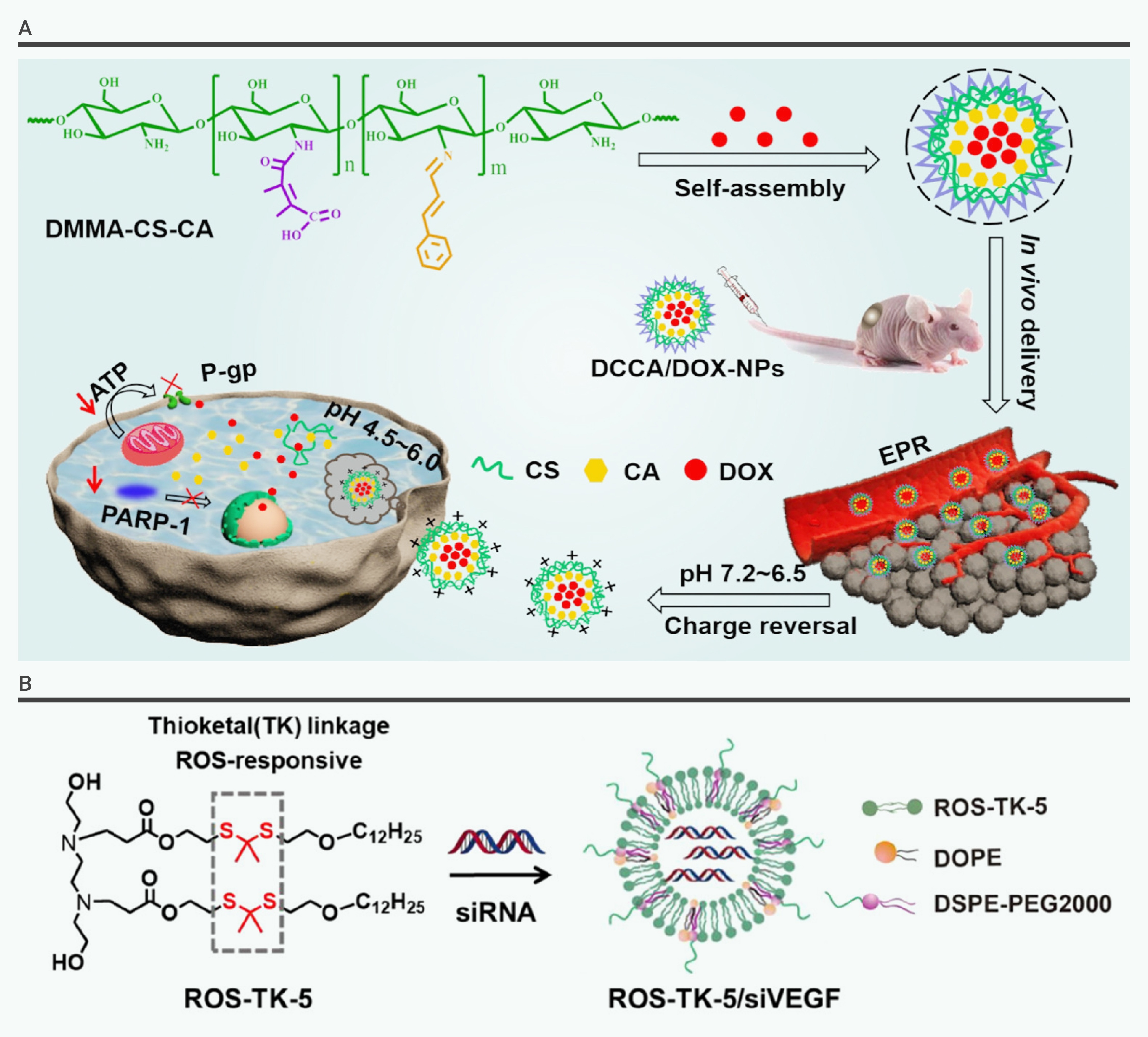
Figure 1.
The internal stimuli-responsive drug delivery system
-
Figure 2.
The external stimuli-responsive drug delivery system
-
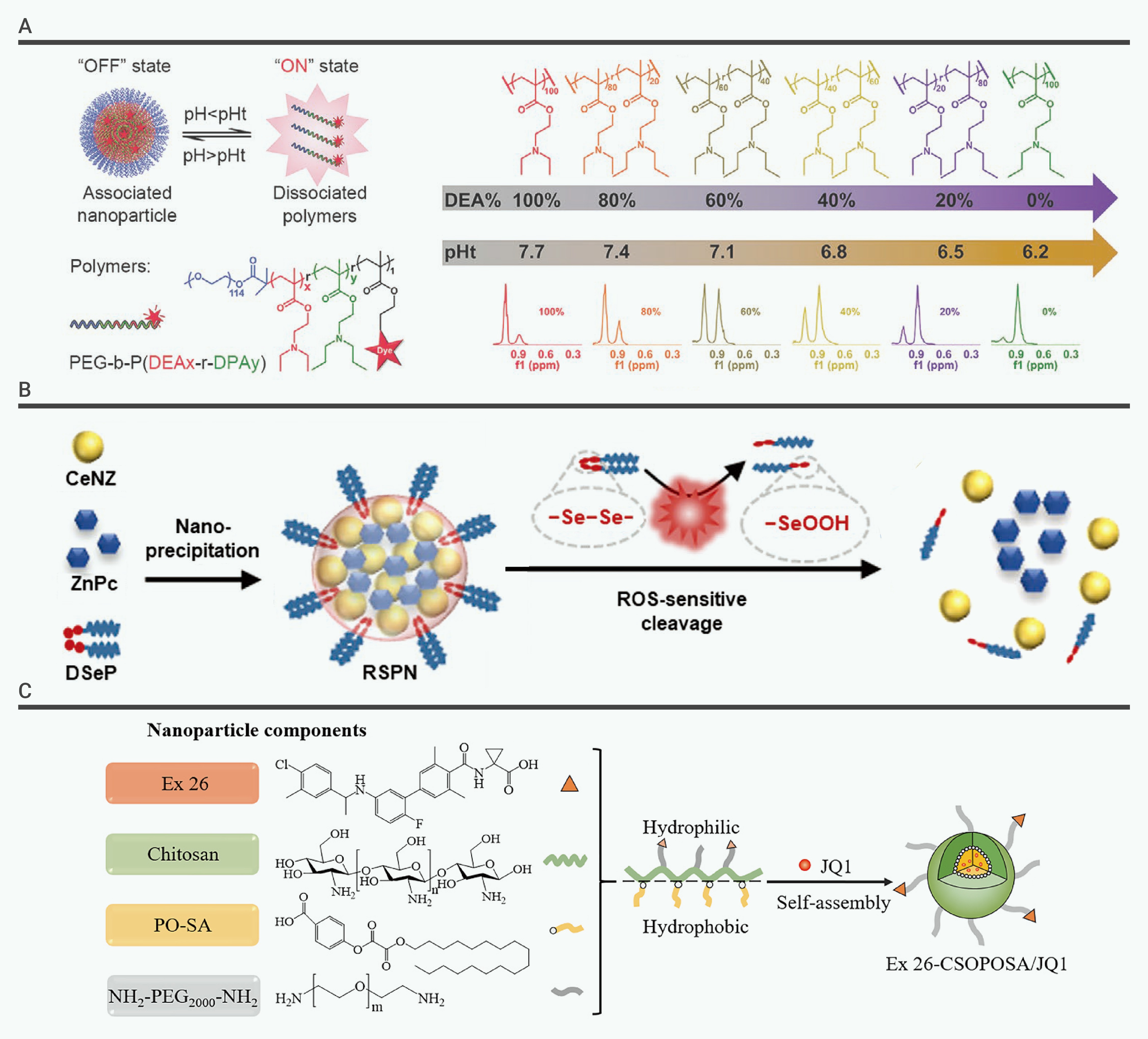
Figure 3.
Drug delivery system constructed with internal stimuli-responsive materials
-
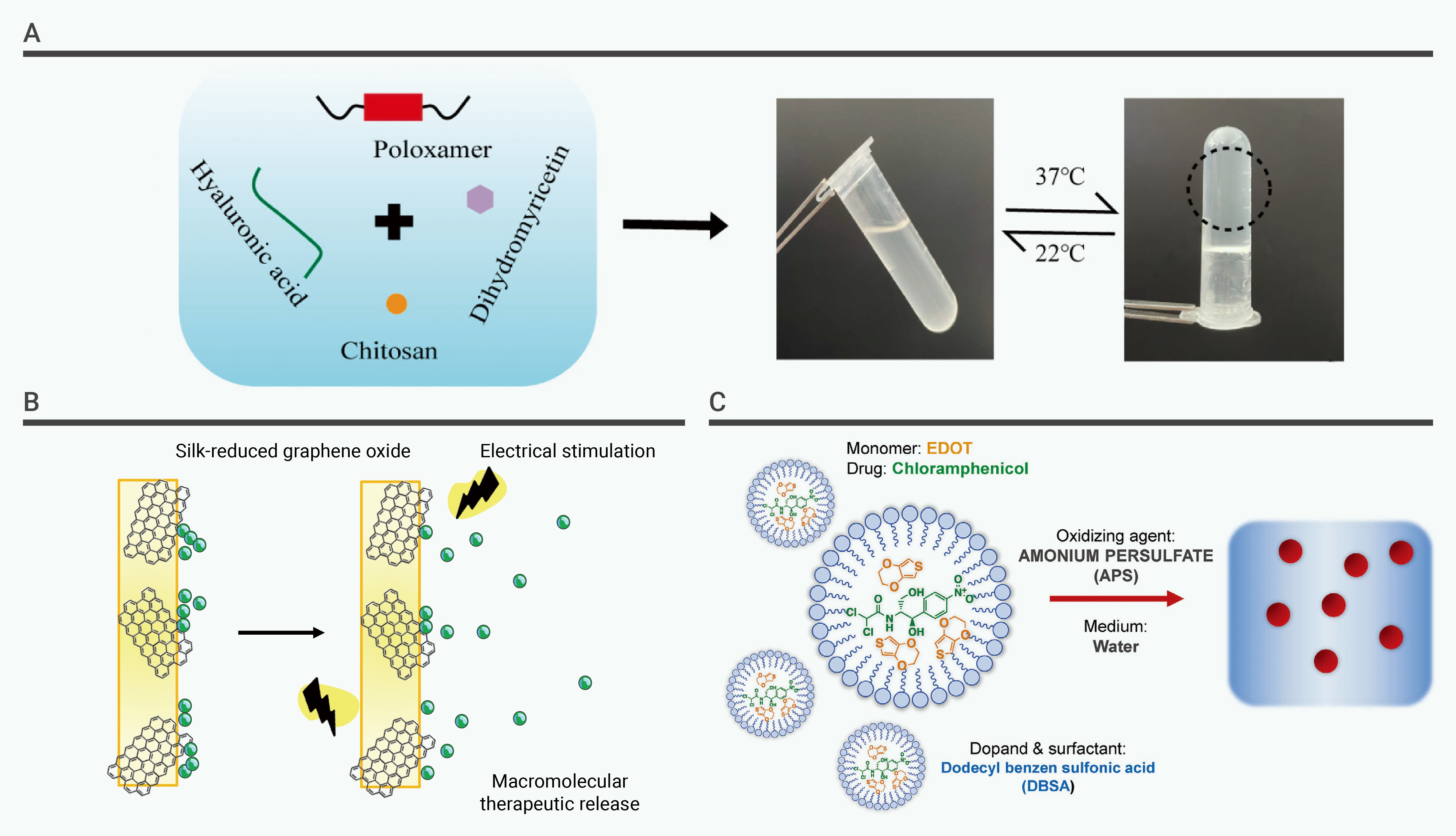
Figure 4.
Drug delivery system constructed with external stimuli-responsive materials
-
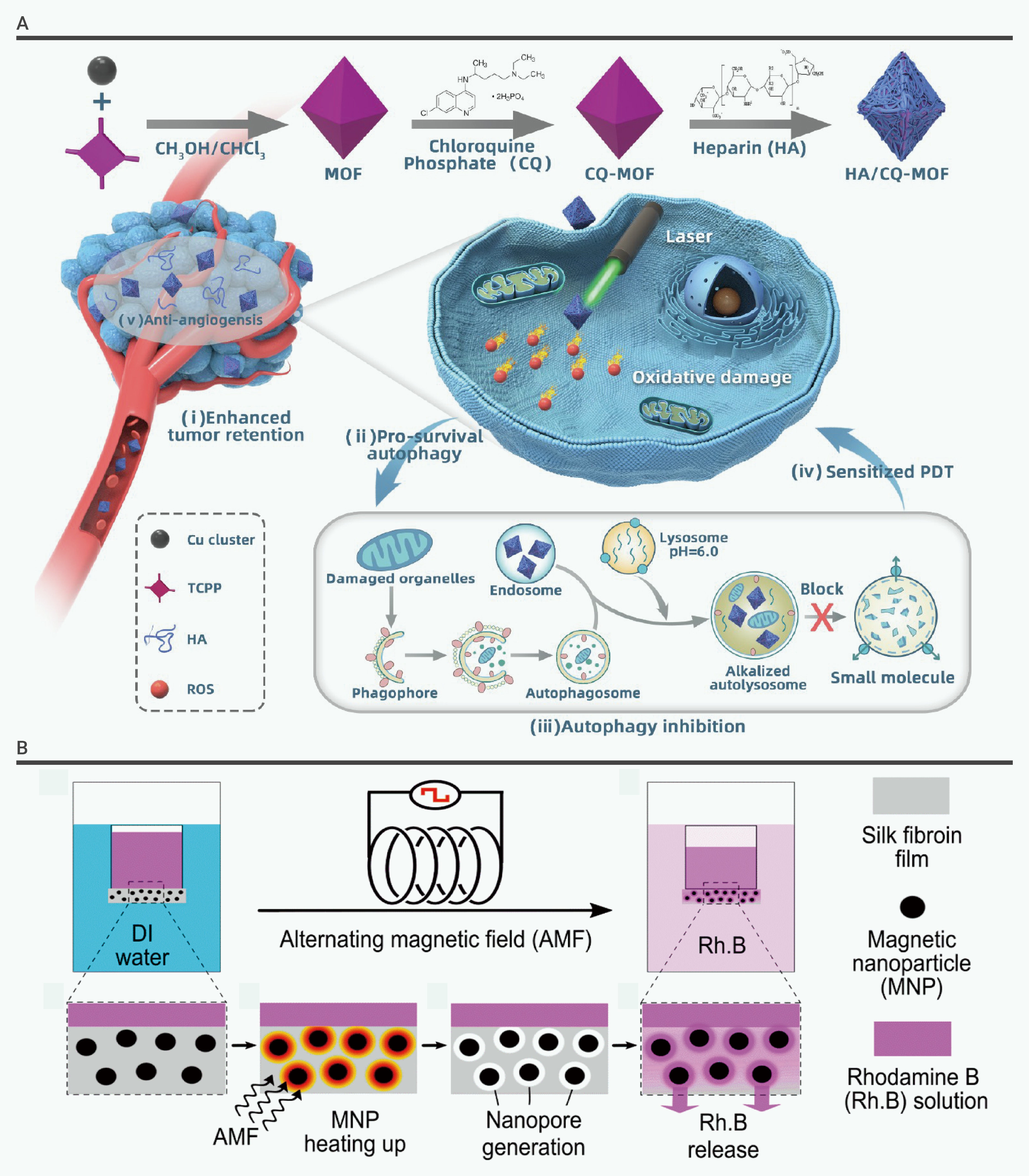
Figure 5.
Drug delivery system constructed with external stimulus-responsive materials
-
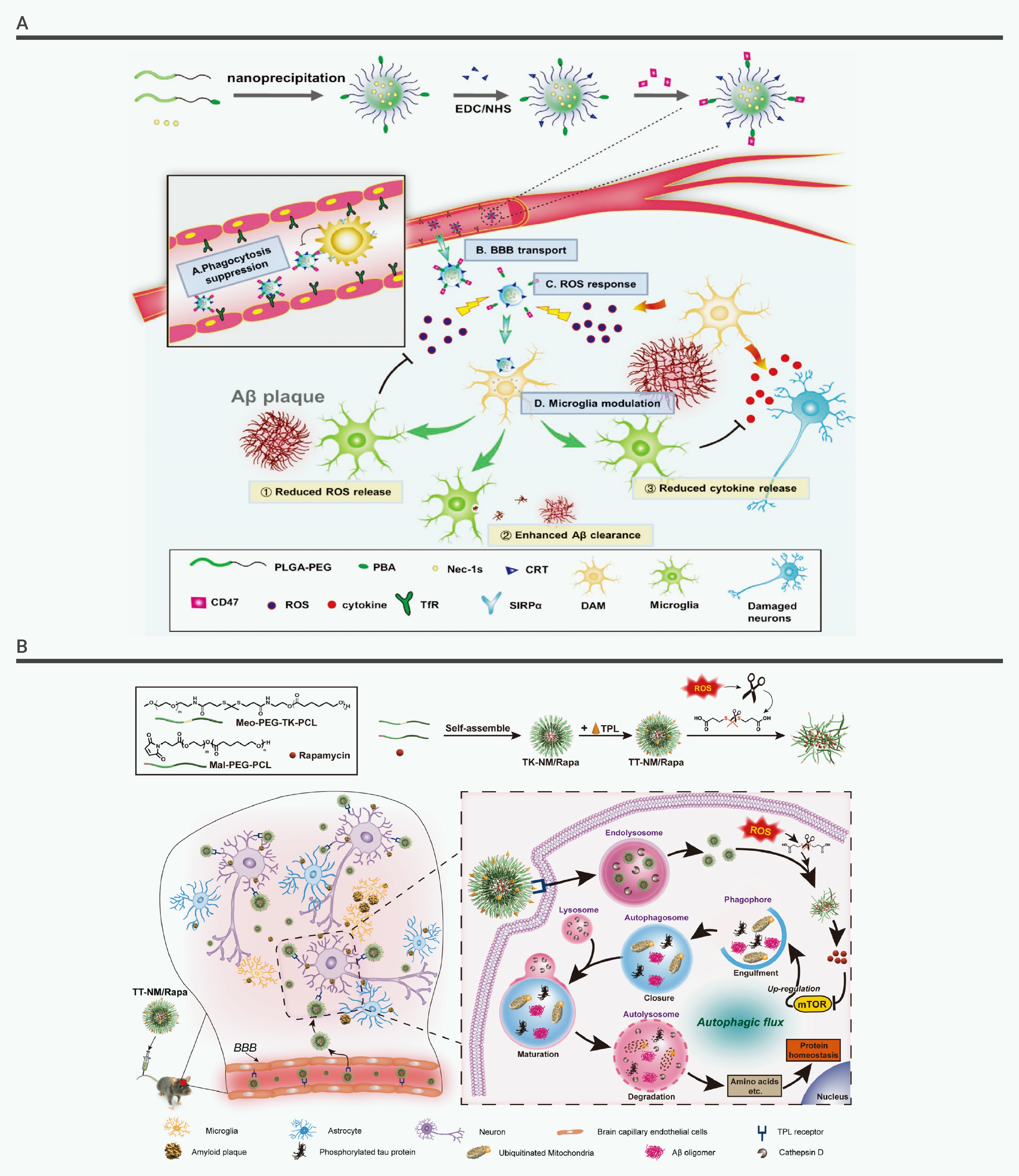
Figure 6.
Internal stimuli-responsive drug release for the treatment of AD
-
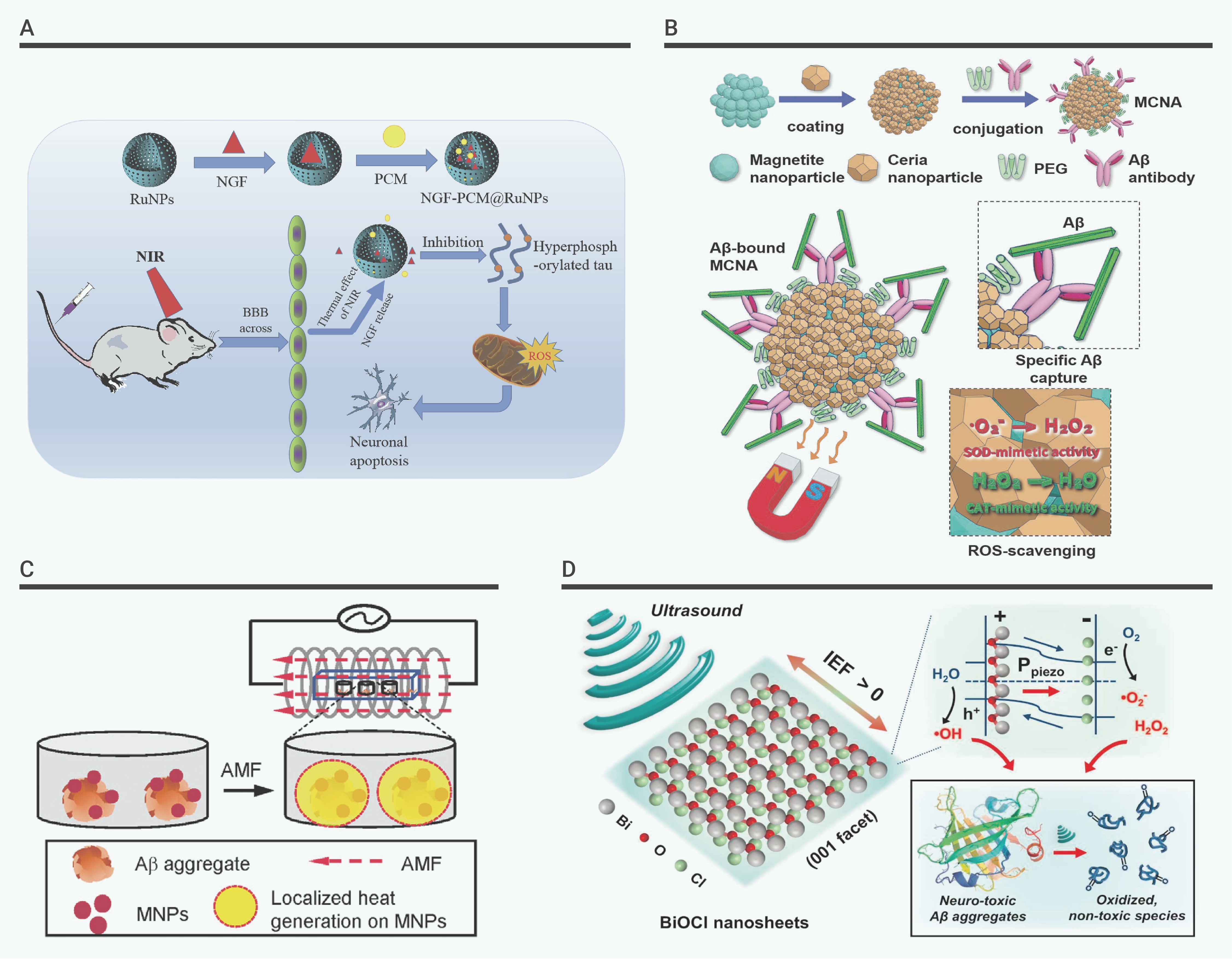
Figure 7.
Exogenous stimuli-responsive drug release for the treatment of AD
-
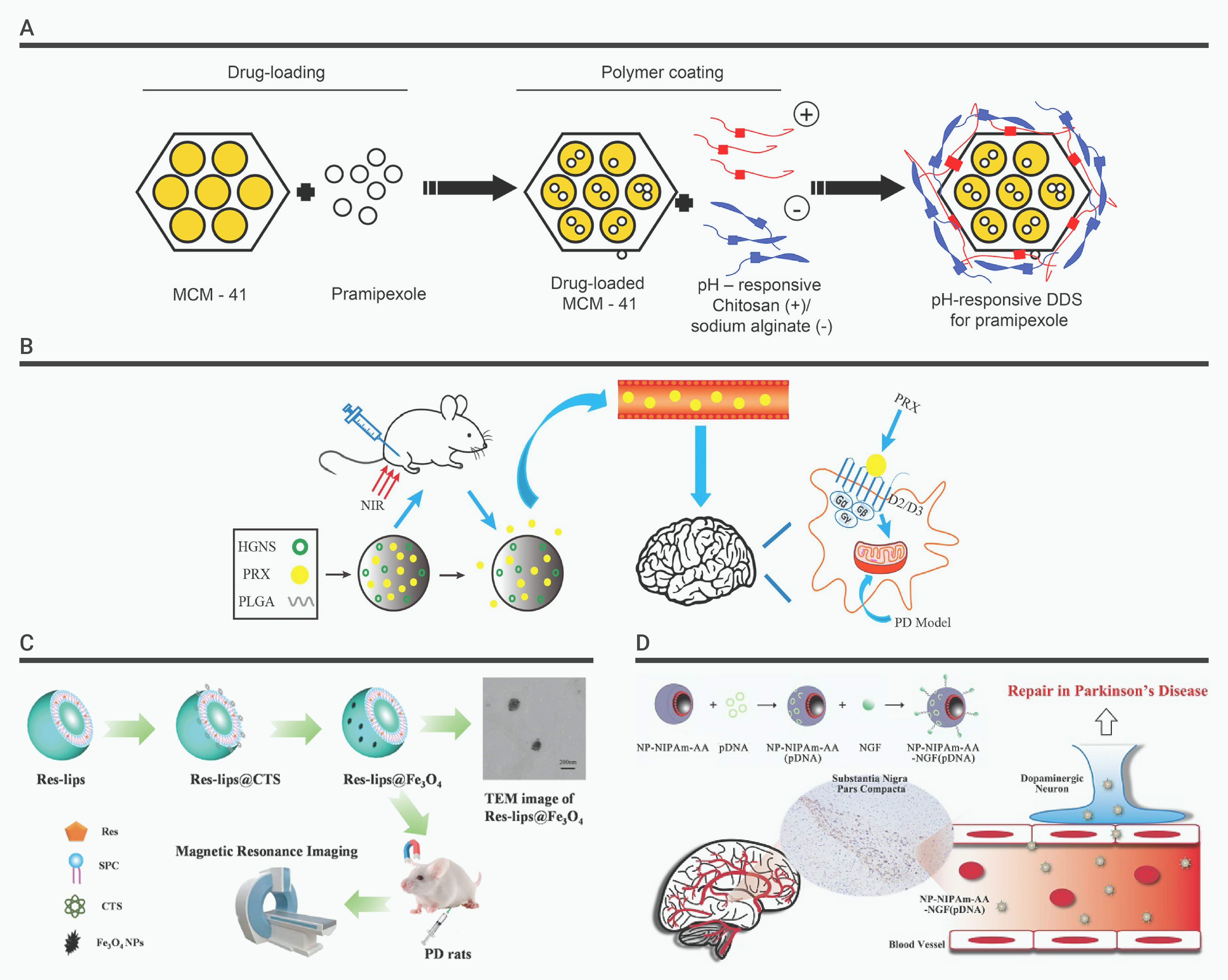
Figure 8.
Stimulus-responsive drug release for the treatment of PD
