
-
GRAPHICAL ABSTRACT
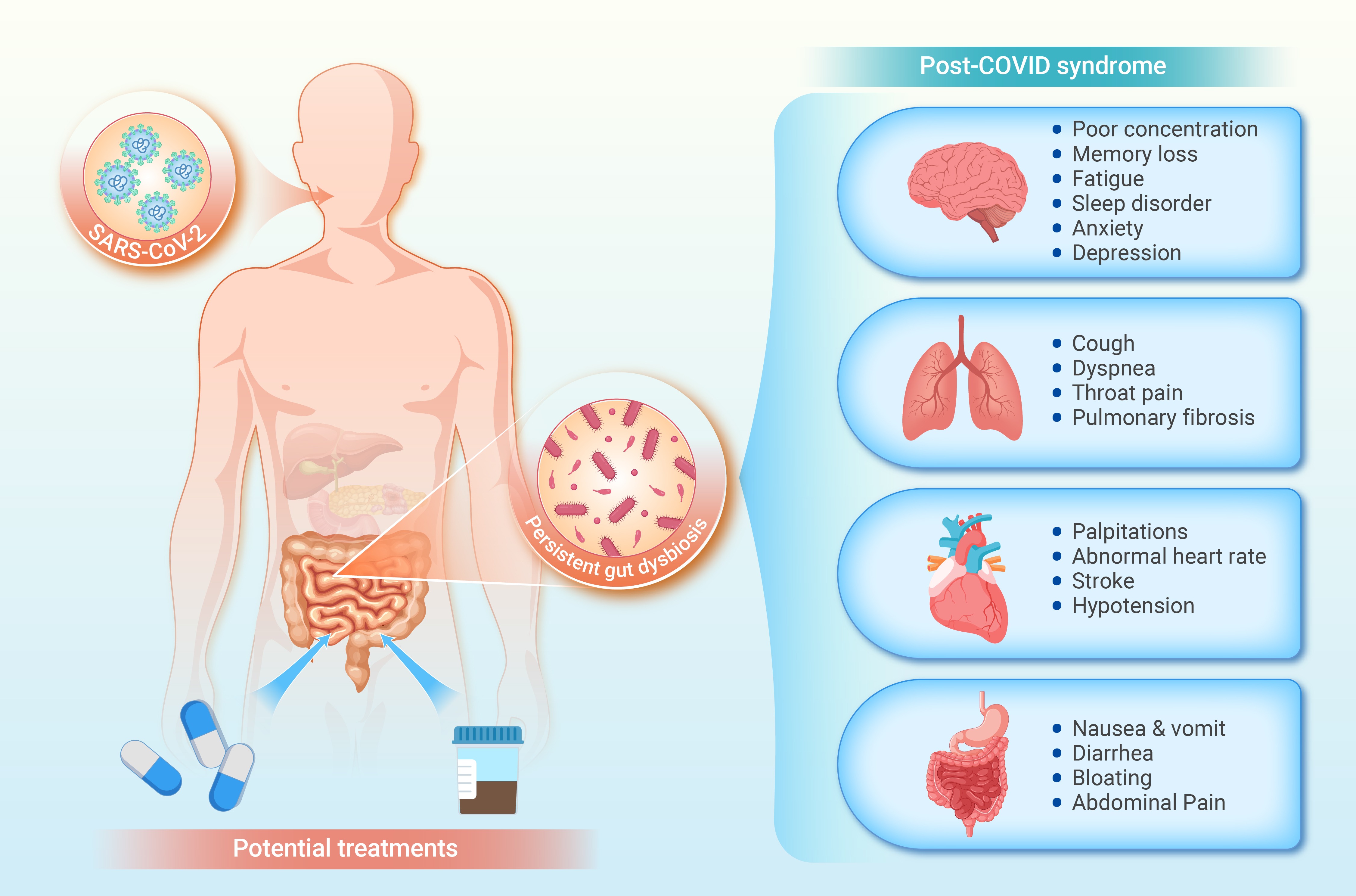
 DownLoad:
Full size image
DownLoad:
Full size image
-
PUBLIC SUMMARY
-
COVID-19 may result in gut dysbiosis, with effects lasting months or longer.
Gut dysbiosis is linked to symptoms like fatigue and cognitive issues in post-COVID syndrome.
Gut microbiome influences systemic health through gut-organ axes in post-COVID syndrome.
Modulating gut microbiota offers potential for treatments of post-COVID syndrome.
-
-
ABSTRACT
Post-COVID syndrome (PCS) is a debilitating condition that affects a significant number of COVID-19 survivors worldwide. PCS is characterized by persistent symptoms such as fatigue, shortness of breath, cognitive impairment, and gastrointestinal issues. The frequent occurrence of gastrointestinal symptoms has prompted researchers to investigate the role of the gut microbiota in the development and progression of PCS. This review synthesizes findings from recent studies, providing a comprehensive overview of gut microbiota changes in both COVID-19 patients and PCS patients. Evidence suggests that COVID-19 can cause lasting alterations in gut microbiota, marked by reduced microbial diversity, an increase in opportunistic pathogens, and a decrease in beneficial bacteria that support immune and metabolic health. In addition, host-microbe interactions such as the gut-lung, gut-brain, and gut-heart axes are explored to understand the impact of gut microbes on systemic health. Finally, potential diagnostic and therapeutic strategies for PCS are discussed, including the use of probiotics, prebiotics, and fecal microbiota transplantation (FMT) to restore microbial balance and alleviate symptoms. -

-
REFERENCES
[1] Soriano J. B., Murthy S., Marshall J. C., et al. (2022). A clinical case definition of post-COVID-19 condition by a Delphi consensus. Lancet Infect. Dis. 22:e102−7. DOI:10.1016/S1473-3099(21)00703-9 [2] Woodrow M., Carey C., Ziauddeen N., et al. (2023). Systematic review of the prevalence of Long COVID. Open Forum Infect. Dis. 10:ofad233. DOI:10.1093/ofid/ofad233 [3] Chen C., Haupert S. R., Zimmermann L., et al. (2022). Global prevalence of post-Coronavirus Disease 2019 (COVID-19) condition or Long COVID: A meta-analysis and systematic review. J. Infect. Dis. 226:1593−607. DOI:10.1093/infdis/jiac136 [4] Sk Abd Razak R., Ismail A., Abdul Aziz A. F., et al. (2024). Post-COVID syndrome prevalence: A systematic review and meta-analysis. BMC Public Health 24:1785. DOI:10.1186/s12889-024-19264-5 [5] Tkacheva O. N., Klimenko N. S., Kashtanova D. A., et al. (2023). Gut microbiome in post-COVID-19 patients is linked to Immune and cardiovascular health status but not COVID-19 severity. Microorganisms 11:1036. DOI:10.3390/microorganisms11041036 [6] Antonelli M., Penfold R. S., Canas L. D. S., et al. (2023). SARS-CoV-2 infection following booster vaccination: Illness and symptom profile in a prospective, observational community-based case-control study. J. Infect. 87:506−15. DOI:10.1016/j.jinf.2023.08.009 [7] Davis H. E., McCorkell L., Vogel J. M., et al. (2023). Long COVID: Major findings, mechanisms and recommendations. Nat. Rev. Microbiol. 21:133−46. DOI:10.1038/s41579-022-00846-2 [8] Al-Aly Z., Bowe B. and Xie Y. (2022). Long COVID after breakthrough SARS-CoV-2 infection. Nat. Med. 28:1461−7. DOI:10.1038/s41591-022-01840-0 [9] Mirin A. A. (2022). A preliminary estimate of the economic impact of long COVID in the United States. Fatigue: Biomedicine, Health & Behavior 10:190−9. DOI:10.1080/21641846.2022.2124064 [10] World Health Organization. Public health and social measures during health emergencies. https://www.who.int/initiatives/who-public-health-and-social-measures-initiative. [11] Proal A. D. and VanElzakker M. B. (2021). Long COVID or post-acute sequelae of COVID-19 (PASC): An overview of biological factors that may contribute to persistent symptoms. Front. Microbiol. 12:698169. DOI:10.3389/fmicb.2021.698169 [12] Swank Z., Senussi Y., Manickas-Hill Z., et al. (2023). Persistent circulating severe acute respiratory syndrome coronavirus 2 spike is associated with post-acute coronavirus disease 2019 sequelae. Clin. Infect. Dis. 76:e487−90. DOI:10.1093/cid/ciac722 [13] Liu Q., Mak J. W. Y., Su Q., et al. (2022). Gut microbiota dynamics in a prospective cohort of patients with post-acute COVID-19 syndrome. Gut 71:544−52. DOI:10.1136/gutjnl-2021-325989 [14] Yeoh Y. K., Zuo T., Lui G. C. Y., et al. (2021). Gut microbiota composition reflects disease severity and dysfunctional immune responses in patients with COVID-19. Gut 70:698−706. DOI:10.1136/gutjnl-2020-323020 [15] Zubchenko S., Kril I., Nadizhko O., et al. (2022). Herpesvirus infections and post-COVID-19 manifestations: A pilot observational study. Rheumatol. Int. 42:1523−30. DOI:10.1007/s00296-022-05146-9 [16] Choudhury A., Tariq R., Jena A., et al. (2022). Gastrointestinal manifestations of long COVID: A systematic review and meta-analysis. Ther. Adv. Gastroenterol. 15: 17562848221118403. DOI: 10.1177/17562848221118403 [17] Sender R., Fuchs S. and Milo R. (2016). Revised estimates for the number of human and bacteria cells in the body. PLoS Biol. 14:e1002533. DOI:10.1371/journal.pbio.1002533 [18] Ronan V., Yeasin R. and Claud E. C. (2021). Childhood development and the microbiome—the intestinal microbiota in maintenance of health and development of disease during childhood development. Gastroenterology 160:495−506. DOI:10.1053/j.gastro.2020.08.065 [19] Dominguez-Bello M. G., Costello E. K., Contreras M., et al. (2010). Delivery mode shapes the acquisition and structure of the initial microbiota across multiple body habitats in newborns. Proc. Natl. Acad. Sci. U. S. A. 107:11971−5. DOI:10.1073/pnas.1002601107 [20] Chee W. J. Y., Chew S. Y. and Than L. T. L. (2020). Vaginal microbiota and the potential of Lactobacillus derivatives in maintaining vaginal health. Microb. Cell Fact. 19:203. DOI:10.1186/s12934-020-01464-4 [21] Ahlawat S., Asha and Sharma K. K. (2021). Gut–organ axis: a microbial outreach and networking. Lett. Appl. Microbiol. 72:636−68. DOI:10.1111/lam.13333 [22] Borrego-Ruiz A. and Borrego J. J. (2024). Neurodevelopmental disorders associated with gut microbiome dysbiosis in children. Children 11:796. DOI:10.3390/children11070796 [23] Zhang F., Lau R. I., Liu Q., et al. (2023). Gut microbiota in COVID-19: Key microbial changes, potential mechanisms and clinical applications. Nat. Rev. Gastroenterol. Hepatol. 20:323−37. DOI:10.1038/s41575-022-00698-4 [24] Xu R., Lu R., Zhang T., et al. (2021). Temporal association between human upper respiratory and gut bacterial microbiomes during the course of COVID-19 in adults. Commun. Biol. 4:1−11. DOI:10.1038/s42003-021-01796-w [25] Gaibani P., D’Amico F., Bartoletti M., et al. (2021). The gut microbiota of critically ill patients with COVID-19. Front. Cell. Infect. Microbiol. 11:670424. DOI:10.3389/fcimb.2021.670424 [26] Gu S., Chen Y., Wu Z., et al. (2020). Alterations of the gut microbiota in patients with coronavirus disease 2019 or H1N1 influenza. Clin. Infect. Dis. 71:2669−78. DOI:10.1093/cid/ciaa709 [27] Ren Z., Wang H., Cui G., et al. (2021). Alterations in the human oral and gut microbiomes and lipidomics in COVID-19. Gut 70:1253−65. DOI:10.1136/gutjnl-2020-323826 [28] Zuo T., Zhang F., Lui G. C. Y., et al. (2020). Alterations in gut microbiota of patients with COVID-19 during time of hospitalization. Gastroenterology 159:944−955.e8. DOI:10.1053/j.gastro.2020.05.048 [29] Mizutani T., Ishizaka A., Koga M., et al. (2022). Correlation analysis between gut microbiota alterations and the cytokine response in patients with coronavirus disease during hospitalization. Microbiol. Spectr. 10:e01689−21. DOI:10.1128/spectrum.01689-21 [30] Reinold J., Farahpour F., Fehring C., et al. (2021). A pro-inflammatory gut microbiome characterizes SARS-CoV-2 infected patients and a reduction in the connectivity of an anti-inflammatory bacterial network associates with severe COVID-19. Front. Cell. Infect. Microbiol. 11:747816. DOI:10.3389/fcimb.2021.747816 [31] Tang L., Gu S., Gong Y., et al. (2020). Clinical significance of the correlation between changes in the major intestinal bacteria species and COVID-19 severity. Eng. 6:1178−1184. DOI:10.1016/j.eng.2020.05.013 [32] Tao W., Zhang G., Wang X., et al. (2020). Analysis of the intestinal microbiota in COVID-19 patients and its correlation with the inflammatory factor IL-18. Med. Microecol. 5:100023. DOI:10.1016/j.medmic.2020.100023 [33] Li S., Yang S., Zhou Y., et al. (2021). Microbiome profiling using shotgun metagenomic sequencing identified unique microorganisms in COVID-19 patients with altered gut microbiota. Front. Microbiol. 12:712081. DOI:10.3389/fmicb.2021.712081 [34] Wu Y., Cheng X., Jiang G., et al. (2021). Altered oral and gut microbiota and its association with SARS-CoV-2 viral load in COVID-19 patients during hospitalization. npj Biofilms Microbiomes 7:61. DOI:10.1038/s41522-021-00232-5 [35] Righi E., Dalla Vecchia I., Auerbach N., et al. (2024). Gut microbiome disruption following SARS-CoV-2: a review. Microorganisms 12:131. DOI:10.3390/microorganisms12010131 [36] Rafiqul Islam S. M., Foysal Md. J., Hoque M. N., et al. (2022). Dysbiosis of oral and gut microbiomes in SARS-CoV-2 infected patients in Bangladesh: Elucidating the role of opportunistic gut microbes. Front. Med. 9:821777. DOI:10.3389/fmed.2022.821777 [37] Viana S. D., Nunes S. and Reis F. (2020). ACE2 imbalance as a key player for the poor outcomes in COVID-19 patients with age-related comorbidities – Role of gut microbiota dysbiosis. Ageing Res. Rev. 62:101123. DOI:10.1016/j.arr.2020.101123 [38] Zuo T., Zhan H., Zhang F., et al. (2020). Alterations in fecal fungal microbiome of patients with COVID-19 during time of hospitalization until discharge. Gastroenterology 159:1302−1310.e5. DOI:10.1053/j.gastro.2020.06.048 [39] Lv L., Gu S., Jiang H., et al. (2021). Gut mycobiota alterations in patients with COVID-19 and H1N1 infections and their associations with clinical features. Commun. Biol. 4:1−11. DOI:10.1038/s42003-021-02036-x [40] Roudbary M., Kumar S., Kumar A., et al. (2021). Overview on the prevalence of fungal infections, immune response, and microbiome role in COVID-19 patients. J. Fungi 7:720. DOI:10.3390/jof7090720 [41] Zuo T., Liu Q., Zhang F., et al. (2020). Depicting SARS-CoV-2 faecal viral activity in association with gut microbiota composition in patients with COVID-19. Gut : gutjnl-2020-322294. DOI: 10.1136/gutjnl-2020-322294 [42] Virgin H. W. (2014). The virome in mammalian physiology and disease. Cell 157:142−50. DOI:10.1016/j.cell.2014.02.032 [43] Zuo T., Wu X., Wen W., et al. (2021). Gut microbiome alterations in COVID-19. Genomics Proteomics Bioinformatics 19:679−88. DOI:10.1016/j.gpb.2021.09.004 [44] Zuo T., Liu Q., Zhang F., et al. (2021). Temporal landscape of human gut RNA and DNA virome in SARS-CoV-2 infection and severity. Microbiome 9:91. DOI:10.1186/s40168-021-01008-x [45] Cao J., Wang C., Zhang Y., et al. (2021). Integrated gut virome and bacteriome dynamics in COVID-19 patients. Gut Microbes 13:1−21. DOI:10.1080/19490976.2021.1887722 [46] Karami Z., Knoop B. T., Dofferhoff A. S. M., et al. (2021). Few bacterial co-infections but frequent empiric antibiotic use in the early phase of hospitalized patients with COVID-19: Results from a multicentre retrospective cohort study in The Netherlands. Infect. Dis. 53:102−10. DOI:10.1080/23744235.2020.1839672 [47] Langford B. J., So M., Raybardhan S., et al. (2021). Antibiotic prescribing in patients with COVID-19: rapid review and meta-analysis. Clin. Microbiol. Infect. 27:520−31. DOI:10.1016/j.cmi.2020.12.018 [48] Alshaikh F. S., Godman B., Sindi O. N., et al. (2022). Prevalence of bacterial coinfection and patterns of antibiotics prescribing in patients with COVID-19: A systematic review and meta-analysis. PLoS ONE 17:e0272375. DOI:10.1371/journal.pone.0272375 [49] Nandi A., Pecetta S. and Bloom D. E. (2023). Global antibiotic use during the COVID-19 pandemic: analysis of pharmaceutical sales data from 71 countries, 2020–2022. eClinicalMedicine 57:101848. DOI:10.1016/j.eclinm.2023.101848 [50] Modi S. R., Collins J. J. and Relman D. A. (2014). Antibiotics and the gut microbiota. J. Clin. Invest. 124:4212−8. DOI:10.1172/JCI72333 [51] Kim S., Covington A. and Pamer E. G. (2017). The intestinal microbiota: Antibiotics, colonization resistance, and enteric pathogens. Immunol. Rev. 279:90−105. DOI:10.1111/imr.12563 [52] Bernard-Raichon L., Venzon M., Klein J., et al. (2022). Gut microbiome dysbiosis in antibiotic-treated COVID-19 patients is associated with microbial translocation and bacteremia. Nat. Commun. 13:5926. DOI:10.1038/s41467-022-33395-6 [53] Taur Y., Xavier J. B., Lipuma L., et al. (2012). Intestinal domination and the risk of bacteremia in patients undergoing allogeneic hematopoietic stem cell transplantation. Clin. Infect. Dis. 55:905−14. DOI:10.1093/cid/cis580 [54] Hegazy M., Ashoush O. A., Hegazy M. T., et al. (2022). Beyond probiotic legend: ESSAP gut microbiota health score to delineate SARS-CoV-2 infection severity. Br. J. Nutr. 127:1180−9. DOI:10.1017/S0007114521001926 [55] Sun G., Lin K., Ai J., et al. (2024). The efficacy of antivirals, corticosteroids, and monoclonal antibodies as acute COVID-19 treatments in reducing the incidence of long COVID: A systematic review and meta-analysis. Clin. Microbiol. Infect. 30:1505−13. DOI:10.1016/j.cmi.2024.07.006 [56] Narayanan A., Kieri O., Vesterbacka J., et al. (2024). Exploring the interplay between antiretroviral therapy and the gut-oral microbiome axis in people living with HIV. Sci. Rep. 14:17820. DOI:10.1038/s41598-024-68479-4 [57] Maier L., Pruteanu M., Kuhn M., et al. (2018). Extensive impact of non-antibiotic drugs on human gut bacteria. Nature 555:623−8. DOI:10.1038/nature25979 [58] Cheng X., Zhang Y., Li Y., et al. (2022). Meta-analysis of 16S rRNA microbial data identified alterations of the gut microbiota in COVID-19 patients during the acute and recovery phases. BMC Microbiol. 22:274. DOI:10.1186/s12866-022-02686-9 [59] Chen Y., Gu S., Chen Y., et al. (2022). Six-month follow-up of gut microbiota richness in patients with COVID-19. Gut 71:222−5. DOI:10.1136/gutjnl-2021-324090 [60] Cui G. Y., Rao B. C., Zeng Z. H., et al. (2022). Characterization of oral and gut microbiome and plasma metabolomics in COVID-19 patients after 1-year follow-up. Mil. Med. Res. 9:32. DOI:10.1186/s40779-022-00387-y [61] Zhang D., Zhou Y., Ma Y., et al. (2023). Gut microbiota dysbiosis correlates with long COVID-19 at one-year after discharge. J. Korean Med. Sci. 38:e120. DOI:10.3346/jkms.2023.38.e120 [62] Zhang F., Wan Y., Zuo T., et al. (2022). Prolonged impairment of short-chain fatty acid and L-isoleucine biosynthesis in gut microbiome in patients with COVID-19. Gastroenterology 162:548−561.e4. DOI:10.1053/j.gastro.2021.10.013 [63] Yao Y., Cai X., Fei W., et al. (2022). The role of short-chain fatty acids in immunity, inflammation and metabolism. Crit. Rev. Food Sci. Nutr. 62:1−12. DOI:10.1080/10408398.2020.1854675 [64] Liu J., Tan Y., Cheng H., et al. (2022). Functions of gut microbiota metabolites, current status and future perspectives. Aging Dis. 13:1106−26. DOI:10.14336/AD.2022.0104 [65] Kearns R. (2024). The kynurenine pathway in gut permeability and inflammation. Inflammation. DOI: 10.1007/s10753-024-02135-x. [66] Xue C., Li G., Zheng Q., et al. (2023). Tryptophan metabolism in health and disease. Cell Metab. 35:1304−26. DOI:10.1016/j.cmet.2023.06.004 [67] Cysique L. A., Jakabek D., Bracken S. G., et al. (2023). The kynurenine pathway relates to post-acute COVID-19 objective cognitive impairment and PASC. Ann. Clin. Transl. Neurol. 10:1338−52. DOI:10.1002/acn3.51825 [68] Agus A., Planchais J. and Sokol H. (2018). Gut microbiota regulation of tryptophan metabolism in health and disease. Cell Host Microbe 23:716−24. DOI:10.1016/j.chom.2018.05.003 [69] Wong A. C., Devason A. S., Umana I. C., et al. (2023). Serotonin reduction in post-acute sequelae of viral infection. Cell 186:4851−4867.e20. DOI:10.1016/j.cell.2023.09.013 [70] Alenina N. and Klempin F. (2015). The role of serotonin in adult hippocampal neurogenesis. Behav. Brain Res. 277:49−57. DOI:10.1016/j.bbr.2014.07.038 [71] López-Hernández Y., Monárrez-Espino J., López D. A. G., et al. (2023). The plasma metabolome of long COVID patients two years after infection. Sci. Rep. 13:12420. DOI:10.1038/s41598-023-39049-x [72] Mahdi A., Zhao A., Fredengren E., et al. (2023). Dysregulations in hemostasis, metabolism, immune response, and angiogenesis in post-acute COVID-19 syndrome with and without postural orthostatic tachycardia syndrome: A multi-omic profiling study. Sci. Rep. 13:20230. DOI:10.1038/s41598-023-47539-1 [73] Sadlier C., Albrich W. C., Neogi U., et al. (2022). Metabolic rewiring and serotonin depletion in patients with postacute sequelae of COVID-19. Allergy 77:1623−5. DOI:10.1111/all.15253 [74] Zhou Y., Zhang J., Zhang D., et al. (2021). Linking the gut microbiota to persistent symptoms in survivors of COVID-19 after discharge. J. Microbiol. 59:941−8. DOI:10.1007/s12275-021-1206-5 [75] Vestad B., Ueland T., Lerum T. V., et al. (2022). Respiratory dysfunction three months after severe COVID‐19 is associated with gut microbiota alterations. J. Intern. Med. 291:801−12. DOI:10.1111/joim.13458 [76] Österdahl M. F., Whiston R., Sudre C. H., et al. (2023). Metabolomic and gut microbiome profiles across the spectrum of community-based COVID and non-COVID disease. Sci. Rep. 13:10407. DOI:10.1038/s41598-023-34598-7 [77] Hamrefors V., Kahn F., Holmqvist M., et al. (2024). Gut microbiota composition is altered in postural orthostatic tachycardia syndrome and post-acute COVID-19 syndrome. Sci. Rep. 14:3389. DOI:10.1038/s41598-024-53784-9 [78] Zhang D., Weng S., Xia C., et al. (2023). Gastrointestinal symptoms of long COVID-19 related to the ectopic colonization of specific bacteria that move between the upper and lower alimentary tract and alterations in serum metabolites. BMC Med. 21:264. DOI:10.1186/s12916-023-02972-x [79] Su Q., Lau R. I., Liu Q., et al. (2023). Post-acute COVID-19 syndrome and gut dysbiosis linger beyond 1 year after SARS-CoV-2 clearance. Gut 72:1230−2. DOI:10.1136/gutjnl-2022-328319 [80] Su Q., Lau R. I., Liu Q., et al. (2024). The gut microbiome associates with phenotypic manifestations of post-acute COVID-19 syndrome. Cell Host Microbe 32:651−660.e4. DOI:10.1016/j.chom.2024.04.005 [81] Wu C., Jiang M. L., Jiang R., et al. (2023). The roles of fungus in CNS autoimmune and neurodegeneration disorders. Front. Immunol. 13:1077335. DOI:10.3389/fimmu.2022.1077335 [82] Giron L. B., Peluso M. J., Ding J., et al. (2022). Markers of fungal translocation are elevated during post-acute sequelae of SARS-CoV-2 and induce NF-κB signaling. JCI Insight 7:e160989. DOI:10.1172/jci.insight.160989 [83] Guo C., Yi B., Wu J., et al. (2023). The microbiome in post-acute infection syndrome (PAIS). Comput. Struct. Biotechnol. J. 21:3904−11. DOI:10.1016/j.csbj.2023.08.002 [84] Haffke M., Freitag H., Rudolf G., et al. (2022). Endothelial dysfunction and altered endothelial biomarkers in patients with post-COVID-19 syndrome and chronic fatigue syndrome (ME/CFS). J. Transl. Med. 20:138. DOI:10.1186/s12967-022-03346-2 [85] Jurek J. M. and Castro-Marrero J. (2024). A narrative review on gut microbiome disturbances and microbial preparations in myalgic encephalomyelitis/chronic fatigue syndrome: Implications for long COVID. Nutrients 16:1545. DOI:10.3390/nu16111545 [86] König R. S., Albrich W. C., Kahlert C. R., et al. (2022). The gut microbiome in myalgic encephalomyelitis (ME)/chronic fatigue syndrome (CFS). Front. Immunol. 12:628741. DOI:10.3389/fimmu.2021.628741 [87] Guo C., Che X., Briese T., et al. (2023). Deficient butyrate-producing capacity in the gut microbiome is associated with bacterial network disturbances and fatigue symptoms in ME/CFS. Cell Host Microbe 31:288−304.e8. DOI:10.1016/j.chom.2023.01.004 [88] Giloteaux L., Goodrich J. K., Walters W. A., et al. (2016). Reduced diversity and altered composition of the gut microbiome in individuals with myalgic encephalomyelitis/chronic fatigue syndrome. Microbiome 4:30. DOI:10.1186/s40168-016-0171-4 [89] Maes M., Mihaylova I. and Leunis J. C. (2007). Increased serum IgA and IgM against LPS of enterobacteria in chronic fatigue syndrome (CFS): Indication for the involvement of gram-negative enterobacteria in the etiology of CFS and for the presence of an increased gut-intestinal permeability. J. Affect. Disord. 99:237−40. DOI:10.1016/j.jad.2006.08.021 [90] Armstrong C. W., McGregor N. R., Lewis D. P., et al. (2015). Metabolic profiling reveals anomalous energy metabolism and oxidative stress pathways in chronic fatigue syndrome patients. Metabolomics 11:1626−39. DOI:10.1007/s11306-015-0816-5 [91] Nagy-Szakal D., Barupal D. K., Lee B., et al. (2018). Insights into myalgic encephalomyelitis/chronic fatigue syndrome phenotypes through comprehensive metabolomics. Sci. Rep. 8:10056. DOI:10.1038/s41598-018-28477-9 [92] Budden K. F., Gellatly S. L., Wood D. L. A., et al. (2017). Emerging pathogenic links between microbiota and the gut–lung axis. Nat. Rev. Microbiol. 15:55−63. DOI:10.1038/nrmicro.2016.142 [93] Wang L., Cai Y., Garssen J., et al. (2023). The bidirectional gut–lung axis in chronic obstructive pulmonary disease. Am. J. Respir. Crit. Care Med. 207:1145−60. DOI:10.1164/rccm.202206-1066TR [94] Allali I., Bakri Y., Amzazi S., et al. (2021). Gut-lung axis in COVID-19. Can. J. Infect. Dis. Med. Microbiol. DOI:10.1155/2021/6655380 [95] Vutcovici M., Bitton A., Ernst P., et al. (2016). Inflammatory bowel disease and risk of mortality in COPD. Eur. Respir. J. 47:1357−64. DOI:10.1183/13993003.01945-2015 [96] Sencio V., Barthelemy A., Tavares L. P., et al. (2020). Gut dysbiosis during influenza contributes to pulmonary pneumococcal superinfection through altered short-chain fatty acid production. Cell Rep. 30:2934−2947.e6. DOI:10.1016/j.celrep.2020.02.013 [97] Vaughan A., Frazer Z. A., Hansbro P. M., et al. (2019). COPD and the gut-lung axis: the therapeutic potential of fibre. J. Thorac. Dis. 11:S2173−80. DOI:10.21037/jtd.2019.10.40 [98] Tian X., Hellman J., Horswill A. R., et al. (2019). Elevated gut microbiome-derived propionate levels are associated with reduced sterile lung inflammation and bacterial immunity in mice. Front. Microbiol. 10:159. DOI:10.3389/fmicb.2019.00159 [99] Chen D., Qiu Y. B., Gao Z. Q., et al. (2020). Sodium propionate attenuates the lipopolysaccharide-induced epithelial-mesenchymal transition via the PI3K/AKT/mTOR signaling pathway. J. Agric. Food Chem. 68:6554−63. DOI:10.1021/acs.jafc.0c01302 [100] Scheithauer T. P. M., Montijn R. C., Mieremet A. (2024). Gut microbe-host interactions in post-COVID syndrome: A debilitating or restorative partnership. Gut Microbes 16:2402544. DOI:10.1080/19490976.2024.2402544 [101] Cho S. J., Hong K. S., Schenck E., et al. (2021). Decreased IDO1-dependent tryptophan metabolism in aged lung during influenza. Eur. Respir. J. 57:2000443. DOI:10.1183/13993003.00443-2020 [102] Agustí A., García-Pardo M. P., López-Almela I., et al. (2018). Interplay between the gut-brain axis, obesity and cognitive function. Front. Neurosci. 12:155. DOI:10.3389/fnins.2018.00155 [103] Cryan J. F., O’Riordan K. J., Cowan C. S. M., et al. (2019). The microbiota-gut-brain axis. Physiol. Rev. 99:1877−2013. DOI:10.1152/physrev.00018.2018 [104] Berthoud H. R. (2008). Vagal and hormonal gut-brain communication: From satiation to satisfaction. Neurogastroenterol. Motil. 20:64–72. DOI:10.1111/j.1365-2982.2008.01104.x [105] Diaz Heijtz R., Wang S., Anuar F., et al. (2011). Normal gut microbiota modulates brain development and behavior. Proc. Natl. Acad. Sci. U.S.A. 108:3047−52. DOI:10.1073/pnas.1010529108 [106] Bernstein C. N. (2017). The brain-gut axis and stress in inflammatory bowel disease. Gastroenterol. Clin. North Am. 46:839−46. DOI:10.1016/j.gtc.2017.08.006 [107] Kennedy P. J., Cryan J. F., Dinan T. G., et al. (2014). Irritable bowel syndrome: A microbiome-gut-brain axis disorder. World J. Gastroenterol. 20:14105−25. DOI:10.3748/wjg.v20.i39.14105 [108] Sanna S., van Zuydam N. R., Mahajan A., et al. (2019). Causal relationships among the gut microbiome, short-chain fatty acids and metabolic diseases. Nat. Genet. 51:600−5. DOI:10.1038/s41588-019-0350-x [109] Torres-Fuentes C., Schellekens H., Dinan T. G., et al. (2017). The microbiota-gut-brain axis in obesity. Lancet Gastroenterol. Hepatol. 2:747−56. DOI:10.1016/S2468-1253(17)30147-4 [110] Zhang L., Wang Y., Xiayu X., et al. (2017). Altered gut microbiota in a mouse model of Alzheimer’s disease. J. Alzheimers Dis. 60:1241−57. DOI:10.3233/JAD-170020 [111] Hsiao E. Y., McBride S. W., Hsien S., et al. (2013). Microbiota modulate behavioral and physiological abnormalities associated with neurodevelopmental disorders. Cell 155:1451−63. DOI:10.1016/j.cell.2013.11.024 [112] Rao M., Gershon M. D. (2016). The bowel and beyond: The enteric nervous system in neurological disorders. Nat. Rev. Gastroenterol. Hepatol. 13:517−28. DOI:10.1038/nrgastro.2016.107 [113] Rutsch A., Kantsjö J. B., Ronchi F. (2020). The gut-brain axis: How microbiota and host inflammasome influence brain physiology and pathology. Front. Immunol. 11:604179. DOI:10.3389/fimmu.2020.604179 [114] Roh E., Choi K. M. (2023). Hormonal gut–brain signaling for the treatment of obesity. Int. J. Mol. Sci. 24:3384. DOI:10.3390/ijms24043384 [115] Krishnan K., Lin Y., Prewitt K. R. M., et al. (2022). Multidisciplinary approach to brain fog and related persisting symptoms post COVID-19. J. Health Serv. Psychol. 48:31−8. DOI:10.1007/s42843-022-00056-7 [116] Hamming I., Timens W., Bulthuis M., et al. (2004). Tissue distribution of ACE2 protein, the functional receptor for SARS coronavirus. A first step in understanding SARS pathogenesis. J. Pathol. 203:631−7. DOI:10.1002/path.1570 [117] Plummer A. M., Matos Y. L., Lin H. C., et al. (2023). Gut-brain pathogenesis of post-acute COVID-19 neurocognitive symptoms. Front. Neurosci. 17:1232480. DOI:10.3389/fnins.2023.1232480 [118] Elyaspour Z., Zibaeenezhad M. J., Razmkhah M., et al. (2021). Is it all about endothelial dysfunction and thrombosis formation? The secret of COVID-19. Clin. Appl. Thromb. Hemost. 27:10760296211042940. DOI:10.1177/10760296211042940 [119] Zhang H., Li H. B., Lyu J. R., et al. (2020). Specific ACE2 expression in small intestinal enterocytes may cause gastrointestinal symptoms and injury after 2019-nCoV infection. Int. J. Infect. Dis. 96:19−24. DOI:10.1016/j.ijid.2020.04.027 [120] Tyagi K., Rai P., Gautam A., et al. (2023). Neurological manifestations of SARS-CoV-2: Complexity, mechanism and associated disorders. Eur. J. Med. Res. 28:307. DOI:10.1186/s40001-023-01293-2 [121] Almutairi M. M., Sivandzade F., Albekairi T. H., et al. (2021). Neuroinflammation and its impact on the pathogenesis of COVID-19. Front. Med. 8:745789. DOI:10.3389/fmed.2021.745789 [122] Boldrini M., Canoll P. D., Klein R. S. (2021). How COVID-19 affects the brain. JAMA Psychiatry 78:682−3. DOI:10.1001/jamapsychiatry.2021.0500 [123] Tada H., Fujino N., Hayashi K., et al. (2022). Human genetics and its impact on cardiovascular disease. J. Cardiol. 79:233−9. DOI:10.1016/j.jjcc.2021.09.005 [124] CDC (2024). Heart disease risk factors. Heart Disease. https://www.cdc.gov/heart-disease/risk-factors/index.html. [125] Yusuf S., Joseph P., Rangarajan S., et al. (2020). Modifiable risk factors, cardiovascular disease, and mortality in 155,722 individuals from 21 high-income, middle-income, and low-income countries (PURE): A prospective cohort study. Lancet 395:795−808. DOI:10.1016/S0140-6736(19)32008-2 [126] Badimon L., Chagas P., Chiva-Blanch G. (2019). Diet and cardiovascular disease: Effects of foods and nutrients in classical and emerging cardiovascular risk factors. Curr. Med. Chem. 26:3639−51. DOI:10.2174/0929867324666170428103206 [127] Rahman M. M., Islam F., -Or-Rashid M. H., et al. (2022). The gut microbiota (microbiome) in cardiovascular disease and its therapeutic regulation. Front. Cell Infect. Microbiol. 12:903570. DOI:10.3389/fcimb.2022.903570 [128] Witkowski M., Weeks T. L., Hazen S. L. (2020). Gut microbiota and cardiovascular disease. Circ. Res. 127:553−70. DOI:10.1161/CIRCRESAHA.120.316242 [129] Battson M. L., Lee D. M., Weir T. L., et al. (2018). The gut microbiota as a novel regulator of cardiovascular function and disease. J. Nutr. Biochem. 56:1−15. DOI:10.1016/j.jnutbio.2017.12.010 [130] Razavi A. C., Potts K. S., Kelly T. N., et al. (2019). Sex, gut microbiome, and cardiovascular disease risk. Biol. Sex Differ. 10:29. DOI:10.1186/s13293-019-0240-z [131] Chen X. F., Chen X., Tang X. (2020). Short-chain fatty acid, acylation and cardiovascular diseases. Clin. Sci. (Lond.) 134:657−76. DOI:10.1042/CS20200128 [132] Yang S., Li X., Yang F., et al. (2019). Gut microbiota-dependent marker TMAO in promoting cardiovascular disease: Inflammation mechanism, clinical prognostic, and potential as a therapeutic target. Front. Pharmacol. 10:1360. DOI:10.3389/fphar.2019.01360 [133] Kashtanova D., Tkacheva O., Popenko A., et al. (2017). Gut microbiota and vascular biomarkers in patients without clinical cardiovascular diseases. Artery Res. 18:41−8. DOI:10.1016/j.artres.2017.02.007 [134] van den Munckhof I. C. L., Kurilshikov A., ter Horst R., et al. (2018). Role of gut microbiota in chronic low-grade inflammation as potential driver for atherosclerotic cardiovascular disease: A systematic review of human studies. Obes. Rev. 19:1719−34. DOI:10.1111/obr.12750 [135] Larsen J. M. (2017). The immune response to Prevotella bacteria in chronic inflammatory disease. Immunology 151:363−74. DOI:10.1111/imm.12760 [136] Xu Y., Liu X., Liu X., et al. (2021). The roles of the gut microbiota and chronic low-grade inflammation in older adults with frailty. Front. Cell. Infect. Microbiol. 11:675414. DOI:10.3389/fcimb.2021.675414 [137] Atay A. E., Esen B., Akbas H., et al. (2017). Serum ICAM-1 level and ICAM-1 gene 1462A>G (K469E) polymorphism on microalbuminuria in nondiabetic, nonhypertensive and normolipidemic obese patients: Genetical background of microalbuminuria in obesity. Nefrología 37:381−8. DOI:10.1016/j.nefro.2016.11.025 [138] Koc H. C., Xiao J., Liu W., et al. (2022). Long COVID and its management. Int. J. Biol. Sci. 18:4768−80. DOI:10.7150/ijbs.75056 [139] Tian Y., Ran H., Wen X., et al. (2023). Probiotics improve symptoms of patients with COVID-19 through gut-lung axis: A systematic review and meta-analysis. Front. Nutr. 10:1179432. DOI:10.3389/fnut.2023.1179432 [140] Tomkinson S., Triscott C., Schenk E., et al. (2023). The potential of probiotics as ingestible adjuvants and immune modulators for antiviral immunity and management of SARS-CoV-2 infection and COVID-19. Pathogens 12:928. DOI:10.3390/pathogens12070928 [141] Santinelli L., Laghi L., Innocenti G. P., et al. (2022). Oral bacteriotherapy reduces the occurrence of chronic fatigue in COVID-19 patients. Front. Nutr. 8:756177. DOI:10.3389/fnut.2021.756177 [142] Marinoni B., Rimondi A., Bottaro F., et al. (2023). The role of VSL#3® in the treatment of fatigue and other symptoms in long COVID-19 syndrome: A randomized, double-blind, placebo-controlled pilot study (DELong#3). (Psychiatry and Clinical Psychology). DOI:10.1101/2023.06.28.23291986 [143] Rathi A., Jadhav S. B., Shah N. (2021). A randomized controlled trial of the efficacy of systemic enzymes and probiotics in the resolution of post-COVID fatigue. Medicines 8:47. DOI:10.3390/medicines8090047 [144] Lau R. I., Su Q., Lau I. S. F., et al. (2023). A synbiotic preparation (SIM01) for post-acute COVID-19 syndrome in Hong Kong (RECOVERY): A randomized, double-blind, placebo-controlled trial. Lancet Infect. Dis 24:256−65. DOI:10.1016/S1473-3099(23)00685-0 [145] Ranisavljev M., Stajer V., Todorovic N., et al. (2024). The effects of 3-month supplementation with synbiotic on patient-reported outcomes, exercise tolerance, and brain and muscle metabolism in adult patients with post-COVID-19 chronic fatigue syndrome (STOP-FATIGUE): A randomized placebo-controlled clinical trial. Eur. J. Nutr. 64:28. DOI:10.1007/s00394-024-03546-0 [146] Warda A. K., Rea K., Fitzgerald P., et al. (2019). Heat-killed Lactobacilli alter both microbiota composition and behaviour. Behav. Brain Res. 362:213−23. DOI:10.1016/j.bbr.2018.12.047 [147] Ross K. (2023). Psychobiotics: Are they the future intervention for managing depression and anxiety. A literature review. EXPLORE 19:669−80. DOI:10.1016/j.explore.2023.02.007 [148] Zielińska D., Karbowiak M., Brzezicka A. (2022). The role of psychobiotics to ensure mental health during the COVID-19 pandemic—A current state of knowledge. Int. J. Environ. Res. Public Health 19:11022. DOI:10.3390/ijerph191711022 [149] Miyaoka T., Kanayama M., Wake R., et al. (2018). Clostridium butyricum MIYAIRI 588 as adjunctive therapy for treatment-resistant major depressive disorder: A prospective open-label trial. Clin. Neuropharmacol. 41:151. DOI:10.1097/WNF.0000000000000299 [150] Mohammadi A. A., Jazayeri S., Khosravi-Darani K., et al. (2016). The effects of probiotics on mental health and hypothalamic–pituitary–adrenal axis: A randomized, double-blind, placebo-controlled trial in petrochemical workers. Nutr. Neurosci. 19:387−95. DOI:10.1179/1476830515Y.0000000023 [151] Eskandarzadeh S., Effatpanah M., Khosravi-Darani K., et al. (2021). Efficacy of a multispecies probiotic as adjunctive therapy in generalized anxiety disorder: A double-blind, randomized, placebo-controlled trial. Nutr. Neurosci. 24:102−8. DOI:10.1080/1028415X.2019.1598669 [152] Ait-Belgnaoui A., Colom A., Braniste V., et al. (2014). Probiotic gut effect prevents the chronic psychological stress-induced brain activity abnormality in mice. Neurogastroenterol. Motil. 26:510−20. DOI:10.1111/nmo.12295 [153] Akkasheh G., Kashani-Poor Z., Tajabadi-Ebrahimi M., et al. (2016). Clinical and metabolic response to probiotic administration in patients with major depressive disorder: A randomized, double-blind, placebo-controlled trial. Nutrition 32:315−20. DOI:10.1016/j.nut.2015.09.003 [154] Marotta A., Sarno E., Del Casale A., et al. (2019). Effects of probiotics on cognitive reactivity, mood, and sleep quality. Front. Psychiatry 10:164. DOI:10.3389/fpsyt.2019.00164 [155] Lalitsuradej E., Sirilun S., Sittiprapaporn P., et al. (2022). The effects of synbiotics administration on stress-related parameters in Thai subjects—A preliminary study. Foods 11:759. DOI:10.3390/foods11050759 [156] d’Ettorre G., Ceccarelli G., Marazzato M., et al. (2020). Challenges in the management of SARS-CoV-2 infection: The role of oral bacteriotherapy as a complementary therapeutic strategy to avoid the progression of COVID-19. Front. Med. 7:389. DOI:10.3389/fmed.2020.00389 [157] Thomas R., Williams M., Aldous J., et al. (2022). A randomized, double-blind, placebo-controlled trial evaluating concentrated phytochemical-rich nutritional capsules in addition to a probiotic capsule on clinical outcomes among individuals with COVID-19—The UK Phyto-V study. COVID 2:433−49. DOI:10.3390/covid2040031 [158] Obermoser K., Brigo N., Schroll A., et al. (2023). Positive effects of probiotic therapy in patients with post-infectious fatigue. Metabolites 13:639. DOI:10.3390/metabo13050639 [159] Aguilar-Toalá J. E., Garcia-Varela R., Garcia H. S., et al. (2018). Postbiotics: An evolving term within the functional foods field. Trends Food Sci. Technol. 75:105−14. DOI:10.1016/j.jpgs.2018.03.009 [160] Sorboni S. G., Moghaddam H. S., Jafarzadeh-Esfehani R., et al. (2022). A comprehensive review on the role of the gut microbiome in human neurological disorders. Clin. Microbiol. Rev. 35:e00338−20. DOI:10.1128/CMR.00338-20 [161] Lin H. V., Frassetto A., Jr E. J. K., et al. (2012). Butyrate and propionate protect against diet-induced obesity and regulate gut hormones via free fatty acid receptor 3-independent mechanisms. PLOS ONE 7:e35240. DOI:10.1371/journal.pone.0035240 [162] Cuevas-González P. F., Liceaga A. M., and Aguilar-Toalá J. E. (2020). Postbiotics and paraprobiotics: From concepts to applications. Food Res. Int. 136:109502. DOI:10.1016/j.foodres.2020.109502 [163] van de Wouw M., Boehme M., Lyte J. M., et al. (2018). Short-chain fatty acids: microbial metabolites that alleviate stress-induced brain-gut axis alterations. J. Physiol. 596:4923−44. DOI:10.1113/JP276431 [164] Nishida K., Sawada D., Kuwano Y., et al. (2017). Daily administration of paraprobiotic Lactobacillus gasseri CP2305 ameliorates chronic stress-associated symptoms in Japanese medical students. J. Funct. Foods 36:112−21. DOI:10.1016/j.jff.2017.06.031 [165] Liu W. H., Chuang H. L., Huang Y. T., et al. (2016). Alteration of behavior and monoamine levels attributable to Lactobacillus plantarum PS128 in germ-free mice. Behav. Brain Res. 298:202−9. DOI:10.1016/j.bbr.2015.10.046 [166] Zhernakova A., Kurilshikov A., Bonder M. J., et al. (2016). Population-based metagenomics analysis reveals markers for gut microbiome composition and diversity. Science 352:565−9. DOI:10.1126/science.aad3369 [167] Yang D., Zhao D., Ali Shah S. Z., et al. (2019). The role of the gut microbiota in the pathogenesis of Parkinson’s disease. Front. Neurol. 10:1155. DOI:10.3389/fneur.2019.01155 [168] Cattaneo A., Cattane N., Galluzzi S., et al. (2017). Association of brain amyloidosis with pro-inflammatory gut bacterial taxa and peripheral inflammation markers in cognitively impaired elderly. Neurobiol. Aging 49:60−8. DOI:10.1016/j.neurobiolaging.2016.08.019 [169] Hegelmaier T., Lebbing M., Duscha A., et al. (2020). Interventional influence of the intestinal microbiome through dietary intervention and bowel cleansing might improve motor symptoms in Parkinson’s disease. Cells 9:376. DOI:10.3390/cells9020376 [170] Zhao H., Gao X., Xi L., et al. (2019). Mo1667 fecal microbiota transplantation for children with autism spectrum disorder. Gastrointest. Endosc. 89:AB512−3. DOI:10.1016/j.gie.2019.03.857 [171] Reitz C. (2012). Alzheimer’s disease and the amyloid cascade hypothesis: A critical review. Int. J. Alzheimers Dis. 2012:1−11. DOI:10.1155/2012/369808 [172] Biliński J., Winter K., Jasiński M., et al. (2022). Rapid resolution of COVID-19 after faecal microbiota transplantation. Gut 71:230−2. DOI:10.1136/gutjnl-2021-325010 -
ABOUT THIS ARTICLE
Cite this article:
Zhai Y., Li X., Lu J., et al. (2025). The gut microbiome in post-COVID syndrome patients. The Innovation Medicine 3:100123. https://doi.org/10.59717/j.xinn-med.2025.100123
Welcome!
To request copyright permission to republish or share portions of our works, please visit Copyright Clearance Center's (CCC) Marketplace website at marketplace.copyright.com.
Export File
Citation
Zhai Y., Li X., Lu J., et al. (2025). The gut microbiome in post-COVID syndrome patients. The Innovation Medicine 3:100123. https://doi.org/10.59717/j.xinn-med.2025.100123

Format
Content
-
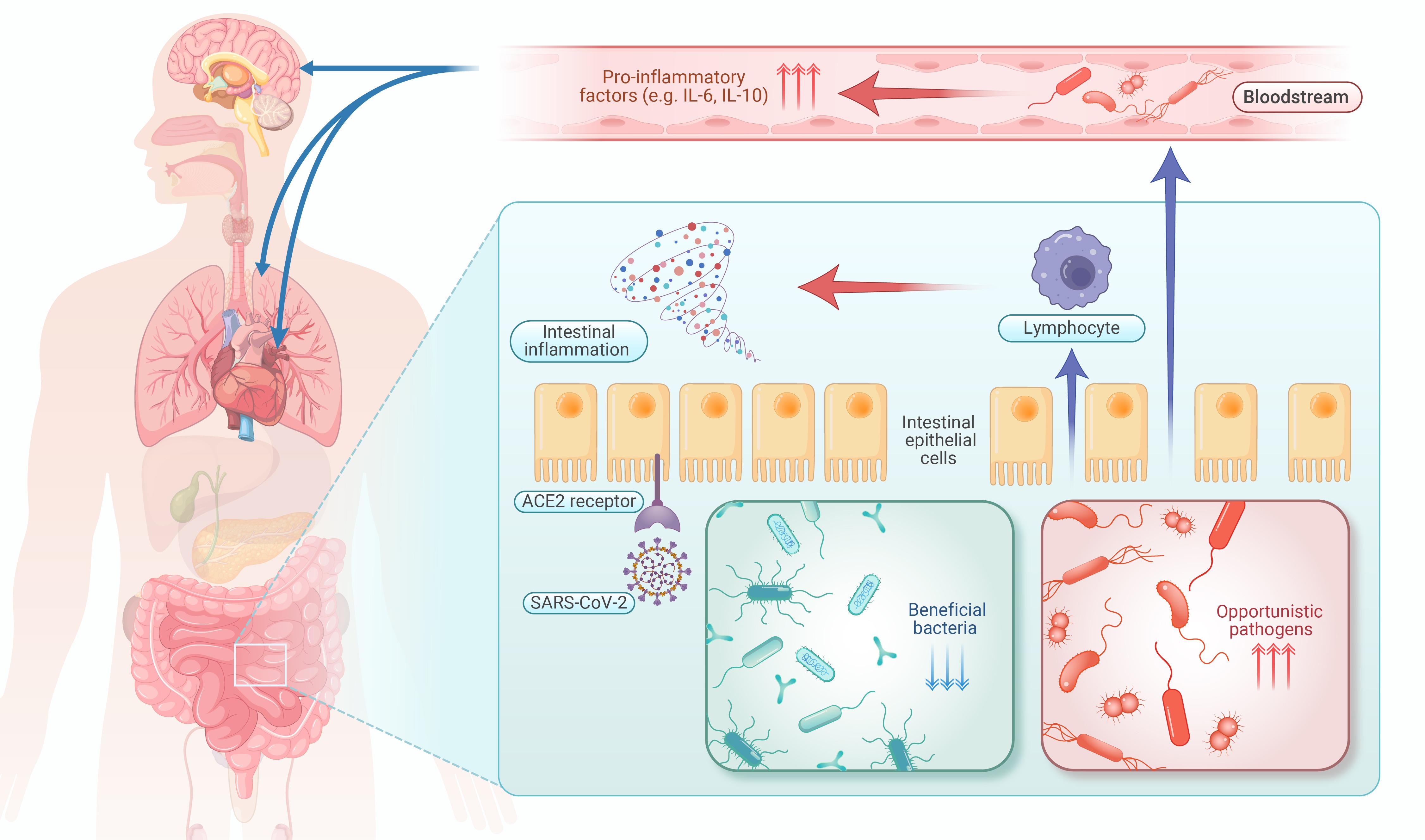
Figure 1.
Possible role of gut microbiome in post-COVID syndrome
-
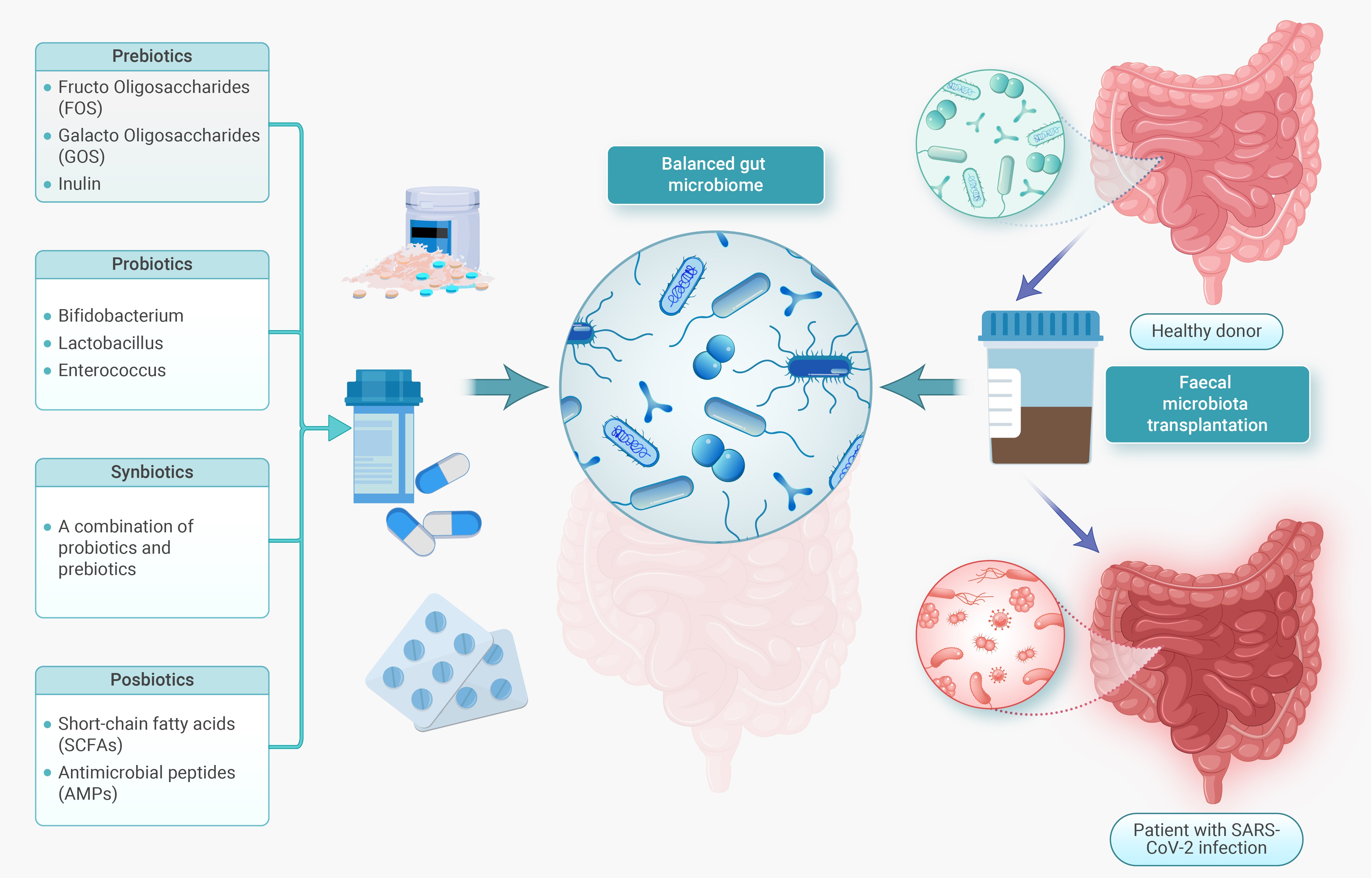
Figure 2.
Potential microbiota-based interventions in PCS
