
Healing beyond tradition: Advanced regenerative therapies for joint-related sports injuries
-
GRAPHICAL ABSTRACT
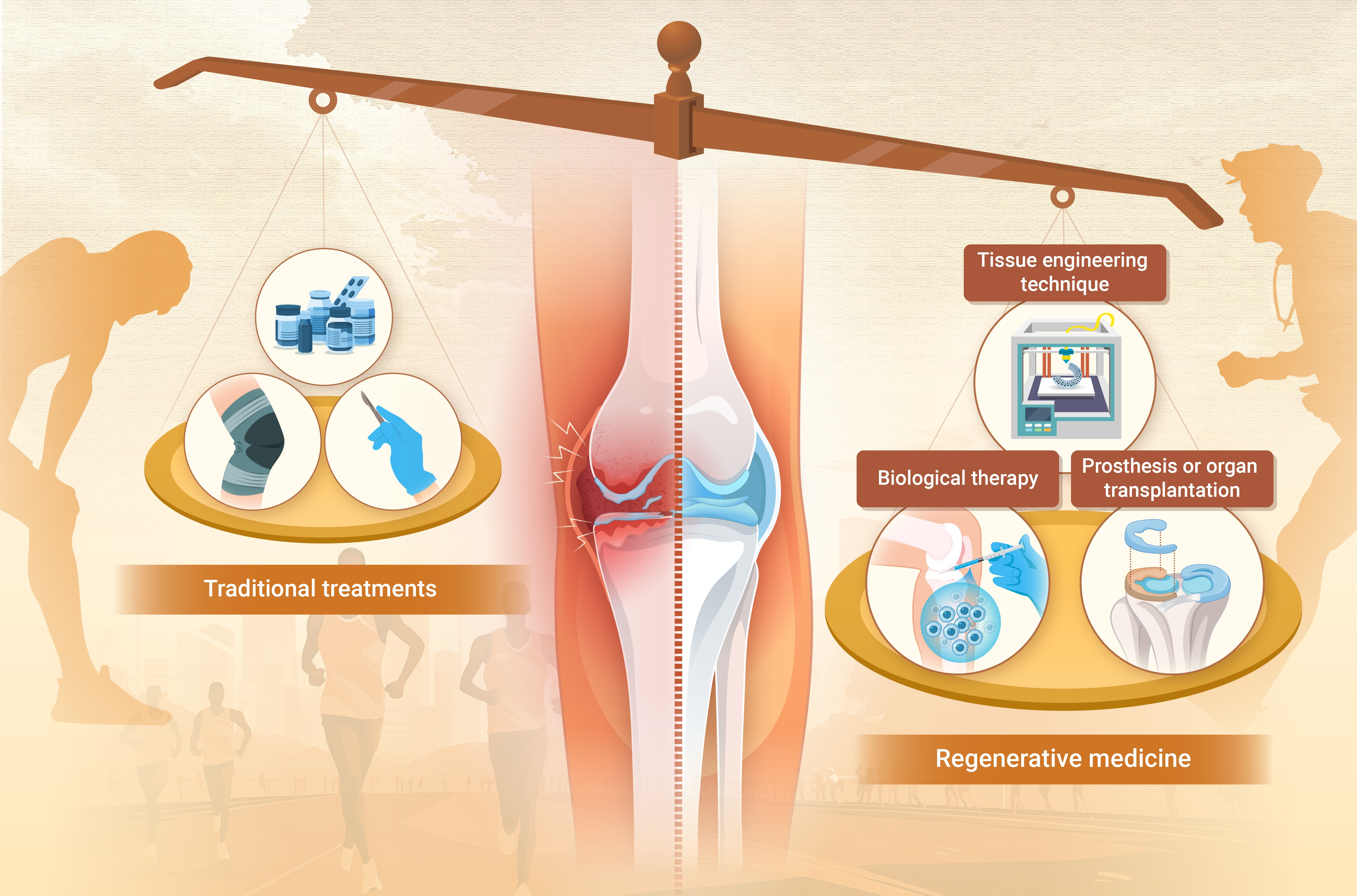
 DownLoad:
Full size image
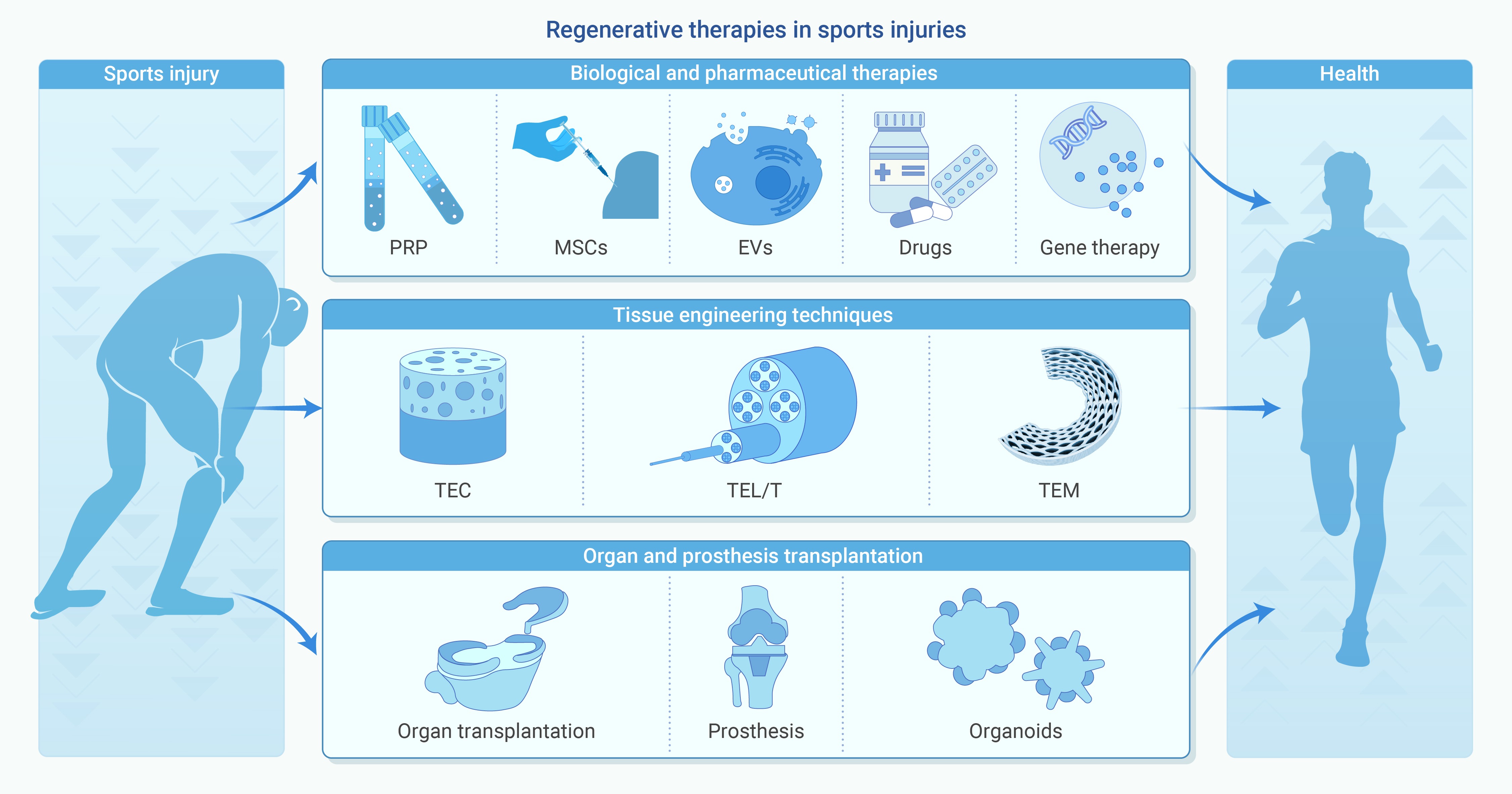
DownLoad:
Full size image
-
PUBLIC SUMMARY
-
Regenerative therapies promote soft tissue repair in sports injuries beyond symptom relief.
Biological therapies reduce inflammation and promote cell differentiation, facilitating tissue regeneration.
Tissue engineering and organoids offer structural and functional reconstruction of tissues.
Clinical translation requires standardization, long-term validation, and personalized treatment strategies.
-
-
ABSTRACT
Sports injuries are one of the most common diseases, and the repair and regeneration of joint soft tissues (ligaments, tendons, cartilage, etc.) involved in them are challenging. Traditional treatment strategies, such as drugs, physical therapy, and surgery, are difficult to achieve tissue regeneration and reconstruction of natural physiological functions. Regenerative medicine offers promising solutions, including biotherapy, tissue engineering, prosthesis or organ transplantation. These approaches aim to regenerate damaged tissues, reduce recovery time, and improve functional outcomes. Stem cells from different sources and their constructed organoids, novel biomaterials, 3D printing and other innovative technologies have been used. Individual variability, uncertain long-term efficacy, and high costs remain obstacles for clinical application. Further research is needed to explore the molecular mechanisms underlying regenerative therapies and orchestrate biological, physical and chemical factors. -

-
REFERENCES
[1] Andia I. and Maffulli N. (2017). Biological therapies in regenerative sports medicine. Sports Med. 47:807−828. DOI:10.1007/s40279-016-0620-z [2] Wang L., Jiang J., Lin H., et al. (2022). Advances in regenerative sports medicine research. Front. Bioeng. Biotechnol. 10:908751. DOI:10.3389/fbioe.2022.908751 [3] Kwon H., Brown W. E., Lee C. A., et al. (2019). Surgical and tissue engineering strategies for articular cartilage and meniscus repair. Nat. Rev. Rheumatol. 15:550−570. DOI:10.1038/s41584-019-0255-1 [4] Truong L. K., Mosewich A. D., Holt C. J., et al. (2020). Psychological, social and contextual factors across recovery stages following a sport-related knee injury: A scoping review. Br. J. Sports Med. 54:1149−1156. DOI:10.1136/bjsports-2019-101206 [5] Zügel M., Maganaris C. N., Wilke J., et al. (2018). Fascial tissue research in sports medicine: from molecules to tissue adaptation, injury and diagnostics: Consensus statement. Br. J. Sports Med. 52:1497. DOI:10.1136/bjsports-2018-099308 [6] Emery C. A. and Pasanen K. (2019). Current trends in sport injury prevention. Best Pract. Res. Clin. Rheumatol. 33:3−15. DOI:10.1016/j.berh.2019.02.009 [7] Prieto-González P., Martínez-Castillo J. L., Fernández-Galván L. M., et al. (2021). Epidemiology of sports-related injuries and associated risk factors in adolescent athletes: An injury surveillance. Int. J. Environ. Res. Public Health 18:4857. DOI:10.3390/ijerph18094857 [8] McCarthy M. M., Voos J. E., Nguyen J. T., et al. (2013). Injury profile in elite female basketball athletes at the Women's National Basketball Association combine. Am. J. Sports Med. 41:645−651. DOI:10.1177/0363546512474223 [9] Evans C. H. and Huard J. (2015). Gene therapy approaches to regenerating the musculoskeletal system. Nat. Rev. Rheumatol. 11:234−242. DOI:10.1038/nrrheum.2015.28 [10] Zhang W.-H., Zhou W., Luo Z.-W., et al. (2024). Anti-aging therapeutics for the musculoskeletal and cardiovascular systems: The role of regular exercise. Innov. Med. 2:100085. DOI:10.59717/j.xinn-med.2024.100085 [11] Leong N. L., Kator J. L., Clemens T. L., et al. (2020). Tendon and ligament healing and current approaches to tendon and ligament regeneration. J. Orthop. Res. 38:7−12. DOI:10.1002/jor.24475 [12] Winkler P. W., Zsidai B., Wagala N. N., et al. (2021). Evolving evidence in the treatment of primary and recurrent posterior cruciate ligament injuries, part 1: Anatomy, biomechanics and diagnostics. Knee Surg. Sports Traumatol. Arthrosc. 29:672−681. DOI:10.1007/s00167-020-06357-y [13] Makris E. A., Hadidi P. and Athanasiou K. A. (2011). The knee meniscus: Structure-function, pathophysiology, current repair techniques, and prospects for regeneration. Biomaterials 32:7411−7431. DOI:10.1016/j.biomaterials.2011.06.037 [14] Vuurberg G., Hoorntje A., Wink L. M., et al. (2018). Diagnosis, treatment and prevention of ankle sprains: update of an evidence-based clinical guideline. Br. J. Sports Med. 52:956. DOI:10.1136/bjsports-2017-098106 [15] Moran T. E., Awowale J. T., Werner B. C., et al. (2020). Associated morbidity after the percutaneous release of the medial collateral ligament for knee arthroscopy. Arthroscopy 36:891−900. DOI:10.1016/j.arthro.2019.08.051 [16] Shi B., Stinson Z., Nault M. L., et al. (2022). Meniscus repair in pediatric athletes. Clin. Sports Med. 41:749−767. DOI:10.1016/j.csm.2022.05.010 [17] Price A. and Beard D. (2014). Arthroscopy for degenerate meniscal tears of the knee. BMJ 348:g2382. DOI:10.1136/bmj.g2382 [18] Roumazeille T., Klouche S., Rousselin B., et al. (2015). Arthroscopic meniscal allograft transplantation with two tibia tunnels without bone plugs: Evaluation of healing on MR arthrography and functional outcomes. Knee Surg. Sports Traumatol. Arthrosc. 23:264−269. DOI:10.1007/s00167-013-2476-1 [19] O'Connor D., Johnston R. V., Brignardello-Petersen R., et al. (2022). Arthroscopic surgery for degenerative knee disease (osteoarthritis including degenerative meniscal tears). Cochrane Database Syst. Rev. 3:Cd014328. DOI:10.1002/14651858.Cd014328 [20] Cossu G., Fears R., Griffin G., et al. (2020). Regenerative medicine: Challenges and opportunities. Lancet 395:1746−1747. DOI:10.1016/s0140-6736(20)31250-2 [21] McKinley K. L., Longaker M. T. and Naik S. (2023). Emerging frontiers in regenerative medicine. Science 380:796−798. DOI:10.1126/science.add6492 [22] Xue Y., Riva N., Zhao L., et al. (2023). Recent advances of exosomes in soft tissue injuries in sports medicine: A critical review on biological and biomaterial applications. J. Control. Release 364:90−108. DOI:10.1016/j.jconrel.2023.10.031 [23] Bennell K. L., Paterson K. L., Metcalf B. R., et al. (2021). Effect of intra-articular platelet-rich plasma vs placebo injection on pain and medial tibial cartilage volume in patients with knee osteoarthritis: The RESTORE randomized clinical trial. JAMA 326:2021−2030. DOI:10.1001/jama.2021.19415 [24] Filbay S. R., Dowsett M., Chaker Jomaa M., et al. (2023). Healing of acute anterior cruciate ligament rupture on MRI and outcomes following non-surgical management with the Cross Bracing Protocol. Br. J. Sports Med. 57:1490−1497. DOI:10.1136/bjsports-2023-106931 [25] Migliorini F., Cuozzo F., Cipollaro L., et al. (2022). Platelet-rich plasma (PRP) augmentation does not result in more favourable outcomes in arthroscopic meniscal repair: A meta-analysis. J. Orthop. Traumatol. 23:8. DOI:10.1186/s10195-022-00630-1 [26] Fan B. S., Liu Y., Zhang J. Y., et al. (2021). Principles for establishment of the stem cell bank and its applications on management of sports injuries. Stem Cell Res. Ther. 12:307. DOI:10.1186/s13287-021-02360-3 [27] Park Y. B., Ha C. W., Rhim J. H., et al. (2018). Stem cell therapy for articular cartilage repair: Review of the entity of cell populations used and the result of the clinical application of each entity. Am. J. Sports Med. 46:2540−2552. DOI:10.1177/0363546517729152 [28] Luo W., Zhang H., Wan R., et al. (2024). Biomaterials-based technologies in skeletal muscle tissue engineering. Adv. Healthc. Mater. 13:e2304196. DOI:10.1002/adhm.202304196 [29] Lee K. I., Lee J. S., Kang K. T., et al. (2018). In vitro and in vivo performance of tissue-engineered tendons for anterior cruciate ligament reconstruction. Am. J. Sports Med. 46:1641−1649. DOI:10.1177/0363546518759729 [30] Schulze-Tanzil G. G. (2022). Healing of ligaments and tendons: Tissue engineering and models. Int. J. Mol. Sci. 23:15503. DOI:10.3390/ijms232415503 [31] Kaarre J., Herman Z. J., Zsidai B., et al. (2023). Meniscus allograft transplantation for biologic knee preservation: gold standard or dilemma. Knee Surg. Sports Traumatol. Arthrosc. 31:3579−3581. DOI:10.1007/s00167-022-07187-w [32] Leite C. B. G., Merkely G., Zgoda M., et al. (2023). Systematic review of clinical results after medial meniscus allograft transplantation reveals improved patient reported outcomes at greater than 5 years follow-up. Arthroscopy 39:802−811. DOI:10.1016/j.arthro.2022.11.033 [33] Mouarbes D., Menetrey J., Marot V., et al. (2019). Anterior cruciate ligament reconstruction: A systematic review and meta-analysis of outcomes for quadriceps tendon autograft versus bone-patellar tendon-bone and hamstring-tendon autografts. Am. J. Sports Med. 47:3531−3540. DOI:10.1177/0363546518825340 [34] Hu Y., Zhang H., Wang S., et al. (2023). Bone/cartilage organoid on-chip: Construction strategy and application. Bioact. Mater. 25:29−41. DOI:10.1016/j.bioactmat.2023.01.016 [35] Steinwachs M. R., Engebretsen L. and Brophy R. H. (2012). Scientific evidence base for cartilage injury and repair in the athlete. Cartilage 3:11s−17s. DOI:10.1177/1947603511415841 [36] Flanigan D. C., Harris J. D., Trinh T. Q., et al. (2010). Prevalence of chondral defects in athletes' knees: A systematic review. Med. Sci. Sports Exerc. 42:1795−1801. DOI:10.1249/MSS.0b013e3181d9eea0 [37] Pathria M. N., Chung C. B. and Resnick D. L. (2016). Acute and stress-related injuries of bone and cartilage: Pertinent anatomy, basic biomechanics, and imaging perspective. Radiology 280:21−38. DOI:10.1148/radiol.16142305 [38] Thibbotuwawa N., Singh S. and Gu Y. (2021). Proteoglycan and collagen contribution to the strain-rate-dependent mechanical behaviour of knee and shoulder cartilage. J. Mech. Behav. Biomed. Mater. 124:104733. DOI:10.1016/j.jmbbm.2021.104733 [39] Hertel J. and Corbett R. O. (2019). An updated model of chronic ankle Instability. J. Athl. Train 54:572−588. DOI:10.4085/1062-6050-344-18 [40] Musahl V. and Karlsson J. (2019). Anterior cruciate ligament tear. N. Engl. J. Med. 380:2341−2348. DOI:10.1056/NEJMcp1805931 [41] Nabian M. H., Zadegan S. A., Zanjani L. O., et al. (2017). Epidemiology of joint dislocations and ligamentous/tendinous injuries among 2,700 patients: Five-year trend of a tertiary center in Iran. Arch. Bone Jt. Surg. 5:426−434. [42] Kaeding C. C., Léger-St-Jean B. and Magnussen R. A. (2017). Epidemiology and diagnosis of anterior cruciate ligament injuries. Clin. Sports Med. 36:1−8. DOI:10.1016/j.csm.2016.08.001 [43] Ladenhauf H. N., Graziano J. and Marx R. G. (2013). Anterior cruciate ligament prevention strategies: Are they effective in young athletes - current concepts and review of literature. Curr. Opin. Pediatr. 25:64−71. DOI:10.1097/MOP.0b013e32835ad208 [44] Kotsifaki R., Korakakis V., King E., et al. (2023). Aspetar clinical practice guideline on rehabilitation after anterior cruciate ligament reconstruction. Br. J. Sports Med. 57:500−514. DOI:10.1136/bjsports-2022-106158 [45] Zitnay J. L., Jung G. S., Lin A. H., et al. (2020). Accumulation of collagen molecular unfolding is the mechanism of cyclic fatigue damage and failure in collagenous tissues. Sci. Adv. 6:eaba2795. DOI:10.1126/sciadv.aba2795 [46] Pitsillides A., Stasinopoulos D. and Giannakou K. (2021). Healing potential of the anterior cruciate ligament in terms of fiber continuity after a complete rupture: A systematic review. J. Bodyw. Mov. Ther. 28:246−254. DOI:10.1016/j.jbmt.2021.06.003 [47] Luvsannyam E., Jain M. S., Leitao A. R., et al. (2022). Meniscus tear: Pathology, incidence, and management. Cureus 14:e25121. DOI:10.7759/cureus.25121 [48] Fox A. J., Wanivenhaus F., Burge A. J., et al. (2015). The human meniscus: A review of anatomy, function, injury, and advances in treatment. Clin. Anat. 28:269−287. DOI:10.1002/ca.22456 [49] Jarraya M., Roemer F. W., Englund M., et al. (2017). Meniscus morphology: Does tear type matter. A narrative review with focus on relevance for osteoarthritis research. Semin. Arthritis Rheum. 46:552−561. DOI:10.1016/j.semarthrit.2016.11.005 [50] Ashraf S., Wibberley H., Mapp P. I., et al. (2011). Increased vascular penetration and nerve growth in the meniscus: a potential source of pain in osteoarthritis. Ann. Rheum. Dis. 70:523−529. DOI:10.1136/ard.2010.137844 [51] Bleakley C. M., O'Connor S. R., Tully M. A., et al. (2010). Effect of accelerated rehabilitation on function after ankle sprain: Randomised controlled trial. BMJ 340:c1964. DOI:10.1136/bmj.c1964 [52] Dubois B. and Esculier J. F. (2020). Soft-tissue injuries simply need PEACE and LOVE. Br. J. Sports Med. 54:72−73. DOI:10.1136/bjsports-2019-101253 [53] Singh D. P., Barani Lonbani Z., Woodruff M. A., et al. (2017). Effects of topical icing on inflammation, angiogenesis, revascularization, and myofiber regeneration in skeletal muscle following contusion injury. Front. Physiol. 8:93. DOI:10.3389/fphys.2017.00093 [54] Alexander J., Allan D. R. and Rhodes D. D. (2022). Cryotherapy in sport: A warm reception for the translation of evidence into applied practice. Res. Sports Med. 30:458−461. DOI:10.1080/15438627.2021.1899920 [55] Ziltener J. L., Leal S. and Fournier P. E. (2010). Non-steroidal anti-inflammatory drugs for athletes: An update. Ann. Phys. Rehabil. Med. 53:278−282,282-278. DOI:10.1016/j.rehab.2010.03.001 [56] Olafsen N. P., Herring S. A. and Orchard J. W. (2018). Injectable corticosteroids in sport. Clin. J. Sport Med. 28:451−456. DOI:10.1097/jsm.0000000000000603 [57] Nichols A. W. (2005). Complications associated with the use of corticosteroids in the treatment of athletic injuries. Clin. J. Sport Med. 15:370−375. DOI:10.1097/01.jsm.0000179233.17885.18 [58] Charles R., Fang L., Zhu R., et al. (2023). The effectiveness of shockwave therapy on patellar tendinopathy, Achilles tendinopathy, and plantar fasciitis: A systematic review and meta-analysis. Front. Immunol. 14:1193835. DOI:10.3389/fimmu.2023.1193835 [59] Szabo D. A., Neagu N., Teodorescu S., et al. (2022). TECAR therapy associated with high-intensity laser therapy (Hilt) and manual therapy in the treatment of muscle disorders: A literature review on the theorised effects supporting their use. J. Clin. Med. 11:6149. DOI:10.3390/jcm11206149 [60] Menezes M. A., Menezes D. A., Vasconcelos L. L., et al. (2022). Is electrical stimulation effective in preventing or treating delayed-onset muscle soreness (DOMS) in athletes and untrained adults. A systematic review with meta-analysis. J. Pain 23:2013−2035. DOI:10.1016/j.jpain.2022.05.004 [61] Steadman J. R., Rodkey W. G. and Rodrigo J. J. (2001). Microfracture: Surgical technique and rehabilitation to treat chondral defects. Clin. Orthop. Relat. Res. 391 Suppl:S362-369. DOI:10.1097/00003086-200110001-00033 [62] Frisbie D. D., Oxford J. T., Southwood L., et al. (2003). Early events in cartilage repair after subchondral bone microfracture. Clin. Orthop. Relat. Res. 407:215−227. DOI:10.1097/00003086-200302000-00031 [63] Nassar J. E., Guerin G., Keel T., et al. (2024). Autologous chondrocyte implantation, matrix-induced autologous chondrocyte implantation, osteochondral autograft transplantation and osteochondral allograft improve knee function and pain with considerations for patient and cartilage defects characteristics: A systematic review and meta-analysis. Knee Surg. Sports Traumatol. Arthrosc. Online ahead of print. DOI:10.1002/ksa.12525 [64] Crecelius C. R., Van Landuyt K. J. and Schaal R. (2021). Postoperative management for articular cartilage surgery in the knee. J. Knee Surg. 34:20−29. DOI:10.1055/s-0040-1718605 [65] Abrams G. D., Frank R. M., Gupta A. K., et al. (2013). Trends in meniscus repair and meniscectomy in the United States, 2005-2011. Am. J. Sports Med. 41:2333−2339. DOI:10.1177/0363546513495641 [66] Johnson M. J., Lucas G. L., Dusek J. K., et al. (1999). Isolated arthroscopic meniscal repair: A long-term outcome study (more than 10 years). Am. J. Sports Med. 27:44−49. DOI:10.1177/03635465990270011501 [67] Sihvonen R., Paavola M., Malmivaara A., et al. (2020). Arthroscopic partial meniscectomy for a degenerative meniscus tear: A 5 year follow-up of the placebo-surgery controlled FIDELITY (Finnish Degenerative Meniscus Lesion Study) trial. Br. J. Sports Med. 54:1332−1339. DOI:10.1136/bjsports-2020-102813 [68] Frizziero A., Ferrari R., Giannotti E., et al. (2012). The meniscus tear. State of the art of rehabilitation protocols related to surgical procedures. Muscles Ligaments Tendons J. 2:295−301. [69] Varone B. B. and Westermann R. W. (2024). Editorial commentary: Complications after meniscal surgery are rare and generally associated with medical comorbidity. Arthroscopy 40:1856−1857. DOI:10.1016/j.arthro.2023.12.017 [70] Nuelle C. W., Balldin B. C. and Slone H. S. (2022). All-inside anterior cruciate ligament reconstruction. Arthroscopy 38:2368−2369. DOI:10.1016/j.arthro.2022.06.001 [71] Monson J., Schoenecker J., Schwery N., et al. (2022). Postoperative rehabilitation and return to sport following multiligament knee reconstruction. Arthrosc. Sports Med. Rehabil. 4:e29−e40. DOI:10.1016/j.asmr.2021.08.020 [72] Eckenrode B. J., Carey J. L., Sennett B. J., et al. (2017). Prevention and management of post-operative complications following ACL reconstruction. Curr. Rev. Musculoskelet. Med. 10:315−321. DOI:10.1007/s12178-017-9427-2 [73] Qiao J. (2023). Win-win cooperation for advancing medical innovation. Innov. Med. 1:100002. DOI:10.59717/j.xinn-med.2023.100002 [74] Goula A., Gkioka V., Michalopoulos E., et al. (2020). Advanced therapy medicinal products challenges and perspectives in regenerative medicine. J. Clin. Med. Res. 12:780−786. DOI:10.14740/jocmr3964 [75] Pretorius J., Habash M., Ghobrial B., et al. (2023). Current status and advancements in platelet-rich plasma therapy. Cureus 15:e47176. DOI:10.7759/cureus.47176 [76] Verma R., Kumar S., Garg P., et al. (2023). Platelet-rich plasma: A comparative and economical therapy for wound healing and tissue regeneration. Cell Tissue Bank. 24:285−306. DOI:10.1007/s10561-022-10039-z [77] Cecerska-Heryc E., Heryc R., Wisniewska M., et al. (2019). Regenerative potential of platelets in patients with chronic kidney disease. Int. Urol. Nephrol. 51:1831−1840. DOI:10.1007/s11255-019-02190-6 [78] Wang X., Fok M. R., Pelekos G., et al. (2022). In vitro and ex vivo kinetic release profile of growth factors and cytokines from leucocyte- and platelet-rich fibrin (L-PRF) preparations. Cells 11:2089. DOI:10.3390/cells11132089. [79] Gupta S., Paliczak A. and Delgado D. (2021). Evidence-based indications of platelet-rich plasma therapy. Expert Rev. Hematol. 14:97−108. DOI:10.1080/17474086.2021.1860002 [80] Elphingstone J. W., Alston E. T. and Colorado B. S. (2024). Platelet-rich plasma for nonoperative management of degenerative meniscal tears: A systematic review. J. Orthop. 54:67−75. DOI:10.1016/j.jor.2024.03.009 [81] Jawanda H., Khan Z. A., Warrier A. A., et al. (2024). Platelet-rich plasma, bone marrow aspirate concentrate, and hyaluronic acid injections outperform corticosteroids in pain and function scores at a minimum of 6 months as intra-articular injections for knee osteoarthritis: A systematic review and network meta-analysis. Arthroscopy 40:1623-1636 e1621. DOI:10.1016/j.arthro.2024.01.037 [82] Berrigan W. A., Bailowitz Z., Park A., et al. (2024). A greater platelet dose may yield better clinical outcomes for platelet-rich plasma in the treatment of knee osteoarthritis: A systematic review. Arthroscopy. 4:809−817. DOI:10.1016/j.arthro.2024.03.018 [83] Khalilizad M., Emadian S. T. and Marzban Abbas Abadi M. (2025). Comparative efficacy of different doses of platelet-rich plasma injection in the treatment of knee osteoarthritis: A systematic review and network meta-analysis. J. Orthop. Surg. Res. 20:221. DOI:10.1186/s13018-025-05650-1 [84] Jiang X., Zhang H., Wu Q., et al. (2023). Comparison of three common shoulder injections for rotator cuff tears: a systematic review and network meta-analysis. J. Orthop. Surg. Res. 18:272. DOI:10.1186/s13018-023-03747-z [85] Dakkak M., Saleem A., Patel D., et al. (2024). A systematic review of randomized control trials looking at functional improvement of rotator cuff partial thickness tears following platelet-rich-plasma injection: A comparison of glenohumeral joint vs. subacromial bursa vs. intratendinous injection locations. JSES Int. 8:464−471. DOI:10.1016/j.jseint.2024.01.003 [86] Weninger V., Agocs G., Kovacs N., et al. (2024). Hyaluronate acid plus platelet-rich plasma is superior to steroids for pain relief less than 6 months using injection therapy of partial rotator cuff tears: A systematic review and network meta-analysis. Arthroscopy 41:304−315. DOI:10.1016/j.arthro.2024.03.035 [87] Averell N., Goodwin B., Park S., et al. (2024). The concentration of platelets in PRP does not affect pain outcomes in lateral epicondylitis: A systematic review and meta-analysis. Pain Manag. 14:223−233. DOI:10.1080/17581869.2024.2360381 [88] Andriolo L., Altamura S. A., Reale D., et al. (2019). Nonsurgical treatments of patellar tendinopathy: Multiple injections of platelet-rich plasma are a suitable option: A systematic review and meta-analysis. Am. J. Sports Med. 47:1001−1018. DOI:10.1177/0363546518759674 [89] Cao Y. and Wan Y. D. (2022). Effectiveness of platelet-rich plasma in anterior cruciate ligament reconstruction: A systematic review of randomized controlled trials. Orthop. Surg. 14:2406−2417. DOI:10.1111/os.13279 [90] Fucaloro S. P., Kang A. S., Bragg J. T., et al. (2024). Return to sport after ulnar collateral ligament tears treated with platelet-rich plasma injections is influenced by length of rehabilitation and leukocyte content of injections: A systematic review. Arthroscopy 41:343−356. DOI:10.1016/j.arthro.2024.03.017 [91] Pinzon-Arteaga C. A., Wang Y., Wei Y., et al. (2023). Bovine blastocyst-like structures derived from stem cell cultures. Cell Stem Cell 30:611-616 e617. DOI:10.1016/j.stem.2023.04.003 [92] Chen Y., Sun H., Luo Z., et al. (2024). Crosstalk between CD8(+) T cells and mesenchymal stromal cells in intestine homeostasis and immunity. Adv. Immunol. 162:23−58. DOI:10.1016/bs.ai.2024.02.001 [93] Watanabe Y., Tsuchiya A. and Terai S. (2021). The development of mesenchymal stem cell therapy in the present, and the perspective of cell-free therapy in the future. Clin. Mol. Hepatol. 27:70−80. DOI:10.3350/cmh.2020.0194 [94] Rinonapoli G., Gregori P., Di Matteo B., et al. (2021). Stem cells application in meniscal tears: A systematic review of pre-clinical and clinical evidence. Eur. Rev. Med. Pharmacol. Sci. 25:7754−7764. DOI:10.26355/eurrev_202112_27622 [95] Mazy D., Wang J., Dodin P., et al. (2024). Emerging biologic augmentation strategies for meniscal repair: A systematic review. BMC Musculoskelet. Disord. 25:541. DOI:10.1186/s12891-024-07644-2 [96] Jin W. S., Yin L. X., Sun H. Q., et al. (2024). Mesenchymal stem cells injection is more effective than hyaluronic acid injection in the treatment of knee osteoarthritis with similar safety: A systematic review and meta-analysis. Arthroscopy 41:318−332. DOI:10.1016/j.arthro.2024.07.027 [97] Razak H., Corona K., Totlis T., et al. (2023). Mesenchymal stem cell implantation provides short-term clinical improvement and satisfactory cartilage restoration in patients with knee osteoarthritis but the evidence is limited: A systematic review performed by the early-osteoarthritis group of ESSKA-European knee associates section. Knee Surg. Sports Traumatol. Arthrosc. 31:5306−5318. DOI:10.1007/s00167-023-07575-w [98] Chen X., Zheng J., Yin L., et al. (2024). Transplantation of three mesenchymal stem cells for knee osteoarthritis, which cell and type are more beneficial. A systematic review and network meta-analysis. J. Orthop. Surg. Res. 19:366. DOI:10.1186/s13018-024-04846-1 [99] Bono O. J., Jenkin B., Forlizzi J., et al. (2023). Evidence for utilization of injectable biologic augmentation in primary rotator cuff repair: A systematic review of data from 2010 to 2022. Orthop. J. Sports Med. 11:23259671221150037. DOI:10.1177/23259671221150037 [100] Chun S. W., Kim W., Lee S. Y., et al. (2022). A randomized controlled trial of stem cell injection for tendon tear. Sci. Rep. 12:818. DOI:10.1038/s41598-021-04656-z [101] Randelli P. S., Cucchi D., Fossati C., et al. (2022). Arthroscopic rotator cuff repair augmentation with autologous microfragmented lipoaspirate tissue is safe and effectively improves short-term clinical and functional results: A prospective randomized controlled trial with 24-month follow-up. Am. J. Sports Med. 50:1344−1357. DOI:10.1177/03635465221083324 [102] Vieira M. H. C., de Carvalho Schweich-Adami L., Oliveira R. J., et al. (2024). Effect of cell therapy with adipose-derived stem cells in the treatment of acute rupture of the Achilles tendon in humans. Cell Tissue Bank. 25:831−838. DOI:10.1007/s10561-024-10141-4 [103] Wang H. D., Li Z., Hu X., et al. (2022). Efficacy of stem cell therapy for tendon graft ligamentization after anterior cruciate ligament reconstruction: A systematic review. Orthop. J. Sports Med. 10:23259671221098363. DOI:10.1177/23259671221098363 [104] Baird J. P. E., Anz A., Andrews J., et al. (2022). Cellular augmentation of anterior cruciate ligament surgery is not currently evidence based: A systematic review of clinical studies. Arthroscopy 38:2047−2061. DOI:10.1016/j.arthro.2021.11.056 [105] Lu Y., Zheng J., Lin P., et al. (2024). Tumor microenvironment-derived exosomes: A double-edged sword for advanced T cell-based immunotherapy. ACS Nano. 18:27230−27260. DOI:10.1021/acsnano.4c09190 [106] Huang G., Zheng W., Zhou Y., et al. (2024). Recent advances to address challenges in extracellular vesicle-based applications for lung cancer. Acta Pharm. Sin. B 14:3855−3875. DOI:10.1016/j.apsb.2024.06.010 [107] Deng S., Cao H., Cui X., et al. (2023). Optimization of exosome-based cell-free strategies to enhance endogenous cell functions in tissue regeneration. Acta Biomater. 171:68−84. DOI:10.1016/j.actbio.2023.09.023 [108] Sadeghi S., Tehrani F. R., Tahmasebi S., et al. (2023). Exosome engineering in cell therapy and drug delivery. Inflammopharmacology 31:145−169. DOI:10.1007/s10787-022-01115-7 [109] Dong Z., Fu Y., Cai Z., et al. (2025). Recent advances in adipose-derived mesenchymal stem cell-derived exosomes for regulating macrophage polarization. Front. Immunol. 16:1525466. DOI:10.3389/fimmu.2025.1525466 [110] Kawata K., Koga H., Tsuji K., et al. (2021). Extracellular vesicles derived from mesenchymal stromal cells mediate endogenous cell growth and migration via the CXCL5 and CXCL6/CXCR2 axes and repair menisci. Stem Cell Res. Ther. 12:414. DOI:10.1186/s13287-021-02481-9 [111] Zhang F. X., Dou Y., Zhang B., et al. (2024). Skeletal stem cell-derived exosomes promote meniscal tear healing and ameliorate secondary osteoarthritis. Am. J. Sports Med. 52:2512−2523. DOI:10.1177/03635465241262002 [112] D'Arrigo D., Salerno M., De Marziani L., et al. (2024). A call for standardization for secretome and extracellular vesicles in osteoarthritis: results show disease-modifying potential, but protocols are too heterogeneous-a systematic review. Hum. Cell 37:1243−1275. DOI:10.1007/s13577-024-01084-9 [113] Xavier J., Jerome W., Zaslav K., et al. (2023). Exosome-laden scaffolds for treatment of post-traumatic cartilage injury and osteoarthritis of the knee: A systematic review. Int. J. Mol. Sci. 24:15178. DOI:10.3390/ijms242015178 [114] Zou M., Wang J. and Shao Z. (2023). Therapeutic potential of exosomes in tendon and tendon-bone healing: A systematic review of preclinical studies. J. Funct. Biomater. 14:299. DOI:10.3390/jfb14060299 [115] Kasula V., Padala V., Gupta N., et al. (2024). The use of extracellular vesicles in achilles tendon repair: A systematic review. Biomedicines 12:942. DOI:10.3390/biomedicines12050942 [116] Lu V., Tennyson M., Zhang J., et al. (2021). Mesenchymal stem cell-derived extracellular vesicles in tendon and ligament repair-A systematic review of in vivo studies. Cells 10:2553. DOI:10.3390/cells10102553 [117] Wang W., Chu Y., Zhang P., et al. (2023). Targeting macrophage polarization as a promising therapeutic strategy for the treatment of osteoarthritis. Int. Immunopharmacol. 116:109790. DOI:10.1016/j.intimp.2023.109790 [118] Nishida Y., Hashimoto Y., Orita K., et al. (2020). Intra-articular injection of stromal cell-derived factor 1α promotes meniscal healing via macrophage and mesenchymal stem cell accumulation in a rat meniscal defect model. Int. J. Mol. Sci. 21:5454. DOI:10.3390/ijms21155454 [119] Lee K. I., Gamini R., Olmer M., et al. (2020). Mohawk is a transcription factor that promotes meniscus cell phenotype and tissue repair and reduces osteoarthritis severity. Sci. Transl. Med. 12:eaan7967. DOI:10.1126/scitranslmed.aan7967 [120] Niu J., Feng F., Zhang S., et al. (2023). Thrombospondin-2 couples pressure-promoted chondrogenesis through NF-κB signaling. Tissue Eng. Regen. Med. 20:753−766. DOI:10.1007/s13770-023-00548-7 [121] Alcaraz M. J., Guillen M. I. and Ferrandiz M. L. (2019). Emerging therapeutic agents in osteoarthritis. Biochem. Pharmacol. 165:4−16. DOI:10.1016/j.bcp.2019.02.034 [122] Aman Z. S., DePhillipo N. N., Familiari F., et al. (2022). Acute intervention with selective interleukin-1 inhibitor therapy may reduce the progression of posttraumatic osteoarthritis of the knee: A systematic review of current evidence. Arthroscopy 38:2543−2556. DOI:10.1016/j.arthro.2022.02.009 [123] Oo W. M. and Hunter D. J. (2023). Efficacy, safety, and accuracy of intra-articular therapies for hand osteoarthritis: Current evidence. Drugs Aging 40:1−20. DOI:10.1007/s40266-022-00994-3 [124] Li Y., Mai Y., Cao P., et al. (2022). Relative efficacy and safety of anti-inflammatory biologic agents for osteoarthritis: A conventional and network meta-analysis. J. Clin. Med. 11:3958. DOI:10.3390/jcm11143958 [125] Richette P., Latourte A., Sellam J., et al. (2021). Efficacy of tocilizumab in patients with hand osteoarthritis: double blind, randomised, placebo-controlled, multicentre trial. Ann. Rheum. Dis. 80:349−355. DOI:10.1136/annrheumdis-2020-218547 [126] Mimpen J. Y., Baldwin M. J., Cribbs A. P., et al. (2021). Interleukin-17A causes osteoarthritis-like transcriptional changes in human osteoarthritis-derived chondrocytes and synovial fibroblasts in vitro. Front. Immunol. 12:676173. DOI:10.3389/fimmu.2021.676173 [127] Margheri F., Maggi L., Biagioni A., et al. (2021). Th17 lymphocyte-dependent degradation of joint cartilage by synovial fibroblasts in a humanized mouse model of arthritis and reversal by secukinumab. Eur. J. Immunol. 51:220−230. DOI:10.1002/eji.202048773 [128] Yu L., Luo R., Qin G., et al. (2023). Efficacy and safety of anti-interleukin-1 therapeutics in the treatment of knee osteoarthritis: A systematic review and meta-analysis of randomized controlled trials. J. Orthop. Surg. Res. 18:100. DOI:10.1186/s13018-023-03590-2 [129] Cao Z., Zhou J., Long Z., et al. (2020). Targeting nerve growth factor, a new option for treatment of osteoarthritis: A network meta-analysis of comparative efficacy and safety with traditional drugs. Aging (Albany NY) 13:1051−1070. DOI:10.18632/aging.202232 [130] Du X., Cai L., Xie J., et al. (2023). The role of TGF-beta3 in cartilage development and osteoarthritis. Bone Res. 11:2. DOI:10.1038/s41413-022-00239-4 [131] Schnitzer T., Pueyo M., Deckx H., et al. (2023). Evaluation of S201086/GLPG1972, an ADAMTS-5 inhibitor, for the treatment of knee osteoarthritis in ROCCELLA: A phase 2 randomized clinical trial. Osteoarthritis Cartilage 31:985−994. DOI:10.1016/j.joca.2023.04.001 [132] Wan Y., Li W., Liao Z., et al. (2020). Selective MMP-13 inhibitors: Promising agents for the therapy of osteoarthritis. Curr. Med. Chem. 27:3753−3769. DOI:10.2174/0929867326666181217153118 [133] Wei Y., Luo L., Gui T., et al. (2021). Targeting cartilage EGFR pathway for osteoarthritis treatment. Sci. Transl. Med. 13:eabb3946. DOI:10.1126/scitranslmed.abb3946 [134] Huang Y., Wang Z., Zhong C., et al. (2024). CRISPRa engineered Elite macrophages enable adoptive cell therapy for rheumatoid arthritis. Innov. Med. 2:100050. DOI:10.59717/j.xinn-med.2024.100050 [135] Poutoglidou F., Pourzitaki C., Manthou M. E., et al. (2022). The inhibitory effect of tocilizumab on systemic bone loss and tendon inflammation in a juvenile Collagen-Induced arthritis rat model. Connect Tissue Res. 63:577−589. DOI:10.1080/03008207.2022.2042275 [136] Kusaba Y., Kumagai K., Ishikawa K., et al. (2023). Bevacizumab promotes tenogenic differentiation and maturation of rat tendon-derived cells in vitro. PLoS One 18:e0293463. DOI:10.1371/journal.pone.0293463 [137] Deng X., Li Q., Yuan H., et al. (2024). Galangin promotes tendon repair mediated by tendon-derived stem cells through activating the TGF-β1/Smad3 signaling pathway. Chem. Pharm. Bull. (Tokyo) 72:669−675. DOI:10.1248/cpb.c24-00117 [138] Wu L. M., Wang Y. J., Li S. F., et al. (2023). Up-regulation of CREB-1 regulates tendon adhesion in the injury tendon healing through the CREB-1/TGF-β3 signaling pathway. BMC Musculoskelet. Disord. 24:325. DOI:10.1186/s12891-023-06425-7 [139] Tan S. L., Chan C. K., Ahmad T. S., et al. (2024). Growth differentiation factor 5(GDF5)-induced mesenchymal stromal cells enhances tendon healing. Tissue Eng. Part. C Methods 30:431−442. DOI:10.1089/ten.TEC.2024.0230 [140] Kobayashi Y., Kida Y., Kabuto Y., et al. (2021). Healing effect of subcutaneous administration of granulocyte colony-stimulating factor on acute rotator cuff injury in a rat model. Tissue Eng. Part A 27:1205−1212. DOI:10.1089/ten.tea.2020.0239.A [141] Wu Y., Shao Y., Xie D., et al. (2022). Effect of secretory leucocyte protease inhibitor on early tendon-to-bone healing after anterior cruciate ligament reconstruction in a rat model. Bone Joint Res. 11:503−512. DOI:10.1302/2046-3758.117.Bjr-2021-0358.R2 [142] Zhou Z., Song W., Zhang G., et al. (2022). The recombinant human fibroblast growth factor-18 (sprifermin) improves tendon-to-bone healing by promoting chondrogenesis in a rat rotator cuff repair model. J. Shoulder Elbow Surg. 31:1617−1627. DOI:10.1016/j.jse.2022.01.137 [143] Kang K., Geng Q., Cui L., et al. (2022). Upregulation of Runt related transcription factor 1 (RUNX1) contributes to tendon-bone healing after anterior cruciate ligament reconstruction using bone mesenchymal stem cells. J. Orthop. Surg. Res. 17:266. DOI:10.1186/s13018-022-03152-y [144] Mendell J. R., Al-Zaidy S. A., Rodino-Klapac L. R., et al. (2021). Current clinical applications of in vivo gene therapy with AAVs. Mol. Ther. 29:464−488. DOI:10.1016/j.ymthe.2020.12.007 [145] Salzman R., Cook F., Hunt T., et al. (2018). Addressing the value of gene therapy and enhancing patient access to transformative treatments. Mol. Ther. 26:2717−2726. DOI:10.1016/j.ymthe.2018.10.017 [146] Schambach A., Buchholz C. J., Torres-Ruiz R., et al. (2024). A new age of precision gene therapy. Lancet 403:568−582. DOI:10.1016/S0140-6736(23)01952-9 [147] Hollander J. M., Goraltchouk A., Liu J., et al. (2024). Single injection AAV2-FGF18 gene therapy reduces cartilage loss and subchondral bone damage in a mechanically induced model of osteoarthritis. Curr. Gene Ther. 24:331−345. DOI:10.2174/0115665232275532231213063634 [148] Hollander J. M., Goraltchouk A., Rawal M., et al. (2023). Adeno-associated virus-delivered fibroblast growth factor 18 gene therapy promotes cartilage anabolism. Cartilage 14:492−505. DOI:10.1177/19476035231158774 [149] Colazo J. M., Hoogenboezem E. N., Keech M. C., et al. (2023). Albumin-binding RNAi conjugate for carrier free treatment of arthritis. bioRxiv. DOI:10.1101/2023.05.31.542971 [150] Wen X., Fang G., Li H., et al. (2024). CircIRAK3 exerts negative feedback regulation on inflammation by binding to HNRNP U and destabilizing proinflammatory cytokine mRNA in osteoarthritis and chondrogenesis. Int. J. Biol. Macromol. 256:128453. DOI:10.1016/j.ijbiomac.2023.128453 [151] Xu Y., Mao G., Long D., et al. (2022). Circular RNA CREBBP modulates cartilage degradation by activating the Smad1/5 pathway through the TGFβ2/ALK1 axis. Exp. Mol. Med. 54:1727−1740. DOI:10.1038/s12276-022-00865-2 [152] Liu Y., Liu K., Tang C., et al. (2020). Long non-coding RNA XIST contributes to osteoarthritis progression via miR-149-5p/DNMT3A axis. Biomed. Pharmacother. 128:110349. DOI:10.1016/j.biopha.2020.110349 [153] Tang Z., Gong Z. and Sun X. (2022). Long non-coding RNA musculin antisense RNA 1 promotes proliferation and suppresses apoptosis in osteoarthritic chondrocytes via the microRNA-369-3p/Janus kinase-2/ signal transducers and activators of transcription 3 axis. Bioengineered 13:1554−1564. DOI:10.1080/21655979.2021.2013028 [154] Zhao Z., Liu J., Deng Z., et al. (2022). LncRNA MALAT1 promotes tenogenic differentiation of tendon-derived stem cells via regulating the miR-378a-3p/MAPK1 axis. Bioengineered 13:13213−13223. DOI:10.1080/21655979.2022.2076507 [155] Han W., Tao X., Weng T., et al. (2022). Circular RNA PVT1 inhibits tendon stem/progenitor cell senescence by sponging microRNA-199a-5p. Toxicol. In Vitro 79:105297. DOI:10.1016/j.tiv.2021.105297 [156] Liu Y. J., Wang H. J., Xue Z. W., et al. (2021). Long noncoding RNA H19 accelerates tenogenic differentiation by modulating miR-140-5p/VEGFA signaling. Eur. J. Histochem. 65:3297. DOI:10.4081/ejh.2021.3297. [157] Hosseinkhani H., Domb A. J., Sharifzadeh G., et al. (2023). Gene therapy for regenerative medicine. Pharmaceutics 15:856. DOI:10.3390/pharmaceutics15030856 [158] Xu X., Xu L., Xia J., et al. (2023). Harnessing knee joint resident mesenchymal stem cells in cartilage tissue engineering. Acta Biomater. 168:372−387. DOI:10.1016/j.actbio.2023.07.024 [159] Kim W., Jang C. H. and Kim G. (2022). Bone tissue engineering supported by bioprinted cell constructs with endothelial cell spheroids. Theranostics 12:5404−5417. DOI:10.7150/thno.74852 [160] Tevlin R., desJardins-Park H., Huber J., et al. (2022). Musculoskeletal tissue engineering: Adipose derived stromal cell implementation for the treatment of osteoarthritis. Biomaterials 286:121544. DOI:10.1016/j.biomaterials.2022.121544 [161] Koushik T. M., Miller C. M. and Antunes E. (2023). Bone tissue engineering scaffolds: Function of multi-material hierarchically structured scaffolds. Adv. Healthc. Mater. 12:e2202766. DOI:10.1002/adhm.202202766 [162] Shan B. H. and Wu F. G. (2024). Hydrogel-based growth factor delivery platforms: Strategies and recent advances. Adv. Mater. 36:e2210707. DOI:10.1002/adma.202210707 [163] Sun K., Guo J., Guo Z., et al. (2023). The roles of the hippo-YAP signalling pathway in cartilage and osteoarthritis. Ageing Res. Rev. 90:102015. DOI:10.1016/j.arr.2023.102015 [164] Haq-Siddiqi N. A., Britton D. and Kim Montclare J. (2023). Protein-engineered biomaterials for cartilage therapeutics and repair. Adv. Drug Deliv. Rev. 192:114647. DOI:10.1016/j.addr.2022.114647 [165] Xu W., Zhu J., Hu J., et al. (2022). Engineering the biomechanical microenvironment of chondrocytes towards articular cartilage tissue engineering. Life Sci. 309:121043. DOI:10.1016/j.lfs.2022.121043 [166] Bhattacharjee M., Coburn J., Centola M., et al. (2015). Tissue engineering strategies to study cartilage development, degeneration and regeneration. Adv. Drug Deliv. Rev. 84:107−122. DOI:10.1016/j.addr.2014.08.010 [167] Roseti L., Desando G., Cavallo C., et al. (2019). Articular cartilage regeneration in osteoarthritis. Cells 8:1305. DOI:10.3390/cells8111305 [168] Zelinka A., Roelofs A. J., Kandel R. A., et al. (2022). Cellular therapy and tissue engineering for cartilage repair. Osteoarthritis Cartilage 30:1547−1560. DOI:10.1016/j.joca.2022.07.012 [169] Yin B., Ni J., Witherel C. E., et al. (2022). Harnessing tissue-derived extracellular vesicles for osteoarthritis theranostics. Theranostics 12:207−231. DOI:10.7150/thno.62708 [170] Zhao X., Hu D. A., Wu D., et al. (2021). Applications of biocompatible scaffold materials in stem cell-based cartilage tissue engineering. Front. Bioeng. Biotechnol. 9:603444. DOI:10.3389/fbioe.2021.603444 [171] Zhou Z., Cui J., Wu S., et al. (2022). Silk fibroin-based biomaterials for cartilage/osteochondral repair. Theranostics 12:5103−5124. DOI:10.7150/thno.74548 [172] Zhang Y. J., Li D., Liu Y., et al. (2024). 3D-bioprinted anisotropic bicellular living hydrogels boost osteochondral regeneration via reconstruction of cartilage-bone interface. The Innovation 5:100542. DOI:10.1016/j.xinn.2023.100542 [173] Keller L., Pijnenburg L., Idoux-Gillet Y., et al. (2019). Preclinical safety study of a combined therapeutic bone wound dressing for osteoarticular regeneration. Nat. Commun. 10:2156. DOI:10.1038/s41467-019-10165-5 [174] Zhao C., Li X., Han X., et al. (2024). Molecular co-assembled strategy tuning protein conformation for cartilage regeneration. Nat. Commun. 15:1488. DOI:10.1038/s41467-024-45703-3 [175] Humphreys P. E. A., Woods S., Bates N., et al. (2023). Optogenetic manipulation of BMP signaling to drive chondrogenic differentiation of hPSCs. Cell Rep. 42:113502. DOI:10.1016/j.celrep.2023.113502 [176] Nordberg R. C., Bielajew B. J., Takahashi T., et al. (2024). Recent advancements in cartilage tissue engineering innovation and translation. Nat. Rev. Rheumatol. 20:323−346. DOI:10.1038/s41584-024-01118-4 [177] Du M., Liu K., Lai H., et al. (2024). Functional meniscus reconstruction with biological and biomechanical heterogeneities through topological self-induction of stem cells. Bioact. Mater. 36:358−375. DOI:10.1016/j.bioactmat.2024.03.005 [178] Gee S. M. and Posner M. (2021). Meniscus anatomy and basic science. Sports Med. Arthrosc. Rev. 29:e18−e23. DOI:10.1097/JSA.0000000000000327 [179] Ma Z., Vyhlidal M. J., Li D. X., et al. (2022). Mechano-bioengineering of the knee meniscus. Am. J. Physiol. Cell Physiol. 323:C1652−C1663. DOI:10.1152/ajpcell.00336.2022 [180] Xia B., Kim D. H., Bansal S., et al. (2021). Development of a decellularized meniscus matrix-based nanofibrous scaffold for meniscus tissue engineering. Acta Biomater. 128:175−185. DOI:10.1016/j.actbio.2021.03.074 [181] Zhang Z. Z., Chen Y. R., Wang S. J., et al. (2019). Orchestrated biomechanical, structural, and biochemical stimuli for engineering anisotropic meniscus. Sci. Transl. Med. 11:eaao0750. DOI:10.1126/scitranslmed.aao0750 [182] Li Z., Wu N., Cheng J., et al. (2020). Biomechanically, structurally and functionally meticulously tailored polycaprolactone/silk fibroin scaffold for meniscus regeneration. Theranostics 10:5090−5106. DOI:10.7150/thno.44270 [183] Gracey E., Burssens A., Cambre I., et al. (2020). Tendon and ligament mechanical loading in the pathogenesis of inflammatory arthritis. Nat. Rev. Rheumatol. 16:193−207. DOI:10.1038/s41584-019-0364-x [184] Frank C. B. (2004). Ligament structure, physiology and function. J. Musculoskelet. Neuronal Interact. 4:199−201. [185] Vogel V. (2018). Unraveling the mechanobiology of extracellular matrix. Annu. Rev. Physiol. 80:353−387. DOI:10.1146/annurev-physiol-021317-121312 [186] Silva M., Ferreira F. N., Alves N. M., et al. (2020). Biodegradable polymer nanocomposites for ligament/tendon tissue engineering. J. Nanobiotechnology 18:23. DOI:10.1186/s12951-019-0556-1 [187] Brindisino F., Salomon M., Giagio S., et al. (2021). Rotator cuff repair vs. nonoperative treatment: A systematic review with meta-analysis. J. Shoulder Elbow Surg. 30:2648−2659. DOI:10.1016/j.jse.2021.04.040 [188] Ainsworth R. and Lewis J. S. (2007). Exercise therapy for the conservative management of full thickness tears of the rotator cuff: a systematic review. Br. J. Sports Med. 41:200−210. DOI:10.1136/bjsm.2006.032524 [189] Zhao J. (2020). Editorial Commentary: Make use of the native insertion structure in rotator cuff repair. Arthroscopy 36:1843−1844. DOI:10.1016/j.arthro.2020.03.035 [190] Wang Y., Jin S., Luo D., et al. (2021). Functional regeneration and repair of tendons using biomimetic scaffolds loaded with recombinant periostin. Nat. Commun. 12:1293. DOI:10.1038/s41467-021-21545-1 [191] Sun Y., Sheng R., Cao Z., et al. (2024). Bioactive fiber-reinforced hydrogel to tailor cell microenvironment for structural and functional regeneration of myotendinous junction. Sci. Adv. 10:eadm7164. DOI:10.1126/sciadv.adm7164 [192] Du L., Wu J., Han Y., et al. (2024). Immunomodulatory multicellular scaffolds for tendon-to-bone regeneration. Sci. Adv. 10:eadk6610. DOI:10.1126/sciadv.adk6610 [193] Burk J., Plenge A., Brehm W., et al. (2016). Induction of tenogenic differentiation mediated by extracellular tendon matrix and short-term cyclic stretching. Stem Cells Int. 2016:7342379. DOI:10.1155/2016/7342379 [194] Huang T., Xu H., Wang H., et al. (2023). Artificial intelligence for medicine: Progress, challenges, and perspectives. Innov. Med. 1:100030. DOI:10.59717/j.xinn-med.2023.100030 [195] Lei C., Song J. H., Li S., et al. (2023). Advances in materials-based therapeutic strategies against osteoporosis. Biomaterials 296:122066. DOI:10.1016/j.biomaterials.2023.122066 [196] Bagherpour R., Bagherpour G. and Mohammadi P. (2025). Application of artificial intelligence in tissue engineering. Tissue Eng. Part B Rev. 31:31−43. DOI:10.1089/ten.TEB.2024.0022 [197] Ahmed A. F., Rinaldi J., Noorzad A. S., et al. (2022). Return to sports following meniscal allograft transplantation is possible but remains questionable: A systematic review. Arthroscopy 38:1351−1361. DOI:10.1016/j.arthro.2021.11.003 [198] Beer A. J., Tauro T. M., Redondo M. L., et al. (2019). Use of allografts in orthopaedic surgery: Safety, procurement, storage, and outcomes. Orthop. J. Sports Med. 7:2325967119891435. DOI:10.1177/2325967119891435 [199] Bracey D. N., Cignetti N. E., Jinnah A. H., et al. (2020). Bone xenotransplantation: A review of the history, orthopedic clinical literature, and a single-center case series. Xenotransplantation 27:e12600. DOI:10.1111/xen.12600 [200] Su L., Garcia-Mansilla I., Kelley B., et al. (2022). Clinical outcomes of meniscal allograft transplantation with respect to the minimal clinically important difference. Am. J. Sports Med. 50:3440−3446. DOI:10.1177/03635465211036116 [201] Romandini I., Grassi A., Andrea Lucidi G., et al. (2024). 10-year survival and clinical improvement of meniscal allograft transplantation in early to moderate knee osteoarthritis. Am. J. Sports Med. 52:1997−2007. DOI:10.1177/03635465241253849 [202] Bozkurt M. (2022). Lateral meniscus allograft transplantation in combination with BMAC (Bone Marrow Aspirate Concentrate) injection: Biologic augmentation of the allograft. Arthrosc. Tech. 11:e767−e773. DOI:10.1016/j.eats.2021.12.035 [203] Struijk C., Van Genechten W., Verdonk P., et al. (2022). Human meniscus allograft augmentation by allogeneic mesenchymal stromal/stem cell injections. J. Orthop. Res. 40:712−726. DOI:10.1002/jor.25074 [204] Jiang D., Zhao L. H., Tian M., et al. (2012). Meniscus transplantation using treated xenogeneic meniscal tissue: viability and chondroprotection study in rabbits. Arthroscopy 28:1147−1159. DOI:10.1016/j.arthro.2012.01.001 [205] Yoon S., Min Y., Park C., et al. (2022). Innate immune response analysis in meniscus xenotransplantation using normal and triple knockout Jeju native pigs. Int. J. Mol. Sci. 23:10416. DOI:10.3390/ijms231810416 [206] Jiang D., Zhang Z. Z., Zhao F., et al. (2018). The radiated deep-frozen xenogenic meniscal tissue regenerated the total meniscus with chondroprotection. Sci. Rep. 8:9041. DOI:10.1038/s41598-018-27016-w [207] Wang X., Ren Z., Liu Y., et al. (2023). Characteristics and clinical outcomes after osteochondral allograft transplantation for treating articular cartilage defects: Systematic review and single-arm meta-analysis of studies from 2001 to 2020. Orthop. J. Sports Med. 11:23259671231199418. DOI:10.1177/23259671231199418 [208] Butler J. J., Rizla M. R. M., Egol A. J., et al. (2024). Particulated juvenile cartilage allograft for the treatment of osteochondral lesions of the talus is associated with a high complication rate and a high failure rate at short-term follow-up: A systematic review. Knee Surg. Sports Traumatol. Arthrosc. 32:529−541. DOI:10.1002/ksa.12069 [209] Migliorini F., Maffulli N., Baroncini A., et al. (2022). Allograft versus autograft osteochondral transplant for chondral defects of the talus: Systematic review and meta-analysis. Am. J. Sports Med. 50:3447−3455. DOI:10.1177/03635465211037349 [210] Trofa D. P., Hong I. S., Lopez C. D., et al. (2023). Isolated osteochondral autograft versus allograft transplantation for the treatment of symptomatic cartilage lesions of the knee: A systematic review and meta-analysis. Am. J. Sports Med. 51:812−824. DOI:10.1177/03635465211053594 [211] Jiang N., Chen H., Zhang J., et al. (2023). Decellularized-disc based allograft and xenograft prosthesis for the long-term precise reconstruction of temporomandibular joint disc. Acta Biomater. 159:173−187. DOI:10.1016/j.actbio.2023.01.042 [212] Vernice N. A., Dong X., Matavosian A. A., et al. (2024). Bioengineering full-scale auricles using 3D-printed external scaffolds and decellularized cartilage xenograft. Acta Biomater. 179:121−129. DOI:10.1016/j.actbio.2024.03.012 [213] Tritschler H., Fischer K., Seissler J., et al. (2021). New insights into xenotransplantation for cartilage repair: Porcine multi-genetically modified chondrocytes as a promising cell source. Cells 10:2152. DOI:10.3390/cells10082152 [214] Villatte G., Erivan R., Nourissat G., et al. (2022). Allograft and autograft provide similar retear rates for the management of large and massive rotator cuff tears: A review and meta-analysis. Knee Surg. Sports Traumatol. Arthrosc. 30:2039−2059. DOI:10.1007/s00167-021-06745-y [215] Xu M., Zhang H., Yang X., et al. (2020). The influence of donor and recipient characteristics on allograft tendons: A systematic review. Cell Tissue Bank. 21:17−29. DOI:10.1007/s10561-019-09803-5 [216] Belk J. W., Littlefield C. P., Smith J. H., et al. (2024). Autograft demonstrates superior outcomes for revision anterior cruciate ligament reconstruction when compared with allograft: A systematic review. Am. J. Sports Med. 52:859−867. DOI:10.1177/03635465231152232 [217] Migliorini F., Pintore A., Oliva F., et al. (2023). Allografts as alternative to autografts in primary posterior cruciate ligament reconstruction: A systematic review and meta-analysis. Knee Surg. Sports Traumatol. Arthrosc. 31:2852−2860. DOI:10.1007/s00167-022-07258-y [218] Peebles L. A., Blackwood N. O., Verma A., et al. (2024). Medial ulnar collateral ligament reconstruction with allograft provides excellent clinical outcomes, high rates of return to play, and a low incidence of postoperative complications: A systematic review. Arthroscopy 41:1113−1122. DOI:10.1016/j.arthro.2024.03.045 [219] Blackwood N. O., Blitz J. A., Vopat B., et al. (2024). Medial collateral ligament reconstruction with autograft versus allograft: A systematic review. Am. J. Sports Med. 52:3419−3426. DOI:10.1177/03635465231225982 [220] Stone K. R., Walgenbach A. W., Turek T. J., et al. (2023). Xenograft bone-patellar tendon-bone ACL reconstruction: A case series at 20-year follow-up as proof of principle. J. Exp. Orthop. 10:91. DOI:10.1186/s40634-023-00651-7 [221] Lombardi J. A., Hoonjan A., Rodriguez N., et al. (2020). Porcine bone-patellar tendon-bone xenograft in a caprine model of anterior cruciate ligament repair. J. Orthop. Surg. (Hong Kong) 28:2309499020939737. DOI:10.1177/2309499020939737. [222] Hexter A. T., Hing K. A., Haddad F. S., et al. (2020). Decellularized porcine xenograft for anterior cruciate ligament reconstruction: A histological study in sheep comparing cross-pin and cortical suspensory femoral fixation. Bone Joint Res. 9:293−301. DOI:10.1302/2046-3758.96.BJR-2020-0030.R2 [223] Li S., Wang S., Liu W., et al. (2022). Current strategies for enhancement of the bioactivity of artificial ligaments: A mini-review. J. Orthop. Translat. 36:205−215. DOI:10.1016/j.jot.2022.07.011 [224] Edgar L., Pu T., Porter B., et al. (2020). Regenerative medicine, organ bioengineering and transplantation. Br. J. Surg. 107:793−800. DOI:10.1002/bjs.11686 [225] Winkler P. W., Rothrauff B. B., Buerba R. A., et al. (2020). Meniscal substitution, a developing and long-awaited demand. J. Exp. Orthop. 7:55. DOI:10.1186/s40634-020-00270-6 [226] van Tienen T. G., van Minnen B., Defoort K. C., et al. (2023). The first-generation anatomical medial meniscus prosthesis led to unsatisfactory results: A first-in-human study. Knee Surg. Sports Traumatol. Arthrosc. 31:2526−2533. DOI:10.1007/s00167-022-07205-x [227] van Minnen B. S. and van Tienen T. G. (2024). The current state of meniscus replacements. Curr. Rev. Musculoskelet. Med. 17:293−302. DOI:10.1007/s12178-024-09902-1 [228] McKeon B. P., Zaslav K. R., Alfred R. H., et al. (2020). Preliminary results from a US clinical trial of a novel synthetic polymer meniscal implant. Orthop. J. Sports Med. 8:2325967120952414. DOI:10.1177/2325967120952414 [229] Haspl M., Trsek D., Lovric D., et al. (2021). Functional and magnetic resonance imaging outcome after polyurethane meniscal scaffold implantation following partial meniscectomy. Int. Orthop. 45:971−975. DOI:10.1007/s00264-020-04844-y [230] Lucidi G. A., Grassi A., Agostinone P., et al. (2022). Risk factors affecting the survival rate of collagen meniscal implant for partial meniscal deficiency: An analysis of 156 consecutive cases at a mean 10 years of follow-up. Am. J. Sports Med. 50:2900−2908. DOI:10.1177/03635465221112635 [231] Dong J., Huang M., Lin J., et al. (2024). Outcome comparison of meniscal allograft transplantation (MAT) and meniscal scaffold implantation (MSI): A systematic review. Int. J. Surg. 110:5112−5123. DOI:10.1097/js9.0000000000001587 [232] Chubinskaya S., Di Matteo B., Lovato L., et al. (2019). Agili-C implant promotes the regenerative capacity of articular cartilage defects in an ex vivo model. Knee Surg. Sports Traumatol. Arthrosc. 27:1953−1964. DOI:10.1007/s00167-018-5263-1 [233] Brusalis C. M., Greditzer H. G. t., Fabricant P. D., et al. (2020). BioCartilage augmentation of marrow stimulation procedures for cartilage defects of the knee: Two-year clinical outcomes. Knee 27:1418−1425. DOI:10.1016/j.knee.2020.07.087 [234] Fletcher W. R., Collins T., Fox A., et al. (2024). Mid-term efficacy of the Cartiva synthetic cartilage implant in symptomatic hallux rigidus. Bone Jt. Open 5:799−805. DOI:10.1302/2633-1462.59.BJO-2024-0031.R1 [235] Papadopoulos G., Griffin S., Rathi H., et al. (2022). Cost-effectiveness analysis of arthroscopic injection of a bioadhesive hydrogel implant in conjunction with microfracture for the treatment of focal chondral defects of the knee - an Australian perspective. J. Med. Econ. 25:712−721. DOI:10.1080/13696998.2022.2078574 [236] Gudas R., Staškūnas M., Mačiulaitis J., et al. (2024). Arthroscopic implantation of a cell-free bilayer scaffold for the treatment of knee chondral lesions: A 2-year prospective study. Cartilage 16:5−16. DOI:10.1177/19476035241232061 [237] Wang M., Wu Y., Li G., et al. (2024). Articular cartilage repair biomaterials: Strategies and applications. Mater. Today Bio. 24:100948. DOI:10.1016/j.mtbio.2024.100948 [238] Wang L., Guo X., Chen J., et al. (2022). Key considerations on the development of biodegradable biomaterials for clinical translation of medical devices: With cartilage repair products as an example. Bioact. Mater. 9:332−342. DOI:10.1016/j.bioactmat.2021.07.031 [239] Kolar M. and Drobnič M. (2023). Multilayered biomimetic scaffolds for cartilage repair of the talus. A systematic review of the literature. Foot Ankle Surg. 29:2−8. DOI:10.1016/j.fas.2022.10.007 [240] Andriolo L., Reale D., Di Martino A., et al. (2021). Cell-free scaffolds in cartilage knee surgery: A systematic review and meta-analysis of clinical evidence. Cartilage 12:277−292. DOI:10.1177/1947603519852406 [241] Tang Y., Wang Z., Xiang L., et al. (2022). Functional biomaterials for tendon/ligament repair and regeneration. Regen. Biomater. 9:rbac062. DOI:10.1093/rb/rbac062 [242] Fukubayashi T. and Ikeda K. (2017). A review on Gore-Tex® artificial ligament-special emphasis on tunnel osteolysis. J. Long Term Eff. Med. Implants 27:111−121. DOI:10.1615/JLongTermEffMedImplants.v27.i2-4.20 [243] Civera M., Devietti Goggia E., De Ros M., et al. (2022). Implantable medical devices for tendon and ligament repair: a review of patents and commercial products. Expert Rev. Med. Devices 19:825−845. DOI:10.1080/17434440.2022.2142557 [244] Chuckpaiwong B., Glisson R. R., Usuelli F. G., et al. (2023). Biomechanical comparison of nonlocked minimally invasive and locked open achilles tendon simulated rupture repairs. Foot Ankle Int. 44:913−921. DOI:10.1177/10711007231178819 [245] Saad Berreta R., Villarreal-Espinosa J. B., Pallone L., et al. (2024). Anterior cruciate ligament repair results in similar patient reported outcome measures as anterior cruciate ligament reconstruction: A systematic review of prospective comparative studies. Arthroscopy 12:S0749-8063(24)00673-X. DOI:10.1016/j.arthro.2024.09.008 [246] Han X. X., Cai C. H., Deng W., et al. (2024). Landscape of human organoids: Ideal model in clinics and research. The Innovation 5:100620. DOI:10.1016/j.xinn.2024.100620 [247] Yi S. A., Zhang Y., Rathnam C., et al. (2021). Bioengineering approaches for the advanced organoid research. Adv. Mater. 33:e2007949. DOI:10.1002/adma.202007949 [248] Hofer M. and Lutolf M. P. (2021). Engineering organoids. Nat. Rev. Mater. 6:402−420. DOI:10.1038/s41578-021-00279-y [249] Huang J., Zhang L., Lu A., et al. (2023). Organoids as innovative models for bone and joint diseases. Cells 12:1590. DOI:10.3390/cells12121590 [250] Mulaudzi P. E., Abrahamse H. and Crous A. (2024). Insights on three dimensional organoid studies for stem cell therapy in regenerative medicine. Stem Cell Rev. Rep. 20:509−523. DOI:10.1007/s12015-023-10655-6 [251] Tang X. Y., Wu S., Wang D., et al. (2022). Human organoids in basic research and clinical applications. Signal Transduct. Target Ther. 7:168. DOI:10.1038/s41392-022-01024-9 [252] Silva-Pedrosa R., Salgado A. J. and Ferreira P. E. (2023). Revolutionizing disease modeling: The emergence of organoids in cellular systems. Cells 12:930. DOI:10.3390/cells12060930 [253] Fan C., He J., Xu S., et al. (2023). Advances in biomaterial-based cardiac organoids. Biomater. Adv. 153:213502. DOI:10.1016/j.bioadv.2023.213502 [254] Yang Z., Wang B., Liu W., et al. (2023). In situ self-assembled organoid for osteochondral tissue regeneration with dual functional units. Bioact. Mater. 27:200−215. DOI:10.1016/j.bioactmat.2023.04.002 [255] Wei X., Qiu J., Lai R., et al. (2024). A human organoid drug screen identifies α2-adrenergic receptor signaling as a therapeutic target for cartilage regeneration. Cell Stem Cell 31:1813−1830.e1818. DOI:10.1016/j.stem.2024.09.001 [256] Abe K., Yamashita A., Morioka M., et al. (2023). Engraftment of allogeneic iPS cell-derived cartilage organoid in a primate model of articular cartilage defect. Nat. Commun. 14:804. DOI:10.1038/s41467-023-36408-0 [257] Crispim J. F. and Ito K. (2021). De novo neo-hyaline-cartilage from bovine organoids in viscoelastic hydrogels. Acta Biomater. 128:236−249. DOI:10.1016/j.actbio.2021.04.008 [258] Yan Z., Yin H., Brochhausen C., et al. (2020). Aged tendon stem/progenitor cells are less competent to form 3D tendon organoids due to cell autonomous and matrix production deficits. Front. Bioeng. Biotechnol. 8:406. DOI:10.3389/fbioe.2020.00406 [259] Graca A. L., Kroner-Weigl N., Reyes Alcaraz V., et al. (2024). Demonstration of self-assembled cell sheet culture and manual generation of a 3D tendon/ligament-like organoid by using human dermal fibroblasts. J. Vis. Exp. DOI:10.3791/66047 -
ABOUT THIS ARTICLE
Cite this article:
Du M., Gu Y., Xu T., et al. (2025). Healing beyond tradition: Advanced regenerative therapies for joint-related sports injuries. The Innovation Medicine 3:100132. https://doi.org/10.59717/j.xinn-med.2025.100132
Welcome!
To request copyright permission to republish or share portions of our works, please visit Copyright Clearance Center's (CCC) Marketplace website at marketplace.copyright.com.
Export File
Citation
Du M., Gu Y., Xu T., et al. (2025). Healing beyond tradition: Advanced regenerative therapies for joint-related sports injuries. The Innovation Medicine 3:100132. https://doi.org/10.59717/j.xinn-med.2025.100132

Format
Content



-
Figure 1.
Application of regenerative therapies in joint-related sports injuries
-
Figure 2.
Traditional clinical therapies for joint sports injuries.
-
Figure 3.
Impact of mohawk on meniscus repair
-
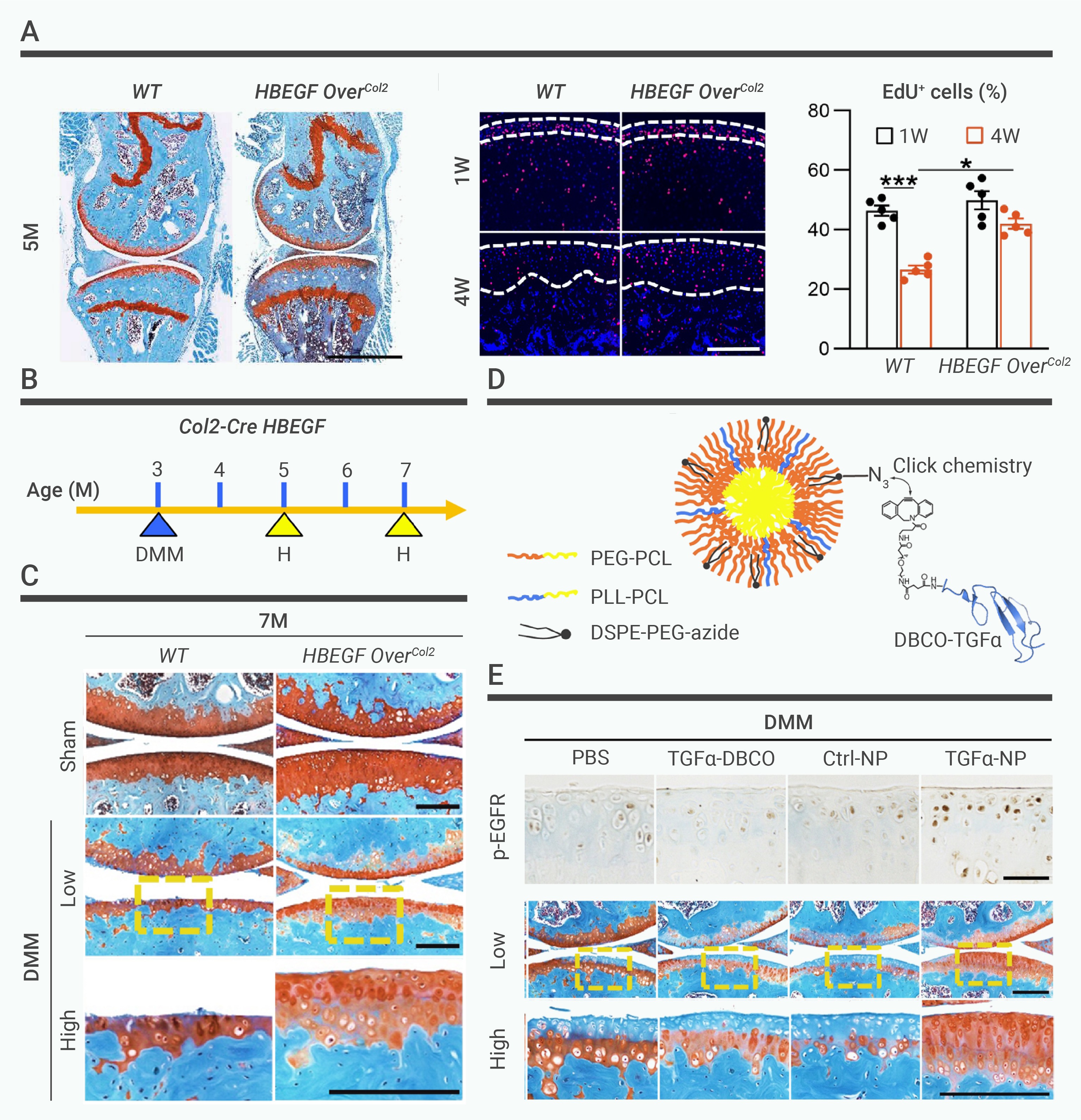
Figure 4.
Impact of EGFR on cartilage Repair
-
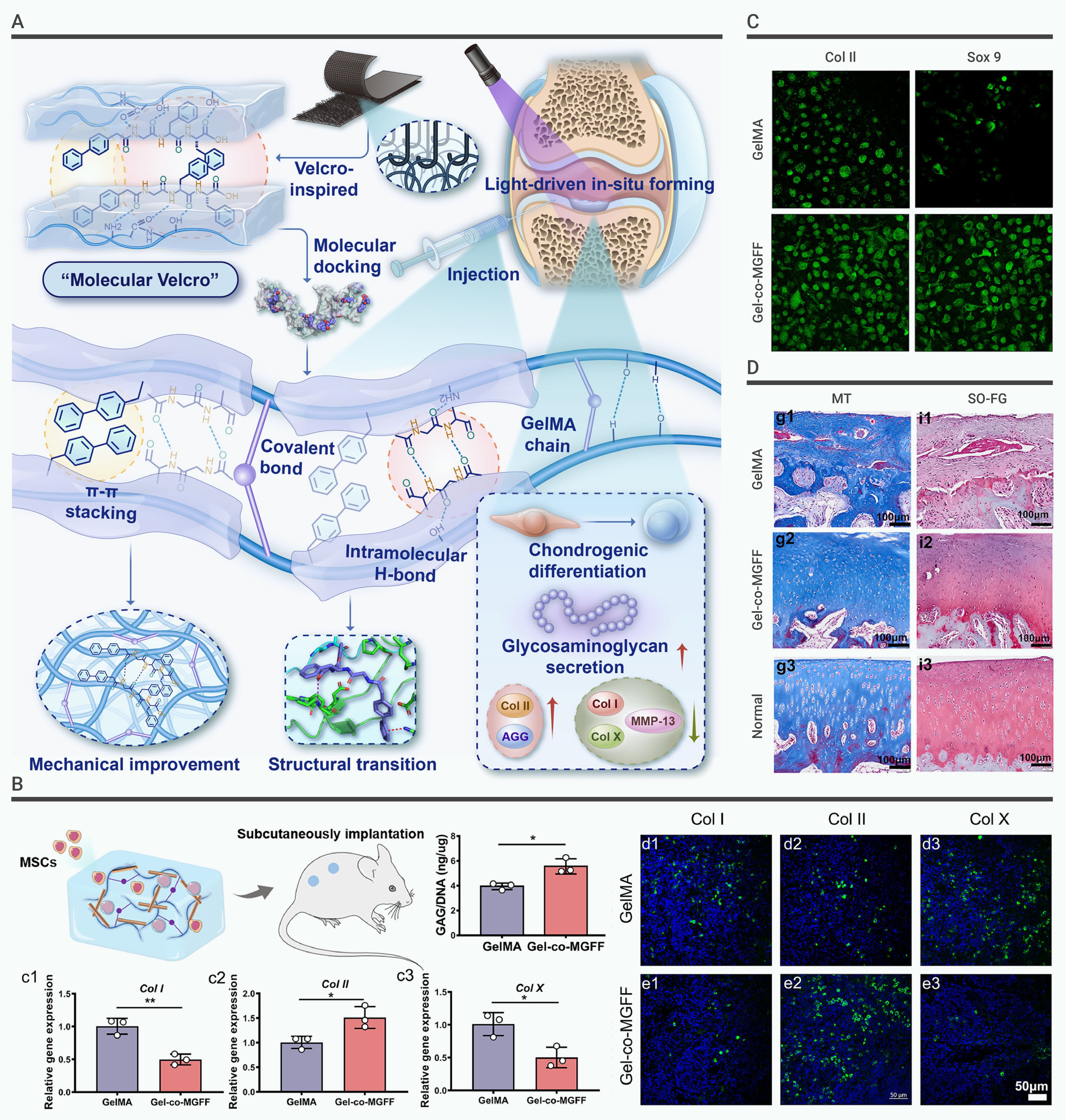
Figure 5.
A molecular co-assembly approach to modulate protein conformation for cartilage repair
-
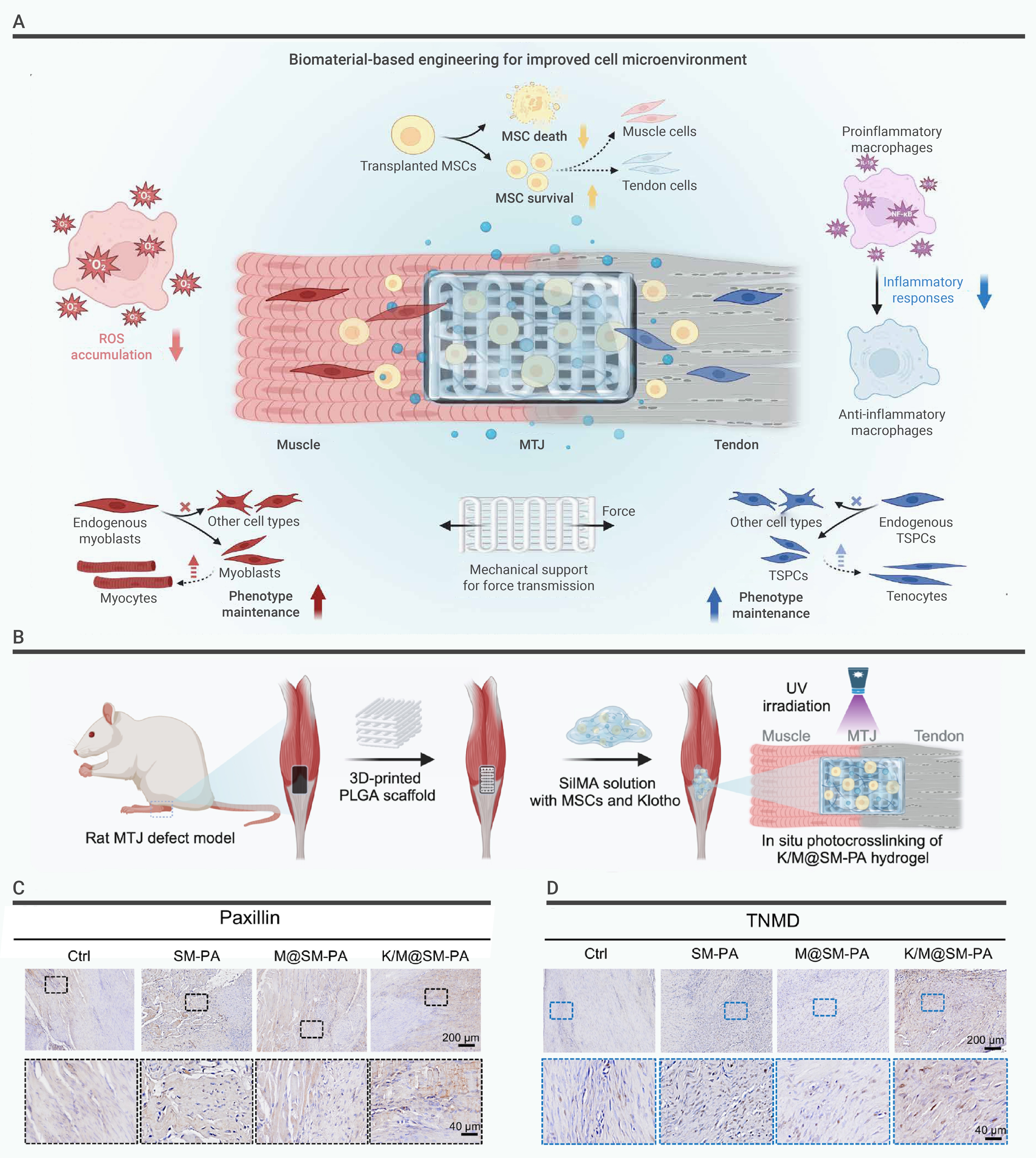
Figure 6.
Bioactive fiber reinforced hydrogel can customize the cell microenvironment for the structural and functional regeneration of myotendinous junction
-
Figure 7.
A human organoid drug screen identifies α2-adrenergic receptor signaling as a therapeutic target for cartilage regeneration
