
First-in-human study of high-dose gp96 in ALS: Safety and preliminary immunological effects
-
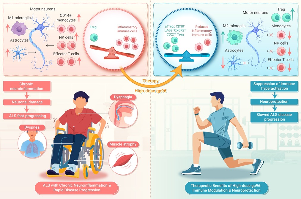
GRAPHICAL ABSTRACT
 DownLoad:
Full size image
DownLoad:
Full size image
-
PUBLIC SUMMARY
-
High-dose gp96 suppresses inflammatory CD14 monocytes, natural killer (NK) cells, and T cells in ALS patients.
gp96 boosts regulatory T cells (Treg) and activated Treg (aTreg), enhancing their suppressive function.
40% (4/10) of patients showed slower ALS Functional Rating Scale-Revised (ALSFRS-R) decline with gp96 therapy.
gp96 efficacy links to Treg activation and reduced innate and effector T cell activity.
-
-
ABSTRACT
Amyotrophic lateral sclerosis (ALS) is a fatal neurodegenerative disease associated with immune dysregulation. Based on the robust therapeutic effect and immunoregulatory capability of gp96 administration in the SOD1G93A mouse model, we conducted a prospective one-arm, open-label, proof-of-concept clinical trial in Hainan, China. High-dose gp96 was well tolerated in all 10 enrolled patients with ALS, and pronouncedly reduced the levels of inflammatory immune cells—CD14+ monocytes, NK cells, and CD4+ and CD8+ effector T cells—to levels comparable to those seen in healthy individuals. Treatment with gp96 also enhanced the suppressive function of Tregs and promoted the expansion of both activated Tregs and the CD38+LAG3+CXCR3hiCD27hi Treg subset. All participants experienced a slower decline in ALSFRS-R scores during the first 16 weeks post-treatment, and this slower decline was sustained in 40% (4/10) of patients at the end of follow-up in week 72. Treatment duration and Treg activation were positively correlated with the efficacy of gp96 treatment in alleviating the disease progression rate. These preliminary results emphasize the importance of the early identification of fast-progressing ALS patients and the need for long-term treatment. Our findings support further evaluation of gp96 as an immunomodulatory agent against ALS in randomized controlled trials with standardized outcome measures. -

-
REFERENCES
[1] Corcia P., Beltran S., Bakkouche S.E., et al. (2021). Therapeutic news in ALS. Rev. Neurol. 177:544−549. DOI:10.1016/j.neurol.2020.12.003 [2] Mejzini R., Flynn L.L., Pitout I.L., et al. (2019). ALS genetics, mechanisms, and therapeutics: Where are we now. Front. Neurosci. 13:1310. DOI:10.3389/fnins.2019.01310 [3] Beers D.R. and Appel S.H. (2019). Immune dysregulation in amyotrophic lateral sclerosis: Mechanisms and emerging therapies. Lancet Neurol. 18:211−220. DOI:10.1016/S1474-4422(18)30394-6 [4] Mccauley M.E. and Baloh R.H. (2019). Inflammation in ALS/FTD pathogenesis. Acta. Neuropathol. 137:715−730. DOI:10.1007/s00401-018-1933-9 [5] Vahsen B.F., Nalluru S., Morgan G.R., et al. (2023). C9orf72-ALS human iPSC microglia are pro-inflammatory and toxic to co-cultured motor neurons via MMP9. Nat. Commun. 14:5898. DOI:10.1038/s41467-023-41603-0 [6] Guo S., Wang H. and Yin Y. (2022). Microglia polarization from m1 to m2 in neurodegenerative diseases. Front. Aging Neurosci. 14:815347. DOI:10.3389/fnagi.2022.815347 [7] Yamanaka K. and Komine O. (2018). The multi-dimensional roles of astrocytes in ALS. Neurosci. Res. 126:31−38. DOI:10.1016/j.neures.2017.09.011 [8] Ralli M., Lambiase A., Artico M., et al. (2019). Amyotrophic lateral sclerosis: Autoimmune pathogenic mechanisms, clinical features, and therapeutic perspectives. Isr. Med. Assoc. J. 21:438−443. https://www.ima.org.il/FilesUploadPublic/IMAJ/0/371/185509.pdf [9] Alipour P., Senkevich K., Ross J.P., et al. (2022). Investigation of the causal relationship between ALS and autoimmune disorders: A mendelian randomization study. BMC Med. 20:382. DOI:10.1186/s12916-022-02578-9 [10] Garofalo S., Cocozza G., Bernardini G., et al. (2022). Blocking immune cell infiltration of the central nervous system to tame neuroinflammation in amyotrophic lateral sclerosis. Brain Behav. Immun. 105:1−14. DOI:10.1016/j.bbi.2022.06.004 [11] Ramachandran S., Grozdanov V., Leins B., et al. (2023). Low t-cell reactivity to TDP-43 peptides in ALS. Front. Immunol. 14:1193507. DOI:10.3389/fimmu.2023.1193507 [12] De Marchi F., Munitic I., Vidatic L., et al. (2023). Overlapping neuroimmune mechanisms and therapeutic targets in neurodegenerative disorders. Biomedicines 11:2793. DOI:10.3390/biomedicines11102793 [13] Balendra R. and Isaacs A.M. (2018). C9orf72-mediated ALS and FTD: Multiple pathways to disease. Nat. Rev. Neurol. 14:544−558. DOI:10.1038/s41582-018-0047-2 [14] Rubino V., La Rosa G., Pipicelli L., et al. (2023). Insights on the multifaceted roles of wild-type and mutated superoxide dismutase 1 in amyotrophic lateral sclerosis pathogenesis. Antioxidants (Basel) 12:1747. DOI:10.3390/antiox12091747 [15] Dubowsky M., Theunissen F., Carr J.M., et al. (2023). The molecular link between TDP-43, endogenous retroviruses and inflammatory neurodegeneration in amyotrophic lateral sclerosis: A potential target for triumeq, an antiretroviral therapy. Mol. Neurobiol. 60:6330−6345. DOI:10.1007/s12035-023-03472-y [16] Beers D.R., Zhao W., Wang J., et al. (2017). ALS patients' regulatory t lymphocytes are dysfunctional, and correlate with disease progression rate and severity. JCI Insight 2:e89530. DOI:10.1172/jci.insight.89530 [17] Yazdani S., Seitz C., Cui C., et al. (2022). T cell responses at diagnosis of amyotrophic lateral sclerosis predict disease progression. Nat. Commun. 13:6733. DOI:10.1038/s41467-022-34526-9 [18] Sheean R.K., Mckay F.C., Cretney E., et al. (2018). Association of regulatory T-cell expansion with progression of amyotrophic lateral sclerosis: A study of humans and a transgenic mouse model. JAMA Neurol. 75:681−689. DOI:10.1001/jamaneurol.2018.0035 [19] Sheean R.K., Weston R.H., Perera N.D., et al. (2015). Effect of thymic stimulation of CD4+ t cell expansion on disease onset and progression in mutant SOD1 mice. J. Neuroinflammation 12:40. DOI:10.1186/s12974-015-0254-3 [20] Thonhoff J.R., Berry J.D., Macklin E.A., et al. (2022). Combined regulatory t-lymphocyte and IL-2 treatment is safe, tolerable, and biologically active for 1 year in persons with amyotrophic lateral sclerosis. Neurol. Neuroimmunol. Neuroinflamm. 9:e200019. DOI:10.1212/NXI.0000000000200019 [21] Chandawarkar R. Y., Wagh M. S. and Srivastava P. K. (1999). The dual nature of specific immunological activity of tumor-derived gp96 preparations. J. Exp. Med. 189:1437−1442. DOI:10.1084/jem.189.9.1437 [22] Kovalchin J. T., Mendonca C., Wagh M. S., et al. (2006). In vivo treatment of mice with heat shock protein, gp 96, improves survival of skin grafts with minor and major antigenic disparity. Transpl. Immunol. 15:179−185. DOI:10.1016/j.trim.2005.07.003 [23] Chandawarkar R. Y., Wagh M. S., Kovalchin J. T., et al. (2004). Immune modulation with high-dose heat-shock protein gp96: Therapy of murine autoimmune diabetes and encephalomyelitis. Int. Immunol. 16:615−624. DOI:10.1093/intimm/dxh063 [24] Liu Z., Li X., Qiu L., et al. (2009). Treg suppress CTL responses upon immunization with HSP gp96. Eur. J. Immunol. 39:3110−3120. DOI:10.1002/eji.200939593 [25] Li X., Liu Z., Yan X., et al. (2013). Induction of regulatory T cells by high-dose gp96 suppresses murine liver immune hyperactivation. PLoS One 8:e68997. DOI:10.1371/journal.pone.0068997 [26] Xu Y., Liu E., Xie X., et al. (2021). Induction of foxp3 and activation of tregs by HSP gp96 for treatment of autoimmune diseases. iScience 24:103445. DOI:10.1016/j.isci.2021.103445 [27] Kinner-Bibeau L.B., Sedlacek A.L., Messmer M.N., et al. (2017). HSPs drive dichotomous t-cell immune responses via DNA methylome remodelling in antigen presenting cells. Nat. Commun. 8:15648. DOI:10.1038/ncomms15648 [28] Randazzo M., Terness P., Opelz G., et al. (2012). Active-specific immunotherapy of human cancers with the heat shock protein gp96-revisited. Int. J. Cancer 130:2219−2231. DOI:10.1002/ijc.27332 [29] Meng S. D., Song J., Rao Z., et al. (2002). Three-step purification of gp96 from human liver tumor tissues suitable for isolation of gp96-bound peptides. J. Immunol. Methods 264:29−35. DOI:10.1016/s0022-1759(02)00093-5 [30] Maclean M., Lopez-Diez R., Vasquez C., et al. (2022). Neuronal-glial communication perturbations in murine SOD1(g93a) spinal cord. Commun. Biol. 5:177. DOI:10.1038/s42003-022-03128-y [31] Mukherjee K. and Bieker J.J. (2022). Correction notice: isolation of healthy f4/80+ macrophages from embryonic day e13.5 mouse fetal liver using magnetic nanoparticles for single cell sequencing. Bio. Protoc. 12:e4329. DOI:10.21769/BioProtoc.4329. [32] Crane C.A, Han S.J, Ahn B., et al. (2013). Individual patient-specific immunity against high-grade glioma after vaccination with autologous tumor derived peptides bound to the 96 KD chaperone protein. Clin. Cancer Res. 19:205−214. DOI:10.1158/1078-0432.CCR-11-3358 [33] Pilla L., Patuzzo R., Rivoltini L., et al. (2006). A phase II trial of vaccination with autologous, tumor-derived heat-shock protein peptide complexes Gp96, in combination with GM-CSF and interferon-alpha in metastatic melanoma patients. Cancer Immunol. Immunother. 55:958−968. DOI:10.1007/s00262-005-0084-8 [34] Liu J., Wang J., Xu J., et al. (2021). Comprehensive investigations revealed consistent pathophysiological alterations after vaccination with COVID-19 vaccines. Cell. Discov. 7:99. DOI:10.1038/s41421-021-00329-3 [35] Du Y, Zhao W., Thonhoff J.R., et al. (2020). Increased activation ability of monocytes from ALS patients. Exp. Neurol. 328:113259. DOI:10.1016/j.expneurol.2020.113259 [36] Quek H., Cuni-Lopez C., Stewart R., et al. (2022). ALS monocyte-derived microglia-like cells reveal cytoplasmic TDP-43 accumulation, DNA damage, and cell-specific impairment of phagocytosis associated with disease progression. J. Neuroinflammation 19:58. DOI:10.1186/s12974-022-02421-1 [37] Szklarczyk D., Gable A.L., Lyon D., et al. (2019). STRING v11: Protein-protein association networks with increased coverage, supporting functional discovery in genome-wide experimental datasets. Nucleic Acids Res. 47:D607−D613. DOI:10.1093/nar/gky1131 [38] Zhang J.Y., Wang X.M., Xing X., et al. (2020). Single-cell landscape of immunological responses in patients with COVID-19. Nat. Immunol. 21:1107−1118. DOI:10.1038/s41590-020-0762-x [39] Terekhova M., Swain A., Bohacova P., et al. (2023). Single-cell atlas of healthy human blood unveils age-related loss of NKG2c(+)GZMB(-)CD8(+) memory t cells and accumulation of type 2 memory T cells. Immunity 56:2836−2854.e9. DOI:10.1016/j.immuni.2023.10.013 [40] Xydia M., Rahbari R., Ruggiero E., et al. (2021). Common clonal origin of conventional T cells and induced regulatory T cells in breast cancer patients. Nat. Commun. 12:1119. DOI:10.1038/s41467-021-21297-y [41] Luo Y., Xu C., Wang B., et al. (2021). Single-cell transcriptomic analysis reveals disparate effector differentiation pathways in human T(reg) compartment. Nat. Commun. 12:3913. DOI:10.1038/s41467-021-24213-6 [42] Li Y., Qin S., Dong L., et al. (2024). Long-term effects of omicron BA.2 breakthrough infection on immunity-metabolism balance: A 6-month prospective study. Nat. Commun. 15:2444. DOI:10.1038/s41467-024-46692-z [43] Glatigny S., Duhen R., Arbelaez C., et al. (2015). Integrin alpha l controls the homing of regulatory T cells during CNS autoimmunity in the absence of integrin alpha 4. Sci. Rep. 5:7834. DOI:10.1038/srep07834 [44] Miyara M., Yoshioka Y., Kitoh A., et al. (2009). Functional delineation and differentiation dynamics of human CD4+ T cells expressing the FoxP3 transcription factor. Immunity 30:899−911. DOI:10.1016/j.immuni.2009.03.019 [45] Cossarizza A., Chang H.D., Radbruch A., et al. (2021). Guidelines for the use of flow cytometry and cell sorting in immunological studies (third edition). Eur. J. Immunol. 51:2708−3145. DOI:10.1002/eji.202170126 [46] Guida N., Sanguigno L., Mascolo L., et al. (2021). The transcriptional complex sp1/KMT2a by up-regulating restrictive element 1 silencing transcription factor accelerates methylmercury-induced cell death in motor neuron-like NSC34 cells overexpressing SOD1-g93a. Front. Neurosci. 15:771580. DOI:10.3389/fnins.2021.771580 [47] Ohgomori T., Yamasaki R., Takeuchi H., et al. (2017). Differential activation of neuronal and glial STAT3 in the spinal cord of the SOD1(g93a) mouse model of amyotrophic lateral sclerosis. Eur. J. Neurosci. 46:2001−2014. DOI:10.1111/ejn.13650 [48] Ong S.M., Teng K., Newell E., et al. (2019). A novel, five-marker alternative to CD16-CD14 gating to identify the three human monocyte subsets. Front. Immunol. 10:1761. DOI:10.3389/fimmu.2019.01761 [49] Campisi L., Chizari S., Ho J., et al. (2022). Author correction: Clonally expanded CD8 T cells characterize amyotrophic lateral sclerosis-4. Nature. 608:E34. DOI:10.1038/s41586-022-05184-0 [50] Merah-Mourah F., Cohen S.O., Charron D., et al. (2020). Identification of novel human monocyte subsets and evidence for phenotypic groups defined by interindividual variations of expression of adhesion molecules. Sci. Rep. 10:4397. DOI:10.1038/s41598-020-61022-1 [51] de Carvalho M. (2023). Advances in amyotrophic lateral sclerosis research in 2022. Lancet Neurol. 22:21−22. DOI:10.1016/S1474-4422(22)00490-2 [52] Okamura T., Sumitomo S., Morita K., et al. (2015). TGF-β3-expressing CD4+CD25(-)LAG3+ regulatory T cells control humoral immune responses. Nat. Commun. 6:6329. DOI:10.1038/ncomms7329 [53] Sarkar I., Basak D., Ghosh P., et al. (2025). CD38-mediated metabolic reprogramming promotes the stability and suppressive function of regulatory T cells in tumor. Sci. Adv. 11:eadt2117. DOI:10.1126/sciadv.adt2117 [54] Wang J., Zhao X. and Wan Y. Y. (2023). Intricacies of TGF-β signaling in Treg and Th17 cell biology. Cell Mol. Immunol. 20:1002−1022. DOI:10.1038/s41423-023-01036-7 [55] Shouse A. N., LaPorte K. M. and Malek, T. R. (2024). Interleukin-2 signaling in the regulation of T cell biology in autoimmunity and cancer. Immunity 57:414−428. DOI:10.1016/j.immuni.2024.02.001 [56] Nayak D. A., Sedlacek A. L., Cillo A. R., et al. (2024). CD91 and its ligand gp96 confer cross-priming capabilities to multiple APCs during immune responses to nascent, emerging tumors. Cancer Immunol. Res. 12:1663−1676. DOI:10.1158/2326-6066.CIR-24-0326 -
ABOUT THIS ARTICLE
Cite this article:
Li X., Cheng F., Wang Z., et al. (2025). First-in-human study of high-dose gp96 in ALS: Safety and preliminary immunological effects. The Innovation Medicine 3:100157. https://doi.org/10.59717/j.xinn-med.2025.100157
Welcome!
To request copyright permission to republish or share portions of our works, please visit Copyright Clearance Center's (CCC) Marketplace website at marketplace.copyright.com.
Article Metrics
Relative Articles
Cited by
Export File
Citation
Li X., Cheng F., Wang Z., et al. (2025). First-in-human study of high-dose gp96 in ALS: Safety and preliminary immunological effects. The Innovation Medicine 3:100157. https://doi.org/10.59717/j.xinn-med.2025.100157

Format
Content
-
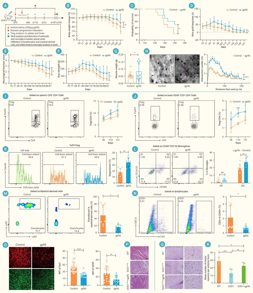
Figure 1.
Effect of high-dose gp96 treatment on Treg activation, inflammatory astrocyte and microglia, and disease progression in SOD1G93A mice
-
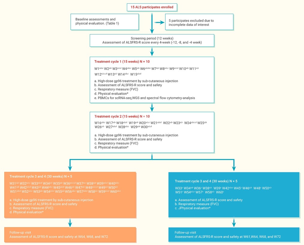
Figure 2.
Trial profile
-
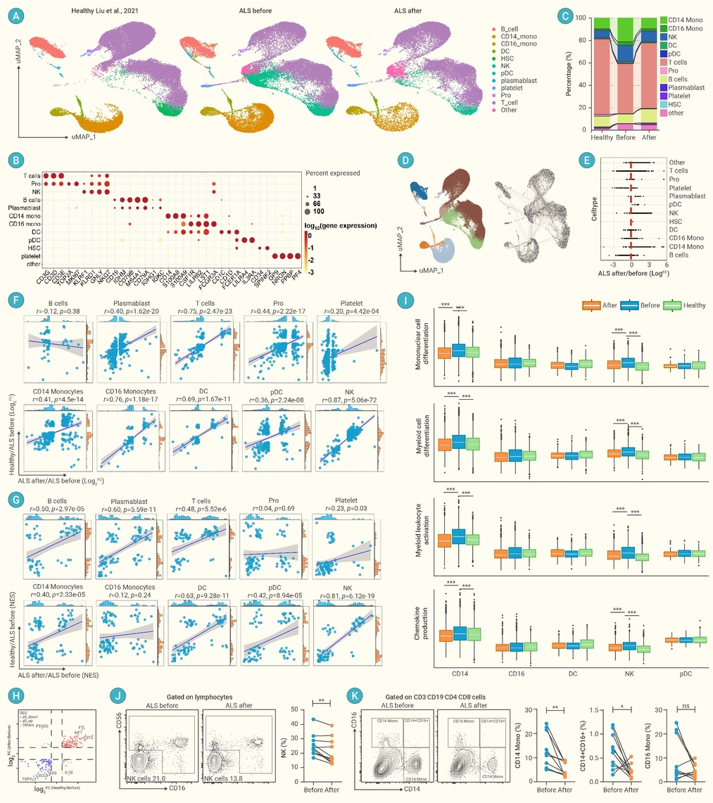
Figure 3.
Characterization of innate immune cells in healthy controls and ALS participants before and following gp96 treatment
-
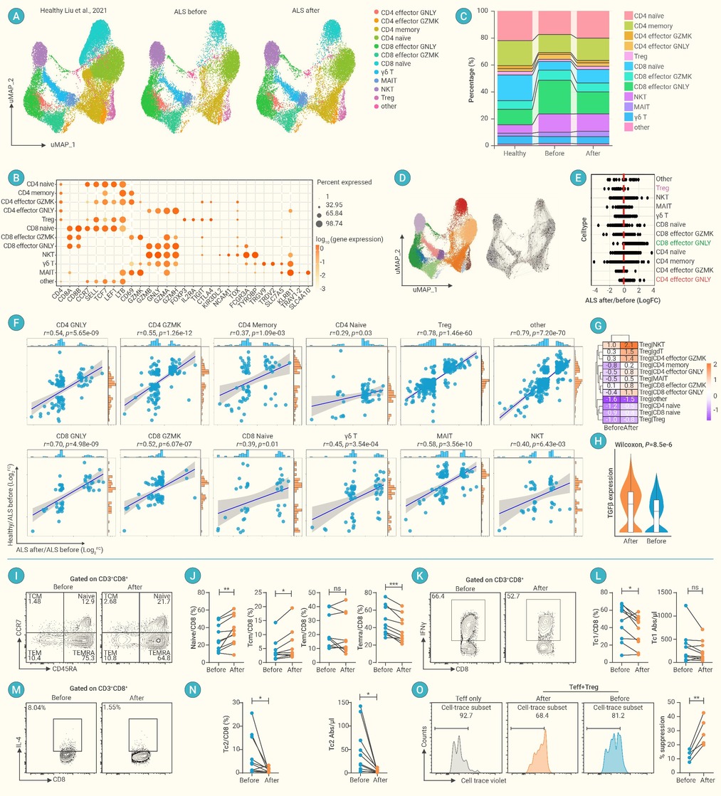
Figure 4.
Immunological features of T cell subsets in ALS participants before and after treatment with high-dose gp96
-
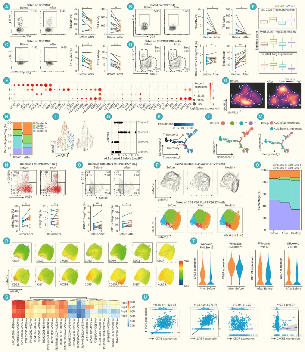
Figure 5.
Immunological features of CD4+T cell and Treg
-
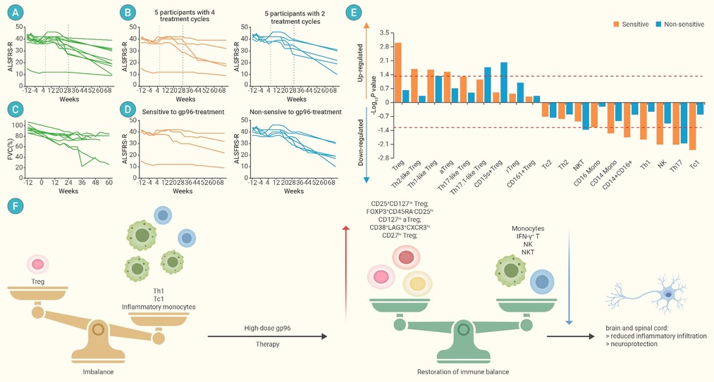
Figure 6.
Therapeutic efficiency analysis of high-dose gp96 treatment for ALS
