
Heterogeneity, immunological mechanisms and translational prospects of tertiary lymphoid structures in cancer therapy
-
GRAPHICAL ABSTRACT
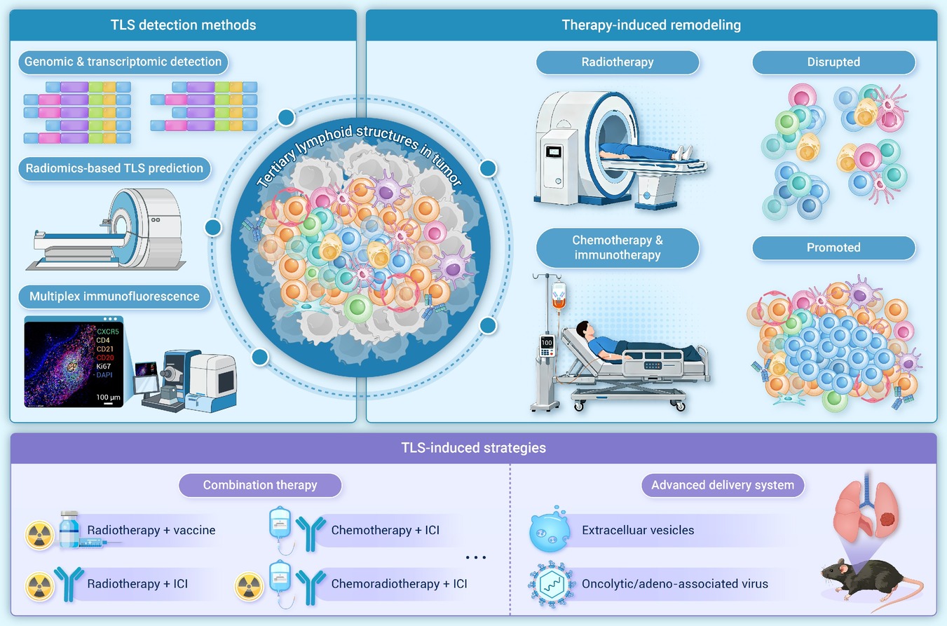
 DownLoad:
Full size image
DownLoad:
Full size image
-
PUBLIC SUMMARY
-
Tertiary lymphoid structures are organized immune niches that can shape cancer therapy outcomes.
Their impact depends on maturation, location, and effector-versus-suppressive immune balance.
Chemotherapy, radiotherapy, and immunotherapy can remodel tertiary lymphoid structures over time.
Standardized tertiary lymphoid structure assessment may improve biomarkers and combination therapy.
-
-
ABSTRACT
Tumor-associated tertiary lymphoid structures (TA-TLS) represent ectopic lymphoid neogenesis within chronically inflamed tumors and are increasingly linked to response to immune checkpoint blockade (ICB) and other anticancer therapies. However, TLS are not uniformly beneficial: their immune outputs vary with maturation, spatial niche (intratumoral, invasive margin, or peritumoral), and the balance between effector programs and suppressive modules. This heterogeneity, together with inconsistent definitions and measurement approaches, has limited cross-cohort comparability and slowed translation of TLS into reliable biomarkers and therapeutic targets. In this review, we synthesize key pathways governing TLS induction, formation, and maturation, emphasizing vascular and stromal scaffold requirements that enable lymphocyte recruitment and compartmentalization. We summarize and reconcile existing TLS classification frameworks across tumor types, and integrate mechanistic interactions among T cells, B cells, dendritic cells, myeloid populations, and stromal elements that shape TLS “quality” and immunological function. Importantly, we highlight therapy-induced TLS remodeling as a dynamic process influenced by chemotherapy, radiotherapy dose/fractionation and sequencing, anti-angiogenic strategies, and ICB itself—helping explain divergent clinical associations and suggesting time-ordered opportunities for intervention. Finally, we discuss multimodal approaches for standardized TLS assessment and outline emerging strategies to safely engineer high-quality TLS to enhance antitumor immunity and rationalize combination regimens. -

-
REFERENCES
[1] Wang Q. and Wu X. (2017). Primary and acquired resistance to PD-1/PD-L1 blockade in cancer treatment. Int. Immunopharmacol. 46:210−219. DOI:10.1016/j.intimp.2017.03.015 [2] Chen D.S. and Mellman I. (2013). Oncology meets immunology: The cancer-immunity cycle. Immunity 39:1−10. DOI:10.1016/j.immuni.2013.07.012 [3] Schumacher T.N. and Thommen D.S. (2022). Tertiary lymphoid structures in cancer. Science 375:eabf9419. DOI:10.1126/science.abf9419 [4] Dieu-Nosjean M.-C., Goc J., Giraldo N.A., et al. (2014). Tertiary lymphoid structures in cancer and beyond. Trends Immunol. 35:571−580. DOI:10.1016/j.it.2014.09.006 [5] Teillaud J.-L., Houel A., Panouillot M., et al. (2024). Tertiary lymphoid structures in anticancer immunity. Nat. Rev. Cancer 24:629−646. DOI:10.1038/s41568-024-00728-0 [6] Deng S., Chen Y., Song B., et al. (2025). Tertiary lymphoid structures in cancer: Spatiotemporal heterogeneity immune orchestration, and translational opportunities. J. Hematol. Oncol. 18:97. DOI:10.1186/s13045-025-01754-7 [7] Petitprez F., De Reyniès A., Keung E.Z., et al. (2020). 318 B cells are associated with survival and immunotherapy response in sarcoma. Nature 577:556−560. DOI:10.1038/s41586-019-1906-8 [8] Fridman W.H., Meylan M., Pupier G., et al. (2023). Tertiary lymphoid structures and B cells: An intratumoral immunity cycle. Immunity 56:2254−2269. DOI:10.1016/j.immuni.2023.08.009 [9] Randall T.D., Carragher D.M. and Rangel-Moreno J. (2008). Development of secondary lymphoid organs. Annu. Rev. Immunol. 26:627−650. DOI:10.1146/annurev.immunol.26.021607.090257 [10] Zhao L., Jin S., Wang S., et al. (2024). Tertiary lymphoid structures in diseases: Immune mechanisms and therapeutic advances. Signal Transduct. Target. Ther. 9:225. DOI:10.1038/s41392-024-01947-5 [11] Johansson-Percival A. and Ganss R. (2021). Therapeutic induction of tertiary lymphoid structures in cancer through stromal remodeling. Front. Immunol. 12:674375. DOI:10.3389/fimmu.2021.674375 [12] Chelvanambi M., Fecek R.J., Taylor J.L., et al. (2021). STING agonist-based treatment promotes vascular normalization and tertiary lymphoid structure formation in the therapeutic melanoma microenvironment. J. Immunother. Cancer 9:e001906. DOI:10.1136/jitc-2020-001906 [13] Sawada J., Kikuchi Y., Duah M., et al. (2025). Simultaneous STING and lymphotoxin-β receptor activation induces B cell responses in tertiary lymphoid structures to potentiate antitumor immunity. Nat. Immunol. 26:1766−1780. DOI:10.1038/s41590-025-02259-8 [14] Jeucken K.C.M., Koning J.J., Mebius R.E., et al. (2019). The role of endothelial cells and TNF-receptor superfamily members in lymphoid organogenesis and function during health and inflammation. Front. Immunol. 10:2700. DOI:10.3389/fimmu.2019.02700 [15] Yu H., Lin L., Zhang Z., et al. (2020). Targeting NF-κB pathway for the therapy of diseases: Mechanism and clinical study. Signal Transduct. Target. Ther. 5:209. DOI:10.1038/s41392-020-00312-6 [16] Bénézech C., Mader E., Desanti G., et al. (2012). Lymphotoxin-β receptor signaling through NF-κB2-RelB pathway reprograms adipocyte precursors as lymph node stromal cells. Immunity 37:721−734. DOI:10.1016/j.immuni.2012.06.010 [17] Sun S.-C. (2017). The non-canonical NF-κB pathway in immunity and inflammation. Nat. Rev. Immunol. 17:545−558. DOI:10.1038/nri.2017.52 [18] Lin L., Hu X., Zhang H., et al. (2019). Tertiary lymphoid organs in cancer immunology: Mechanisms and the new strategy for immunotherapy. Front. Immunol. 10:1398. DOI:10.3389/fimmu.2019.01398 [19] Amisaki M., Zebboudj A., Yano H., et al. (2025). IL-33-activated ILC2s induce tertiary lymphoid structures in pancreatic cancer. Nature 638:1076−1084. DOI:10.1038/s41586-024-08426-5 [20] Grogan J.L. and Ouyang W. (2012). A role for Th17 cells in the regulation of tertiary lymphoid follicles. Eur. J. Immunol. 42:2255−2262. DOI:10.1002/eji.201242656 [21] Chaurio R.A., Anadon C.M., Lee Costich T., et al. (2022). TGF-β-mediated silencing of genomic organizer SATB1 promotes tfh cell differentiation and formation of intra-tumoral tertiary lymphoid structures. Immunity 55:115−128.e9. DOI:10.1016/j.immuni.2021.12.007 [22] Liu W., You W., Lan Z., et al. (2024). An immune cell map of human lung adenocarcinoma development reveals an anti-tumoral role of the tfh-dependent tertiary lymphoid structure. Cell Rep. Med. 5:101448. DOI:10.1016/j.xcrm.2024.101448 [23] Rodriguez A.B., Peske J.D., Woods A.N., et al. (2021). Immune mechanisms orchestrate tertiary lymphoid structures in tumors via cancer-associated fibroblasts. Cell Rep. 36:109422. DOI:10.1016/j.celrep.2021.109422 [24] Shen S., Cui Y., Li M., et al. (2025). Toll-like receptor agonists promote the formation of tertiary lymphoid structure and improve anti-glioma immunity. Neuro-Oncol. 27:140−154. DOI:10.1093/neuonc/noae167 [25] Zhang Y., Liu G., Zeng Q., et al. (2024). CCL19-producing fibroblasts promote tertiary lymphoid structure formation enhancing anti-tumor IgG response in colorectal cancer liver metastasis. Cancer Cell 42:1370−1385.e9. DOI:10.1016/j.ccell.2024.07.006 [26] Ware M.B., Wolfarth A.A., Goon J.B., et al. (2022). The role of interleukin-7 in the formation of tertiary lymphoid structures and their prognostic value in gastrointestinal cancers. J. Immunother. Precis. Oncol. 5:105−117. DOI:10.36401/JIPO-22-10 [27] Rangel-Moreno J., Carragher D.M., De La Luz Garcia-Hernandez M., et al. (2011). The development of inducible bronchus-associated lymphoid tissue depends on IL-17. Nat. Immunol. 12:639−646. DOI:10.1038/ni.2053 [28] Gu X., Li D., Wu P., et al. (2024). Revisiting the CXCL13/CXCR5 axis in the tumor microenvironment in the era of single-cell omics: Implications for immunotherapy. Cancer Lett. 605:217278. DOI:10.1016/j.canlet.2024.217278 [29] Cabrita R., Lauss M., Sanna A., et al. (2020). Tertiary lymphoid structures improve immunotherapy and survival in melanoma. Nature 577:561−565. DOI:10.1038/s41586-019-1914-8 [30] Helmink B.A., Reddy S.M., Gao J., et al. (2020). B cells and tertiary lymphoid structures promote immunotherapy response. Nature 577:549−555. DOI:10.1038/s41586-019-1922-8 [31] Ahmed A., Springfeld C. and Halama N. (2025). De novo induction of tertiary lymphoid structures: An immunotherapeutic strategy in pancreatic cancer. Signal Transduct. Target. Ther. 10:173. DOI:10.1038/s41392-025-02260-5 [32] Vella G., Guelfi S. and Bergers G. (2021). High endothelial venules: A vascular perspective on tertiary lymphoid structures in cancer. Front. Immunol. 12:736670. DOI:10.3389/fimmu.2021.736670 [33] Vella G., Hua Y. and Bergers G. (2023). High endothelial venules in cancer: Regulation, function, and therapeutic implication. Cancer Cell 41:527−545. DOI:10.1016/j.ccell.2023.02.002 [34] Johansson-Percival A., He B., Li Z.-J., et al. (2017). De novo induction of intratumoral lymphoid structures and vessel normalization enhances immunotherapy in resistant tumors. Nat. Immunol. 18:1207−1217. DOI:10.1038/ni.3836 [35] Fleig S., Kapanadze T., Bernier-Latmani J., et al. (2022). Loss of vascular endothelial notch signaling promotes spontaneous formation of tertiary lymphoid structures. Nat. Commun. 13:2022. DOI:10.1038/s41467-022-29701-x [36] Heesters B.A., Chatterjee P., Kim Y.-A., et al. (2013). Endocytosis and recycling of immune complexes by follicular dendritic cells enhances B cell antigen binding and activation. Immunity 38:1164−1175. DOI:10.1016/j.immuni.2013.02.023 [37] Anania J.C., Westin A., Adler J., et al. (2021). A novel image analysis approach reveals a role for complement receptors 1 and 2 in follicular dendritic cell organization in germinal centers. Front. Immunol. 12:655753. DOI:10.3389/fimmu.2021.655753 [38] Chen Y., Wu Y., Yan G., et al. (2024). Tertiary lymphoid structures in cancer: Maturation and induction. Front. Immunol. 15:1369626. DOI:10.3389/fimmu.2024.1369626 [39] Cui X., Gu X., Li D., et al. (2025). Tertiary lymphoid structures as a biomarker in immunotherapy and beyond: Advancing towards clinical application. Cancer Lett. 613:217491. DOI:10.1016/j.canlet.2025.217491 [40] Sautès-Fridman C., Petitprez F., Calderaro J., et al. (2019). Tertiary lymphoid structures in the era of cancer immunotherapy. Nat. Rev. Cancer 19:307−325. DOI:10.1038/s41568-019-0144-6 [41] Le Rochais M., Ghamry-Barrin S., Panouillot M., et al. (2025). Imaging mass cytometry to decipher the maturation of tertiary lymphoid structures. Methods Mol. Biol. 2864:159−179. DOI:10.1007/978-1-0716-4184-2_9 [42] Hayashi Y., Makino T., Sato E., et al. (2023). Density and maturity of peritumoral tertiary lymphoid structures in oesophageal squamous cell carcinoma predicts patient survival and response to immune checkpoint inhibitors. Br. J. Cancer 128:2175−2185. DOI:10.1038/s41416-023-02235-9 [43] Song I.H., Heo S.-H., Bang W.S., et al. (2017). Predictive value of tertiary lymphoid structures assessed by high endothelial venule counts in the neoadjuvant setting of triple-negative breast cancer. Cancer Res. Treat. 49:399−407. DOI:10.4143/crt.2016.215 [44] Vanhersecke L., Bougouin A., Crombé A., et al. (2023). Standardized pathology screening of mature tertiary lymphoid structures in cancers. Lab. Invest. 103:100063. DOI:10.1016/j.labinv.2023.100063 [45] Germain C., Gnjatic S., Tamzalit F., et al. (2014). Presence of B cells in tertiary lymphoid structures is associated with a protective immunity in patients with lung cancer. Am. J. Respir. Crit. Care Med. 189:832−844. DOI:10.1164/rccm.201309-1611OC [46] Siliņa K., Soltermann A., Attar F.M., et al. (2018). Germinal centers determine the prognostic relevance of tertiary lymphoid structures and are impaired by corticosteroids in lung squamous cell carcinoma. Cancer Res. 78:1308−1320. DOI:10.1158/0008-5472.CAN-17-1987 [47] Aoyama S., Noda T., Akita H., et al. (2025). Tumor-associated high endothelial venules are associated with enhanced lymphocyte infiltration and favorable prognosis in resected hepatocellular carcinoma. Cancer Immunol. Immunother. 75:20. DOI:10.1007/s00262-025-04265-z [48] Posch F., Silina K., Leibl S., et al. (2018). Maturation of tertiary lymphoid structures and recurrence of stage II and III colorectal cancer. OncoImmunology 7:e1378844. DOI:10.1080/2162402X.2017.1378844 [49] Lynch K.T., Young S.J., Meneveau M.O., et al. (2021). Heterogeneity in tertiary lymphoid structure B-cells correlates with patient survival in metastatic melanoma. J. Immunother. Cancer 9:e002273. DOI:10.1136/jitc-2020-002273 [50] Werner F., Wagner C., Simon M., et al. (2021). A standardized analysis of tertiary lymphoid structures in human melanoma: Disease progression- and tumor site-associated changes with germinal center alteration. Front. Immunol. 12:675146. DOI:10.3389/fimmu.2021.675146 [51] Osorio J.C., Knorr D.A., Weitzenfeld P., et al. (2025). Fc-optimized CD40 agonistic antibody elicits tertiary lymphoid structure formation and systemic antitumor immunity in metastatic cancer. Cancer Cell 43:1902−1916.e9. DOI:10.1016/j.ccell.2025.07.013 [52] Gunderson A.J., Rajamanickam V., Bui C., et al. (2021). Germinal center reactions in tertiary lymphoid structures associate with neoantigen burden humoral immunity and long-term survivorship in pancreatic cancer. OncoImmunology 10:1900635. DOI:10.1080/2162402X.2021.1900635 [53] Sofopoulos M., Fortis S.P., Vaxevanis C.K., et al. (2019). The prognostic significance of peritumoral tertiary lymphoid structures in breast cancer. Cancer Immunol. Immunother. 68:1733−1745. DOI:10.1007/s00262-019-02407-8 [54] Fang Q., Chen S., Chen X., et al. (2025). Mature tertiary lymphoid structure associated CD103+ CD8+ trm cells determined improved anti-tumor immune in breast cancer. Front. Oncol. 15:1480461. DOI:10.3389/fonc.2025.1480461 [55] Li H., Liu H., Fu H., et al. (2021). Peritumoral tertiary lymphoid structures correlate with protective immunity and improved prognosis in patients with hepatocellular carcinoma. Front. Immunol. 12:648812. DOI:10.3389/fimmu.2021.648812 [56] Long S., Li M., Chen J., et al. (2024). Spatial patterns and MRI-based radiomic prediction of high peritumoral tertiary lymphoid structure density in hepatocellular carcinoma: A multicenter study. J. Immunother. Cancer 12:e009879. DOI:10.1136/jitc-2024-009879 [57] Li H., Wang J., Liu H., et al. (2020). Existence of intratumoral tertiary lymphoid structures is associated with immune cells infiltration and predicts better prognosis in early-stage hepatocellular carcinoma. Aging 12:3451−3472. DOI:10.18632/aging.102821 [58] Zhang T., Lei X., Jia W., et al. (2023). Peritumor tertiary lymphoid structures are associated with infiltrating neutrophils and inferior prognosis in hepatocellular carcinoma. Cancer Med. 12:3068−3078. DOI:10.1002/cam4.5227 [59] Finkin S., Yuan D., Stein I., et al. (2015). Ectopic lymphoid structures function as microniches for tumor progenitor cells in hepatocellular carcinoma. Nat. Immunol. 16:1235−1244. DOI:10.1038/ni.3290 [60] Calderaro J., Petitprez F., Becht E., et al. (2019). Intra-tumoral tertiary lymphoid structures are associated with a low risk of early recurrence of hepatocellular carcinoma. J. Hepatol. 70:58−65. DOI:10.1016/j.jhep.2018.09.003 [61] Li J., Zhang L., Xing H., et al. (2024). The absence of intra-tumoral tertiary lymphoid structures is associated with a worse prognosis and mtor signaling activation in hepatocellular carcinoma with liver transplantation: A multicenter retrospective study. Adv. Sci. 11:e2309348. DOI:10.1002/advs.202309348 [62] Xin S., Wen S., He P., et al. (2024). Density of tertiary lymphoid structures and their correlation with prognosis in non-small cell lung cancer. Front. Immunol. 15:1423775. DOI:10.3389/fimmu.2024.1423775 [63] Xu W., Lu J., Tian X., et al. (2024). Unveiling the impact of tertiary lymphoid structures on immunotherapeutic responses of clear cell renal cell carcinoma. MedComm 5:e461. DOI:10.1002/mco2.461 [64] Zhou C., Chen J., Wang Y., et al. (2026). Clinical and immunological significance of tertiary lymphoid structure maturation heterogeneity in brain metastases of lung adenocarcinoma. J. Transl. Med. 24:396. DOI:10.1186/s12967-026-07852-5 [65] Xu W., Lu J., Liu W.-R., et al. (2023). Heterogeneity in tertiary lymphoid structures predicts distinct prognosis and immune microenvironment characterizations of clear cell renal cell carcinoma. J. Immunother. Cancer 11:e006667. DOI:10.1136/jitc-2023-006667 [66] Deng M., Liu X., Jiang Y., et al. (2024). Tertiary lymphoid structures’ pattern and prognostic value in primary adenocarcinoma of jejunum and ileum. World J. Surg. Oncol. 22:261. DOI:10.1186/s12957-024-03543-x [67] Su G.-L., Zhang M.-J., Li H., et al. (2025). Dissecting tertiary lymphoid structures in cancer: Maturation, localization and density. Theranostics 15:9459−9485. DOI:10.7150/thno.113940 [68] Ding G.-Y., Ma J.-Q., Yun J.-P., et al. (2022). Distribution and density of tertiary lymphoid structures predict clinical outcome in intrahepatic cholangiocarcinoma. J. Hepatol. 76:608−618. DOI:10.1016/j.jhep.2021.10.030 [69] Elfving H., Yu H., Fessehatsion K.K., et al. (2025). Spatial distribution of tertiary lymphoid structures in the molecular and clinical context of non-small cell lung cancer. Cell. Oncol. 48:801−813. DOI:10.1007/s13402-025-01052-x [70] Shu D.H., Ho W.J., Kagohara L.T., et al. (2024). Immunotherapy response induces divergent tertiary lymphoid structure morphologies in hepatocellular carcinoma. Nat. Immunol. 25:2110−2123. DOI:10.1038/s41590-024-01992-w [71] Zhang C., Wang X.-Y., Zuo J.-L., et al. (2023). Localization and density of tertiary lymphoid structures associate with molecular subtype and clinical outcome in colorectal cancer liver metastases. J. Immunother. Cancer 11:e006425. DOI:10.1136/jitc-2022-006425 [72] Shang T., Jiang T., Lu T., et al. (2023). Tertiary lymphoid structures predict the prognosis and immunotherapy response of cholangiocarcinoma. Front. Immunol. 14:1166497. DOI:10.3389/fimmu.2023.1166497 [73] Cakmak P., Lun J.H., Singh A., et al. (2025). Spatial immune profiling defines a subset of human gliomas with functional tertiary lymphoid structures. Immunity 58:2847−2863.e8. DOI:10.1016/j.immuni.2025.09.018 [74] Tang Z., Bai Y., Fang Q., et al. (2025). Spatial transcriptomics reveals tryptophan metabolism restricting maturation of intratumoral tertiary lymphoid structures. Cancer Cell 43:1025−1044.e14. DOI:10.1016/j.ccell.2025.03.011 [75] Du Bois H., Heim T.A. and Lund A.W. (2021). Tumor-draining lymph nodes: At the crossroads of metastasis and immunity. Sci. Immunol. 6:eabg3551. DOI:10.1126/sciimmunol.abg3551 [76] Zhu X., Maier G. and Panda S. (2024). Learning from circadian rhythm to transform cancer prevention prognosis and survivorship care. Trends Cancer 10:196−207. DOI:10.1016/j.trecan.2023.11.002 [77] Lanitis E., Irving M. and Coukos G. (2015). Targeting the tumor vasculature to enhance T cell activity. Curr. Opin. Immunol. 33:55−63. DOI:10.1016/j.coi.2015.01.011 [78] Thiemann S., Man J.H., Chang M.H., et al. (2015). Galectin-1 regulates tissue exit of specific dendritic cell populations. J. Biol. Chem. 290:22662−22677. DOI:10.1074/jbc.M115.644799 [79] Pietrobon V. and Marincola F.M. (2021). Hypoxia and the phenomenon of immune exclusion. J. Transl. Med. 19:9. DOI:10.1186/s12967-020-02667-4 [80] Geindreau M., Ghiringhelli F. and Bruchard M. (2021). Vascular endothelial growth factor, a key modulator of the anti-tumor immune response. Int. J. Mol. Sci. 22:4871. DOI:10.3390/ijms22094871 [81] Rodriguez A.B. and Engelhard V.H. (2020). Insights into tumor-associated tertiary lymphoid structures: Novel targets for antitumor immunity and cancer immunotherapy. Cancer Immunol. Res. 8:1338−1345. DOI:10.1158/2326-6066.cir-20-0432 [82] Asrir A., Tardiveau C., Coudert J., et al. (2022). Tumor-associated high endothelial venules mediate lymphocyte entry into tumors and predict response to PD-1 plus CTLA-4 combination immunotherapy. Cancer Cell 40:318−334.e9. DOI:10.1016/j.ccell.2022.01.002 [83] Li H., Zhang M.-J., Zhang B., et al. (2025). Mature tertiary lymphoid structures evoke intra-tumoral T and B cell responses via progenitor exhausted CD4+ T cells in head and neck cancer. Nat. Commun. 16:4228. DOI:10.1038/s41467-025-59341-w [84] Kinker G.S., Vitiello G.A.F., Diniz A.B., et al. (2023). Mature tertiary lymphoid structures are key niches of tumour-specific immune responses in pancreatic ductal adenocarcinomas. Gut 72:1927−1941. DOI:10.1136/gutjnl-2022-328697 [85] Crotty S. (2011). Follicular helper CD4 T cells (TFH ). Annu. Rev. Immunol. 29:621−663. DOI:10.1146/annurev-immunol-031210-101400 [86] Esparcia-Pinedo L., Romero-Laorden N. and Alfranca, A. (2023). Tertiary lymphoid structures and B lymphocytes: A promising therapeutic strategy to fight cancer. Front. Immunol. 14:1231315. DOI:10.3389/fimmu.2023.1231315 [87] Vinuesa C.G., Linterman M.A., Yu D., et al. (2016). Follicular helper T cells. Annu. Rev. Immunol. 34:335−368. DOI:10.1146/annurev-immunol-041015-055605 [88] Bao X., Lin X., Xie M., et al. (2024). Mature tertiary lymphoid structures: Important contributors to anti-tumor immune efficacy. Front. Immunol. 15:1413067. DOI:10.3389/fimmu.2024.1413067 [89] Noël G., Fontsa M.L., Garaud S., et al. (2021). Functional Th1-oriented T follicular helper cells that infiltrate human breast cancer promote effective adaptive immunity. J. Clin. Invest. 131:e139905. DOI:10.1172/JCI139905 [90] Ukita M., Hamanishi J., Yoshitomi H., et al. (2022). CXCL13-producing CD4+ T cells accumulate in the early phase of tertiary lymphoid structures in ovarian cancer. JCI Insight 7:e157215. DOI:10.1172/jci.insight.157215 [91] Li J.-P., Wu C.-Y., Chen M.-Y., et al. (2021). PD-1+CXCR5-CD4+ Th-CXCL13 cell subset drives B cells into tertiary lymphoid structures of nasopharyngeal carcinoma. J. Immunother. Cancer 9:e002101. DOI:10.1136/jitc-2020-002101 [92] Im S.J., Obeng R.C., Nasti T.H., et al. (2023). Characteristics and anatomic location of PD-1+TCF1+ stem-like CD8 T cells in chronic viral infection and cancer. Proc. Natl. Acad. Sci. USA. 120:e2221985120. DOI:10.1073/pnas.2221985120 [93] Tanoue K., Ohmura H., Uehara K., et al. (2024). Spatial dynamics of CD39+CD8+ exhausted T cell reveal tertiary lymphoid structures-mediated response to PD-1 blockade in esophageal cancer. Nat. Commun. 15:9033. DOI:10.1038/s41467-024-53262-w [94] Tran K., Kumari A.N., Raghu D., et al. (2024). T cell factor 1 (TCF-1) defines T cell differentiation in colorectal cancer. iScience 27:110754. DOI:10.1016/j.isci.2024.110754 [95] Im S.J., Hashimoto M., Gerner M.Y., et al. (2016). Defining CD8+ T cells that provide the proliferative burst after PD-1 therapy. Nature 537:417−421. DOI:10.1038/nature19330 [96] Siddiqui I., Schaeuble K., Chennupati V., et al. (2019). Intratumoral Tcf1+PD-1+CD8+ T cells with stem-like properties promote tumor control in response to vaccination and checkpoint blockade immunotherapy. Immunity 50:195−211.e10. DOI:10.1016/j.immuni.2018.12.021 [97] Miller B.C., Sen D.R., Al Abosy R., et al. (2019). Subsets of exhausted CD8+ T cells differentially mediate tumor control and respond to checkpoint blockade. Nat. Immunol. 20:326−336. DOI:10.1038/s41590-019-0312-6 [98] Schenkel J.M., Herbst R.H., Canner D., et al. (2021). Conventional type I dendritic cells maintain a reservoir of proliferative tumor-antigen specific TCF-1+ CD8+ T cells in tumor-draining lymph nodes. Immunity 54:2338−2353.e6. DOI:10.1016/j.immuni.2021.08.026 [99] Kasikova L., Rakova J., Hensler M., et al. (2024). Tertiary lymphoid structures and B cells determine clinically relevant T cell phenotypes in ovarian cancer. Nat. Commun. 15:2528. DOI:10.1038/s41467-024-46873-w [100] Joshi N.S., Akama-Garren E.H., Lu Y., et al. (2015). Regulatory T cells in tumor-associated tertiary lymphoid structures suppress anti-tumor T cell responses. Immunity 43:579−590. DOI:10.1016/j.immuni.2015.08.006 [101] Devi-Marulkar P., Fastenackels S., Karapentiantz P., et al. (2022). Regulatory T cells infiltrate the tumor-induced tertiary lymphoïd structures and are associated with poor clinical outcome in NSCLC. Commun. Biol. 5:1416. DOI:10.1038/s42003-022-04356-y [102] Davies A.M. and Sutton B.J. (2015). Human IgG4: A structural perspective. Immunol. Rev. 268:139−159. DOI:10.1111/imr.12349 [103] Wang H., Li J., Wang Y., et al. (2025). IgG4-mediated M2 macrophage polarization in tertiary lymphoid structures of esophageal cancer: Implications for immunosuppression. Front. Immunol. 15:1497783. DOI:10.3389/fimmu.2024.1497783 [104] Su C., Chen S., He X., et al. (2025). Increased IgG4 expression within tertiary lymphoid structures of esophageal cancer and implications for prognosis. Front. Immunol. 16:1654655. DOI:10.3389/fimmu.2025.1654655 [105] Chen C., An M., Zheng H., et al. (2026). B cells disrupt tertiary lymphoid structure formation and suppress anti-tumor immunity. Cancer Cell 44:551−566.e17. DOI:10.1016/j.ccell.2025.12.011 [106] Huang Q., Wang F., Li X., et al. (2025). Multi-omics profiling reveals an immunosuppressive plasma cell subset within tertiary lymphoid structures in cervical cancer. Cancer Immunol. Immunother. 75:26. DOI:10.1007/s00262-025-04268-w [107] Meylan M., Petitprez F., Becht E., et al. (2022). Tertiary lymphoid structures generate and propagate anti-tumor antibody-producing plasma cells in renal cell cancer. Immunity 55:527−541.e5. DOI:10.1016/j.immuni.2022.02.001 [108] Lehmann J., Thelen M., Kreer C., et al. (2024). Tertiary lymphoid structures in pancreatic cancer are structurally homologous, share gene expression patterns and B-cell clones with secondary lymphoid organs but show increased T-cell activation. Cancer Immunol. Res. 13:323−336. DOI:10.1158/2326-6066.cir-24-0299 [109] He M., He Q., Cai X., et al. (2023). Intratumoral tertiary lymphoid structure (TLS) maturation is influenced by draining lymph nodes of lung cancer. J. Immunother. Cancer 11:e005539. DOI:10.1136/jitc-2022-005539 [110] Gao J., Gu D., Yang K., et al. (2025). Infiltrating plasma cells maintain glioblastoma stem cells through IgG-tumor binding. Cancer Cell 43:122−143.e8. DOI:10.1016/j.ccell.2024.12.006 [111] Sammut S.-J., Galson J.D., Minter R., et al. (2024). Predictability of B cell clonal persistence and immunosurveillance in breast cancer. Nat. Immunol. 25:916−924. DOI:10.1038/s41590-024-01821-0 [112] Che Y., Lee J., Abou-Taleb F., et al. (2025). Induced B cell receptor diversity predicts PD-1 blockade immunotherapy response. Proc. Natl. Acad. Sci. USA 122:e2501269122. DOI:10.1073/pnas.2501269122 [113] Goc J., Germain C., Vo-Bourgais T.K.D., et al. (2014). Dendritic cells in tumor-associated tertiary lymphoid structures signal a Th1 cytotoxic immune contexture and license the positive prognostic value of infiltrating CD8+ T cells. Cancer Res. 74:705−715. DOI:10.1158/0008-5472.CAN-13-1342 [114] Berthe J., Poudel P., Segerer F.J., et al. (2024). Exploring the impact of tertiary lymphoid structures maturity in NSCLC: Insights from TLS scoring. Front. Immunol. 15:1422206. DOI:10.3389/fimmu.2024.1422206 [115] Wang M., Shi J., Xu K., et al. (2026). T cell exhaustion and dendritic cell-mediated tertiary lymphoid structures (TLSs) modulation affect response to neoadjuvant chemoradiotherapy in microsatellite stable rectal cancer. Adv. Sci. 13:e14332. DOI:10.1002/advs.202514332 [116] Syding L.A., Plačková K., Pavelková L., et al. (2025). High treg and PMN-MDSC densities are a hallmark of tertiary lymphoid structures in fatal cases of cervical cancer. J. Immunother. Cancer 13:e012613. DOI:10.1136/jitc-2025-012613 [117] Condamine T., Dominguez G.A., Youn J.-I., et al. (2016). Lectin-type oxidized LDL receptor-1 distinguishes population of human polymorphonuclear myeloid-derived suppressor cells in cancer patients. Sci. Immunol. 1:aaf8943−aaf8943. DOI:10.1126/sciimmunol.aaf8943 [118] Zhao F., Hoechst B., Duffy A., et al. (2012). A9 a new marker for monocytic human myeloid‐derived suppressor cells. Immunology 136:176−183. DOI:10.1111/j.1365-2567.2012.03566.x [119] Lesokhin A.M., Hohl T.M., Kitano S., et al. (2012). Monocytic CCR2+ myeloid-derived suppressor cells promote immune escape by limiting activated CD8 T-cell infiltration into the tumor microenvironment. Cancer Res. 72:876−886. DOI:10.1158/0008-5472.CAN-11-1792 [120] Groth C., Hu X., Weber R., et al. (2019). Immunosuppression mediated by myeloid-derived suppressor cells (MDSCs) during tumour progression. Br. J. Cancer 120:16−25. DOI:10.1038/s41416-018-0333-1 [121] Rodríguez P.C. and Ochoa A.C. (2008). Arginine regulation by myeloid derived suppressor cells and tolerance in cancer: Mechanisms and therapeutic perspectives. Immunol. Rev. 222:180−191. DOI:10.1111/j.1600-065X.2008.00608.x [122] Baniyash M. (2004). TCR ζ-chain downregulation: Curtailing an excessive inflammatory immune response. Nat. Rev. Immunol. 4:675−687. DOI:10.1038/nri1434 [123] Kittang A.O., Kordasti S., Sand K.E., et al. (2016). Expansion of myeloid derived suppressor cells correlates with number of T regulatory cells and disease progression in myelodysplastic syndrome. OncoImmunology 5:e1062208. DOI:10.1080/2162402X.2015.1062208 [124] Marcovecchio P.M., Thomas G. and Salek-Ardakani S. (2021). CXCL9-expressing tumor-associated macrophages: New players in the fight against cancer. J. Immunother. Cancer 9:e002045. DOI:10.1136/jitc-2020-002045 [125] Duan Z. and Luo Y. (2021). Targeting macrophages in cancer immunotherapy. Signal Transduct. Target. Ther. 6:127. DOI:10.1038/s41392-021-00506-6 [126] Guedj K., Khallou-Laschet J., Clement M., et al. (2014). M1 macrophages act as LTβR-independent lymphoid tissue inducer cells during atherosclerosis-related lymphoid neogenesis. Cardiovasc. Res. 101:434−443. DOI:10.1093/cvr/cvt263 [127] Labadie B.W.,Bao R. and Luke J.J. (2019). Reimagining IDO pathway inhibition in cancer immunotherapy via downstream focus on the tryptophan–kynurenine–aryl hydrocarbon axis. Clin. Cancer Res. 25:1462−1471. DOI:10.1158/1078-0432.CCR-18-2882 [128] Laviron M., Petit M., Weber-Delacroix E., et al. (2022). Tumor-associated macrophage heterogeneity is driven by tissue territories in breast cancer. Cell Rep. 39:110865. DOI:10.1016/j.celrep.2022.110865 [129] Ma R.-Y., Black A. and Qian B.-Z. (2022). Macrophage diversity in cancer revisited in the era of single-cell omics. Trends Immunol. 43:546−563. DOI:10.1016/j.it.2022.04.008 [130] Sica A., Larghi P., Mancino A., et al. (2008). Macrophage polarization in tumour progression. Semin. Cancer Biol. 18:349−355. DOI:10.1016/j.semcancer.2008.03.004 [131] Xu R., Liu H., Zhu T., et al. (2025). An immunosuppressive tertiary lymphoid structure is associated with adverse prognosis in gastric-type endocervical adenocarcinoma. J. Natl. Cancer Inst. 118:276−288. DOI:10.1093/jnci/djaf310 [132] Petroni G., Scolari F., Scoccianti G., et al. (2025). Th17-like cells and immunosuppressive macrophages infiltrate tertiary lymphoid structures with distinct maturation status in soft-tissue sarcoma. Cell Death Dis. 16:917. DOI:10.1038/s41419-025-08376-4 [133] Sánchez-Alonso S., Setti-Jerez G., Arroyo M., et al. (2020). A new role for circulating T follicular helper cells in humoral response to anti-PD-1 therapy. J. Immunother. Cancer 8:e001187. DOI:10.1136/jitc-2020-001187 [134] Matsumoto K., Noda Y., Hachiya K., et al. (2026). Single-cell spatial analysis identifies ID1-high endothelial cells in tertiary lymphoid structures as predictors of durable response to immunotherapy in non-small cell lung cancer. Cancer Cell Int. 26:65. DOI:10.1186/s12935-025-04161-7 [135] Liu S.-X., Wu T.-W., Luo D.-H., et al. (2025). 26 Interferon-responsive HEVs drive tumor tertiary lymphoid structure formation and predict immunotherapy response in nasopharyngeal carcinoma. Cell Rep. Med. 6:102200. DOI:10.1016/j.xcrm.2025.102200 [136] Onder L., Papadopoulou C., Lütge A., et al. (2025). Fibroblastic reticular cells generate protective intratumoral T cell environments in lung cancer. Cell 188:430−446.e20. DOI:10.1016/j.cell.2024.10.042 [137] Peyraud F., Guégan J.-P., Rey C., et al. (2025). Spatially resolved transcriptomics reveal the determinants of primary resistance to immunotherapy in NSCLC with mature tertiary lymphoid structures. Cell Rep. Med. 6:101934. DOI:10.1016/j.xcrm.2025.101934 [138] O’Connor R.A., Martinez B.R., Koppensteiner L., et al. (2023). Cancer-associated fibroblasts drive CXCL13 production in activated T cells via TGF-beta. Front. Immunol. 14:1221532. DOI:10.3389/fimmu.2023.1221532 [139] Morcrette G., Hirsch T.Z., Badour E., et al. (2019). APC germline hepatoblastomas demonstrate cisplatin-induced intratumor tertiary lymphoid structures. OncoImmunology 8:e1583547. DOI:10.1080/2162402X.2019.1583547 [140] Lanickova T., Hensler M., Kasikova L., et al. (2025). Chemotherapy drives tertiary lymphoid structures that correlate with ICI-responsive TCF1+CD8+ T cells in metastatic ovarian cancer. Clin. Cancer Res. 31:164−180. DOI:10.1158/1078-0432.CCR-24-1594 [141] Lv J., Wei Y., Yin J.-H., et al. (2023). The tumor immune microenvironment of nasopharyngeal carcinoma after gemcitabine plus cisplatin treatment. Nat. Med. 29:1424−1436. DOI:10.1038/s41591-023-02369-6 [142] Tian N., Wang Q., Lv Y., et al. (2025). Mature tertiary lymphoid structures support B cell-mediated antitumour immunity and are disrupted by neoadjuvant therapy in rectal cancer: A multicentre retrospective study. eBioMedicine 122:106030. DOI:10.1016/j.ebiom.2025.106030 [143] Constantinides A., Lansu N., Mosen P., et al. (2025). Treatment of colorectal peritoneal metastases with oxaliplatin induces biomarkers predicting response to immune checkpoint blockade. Transl. Oncol. 59:102464. DOI:10.1016/j.tranon.2025.102464 [144] Xing R., Mei J., Zuo Z., et al. (2025). Enhanced formation of tertiary lymphoid structures shapes the anti-tumor microenvironment in hepatocellular carcinoma after FOLFOX-HAIC therapy. Cell Rep. Med. 6:102298. DOI:10.1016/j.xcrm.2025.102298 [145] Huang Y., Du Z., Lai Z., et al. (2025). Single-nucleus and spatial transcriptome profiling delineates the multicellular ecosystem in hepatocellular carcinoma after hepatic arterial infusion chemotherapy. Adv. Sci. 12:2405749. DOI:10.1002/advs.202405749 [146] Rupp L., Dietsche I., Kießler M., et al. (2024). Neoadjuvant chemotherapy is associated with suppression of the B cell-centered immune landscape in pancreatic ductal adenocarcinoma. Front. Immunol. 15:1378190. DOI:10.3389/fimmu.2024.1378190 [147] Delvecchio F.R., Fincham R.E.A., Spear S., et al. (2021). Pancreatic cancer chemotherapy is potentiated by induction of tertiary lymphoid structures in mice. Cell. Mol. Gastroenterol. Hepatol. 12:1543−1565. DOI:10.1016/j.jcmgh.2021.06.023 [148] Fucikova J., Kepp O., Kasikova L., et al. (2020). Detection of immunogenic cell death and its relevance for cancer therapy. Cell Death Dis. 11:1013. DOI:10.1038/s41419-020-03221-2 [149] Adkins I., Fucikova J., Garg A.D., et al. (2015). Physical modalities inducing immunogenic tumor cell death for cancer immunotherapy. OncoImmunology 3:e968434. DOI:10.4161/21624011.2014.968434 [150] Zhao H., Zhao Y., Zhang S., et al. (2024). Effects of immunogenic cell death-inducing chemotherapeutics on the immune cell activation and tertiary lymphoid structure formation in melanoma. Front. Immunol. 15:1302751. DOI:10.3389/fimmu.2024.1302751 [151] Bertucci F., De Nonneville A., Finetti P., et al. (2023). Predictive power of tertiary lymphoid structure signature for neoadjuvant chemotherapy response and immunotherapy benefit in HER2‐negative breast cancer. Cancer Commun. 43:943−946. DOI:10.1002/cac2.12447 [152] Wei Z., Lin K., Deng W., et al. (2025). Tertiary lymphoid structures are associated with lower axillary residual nodal burden in breast cancer patients after neoadjuvant chemotherapy. Eur. J. Med. Res. 30:1263. DOI:10.1186/s40001-025-03556-6 [153] Cabrero-de las Heras S., Hernández-Yagüe X., González A., et al. (2024). Changes In serum CXCL13 levels are associated with outcomes of colorectal cancer patients undergoing first-line oxaliplatin-based treatment. Biomed. Pharmacother. 176:116857. DOI:10.1016/j.biopha.2024.116857 [154] Benzerdjeb N., Dartigues P., Kepenekian V., et al. (2021). Tertiary lymphoid structures in epithelioid malignant peritoneal mesothelioma are associated with neoadjuvant chemotherapy but not with prognosis. Virchows Arch. 479:765−772. DOI:10.1007/s00428-021-03099-1 [155] Li S., Chen K., Sun Z., et al. (2024). Radiation drives tertiary lymphoid structures to reshape TME for synergized antitumour immunity. Expert Rev. Mol. Med. 26:e30. DOI:10.1017/erm.2024.27 [156] Boivin G., Kalambaden P., Faget J., et al. (2018). Cellular composition and contribution of tertiary lymphoid structures to tumor immune infiltration and modulation by radiation therapy. Front. Oncol. 8:256. DOI:10.3389/fonc.2018.00256 [157] Mills B.N., Qiu H., Drage M.G., et al. (2022). Modulation of the human pancreatic ductal adenocarcinoma immune microenvironment by stereotactic body radiotherapy. Clin. Cancer Res. 28:150−162. DOI:10.1158/1078-0432.CCR-21-2495 [158] Wang Q., Zhong W., Shen X., et al. (2024). Tertiary lymphoid structures predict survival and response to neoadjuvant therapy in locally advanced rectal cancer. Npj Precis. Oncol. 8:61. DOI:10.1038/s41698-024-00533-w [159] Huang H., Zhao G., Wang T., et al. (2024). Survival benefit and spatial properties of tertiary lymphoid structures in esophageal squamous cell carcinoma with neoadjuvant therapies. Cancer Lett. 601:217178. DOI:10.1016/j.canlet.2024.217178 [160] Wang D., Huang L., Qian D., et al. (2023). Low-dose radiotherapy promotes the formation of tertiary lymphoid structures in lung adenocarcinoma. Front. Immunol. 14:1334408. DOI:10.3389/fimmu.2023.1334408 [161] Zhai K., Xie R., Ru K., et al. (2025). Tertiary lymphoid structures correlate with the therapeutic efficacy and prognosis of resectable esophageal squamous cell carcinoma undergoing neoadjuvant chemoradiotherapy plus immunotherapy. Front. Immunol. 16:1616247. DOI:10.3389/fimmu.2025.1616247 [162] Huang J., Theelen W.S.M.E., Belcaid Z., et al. (2025). Combination of pembrolizumab and radiotherapy induces systemic antitumor immune responses in immunologically cold non-small cell lung cancer. Nat. Cancer 6:1676−1692. DOI:10.1038/s43018-025-01018-w [163] Huang Y.-S., Li Z., Xiao Z.-F., et al. (2022). Case report: Radiotherapy plus pneumococcal conjugate vaccine stimulates abscopal immune response in a patient with ALK+ NSCLC. Front. Immunol. 13:950252. DOI:10.3389/fimmu.2022.950252 [164] Shiao S.L., Gouin K.H., Ing N., et al. (2024). Single-cell and spatial profiling identify three response trajectories to pembrolizumab and radiation therapy in triple negative breast cancer. Cancer Cell 42:70−84.e8. DOI:10.1016/j.ccell.2023.12.012 [165] van Hooren L., Handgraaf S.M., Kloosterman D.J., et al. (2023). CD103+ regulatory T cells underlie resistance to radio-immunotherapy and impair CD8+ T cell activation in glioblastoma. Nat. Cancer 4:665−681. DOI:10.1038/s43018-023-00547-6 [166] Deng L., Liang H., Xu M., et al. (2014). STING-dependent cytosolic DNA sensing promotes radiation-induced type I interferon-dependent antitumor immunity in immunogenic tumors. Immunity 41:843−852. DOI:10.1016/j.immuni.2014.10.019 [167] Vanpouille-Box C., Alard A., Aryankalayil M.J., et al. (2017). DNA exonuclease Trex1 regulates radiotherapy-induced tumour immunogenicity. Nat. Commun. 8:15618. DOI:10.1038/ncomms15618 [168] Herrera F.G., Ronet C., Ochoa de Olza M., et al. (2022). Low-dose radiotherapy reverses tumor immune desertification and resistance to immunotherapy. Cancer Discov. 12:108−133. DOI:10.1158/2159-8290.CD-21-0003 [169] Italiano A., Bessede A., Pulido M., et al. (2022). Pembrolizumab in soft-tissue sarcomas with tertiary lymphoid structures: A phase 2 PEMBROSARC trial cohort. Nat. Med. 28:1199−1206. DOI:10.1038/s41591-022-01821-3 [170] Chung S.-Y., Yeh Y.-C., Huang C.-J., et al. (2025). Comparative impact of tertiary lymphoid structures and tumor-infiltrating lymphocytes in cholangiocarcinoma. J. Immunother. Cancer 13:e010173. DOI:10.1136/jitc-2024-010173 [171] Sadeghirad H., Monkman J., Tan C.W., et al. (2024). Spatial dynamics of tertiary lymphoid aggregates in head and neck cancer: Insights into immunotherapy response. J. Transl. Med. 22:677. DOI:10.1186/s12967-024-05409-y [172] Xie Y., Peng H., Hu Y., et al. (2025). Immune microenvironment spatial landscapes of tertiary lymphoid structures in gastric cancer. BMC Med. 23:59. DOI:10.1186/s12916-025-03889-3 [173] Vanhersecke L., Brunet M., Guégan J.-P., et al. (2021). Mature tertiary lymphoid structures predict immune checkpoint inhibitor efficacy in solid tumors independently of PD-L1 expression. Nat. Cancer 2:794−802. DOI:10.1038/s43018-021-00232-6 [174] van Hooren L., Vaccaro A., Ramachandran, M., et al. (2021). Agonistic CD40 therapy induces tertiary lymphoid structures but impairs responses to checkpoint blockade in glioma. Nat. Commun. 12:4127. DOI:10.1038/s41467-021-24347-7 [175] Levy A., Morel D., Texier M., et al. (2025). Monocyte-lineage tumor infiltration predicts immunoradiotherapy response in advanced pretreated soft-tissue sarcoma: Phase 2 trial results. Signal Transduct. Target. Ther. 10:103. DOI:10.1038/s41392-025-02173-3 [176] Hua Y., Vella G., Rambow F., et al. (2022). Cancer immunotherapies transition endothelial cells into high-endothelial venules that generate TCF1+ T lymphocyte niches through a feed-forward loop. Cancer Cell 40:1600−1618.e10. DOI:10.1016/j.ccell.2022.11.002 [177] Eschweiler S., Clarke J., Ramírez-Suástegui C., et al. (2021). Intratumoral follicular regulatory T cells curtail anti-PD-1 treatment efficacy. Nat. Immunol. 22:1052−1063. DOI:10.1038/s41590-021-00958-6 [178] Cheng R., Li S., Ma X., et al. (2025). Intratumoral antigen-presenting cell activation by a nanovesicle for the concurrent tertiary lymphoid structure de novo neogenesis. Sci. Adv. 11:eadr1299. DOI:10.1126/sciadv.adr1299 [179] Ahn W., Han J., Kim N., et al. (2025). Hierarchical protein nano-crystalline hydrogel with extracellular vesicles for ectopic lymphoid structure formation. Biomaterials 318:123166. DOI:10.1016/j.biomaterials.2025.123166 [180] Mi Z., Chen J., Zhang Z., et al. (2025). Synthetic biology–driven induction of mature TLS formation enhances antitumor immunity in colorectal cancer. Sci. Transl. Med. 17:eado8395. DOI:10.1126/scitranslmed.ado8395 [181] Ramachandran M., Vaccaro A., Van De Walle T., et al. (2023). Tailoring vascular phenotype through AAV therapy promotes anti-tumor immunity in glioma. Cancer Cell 41:1134−1151.e10. DOI:10.1016/j.ccell.2023.04.010 [182] Kuwentrai C., Tang W., Lin X., et al. (2025). Injectable hydrogel-based drug formulation for enhancing tertiary lymphoid structure formation and cancer immunotherapy efficacy. J. Controlled Release 384:113897. DOI:10.1016/j.jconrel.2025.113897 [183] Falahat R. and Mulé J.J. (2025). Chemokine/cytokine-releasing biomaterials induce in situ tertiary lymphoid-like structures and enhance antitumor immunity. Proc. Natl. Acad. Sci. USA 122:e2409560122. DOI:10.1073/pnas.2409560122 [184] Kuang G., Zhang Q., Li W., et al. (2024). Biomimetic tertiary lymphoid structures with microporous annealed particle scaffolds for cancer postoperative therapy. ACS Nano 18:9176−9186. DOI:10.1021/acsnano.4c01180 [185] Zhang L., Zhang B., Zhang M.-J., et al. (2025). Trigger inducible tertiary lymphoid structure formation using covalent organic frameworks for cancer immunotherapy. Nat. Commun. 16:44. DOI:10.1038/s41467-024-55430-4 [186] Yang Q.-C., Wang Y.-Y., Wang S., et al. (2025). Engineered bacterial membrane biomimetic covalent organic framework as nano-immunopotentiator for cancer immunotherapy. Bioact. Mater. 47:283−294. DOI:10.1016/j.bioactmat.2025.01.018 [187] Wang F., Zeng Y., Yan M., et al. (2026). Self-transforming hydrogel mimicking tertiary lymph nodes to activate cGAS-STING pathway for enhanced antitumor immunotherapy. Sci. Adv. 12:eadz5078. DOI:10.1126/sciadv.adz5078 [188] Li M., Lu L., Bao Q., et al. (2025). Radiotherapy-derived engineered stem cell exosomes improve anti-glioma immunotherapy by promoting the formation of tertiary lymphoid structure and improve the release of type I interferon. J. Nanobiotechnology 23:239. DOI:10.1186/s12951-025-03301-5 [189] Zhang M.-J., Lin W.-P., Wang Q., et al. (2025). Oncolytic herpes simplex virus propagates tertiary lymphoid structure formation via CXCL10/CXCR3 to boost antitumor immunity. Cell Prolif. 58:e13740. DOI:10.1111/cpr.13740 [190] He T., Hao Z., Lin M., et al. (2022). Oncolytic adenovirus promotes vascular normalization and nonclassical tertiary lymphoid structure formation through STING-mediated DC activation. OncoImmunology 11:2093054. DOI:10.1080/2162402X.2022.2093054 [191] Hu L., Li T., Deng S., et al. (2025). Tertiary lymphoid structure formation induced by LIGHT-engineered and photosensitive nanoparticles-decorated bacteria enhances immune response against colorectal cancer. Biomaterials 314:122846. DOI:10.1016/j.biomaterials.2024.122846 [192] Zhang N., Liu X., Qin J., et al. (2023). LIGHT/TNFSF14 promotes CAR-T cell trafficking and cytotoxicity through reversing immunosuppressive tumor microenvironment. Mol. Ther. 31:2575−2590. DOI:10.1016/j.ymthe.2023.06.015 [193] Li C., Clauson R., Bugada L.F., et al. (2024). Antigen-clustered nanovaccine achieves long-term tumor remission by promoting B/CD 4 T cell crosstalk. ACS Nano 18:9584−9604. DOI:10.1021/acsnano.3c13038 [194] Wen Z., Liu H., Qiao D., et al. (2023). Nanovaccines fostering tertiary lymphoid structure to attack mimicry nasopharyngeal carcinoma. ACS Nano 17:7194−7206. DOI:10.1021/acsnano.2c09619 [195] Chen G., Li T., Duan R., et al. (2025). Cognate nanovaccine promotes tertiary lymphoid structures function and strengthens immune cell cross-talk by targeting exhausted T cells in nonimmunogenic cancers. ACS Nano 19:21385−21399. DOI:10.1021/acsnano.5c01280 [196] Yang X., Jiang S., Liu F., et al. (2024). HCMV IE1/IE1mut therapeutic vaccine induces tumor regression via intratumoral tertiary lymphoid structure formation and peripheral immunity activation in glioblastoma multiforme. Mol. Neurobiol. 61:5935−5949. DOI:10.1007/s12035-024-03937-8 [197] Zhang Y., Xu J., Fei Z., et al. (2021). 3D printing scaffold vaccine for antitumor immunity. Adv. Mater. 33:2106768. DOI:10.1002/adma.202106768 [198] Aeffner F., Zarella M.D., Buchbinder N., et al. (2019). Introduction to digital image analysis in whole-slide imaging: A white paper from the digital pathology association. J. Pathol. Inform. 10:9. DOI:10.4103/jpi.jpi_82_18 [199] Abels E., Pantanowitz L., Aeffner F., et al. (2019). Computational pathology definitions, best practices, and recommendations for regulatory guidance: A white paper from the digital pathology association. J. Pathol. 249:286−294. DOI:10.1002/path.5331 [200] van Rijthoven M., Obahor S., Pagliarulo F., et al. (2024). Multi-resolution deep learning characterizes tertiary lymphoid structures and their prognostic relevance in solid tumors. Commun. Med. 4:5. DOI:10.1038/s43856-023-00421-7 [201] Chen Y., Sun Z., Yin J., et al. (2024). Digital assessment of tertiary lymphoid structures and therapeutic responses in gastric cancer: A multicentric retrospective study. Int. J. Surg. 110:6732−6747. DOI:10.1097/JS9.0000000000001834 [202] Zhao Z., Chen D., Wang R., et al. (2026). Multiview deep-learning-enabled histopathology for prognostic and therapeutic stratification in stage II colorectal cancer: A retrospective multicenter study. PLOS Med. 23:e1004614. DOI:10.1371/journal.pmed.1004614 [203] Le Rochais M., Brahim I., Zeghlache R., et al. (2025). Automated classification of tertiary lymphoid structures in colorectal cancer using TLS-PAT artificial intelligence tool. Sci. Rep. 15:9845. DOI:10.1038/s41598-025-94664-0 [204] Suzuki H., Hamada K., Hamanishi J., et al. (2025). Artificial intelligence-based spatial analysis of tertiary lymphoid structures and clinical significance for endometrial cancer. Cancer Immunol. Immunother. 74:84. DOI:10.1007/s00262-024-03929-6 [205] Xia P., Chen D., An H., et al. (2025). Learnable prototype-guided multiple instance learning for detecting tertiary lymphoid structures in multi-cancer whole-slide pathological images. Med. Image Anal. 104:103652. DOI:10.1016/j.media.2025.103652 [206] Messina J.L., Fenstermacher D.A., Eschrich S., et al. (2012). 12-chemokine gene signature identifies lymph node-like structures in melanoma: Potential for patient selection for immunotherapy. Sci. Rep. 2:765. DOI:10.1038/srep00765 [207] Tokunaga R., Nakagawa S., Sakamoto Y., et al. (2020). 12‐chemokine signature a predictor of tumor recurrence in colorectal cancer. Int. J. Cancer 147:532−541. DOI:10.1002/ijc.32982 [208] Li R., Berglund A., Zemp L., et al. (2021). The 12-CK score: Global measurement of tertiary lymphoid structures. Front. Immunol. 12:694079. DOI:10.3389/fimmu.2021.694079 [209] Li K., Guo Q., Zhang X., et al. (2020). Oral cancer-associated tertiary lymphoid structures: Gene expression profile and prognostic value. Clin. Exp. Immunol. 199:172−181. DOI:10.1111/cei.13389 [210] Hou Y., Qiao S., Li M., et al. (2023). The gene signature of tertiary lymphoid structures within ovarian cancer predicts the prognosis and immunotherapy benefit. Front. Genet. 13:1090640. DOI:10.3389/fgene.2022.1090640 [211] Wang X., Venet D., Lifrange F., et al. (2024). Spatial transcriptomics reveals substantial heterogeneity in triple-negative breast cancer with potential clinical implications. Nat. Commun. 15:10232. DOI:10.1038/s41467-024-54145-w [212] MacFawn I.P., Magnon G., Gorecki G., et al. (2024). The activity of tertiary lymphoid structures in high grade serous ovarian cancer is governed by site, stroma, and cellular interactions. Cancer Cell 42:1864−1881.e5. DOI:10.1016/j.ccell.2024.09.007 [213] Zhang Y., Yu B., Ming W., et al. (2024). SpaTopic: A statistical learning framework for exploring tumor spatial architecture from spatially resolved transcriptomic data. Sci. Adv. 10:eadp4942. DOI:10.1126/sciadv.adp4942 [214] Lin J., Jiang S., Chen B., et al. (2025). Tertiary lymphoid structures are linked to enhanced antitumor immunity and better prognosis in muscle‐invasive bladder cancer. Adv. Sci. 12:2410998. DOI:10.1002/advs.202410998 [215] Groeneveld C.S., Fontugne J., Cabel L., et al. (2021). Tertiary lymphoid structures marker CXCL13 is associated with better survival for patients with advanced-stage bladder cancer treated with immunotherapy. Eur. J. Cancer 148:181−189. DOI:10.1016/j.ejca.2021.01.036 [216] Du W., Xiao B., Yang X., et al. (2025). Tertiary lymphoid structures gene signature predicts response to immunotherapy plus chemotherapy in advanced non-small cell lung cancer. Cancer Immunol. Immunother. 74:307. DOI:10.1007/s00262-025-04165-2 [217] Sala E., Mema E., Himoto Y., et al. (2017). Unravelling tumour heterogeneity using next-generation imaging: Radiomics, radiogenomics, and habitat imaging. Clin. Radiol. 72:3−10. DOI:10.1016/j.crad.2016.09.013 [218] Lambin P., Leijenaar R.T.H., Deist T.M., et al. (2017). Radiomics: The bridge between medical imaging and personalized medicine. Nat. Rev. Clin. Oncol. 14:749−762. DOI:10.1038/nrclinonc.2017.141 [219] Sun R., Henry T., Laville A., et al. (2022). Imaging approaches and radiomics: Toward a new era of ultraprecision radioimmunotherapy. J. Immunother. Cancer 10:e004848. DOI:10.1136/jitc-2022-004848 [220] Zhao X., Wang Y., Xue M., et al. (2024). Preoperative assessment of tertiary lymphoid structures in stage I lung adenocarcinoma using CT radiomics: A multicenter retrospective cohort study. Cancer Imaging 24:167. DOI:10.1186/s40644-024-00813-5 [221] Xu Y., Li Z., Yang Y., et al. (2023). A CT-based radiomics approach to predict intra-tumoral tertiary lymphoid structures and recurrence of intrahepatic cholangiocarcinoma. Insights Imaging 14:173. DOI:10.1186/s13244-023-01527-1 [222] Yu Y., Yang T., Ma P., et al. (2025). Determining the status of tertiary lymphoid structures in invasive pulmonary adenocarcinoma based on chest CT radiomic features. Insights Imaging 16:28. DOI:10.1186/s13244-025-01906-w [223] Wu J., Zuo Z., Na L., et al. (2025). Machine learning-driven prediction of intratumoral tertiary lymphoid structures in hepatocellular carcinoma using contrast-enhanced CT imaging and integrated clinical data. Front. Oncol. 15:1652509. DOI:10.3389/fonc.2025.1652509 [224] Silva S.B., Wanderley C.W.S., Gomes Marin J.F., et al. (2022). Tumor glycolytic profiling through 18F-FDG PET/CT predicts immune checkpoint inhibitor efficacy in advanced NSCLC. Ther. Adv. Med. Oncol. 14:17588359221138386. DOI:10.1177/17588359221138386. [225] Siska P.J., Singer K., Evert K., et al. (2020). The immunological warburg effect: Can a metabolic-tumor-stroma score (MeTS) guide cancer immunotherapy. Immunol. Rev. 295:187−202. DOI:10.1111/imr.12846 [226] Yan Y., Zou X., Lin X., et al. (2025). Assessment of tertiary lymphoid structures via 18F-FDG PET/CT scan in patients with pancreatic adenocarcinoma. Cancer Immunol. Immunother. 74:345. DOI:10.1007/s00262-025-04205-x [227] Long S., Li M., Chen J., et al. (2025). Transfer learning radiomic model predicts intratumoral tertiary lymphoid structures in hepatocellular carcinoma: A multicenter study. J. Immunother. Cancer 13:e011126. DOI:10.1136/jitc-2024-011126 [228] Wang T., Wang L., Zhong H., et al. (2025). MRI-derived radiomics model for predicting intratumoral tertiary lymphoid structures in soft tissue sarcoma. Insights Imaging 16:201. DOI:10.1186/s13244-025-02086-3 [229] Li K., Ji J., Li S., et al. (2024). Analysis of the correlation and prognostic significance of tertiary lymphoid structures in breast cancer: A radiomics-clinical integration approach. J. Magn. Reson. Imaging 59:1206−1217. DOI:10.1002/jmri.28900 [230] Lin Y., Yu Y., Wang Q., et al. (2025). Machine learning model for predicting tertiary lymphoid structures and treatment response in triple-negative breast cancer. Npj Precis. Oncol. 9:216. DOI:10.1038/s41698-025-01012-6 [231] Xu Y., Li Z., Zhi W., et al. (2025). An MRI-based model for preoperative prediction of tertiary lymphoid structures in patients with gallbladder cancer. Insights Imaging 16:189. DOI:10.1186/s13244-025-02007-4 [232] Liu L., Gao F., Li Y., et al. (2025). An interpretable radiomics-based model using susceptibility-weighted imaging for non-invasive prediction of tertiary lymphoid structures in hepatocellular carcinoma. J. Hepatocell. Carcinoma 12:2197−2211. DOI:10.2147/JHC.S551462 [233] Ma L., Liao S., Zhang X., et al. (2025). Application of intravoxel incoherent motion in the prediction of intra-tumoral tertiary lymphoid structures in hepatocellular carcinoma. J. Hepatocell. Carcinoma 12:383−398. DOI:10.2147/JHC.S508357 [234] Li Y., Li X., Xiao X., et al. (2025). A novel hybrid model for predicting tertiary lymphoid structures and targeted immunotherapy outcomes in hepatocellular carcinoma: A multicenter retrospective study. Eur. Radiol. 35:3206−3222. DOI:10.1007/s00330-024-11255-9 [235] Xu Y., Li Z., Yang Y., et al. (2024). Association between MRI radiomics and intratumoral tertiary lymphoid structures in intrahepatic cholangiocarcinoma and its prognostic significance. J. Magn. Reson. Imaging 60:715−728. DOI:10.1002/jmri.29128 [236] Meyer M., Ronald J., Vernuccio F., et al. (2019). Reproducibility of CT radiomic features within the same patient: Influence of radiation dose and CT reconstruction settings. Radiology 293:583−591. DOI:10.1148/radiol.2019190928 [237] Midya A., Chakraborty J., Gönen M., et al. (2018). Influence of CT acquisition and reconstruction parameters on radiomic feature reproducibility. J. Med. Imaging 5:011020. DOI:10.1117/1.JMI.5.1.011020 [238] Stamoulou E., Spanakis C., Manikis G.C., et al. (2022). Harmonization strategies in multicenter MRI-based radiomics. J. Imaging 8:303. DOI:10.3390/jimaging8110303 [239] Crombé A., Kind M., Fadli D., et al. (2020). Intensity harmonization techniques influence radiomics features and radiomics-based predictions in sarcoma patients. Sci. Rep. 10:15496. DOI:10.1038/s41598-020-72535-0 [240] Tan C., Huang J., Gao N., et al. (2025). Dynamic remodeling of tertiary lymphoid structures in response to cancer therapy: A recent review. Cancer Immunol. Immunother. CII 74:313. DOI:10.1007/s00262-025-04183-0 -
ABOUT THIS ARTICLE
Cite this article:
Zheng J., Huang H., Li W., et al. (2026). Heterogeneity, immunological mechanisms and translational prospects of tertiary lymphoid structures in cancer therapy. The Innovation Oncology 1:100011. https://doi.org/10.59717/j.xinn-oncol.2026.100011
Welcome!
To request copyright permission to republish or share portions of our works, please visit Copyright Clearance Center's (CCC) Marketplace website at marketplace.copyright.com.
Export File
Citation
Zheng J., Huang H., Li W., et al. (2026). Heterogeneity, immunological mechanisms and translational prospects of tertiary lymphoid structures in cancer therapy. The Innovation Oncology 1:100011. https://doi.org/10.59717/j.xinn-oncol.2026.100011

Format
Content
-
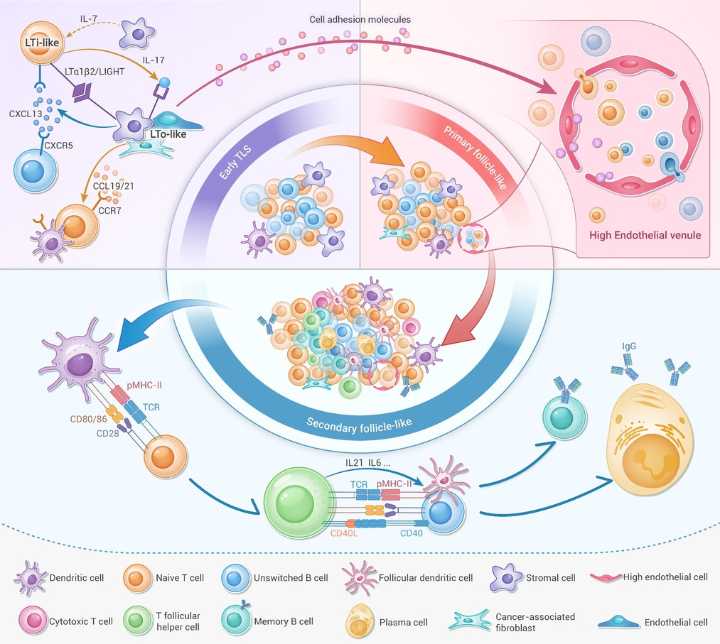
Figure 1.
Initiation, formation, and maturation of tumor-associated tertiary lymphoid structures (TA-TLSs)
-
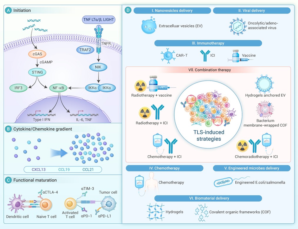
Figure 2.
TA-TLS induction: key intervention nodes (A–C) and representative strategies (I–VII)
