
Cancer cell membranes, tumor extracellular vesicles, and subcellular organelle targeting as promising biomimetic drug delivery systems
-
GRAPHICAL ABSTRACT
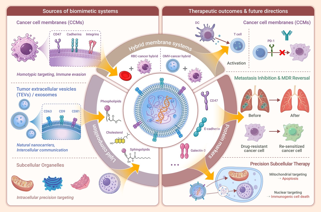
 DownLoad:
Full size image
-
PUBLIC SUMMARY
-
CCMs and TEVs provide biomimetic platforms with complementary advantages in tumor targeting and biocompatibility.
Hybrid membrane systems synergistically enhance circulation, immune evasion, and antitumor efficacy.
Subcellular organelle targeting integrated with membrane systems enables sequential precision delivery.
-
-
ABSTRACT
Conventional chemotherapy suffers from severe off-target toxicity and suboptimal efficacy. This review systematically summaries cancer cell membranes (CCMs), tumor extracellular vesicles (TEVs), and subcellular organelle targeting as biomimetic advanced drug delivery systems (ADDS) for precision cancer therapy. CCMs exhibit unique homotypic targeting, immune evasion, and enhanced tumor accumulation, while hybrid membrane systems further improve circulation and targeting specificity. TEVs, particularly exosomes, offer naturally occurring nanoscale carriers with inherent biocompatibility and intercellular communication capabilities. We critically compare the lipid and protein compositions between CCMs and TEVs, and explore emerging applications in metastasis inhibition, multidrug resistance reversal, and antitumor immunity. Beyond membrane-based strategies, we investigate mitochondria, Golgi apparatus, endoplasmic reticulum, and nucleus targeting for precise intracellular drug delivery. The review identifies critical gaps in standardized isolation protocols and lipid-mediated recognition mechanisms, proposing that hybrid systems integrating CCMs, TEVs, and organelle-targeting moieties represent the future direction for translating precision nanomedicine from bench to bedside. -

-
REFERENCES
[1] McClelland S. and Yeboa D.N. (2023). The emerging paradigm of preoperative stereotactic radiosurgery for resectable brain metastases. JAMA Oncol 9:1073−1074. DOI:10.1001/jamaoncol.2023.1342 [2] Zhou Z., Guan B., Xia H., et al. (2023). Particle radiotherapy in the era of radioimmunotherapy. Cancer Lett 567:216268. DOI:10.1016/j.canlet.2023.216268 [3] Kimura Y., Kawakami H., Tamura S., et al. (2023). Effect of the number of cycles of docetaxel + S-1 therapy on long-term survival in adjuvant chemotherapy for stage III gastric cancer. A pooled analysis of the OGSG0604 and OGSG1002 trials. Gastric Cancer 26:788-797. DOI: 10.1007/s10120-023-01408-y [4] Hamid A.A., Sayegh N., Tombal B., et al. (2023). Metastatic hormone-sensitive prostate cancer: toward an era of adaptive and personalized treatment. Am Soc Clin Oncol Educ Book 43:e390166. DOI:10.1200/EDBK_390166 [5] Park J.C., Bertaux B., Park J., et al. (2023). Current status of human papillomavirus-targeted therapies development in head and neck cancer. JCO Precis Oncol 7:e2300098. DOI:10.1200/PO.23.00098 [6] Hosseini F., Ahmadvand M., Karimi R., et al. (2023). Cancer immunotherapy via stem cell-derived NK cells. Immunotherapy 15:963−973. DOI:10.2217/imt-2022-0224 [7] Feinberg A.P. and Levchenko A. (2023). Epigenetics as a mediator of plasticity in cancer. Science 379:eaaw3835 DOI:10.1126/science.aaw3835 [8] Rojas L.A., Sethna Z., Soares K.C., et al. (2023). Personalized RNA neoantigen vaccines stimulate T cells in pancreatic cancer. Nature 618:144−150. DOI:10.1038/s41586-023-06063-y [9] Rubin R. (2023). Updated labeling for capecitabine, a 25-year-old cancer drug. JAMA 329:201. DOI:10.1001/jama.2022.24814 [10] Alam J., Dilnawaz F., Sahoo S.K., et al. (2022). Curcumin encapsulated into biocompatible co-polymer PLGA nanoparticle enhanced anti-gastric cancer and anti-helicobacter eylori Effect. Asian Pac J Cancer Prev 23:61−70. DOI:10.31557/APJCP.2022.23.1.61 [11] Zhu X. and Li S. (2023). Nanomaterials in tumor immunotherapy: new strategies and challenges. Mol Cancer 22:94. DOI:10.1186/s12943-023-01797-9 [12] Qin X., Yang T., Xu H., et al. (2023). Dying tumor cells-inspired vaccine for boosting humoral and cellular immunity against cancer. J Control Release 359:359−372. DOI:10.1016/j.jconrel.2023.05.044 [13] Dutt Y., Pandey R.P., Dutt M., et al. (2023). Therapeutic applications of nanobiotechnology. J Nanobiotechnology 21:148. DOI:10.1186/s12951-023-01909-z [14] Bremer E., de Bruyn M., Wajant H., et al. (2009). Targeted cancer immunotherapy using ligands of the tumor necrosis factor super-family. Curr Drug Targets 10:94−103. DOI:10.2174/138945009787354593 [15] Yang, V.C. (2019). Personal perspectives and concerns over the so-called nanomedicine. J Control Release 311-312:322-323. DOI: 10.1016/j.jconrel.2019.10.021. [16] Baati T., Chaabani I., Salek A., et al. (2023). Chitosan-coated ultrapure silicon nanoparticles produced by laser ablation: biomedical potential in nano-oncology as a tumor-targeting nanosystem. Nanoscale Adv 5:3044−3052. DOI:10.1039/d3na00253e [17] Li, Z., Zhang S., Fu Z., et al. (2023). Surficial nano-deposition locoregionally yielding bactericidal super CAR-macrophages expedites periprosthetic osseointegration. Sci Adv 9:eadg3365. DOI:10.1126/sciadv.adg3365 [18] Pan, W., Wu B., Nie C., et al. (2023). NIR-II responsive nanohybrids incorporating thermosensitive hydrogel as sprayable dressing for multidrug-resistant-bacteria infected wound management. ACS Nano 17:11253−11267. DOI:10.1021/acsnano.2c10742 [19] Sheffey, V.V., Siew E.B., Tanner E.E.L., et al. (2022). PLGA's plight and the role of stealth surface modification strategies in its use for intravenous particulate drug delivery. Adv Healthc Mater 11:e2101536. DOI:10.1002/adhm.202101536 [20] Aydeger, A., Aysit N., Baydas G., et al. (2023). Design of IKVAV peptide/gold nanoparticle decorated, micro/nano-channeled PCL/PLGA film scaffolds for neuronal differentiation and neurite outgrowth. Biomater Adv 152:213472. DOI:10.1016/j.bioadv.2023.213472 [21] Li, Y., Hu C., Hu B., et al. (2023). Sustained release of dicumarol via novel grafted polymer in electrospun nanofiber membrane for treatment of peritendinous adhesion. Adv Healthc Mater 12:e2203078. DOI:10.1002/adhm.202203078 [22] Lv, Y., Xu Y., Sang X., et al. (2022). PLLA-gelatin composite fiber membranes incorporated with functionalized CeNPs as a sustainable wound dressing substitute promoting skin regeneration and scar remodeling. J Mater Chem B 10:1116−1127. DOI:10.1039/d1tb02677a [23] Shi, D., Beasock D., Fessler A., et al. (2022). To PEGylate or not to PEGylate: Immunological properties of nanomedicine's most popular component, polyethylene glycol and its alternatives. Adv Drug Deliv Rev 180:114079. DOI:10.1016/j.addr.2021.114079 [24] Kierstead, P.H., Okochi H., Venditto V.J., Chuong T.C., et al. (2015). The effect of polymer backbone chemistry on the induction of the accelerated blood clearance in polymer modified liposomes. J Control Release 213:1−9. DOI:10.1016/j.jconrel.2015.06.023 [25] Gilson, R.C., Gunasinghe S.D., Johannes L., et al. (2019). Galectin-3 modulation of T-cell activation: mechanisms of membrane remodelling. Prog Lipid Res 76:101010. DOI:10.1016/j.plipres.2019.101010 [26] Wang, S., Tang Q., Ya H., et al. (2020). Study on the optical and biological properties in vitro of IR808-PEG-FA. J Biomed Mater Res A 108:1816−1823. DOI:10.1002/jbm.a.36946 [27] Aparna, A., Sreehari H., Chandran A., et al. (2022). Ligand-protected nanoclusters and their role in agriculture, sensing and allied applications. Talanta 239:123134. DOI:10.1016/j.talanta.2021.123134 [28] Jiang, X., Zhao Y., Sun S., et al. (2023). Research development of porphyrin-based metal-organic frameworks: targeting modalities and cancer therapeutic applications. J Mater Chem B 11:6172−6200. DOI:10.1039/d3tb00632h [29] Liu, R., Li L., Chen S., et al. (2023). Evolution of protein assemblies driven by the switching of interplay mode. ACS Nano 17:2245−2256. DOI:10.1021/acsnano.2c08583 [30] Pan, C., Ye J., Zhang S., et al. (2023). Production of a promising modular proteinaceous self-assembled delivery system for vaccination. Nanoscale 15:10794−10807. DOI:10.1039/d2nr06718h [31] Beneyto-Calabuig, S., Merbach A.K., Kniffka JA., et al. (2023). Clonally resolved single-cell multi-omics identifies routes of cellular differentiation in acute myeloid leukemia. Cell Stem Cell 30:706−721.e8. DOI:10.1016/j.stem.2023.04.001 [32] Zuarez-Chamba, M., Rajendran S., Herrera-Robledo M., et al. (2022). Bi-based photocatalysts for bacterial inactivation in water: Inactivation mechanisms, challenges, and strategies to improve the photocatalytic activity. Environ Res 209:112834. DOI:10.1016/j.envres.2022.112834 [33] Wen, M., Zhao Y., Qiu P., et al. (2023). Efficient sonodynamic ablation of deep-seated tumors via cancer-cell-membrane camouflaged biocompatible nanosonosensitizers. J Colloid Interface Sci 644:388−396. DOI:10.1016/j.jcis.2023.04.088 [34] Zhang, D., Ye Z., Liu H., et al. (2021). Cell membrane coated smart two-dimensional supraparticle for in vivo homotypic cancer targeting and enhanced combinational theranostics. Nanotheranostics 5:275−287. DOI:10.7150/ntno.57657 [35] Liu, Z., Wang F., Liu X., et al. (2021). Cell membrane-camouflaged liposomes for tumor cell-selective glycans engineering and imaging in vivo. Proc Natl Acad Sci U S A 118:e2022769118. DOI:10.1073/pnas.2022769118 [36] Gong, C., Yu X., Zhang W., et al. (2021). Regulating the immunosuppressive tumor microenvironment to enhance breast cancer immunotherapy using pH-responsive hybrid membrane-coated nanoparticles. J Nanobiotechnology 19:58. DOI:10.1186/s12951-021-00805-8 [37] Chen, K., Zhou A., Zhou X., et al. (2023). An intelligent cell-derived nanorobot bridges synergistic crosstalk between sonodynamic therapy and cuproptosis to promote cancer treatment. Nano Lett 23:3038−3047. DOI:10.1021/acs.nanolett.3c00434 [38] Han, J., Wang S., Fang W., et al. (2025). Long-acting IL-2 release from pressure-fused biomineral tablets promotes antitumor immune response. Nat Cancer 6:1384−1399. DOI:10.1038/s43018-025-00993-4 [39] Meng, Z., Zhang Y., Zhou X., et al. (2022). Nanovaccines with cell-derived components for cancer immunotherapy. Adv Drug Deliv Rev 182:114107. DOI:10.1016/j.addr.2021.114107 [40] Gorey, K.M., Haji-Jama S., Bartfay E., et al. (2014). Lack of access to chemotherapy for colon cancer: multiplicative disadvantage of being extremely poor, inadequately insured and African American. BMC Health Serv Res 14:133. DOI:10.1186/1472-6963-14-133 [41] Wu, C., Zhang F., Li B., et al. (2023). A self-assembly nano-prodrug for combination therapy in triple-negative breast cancer stem cells. Small 19:e2301600. DOI:10.1002/smll.202301600 [42] Salvatore, G., Dolci S., Camaioni A., et al. (2023). Reprogramming human female adipose mesenchymal stem cells into primordial germ cell-like cells. Stem Cell Rev Rep 19:2274−2283. DOI:10.1007/s12015-023-10561-x [43] Zhu, Y., Chen X., and Liao Y. (2023). Mesenchymal stem cells-derived apoptotic extracellular vesicles (ApoEVs): mechanism and application in tissue regeneration. Stem Cells 41:837−849. DOI:10.1093/stmcls/sxad046 [44] Yuan, Y.G., Wang J.L., Zhang Y.X., et al. (2023). Biogenesis, composition and potential therapeutic applications of mesenchymal stem cells derived exosomes in various diseases. Int J Nanomedicine 18:3177−3210. DOI:10.2147/IJN.S407029 [45] Lin, S.Y., Dolfi S.C., Amiri S., et al. (2013). P53 regulates the migration of mesenchymal stromal cells in response to the tumor microenvironment through both CXCL12-dependent and -independent mechanisms. Int J Oncol 43:1817−1823. DOI:10.3892/ijo.2013.2109 [46] Yu, P.F., Huang Y., Xu C.L., et al. (2017). Downregulation of CXCL12 in mesenchymal stromal cells by TGFβ promotes breast cancer metastasis. Oncogene 36:840−849. DOI:10.1038/onc.2016.252 [47] Jung, Y., Kim J.K., Shiozawa Y., et al. (2013). Recruitment of mesenchymal stem cells into prostate tumours promotes metastasis. Nat Commun 4:1795. DOI:10.1038/ncomms2766 [48] Quante, M., Tu S.P., Tomita H., et al. (2011). Bone marrow-derived myofibroblasts contribute to the mesenchymal stem cell niche and promote tumor growth. Cancer Cell 19:257−272. DOI:10.1016/j.ccr.2011.01.020 [49] Ren, G., Zhao X., Wang Y., et al. (2012). CCR2-dependent recruitment of macrophages by tumor-educated mesenchymal stromal cells promotes tumor development and is mimicked by TNFα. Cell Stem Cell 11:812−824. DOI:10.1016/j.stem.2012.08.013 [50] Lin, L.Y., Du L.M., Cao K., et al. (2016). Tumour cell-derived exosomes endow mesenchymal stromal cells with tumour-promotion capabilities. Oncogene 35:6038−6042. DOI:10.1038/onc.2016.131 [51] Mishra, P.J., Mishra P.J., Humeniuk R., et al. (2008). Carcinoma-associated fibroblast-like differentiation of human mesenchymal stem cells. Cancer Res 68:4331−4339. DOI:10.1158/0008-5472.CAN-08-0943 [52] McLean, K., Gong Y., Choi Y., et al. (2011). Human ovarian carcinoma–associated mesenchymal stem cells regulate cancer stem cells and tumorigenesis via altered BMP production. J Clin Invest 121:3206−3219. DOI:10.1172/JCI45273 [53] De Boeck, A., Pauwels P., Hensen K., et al. (2013). Bone marrow-derived mesenchymal stem cells promote colorectal cancer progression through paracrine neuregulin 1/HER3 signalling. Gut 62:550−560. DOI:10.1136/gutjnl-2011-301393 [54] Beckermann, B.M., Kallifatidis G., Groth A., et al. (2008). VEGF expression by mesenchymal stem cells contributes to angiogenesis in pancreatic carcinoma. Br J Cancer 99:622−631. DOI:10.1038/sj.bjc.6604508 [55] Zheng, J., Wang Q., Leng W., et al. (2018). Bone marrow-derived mesenchymal stem cell-conditioned medium attenuates tubulointerstitial fibrosis by inhibiting monocyte mobilization in an irreversible model of unilateral ureteral obstruction. Mol Med Rep 17:7701−7707. DOI:10.3892/mmr.2018.8848 [56] Cho, D.I., Kim M.R., Jeong H.Y., et al. (2014). Mesenchymal stem cells reciprocally regulate the M1/M2 balance in mouse bone marrow-derived macrophages. Exp Mol Med 46:e70. DOI:10.1038/emm.2013.135 [57] Bianchi, G., Borgonovo G., Pistoia V., et al. (2011). Immunosuppressive cells and tumour microenvironment: focus on mesenchymal stem cells and myeloid derived suppressor cells. Histol Histopathol 26:941−951. DOI:10.14670/HH-26.941 [58] Hu, Y.L., Fu YH., Tabata Y., et al. (2010). Mesenchymal stem cells: a promising targeted-delivery vehicle in cancer gene therapy. J Control Release 147:154−162. DOI:10.1016/j.jconrel.2010.05.015 [59] von Einem, J.C., Guenther C ., Volk H.D., et al. (2019). Treatment of advanced gastrointestinal cancer with genetically modified autologous mesenchymal stem cells: Results from the phase 1/2 TREAT-ME-1 trial. Int J Cancer 145:1538−1546. DOI:10.1002/ijc.32230 [60] Niess, H., von Einem J.C., Thomas M.N., et al. (2015). Treatment of advanced gastrointestinal tumors with genetically modified autologous mesenchymal stromal cells (TREAT-ME1): study protocol of a phase I/II clinical trial. BMC Cancer 15:237. DOI:10.1186/s12885-015-1241-x [61] Xia, Y., Rao L., Yao H., et al. (2020). Engineering macrophages for cancer immunotherapy and drug delivery. Adv Mater 32:e2002054. DOI:10.1002/adma.202002054 [62] Guo, L., Zhang Y., Yang Z., et al. (2019). Tunneling nanotubular expressways for ultrafast and accurate M1 macrophage delivery of anticancer drugs to metastatic ovarian carcinoma. ACS Nano 13:1078−1096. DOI:10.1021/acsnano.8b08872 [63] Fu, J., Wang D., Mei D., et al. (2015). Macrophage mediated biomimetic delivery system for the treatment of lung metastasis of breast cancer. J Control Release 204:11−19. DOI:10.1016/j.jconrel.2015.01.039 [64] Ren, K., Qiu Y., Yu Q., et al. (2021). Macrophage-mediated multi-mode drug release system for photothermal combined with anti-inflammatory therapy against postoperative recurrence of triple negative breast cancer. Int J Pharm 607:120975. DOI:10.1016/j.ijpharm.2021.120975 [65] Balasubramanian, V., Correia A., Zhang H., et al. (2017). Biomimetic engineering using cancer cell membranes for designing compartmentalized nanoreactors with organelle-like functions. Adv Mater 29:1605375. DOI:10.1002/adma.201605375 [66] Kumar, P., Treuren T.V., Ranjan A.P., et al. (2019). In vivo imaging and biodistribution of near infrared dye loaded brain-metastatic-breast-cancer-cell-membrane coated polymeric nanoparticles. Nanotechnology 30:265101. DOI:10.1088/1361-6528/ab0f46 [67] Xue, C.R., Wang K., Zhang M.Z., et al. (2022). Tracking Neural Stem Cells in vivo: Achievements and Limitations. Stem Cell Rev Rep 18:1774−1788. DOI:10.1007/s12015-022-10333-z [68] Ding, P., Wang Z., Wu Z., et al. (2020). Natural biointerface based on cancer cell membranes for specific capture and release of circulating tumor cells. ACS Appl Mater Interfaces 12:20263−20270. DOI:10.1021/acsami.0c03355 [69] Zhang, J., Miao Y., Ni W., et al. (2019). Cancer cell membrane coated silica nanoparticles loaded with ICG for tumour specific photothermal therapy of osteosarcoma. Artif Cells Nanomed Biotechnol 47:2298−2305. DOI:10.1080/21691401.2019.1622554 [70] Chen, M., Chen M., and He J. (2019). Cancer cell membrane cloaking nanoparticles for targeted co-delivery of doxorubicin and PD-L1 siRNA. Artif Cells Nanomed Biotechnol 47:1635−1641. DOI:10.1080/21691401.2019.1608219 [71] Piprek, R.P., Kloc M., Mizia P., et al. (2020). The central role of cadherins in gonad development, reproduction, and fertility. Int J Mol Sci 21:8264. DOI:10.3390/ijms21218264 [72] Kouo, T., Huang L., Pucsek A.B., et al. (2015). Galectin-3 shapes antitumor immune responses by suppressing CD8+ T cells via LAG-3 and inhibiting expansion of plasmacytoid dendritic cells. Cancer Immunol Res 3:412−423. DOI:10.1158/2326-6066.CIR-14-0150 [73] Cereijido, M., Contreras R.G., Shoshani L., et al. (2012). The Na+-K+-ATPase as self-adhesion molecule and hormone receptor. Am J Physiol Cell Physiol 302:C473−C481. DOI:10.1152/ajpcell.00083.2011 [74] Gahmberg, C.G., Grönholm M., Madhavan S., et al. (2019). Regulation of cell adhesion: a collaborative effort of integrins, their ligands, cytoplasmic actors, and phosphorylation. Q Rev Biophys 52:e10. DOI:10.1017/S0033583519000088 [75] Alami, J., Williams B.R. and Yeger H. (2003). Differential expression of E-cadherin and beta catenin in primary and metastatic Wilms's tumours. Mol Pathol 56:218−225. DOI:10.1136/mp.56.4.218 [76] Wu, M., Liu X., Bai H., et al. (2019). Surface-layer protein-enhanced immunotherapy based on cell membrane-coated nanoparticles for the effective inhibition of tumor growth and metastasis. ACS Appl Mater Interfaces 11:9850−9859. DOI:10.1021/acsami.9b00294 [77] Yamamoto, H., Watanabe Y., Maehata T., et al. (2020). Microsatellite instability in cancer: a novel landscape for diagnostic and therapeutic approach. Arch Toxicol 94:3349−3357. DOI:10.1007/s00204-020-02833-z [78] Wallace, L., Cherian A.M., Adamson P., et al. (2018). Comparison of pre- and post-translational expressions of COXIV-1 and MT-ATPase 6 genes in colorectal adenoma-carcinoma tissues. J Carcinog Mutagen 9:e295−e300. DOI:10.4172/2157-2518.1000319 [79] Przybyla, A., Zhang T., Li R., et al. (2019). Natural T cell autoreactivity to melanoma antigens: clonally expanded melanoma-antigen specific CD8 + memory T cells can be detected in healthy humans. Cancer Immunol Immunother 68:709-720. DOI: 10.1007/s00262-018-02292-7 [80] Fang, R.H., Hu C.M., Luk B.T., et al. (2014). Cancer cell membrane-coated nanoparticles for anticancer vaccination and drug delivery. Nano Lett 14:2181−2188. DOI:10.1021/nl500618u [81] Feng, Q., Yang X., Hao Y., et al. (2019). Cancer cell membrane-biomimetic nanoplatform for enhanced sonodynamic therapy on breast cancer via autophagy regulation strategy. ACS Appl Mater Interfaces 11:32729−32738. DOI:10.1021/acsami.9b10948 [82] Han, J., Wu Z., Zhan S., et al. (2025). Biorhythm-mimicking growth hormone patch. Nat Mater 24:1283−1294. DOI:10.1038/s41563-025-02188-9 [83] Reya, T., Morrison S.J., Clarke M.F., et al. (2001). Stem cells, cancer, and cancer stem cells. Nature 414:105−111. DOI:10.1038/35102167 [84] Gao, C., Lin Z., Jurado-Sánchez B., et al. (2016). Stem cell membrane-coated nanogels for highly efficient in vivo tumor targeted drug delivery. Small 12:4056−4062. DOI:10.1002/smll.201600624 [85] Zhang, M., Zhang F., Liu T., et al. (2020). Polydopamine nanoparticles camouflaged by stem cell membranes for synergistic chemo-photothermal therapy of malignant bone tumors. Int J Nanomedicine 15:10183−10197. DOI:10.2147/IJN.S282931 [86] Mu, X., Li J., Yan S., et al. (2018). siRNA delivery with stem cell membrane-coated magnetic nanoparticles for imaging-guided photothermal therapy and gene therapy. ACS Biomater Sci Eng 4:3895−3905. DOI:10.1021/acsbiomaterials.8b00858 [87] Wang, L., Liu Z., Zhou Q., et al. (2021). Prodrug nanoparticles rationally integrating stroma modification and chemotherapy to treat metastatic pancreatic cancer. Biomaterials 278:121176. DOI:10.1016/j.biomaterials.2021.121176 [88] Sharkis, S.J., Jones R.J., Civin C., et al. (2012).Pluripotent stem cell-based cancer therapy: promise and challenges. Sci Transl Med 4:127ps9. DOI:10.1126/scitranslmed.3003920 [89] Zakrzewski, W., Dobrzyński M., Szymonowicz M., et al. (2019). Stem cells: past, present, and future. Stem Cell Res Ther 10:68. DOI:10.1186/s13287-019-1165-5 [90] Chen, H.Y., Deng J., Wang Y., et al. (2020). Hybrid cell membrane-coated nanoparticles: A multifunctional biomimetic platform for cancer diagnosis and therapy. Acta Biomater 112:1−13. DOI:10.1016/j.actbio.2020.05.028 [91] Tang, W., Zhen Z., Wang M., et al. (2016). Red blood cell-facilitated photodynamic therapy for cancer treatment. Adv Funct Mater 26:1757−1768. DOI:10.1002/adfm.201504803 [92] Liu, B., Wang W., Fan J., et al. (2019). RBC membrane camouflaged prussian blue nanoparticles for gamabutolin loading and combined chemo/photothermal therapy of breast cancer. Biomaterials 217:119301. DOI:10.1016/j.biomaterials.2019.119301 [93] Xuan, M., Shao J., Dai L., et al. (2016). Macrophage cell membrane camouflaged Au nanoshells for in vivo prolonged circulation life and enhanced cancer photothermal therapy. ACS Appl Mater Interfaces 8:9610−9618. DOI:10.1021/acsami.6b00853 [94] Parodi, A., Quattrocchi N., van de Ven A.L., et al. (2013). Synthetic nanoparticles functionalized with biomimetic leukocyte membranes possess cell-like functions. Nat Nanotechnol 8:61−68. DOI:10.1038/nnano.2012.212 [95] Liao, Y., Zhang Y., Blum N.T., et al. (2020). Biomimetic hybrid membrane-based nanoplatforms: synthesis, properties and biomedical applications. Nanoscale Horiz 5:1293−1302. DOI:10.1039/d0nh00267d [96] Li, Y., Gan Y., Li C., et al. (2020). Cell membrane-engineered hybrid soft nanocomposites for biomedical applications. J Mater Chem B 8:5578−5596. DOI:10.1039/d0tb00472c [97] Jiang, Q., Liu Y., Guo R., et al. (2019). Erythrocyte-cancer hybrid membrane-camouflaged melanin nanoparticles for enhancing photothermal therapy efficacy in tumors. Biomaterials 192:292−308. DOI:10.1016/j.biomaterials.2018.11.021 [98] Chen, W., Wang Y., Qin M., et al. (2018). Bacteria-driven hypoxia targeting for combined biotherapy and photothermal therapy. ACS Nano 12:5995−6005. DOI:10.1021/acsnano.8b02235 [99] Gao, W., Fang R.H., Thamphiwatana S., et al. (2015). Modulating antibacterial immunity via bacterial membrane-coated nanoparticles. Nano Lett 15:1403−1409. DOI:10.1021/nl504798g [100] Wang, D., Liu C., You S., et al. (2020). Bacterial vesicle-cancer cell hybrid membrane-coated nanoparticles for tumor specific immune activation and photothermal therapy. ACS Appl Mater Interfaces 12:41138−41147. DOI:10.1021/acsami.0c13169 [101] Liu, H., Su Y., Jiang X., et al. (2023). Cell membrane-coated nanoparticles: a novel multifunctional biomimetic drug delivery system. Drug Deliv Transl Res 13:716−737. DOI:10.1007/s13346-022-01252-0 [102] Zhu, L., Yu X., Cao T., et al. (2023). Immune cell membrane-based biomimetic nanomedicine for treating cancer metastasis. Acta Pharm Sin B 13:2464−2482. DOI:10.1016/j.apsb.2023.03.004 [103] Wu, L., Li Q., Deng J., et al. (2021). Platelet-tumor cell hybrid membrane-camouflaged nanoparticles for enhancing therapy efficacy in glioma. Int J Nanomedicine 16:8433−8446. DOI:10.2147/IJN.S333279 [104] Zhang, D., Liu X., Gao J., et al. (2017).The role of epithelial cell adhesion molecule N-glycosylation on apoptosis in breast cancer cells. Tumour Biol 39:1010428317695973. DOI: 10.1177/1010428317695973 [105] Zhang, W., Yu M., Xi Z., et al. (2019). Cancer cell membrane-camouflaged nanorods with endoplasmic reticulum targeting for improved antitumor therapy. ACS Appl Mater Interfaces 11:46614−46625. DOI:10.1021/acsami.9b18388 [106] Okabe, T., Togo S., Fujimoto Y., et al. (2020). Mesenchymal characteristics and predictive biomarkers on circulating tumor cells for therapeutic strategy. Cancers (Basel) 12:3588. DOI:10.3390/cancers12123588 [107] Chen, Y., Li N., Wang J., et al. (2019). Enhancement of mitochondrial ROS accumulation and radiotherapeutic efficacy using a Gd-doped titania nanosensitizer. Theranostics 9:167−178. DOI:10.7150/thno.28033 [108] Pan, W., Ge Y., Yu Z., et al. (2019). A cancer cell membrane-encapsulated MnO(2) nanoreactor for combined photodynamic-starvation therapy. Chem Commun (Camb) 55:5115−5118. DOI:10.1039/c9cc01386e [109] Kroll, A.V., Fang R.H., Jiang Y., et al. (2017). Nanoparticulate delivery of cancer cell membrane elicits multiantigenic antitumor immunity. Adv Mater. 29:1703969. DOI:10.1002/adma.201703969 [110] Chen, D., Cai L., Guo Y., et al. (2020). Cancer cell membrane-decorated zeolitic-imidazolate frameworks codelivering cisplatin and oleanolic acid induce apoptosis and reversed multidrug resistance on bladder carcinoma cells. ACS Omega 5:995−1002. DOI:10.1021/acsomega.9b02261 [111] Wan, J., Wang J., Rao Z., et al. (2020). A cell membrane vehicle co-delivering sorafenib and doxorubicin remodel the tumor microenvironment and enhance immunotherapy by inducing immunogenic cell death in lung cancer cells. J Mater Chem B 8:7755−7765. DOI:10.1039/d0tb01052a [112] Fang, R.H., Kroll A.V. and Zhang L. (2015). Nanoparticle-based manipulation of antigen-presenting cells for cancer immunotherapy. Small 11:5483−5496. DOI:10.1002/smll.201501284 [113] Zhu, J.Y., Zheng D.W., Zhang M.K., et al. (2016). Preferential cancer cell self-recognition and tumor self-targeting by coating nanoparticles with homotypic cancer cell membranes. Nano Lett 16:5895−5901. DOI:10.1021/acs.nanolett.6b02786 [114] Liu, Y., Bhattarai P., Dai Z., et al. (2019). Photothermal therapy and photoacoustic imaging via nanotheranostics in fighting cancer. Chem Soc Rev 48:2053−2108. DOI:10.1039/c8cs00618k [115] Urabe, F., Kosaka N., Ito K., et al. (2020). Extracellular vesicles as biomarkers and therapeutic targets for cancer. Am J Physiol Cell Physiol 318:C29−C39. DOI:10.1152/ajpcell.00280.2019 [116] Cocucci, E. and Meldolesi J. (2015). Ectosomes and exosomes: shedding the confusion between extracellular vesicles. Trends Cell Biol 25:364−372. DOI:10.1016/j.tcb.2015.01.004 [117] Bergsmedh, A., Szeles A., Henriksson M., et al. (2001). Horizontal transfer of oncogenes by uptake of apoptotic bodies. Proc Natl Acad Sci U S A 98(11):6407−6411. DOI:10.1073/pnas.101129998 [118] Liu, Y., Wen J. and Huang W. (2021). Exosomes in nasopharyngeal carcinoma. Clin Chim Acta 523:355−364. DOI:10.1016/j.cca.2021.10.013 [119] Zhong, Y., Li H., Li P., et al. (2021). Exosomes: A new pathway for cancer drug resistance. Front Oncol 11:743556. DOI:10.3389/fonc.2021.743556 [120] van Niel, G., D'Angelo G. and Raposo G. (2018). Shedding light on the cell biology of extracellular vesicles. Nat Rev Mol Cell Biol 19:213−228. DOI:10.1038/nrm.2017.125 [121] Elsharkasy, O.M., Nordin J.Z., Hagey D.W., et al. (2020). Extracellular vesicles as drug delivery systems: Why and how? Adv Drug Deliv Rev 159:332−343. DOI:10.1016/j.addr.2020.04.004 [122] Hauser, P., Wang S. and Didenko V.V. (2017). Apoptotic bodies: selective detection in extracellular vesicles. Methods Mol Biol 1554:193−200. DOI:10.1007/978-1-4939-6759-9_12 [123] Pullan, J.E., Confeld M.I., Osborn J.K., et al. (2019). Exosomes as drug carriers for cancer therapy. Mol Pharm 16:1789−1798. DOI:10.1021/acs.molpharmaceut.9b00104 [124] Théry, C., Witwer K.W., Aikawa E., et al. (2018). Minimal information for studies of extracellular vesicles 2018 (MISEV2018): a position statement of the International Society for Extracellular Vesicles and update of the MISEV2014 guidelines. J Extracell Vesicles 7:1535750. DOI:10.1080/20013078.2018.1535750 [125] Minciacchi, V.R., Freeman M.R. and Di Vizio D. (2015). Extracellular vesicles in cancer: exosomes, microvesicles and the emerging role of large oncosomes. Semin Cell Dev Biol 40:41−51. DOI:10.1016/j.semcdb.2015.02.010 [126] Meldolesi, J. (2018). Exosomes and ectosomes in intercellular communication. Curr Biol 28:R435−R444. DOI:10.1016/j.cub.2018.01.059 [127] Ratajczak, J., Wysoczynski M., Hayek F., et al. (2006). Membrane-derived microvesicles: important and underappreciated mediators of cell-to-cell communication. Leukemia 20:1487−1495. DOI:10.1038/sj.leu.2404296 [128] Piccin, A., Murphy W.G. and Smith O.P. (2007). Circulating microparticles: pathophysiology and clinical implications. Blood Rev 21:157−171. DOI:10.1016/j.blre.2006.09.001 [129] Pap, E., Pállinger E., Pásztói M., et al. (2009). Highlights of a new type of intercellular communication: microvesicle-based information transfer. Inflamm Res 58:1−8. DOI:10.1007/s00011-008-8210-7 [130] Yu, M., Xie R., Zhang Y., et al. (2018). Phosphatidylserine on microparticles and associated cells contributes to the hypercoagulable state in diabetic kidney disease. Nephrol Dial Transplant 33:2115−2127. DOI:10.1093/ndt/gfy027 [131] Kou, Y., Zou L., Liu R., et al. (2019). Intravascular cells and circulating microparticles induce procoagulant activity via phosphatidylserine exposure in heart failure. J Thromb Thrombolysis 48:187−194. DOI:10.1007/s11239-019-01889-8 [132] Liu, Y., He Z., Zhang Y., et al. (2016). Dissimilarity of increased phosphatidylserine-positive microparticles and associated coagulation activation in acute coronary syndromes. Coron Artery Dis 27:365−375. DOI:10.1097/MCA.0000000000000368 [133] Gleizes, C., Kreutter G., Abbas M., et al. (2016). β cell membrane remodelling and procoagulant events occur in inflammation-driven insulin impairment: a GLP-1 receptor dependent and independent control. J Cell Mol Med 20:231−242. DOI:10.1111/jcmm.12683 [134] Del Conde, I., Shrimpton C.N., Thiagarajan P., et al. (2005). Tissue-factor-bearing microvesicles arise from lipid rafts and fuse with activated platelets to initiate coagulation. Blood 106:1604−1611. DOI:10.1182/blood-2004-03-1095 [135] Akers, J.C., Gonda D., Kim R., et al. (2013). Biogenesis of extracellular vesicles (EV): exosomes, microvesicles, retrovirus-like vesicles, and apoptotic bodies J Neurooncol 113:1−11. DOI:10.1007/s11060-013-1084-8 [136] Al-Nedawi, K., Meehan B., Micallef J., et al. (2008). Intercellular transfer of the oncogenic receptor EGFRvIII by microvesicles derived from tumour cells. Nat Cell Biol 10:619−624. DOI:10.1038/ncb1725 [137] Di Vizio, D., Kim J., Hager M.H., et al. (2009). Oncosome formation in prostate cancer: association with a region of frequent chromosomal deletion in metastatic disease. Cancer Res 69:5601−5609. DOI:10.1158/0008-5472.CAN-08-3860 [138] Cocucci, E., Racchetti G. and Meldolesi J. (2009). Shedding microvesicles: artefacts no more. Trends Cell Biol 19:43−51. DOI:10.1016/j.tcb.2008.11.003 [139] Minciacchi, V.R., You S., Spinelli C., et al. (2015). Large oncosomes contain distinct protein cargo and represent a separate functional class of tumor-derived extracellular vesicles. Oncotarget 6:11327−11341. DOI:10.18632/oncotarget.3598 [140] Morello, M., Minciacchi V.R., de Candia P., et al. (2013). Large oncosomes mediate intercellular transfer of functional microRNA. Cell Cycle 12:3526−3536. DOI:10.4161/cc.26539 [141] Di Vizio, D., Morello M., Dudley A.C., et al. (2012). Large oncosomes in human prostate cancer tissues and in the circulation of mice with metastatic disease. Am J Pathol 181:1573−1584. DOI:10.1016/j.ajpath.2012.07.030 [142] Choi, D.S., Kim D.K., Kim Y.K., et al. (2015). Proteomics of extracellular vesicles: Exosomes and ectosomes. Mass Spectrom Rev 34:474−490. DOI:10.1002/mas.21420 [143] Choi, D.S., Yang J.S., Choi E.J., et al. (2012). The protein interaction network of extracellular vesicles derived from human colorectal cancer cells. J Proteome Res 11:1144−1151. DOI:10.1021/pr200842h [144] Simons, M. and Raposo G. (2009). Exosomes--vesicular carriers for intercellular communication. Curr Opin Cell Biol 21:575-581. DOI: 10.1016/j.ceb.2009.03.007. [145] Raposo, G., Nijman H.W., Stoorvogel W., et al. (1996). B lymphocytes secrete antigen-presenting vesicles. J Exp Med 183:1161−1172. DOI:10.1084/jem.183.3.1161 [146] Pan, B.T., Teng K., Wu C., et al. (1985). Electron microscopic evidence for externalization of the transferrin receptor in vesicular form in sheep reticulocytes. J Cell Biol 101:942−948. DOI:10.1083/jcb.101.3.942 [147] Shetty, A.K. and Upadhya R. (2021). >Extracellular vesicles in health and disease. Aging Dis 12:1358−1362. DOI:10.14336/AD.2021.0827 [148] Perrin, P., Janssen L., Janssen H., et al. (2021). Retrofusion of intralumenal MVB membranes parallels viral infection and coexists with exosome release. Curr Biol 31:3884−3893.e4. DOI:10.1016/j.cub.2021.06.022 [149] Théry, C., Zitvogel L. and Amigorena S. (2002).Exosomes: composition, biogenesis and function Nat Rev Immunol 2:569−579. DOI:10.1038/nri855 [150] Ostrowski, M., Carmo N.B., Krumeich S., et al. (2010). Rab27a and Rab27b control different steps of the exosome secretion pathway. Nat Cell Biol 12:19−30. DOI:10.1038/ncb2000 [151] Than, U.T.T., Guanzon D., Leavesley D., et al. (2017). Association of extracellular membrane vesicles with cutaneous wound healing. Int J Mol Sci 18:956. DOI:10.3390/ijms18050956 [152] Zhang, Y., Liu Y., Liu H., et al. (2019). Exosomes: biogenesis, biologic function and clinical potential. Cell Biosci 9:19. DOI:10.1186/s13578-019-0282-2 [153] Zhou, J., Xu L., Yang P., et al. (2021). The exosomal transfer of human bone marrow mesenchymal stem cell-derived miR-1913 inhibits osteosarcoma progression by targeting NRSN2. Am J Transl Res 13:10178−10192. DOI:10.1016/j.canlet.2020.07.008 [154] Wang, D.D., Qian X., Li H., et al. (2021). Sensing and imaging of exosomal CD26 secreted from cancer cells and 3D colorectal tumor model using a novel near-infrared fluorogenic probe. Mater Sci Eng C Mater Biol Appl 130:112472. DOI:10.1016/j.msec.2021.112472 [155] Joseph, J., Rahmani B., Cole Y., et al. (2022). Can soluble immune checkpoint molecules on exosomes mediate inflammation? J Neuroimmune Pharmacol 17:381−397. DOI:10.1007/s11481-021-10018-3 [156] Mathieu, M., Névo N., Jouve M., et al. (2021). Specificities of exosome versus small ectosome secretion revealed by live intracellular tracking of CD63 and CD9 Nat Commun 12:4389. DOI:10.1038/s41467-021-24384-2 [157] Bhatta, M., Shenoy G.N., Loyall J.L., et al. (2021). Novel phosphatidylserine-binding molecule enhances antitumor T-cell responses by targeting immunosuppressive exosomes in human tumor microenvironments. J Immunother Cancer 9:e003148. DOI:10.1136/jitc-2021-003148 [158] Do Minh, A., Star A.T., Stupak J., et al. (2021). Characterization of extracellular vesicles secreted in lentiviral producing HEK293SF cell cultures. Viruses 13:797. DOI:10.3390/v13050797 [159] Lei, B., Wu X., Xia K., et al. (2021). Exosomal micro-RNA-96 derived from bone marrow mesenchymal stem cells inhibits doxorubicin-induced myocardial toxicity by inhibiting the Rac1/nuclear factor-κB signaling pathway. J Am Heart Assoc 10:e020589. DOI:10.1161/JAHA.120.020589 [160] Tosar, J.P. and Cayota A. (2020). Extracellular tRNAs and tRNA-derived fragments. RNA Biol 17:1149−1167. DOI:10.1080/15476286.2020.1729584 [161] Meng, W., He C., Hao Y., et al. (2020). Prospects and challenges of extracellular vesicle-based drug delivery system: considering cell source. Drug Deliv 27:585−598. DOI:10.1080/10717544.2020.1748758 [162] Xu, X., Tao R., Sun L., et al. (2020). Exosome-transferred hsa_circ_0014235 promotes DDP chemoresistance and deteriorates the development of non-small cell lung cancer by mediating the miR-520a-5p/CDK4 pathway. Cancer Cell Int 20:552. DOI:10.1186/s12935-020-01642-9 [163] Ju, T., Wang S., Wang J., et al. (2020). A study on the effects of tumor-derived exosomes on hepatoma cells and hepatocytes by atomic force microscopy. Anal Methods 12:5458−5467. DOI:10.1039/d0ay01730b [164] Khan, N.Z., Cao T., He J., et al. (2021). Spinal cord injury alters microRNA and CD81+ exosome levels in plasma extracellular nanoparticles with neuroinflammatory potential. Brain Behav Immun 92:165−183. DOI:10.1016/j.bbi.2020.12.007 [165] Wang, Y.C., Xie H., Zhang Y.C., et al. (2021). Exosomal miR-107 antagonizes profibrotic phenotypes of pericytes by targeting a pathway involving HIF-1α/Notch1/PDGFRβ/YAP1/Twist1 axis in vitro. Am J Physiol Heart Circ Physiol 320:H520−H534. DOI:10.1152/ajpheart.00373.2020 [166] Jiang, M.J., Chen Y.Y., Dai J.J., et al. (2020). Dying tumor cell-derived exosomal miR-194-5p potentiates survival and repopulation of tumor repopulating cells upon radiotherapy in pancreatic cancer. Mol Cancer 19:68. DOI:10.1186/s12943-020-01178-6 [167] Song, Y., Wang M., Tong H., et al. (2021). Plasma exosomes from endometrial cancer patients contain LGALS3BP to promote endometrial cancer progression. Oncogene 40:633−646. DOI:10.1038/s41388-020-01555-x [168] Fu, Z., Lu Y.C. and Lai J.J. (2019). Recent advances in biosensors for nucleic acid and exosome detection. Chonnam Med J 55:86−98. DOI:10.4068/cmj.2019.55.2.86 [169] Harrell, C.R., Jovicic N., Djonov V., et al. (2019). Mesenchymal stem cell-derived exosomes and other extracellular vesicles as new remedies in the therapy of inflammatory diseases. Cells 8:1605. DOI:10.3390/cells8121605 [170] Casson, J., Davies O.G., Smith C.A., et al. (2018). Mesenchymal stem cell-derived extracellular vesicles may promote breast cancer cell dormancy. J Tissue Eng 9:2041731418810093. DOI: 10.1177/2041731418810093 [171] Maguire, C.A., Balaj L., Sivaraman S., et al. (2012). Microvesicle-associated AAV vector as a novel gene delivery system. Mol Ther 20:960−971. DOI:10.1038/mt.2011.303 [172] Costa-Verdera, H., Collaud F., Riling C.R., et al. (2021). Hepatic expression of GAA results in enhanced enzyme bioavailability in mice and non-human primates. Nat Commun 12:6393. DOI:10.1038/s41467-021-26744-4 [173] Meyer, N.L. and Chapman M.S. (2022). Adeno-associated virus (AAV) cell entry: structural insights. Trends Microbiol 30:432−451. DOI:10.1016/j.tim.2021.09.005 [174] Matsuki, Y., Yanagawa T., Sumiyoshi H., et al. (2021). Modification of exosomes with carbonate apatite and a glycan polymer improves transduction efficiency and target cell selectivity. Biochem Biophys Res Commun 583:93−99. DOI:10.1016/j.bbrc.2021.10.063 [175] Khan, N., Maurya S., Bammidi S., et al. (2020). AAV6 vexosomes mediate robust suicide gene delivery in a murine model of hepatocellular carcinoma. Mol Ther Methods Clin Dev 17:497−504. DOI:10.1016/j.omtm.2020.03.006 [176] Butreddy, A., Kommineni N. and Dudhipala N. (2021). Exosomes as naturally occurring vehicles for delivery of biopharmaceuticals: Insights from drug delivery to clinical perspectives. Nanomaterials (Basel) 11:1481. DOI:10.3390/nano11061481 [177] Saari, H., Lázaro-Ibáñez E., Viitala T., et al. (2015). Microvesicle- and exosome-mediated drug delivery enhances the cytotoxicity of Paclitaxel in autologous prostate cancer cells. J Control Release 220:727−737. DOI:10.1016/j.jconrel.2015.09.031 [178] Kosaka, N., Iguchi H., Yoshioka Y., et al. (2012). Competitive interactions of cancer cells and normal cells via secretory microRNAs. J Biol Chem 287:1397−1405. DOI:10.1074/jbc.M111.288662 [179] Pascucci, L., Coccè V., Bonomi A., et al. (2014). Paclitaxel is incorporated by mesenchymal stromal cells and released in exosomes that inhibit in vitro tumor growth: a new approach for drug delivery. J Control Release 192:262−270. DOI:10.1016/j.jconrel.2014.07.042 [180] Wiklander, O.P., Nordin J.Z., O'Loughlin A., et al. (2015). Extracellular vesicle in vivo biodistribution is determined by cell source, route of administration and targeting. J Extracell Vesicles 4:26316. DOI:10.3402/jev.v4.26316 [181] Liang, G., Zhu Y., Jaffar Ali D., et al. (2020). >Engineered exosomes for targeted co-delivery of miR-21 inhibitor and chemotherapeutics to reverse drug resistance in colon cancer. J Nanobiotechnology 18:10. DOI:10.1186/s12951-019-0563-2 [182] Sun, Z., Costell M. and Fässler R. (2019). Integrin activation by talin, kindlin and mechanical forces. Nat Cell Biol 21:25−31. DOI:10.1038/s41556-018-0234-9 [183] Bernardes, N. and Fialho A.M. (2018). Perturbing the dynamics and organization of cell membrane components: A new paradigm for cancer-targeted therapies. Int J Mol Sci 19:3871. DOI:10.3390/ijms19123871 [184] Perrotti, F., Rosa C., Cicalini I., et al. (2016). Advances in lipidomics for cancer biomarkers discovery. Int J Mol Sci 17:1992. DOI:10.3390/ijms17121992 [185] Llorente, A., Skotland T., Sylvänne T., et al. (2013). Molecular lipidomics of exosomes released by PC-3 prostate cancer cells. Biochim Biophys Acta 1831:1302−1309. DOI:10.1016/j.bbalip.2013.04.011 [186] Phuyal, S., Skotland T., Pettersen Hessvik N., et al. (2015). The ether lipid precursor hexadecylglycerol stimulates the release and changes the composition of exosomes derived from PC-3 cells. J Biol Chem 290:4225−4237. DOI:10.1074/jbc.M114.593962 [187] Chapuy-Regaud, S., Dubois M., Plisson-Chastang C., et al. (2017). Characterization of the lipid envelope of exosome encapsulated HEV particles protected from the immune response. Biochimie 141:70−79. DOI:10.1016/j.biochi.2017.05.003 [188] Hoejholt, K.L., Mužić T., Jensen S.D., et al. (2019). Calcium electroporation and electrochemotherapy for cancer treatment: Importance of cell membrane composition investigated by lipidomics, calorimetry and in vitro efficacy. Sci Rep 9:4758. DOI:10.1038/s41598-019-41188-z [189] Mullen, T.D. and Obeid L.M. (2012). Ceramide and apoptosis: exploring the enigmatic connections between sphingolipid metabolism and programmed cell death. Anticancer Agents Med Chem 12:340−363. DOI:10.2174/187152012800228661 [190] Ogretmen, B. (2018). Sphingolipid metabolism in cancer signalling and therapy Nat Rev Cancer 18:33−50. DOI:10.1038/nrc.2017.96 [191] Sharma, B. and Kanwar S.S. (2018).Phosphatidylserine: A cancer cell targeting biomarker. Semin Cancer Biol 52:17−25. DOI:10.1016/j.semcancer.2017.08.012 [192] Gruner, S.M., Cullis P.R., Hope M.J., et al. (1985). Lipid polymorphism: the molecular basis of nonbilayer phases. Annu Rev Biophys Biophys Chem 14:211−238. DOI:10.1146/annurev.bb.14.060185.001235 [193] Cullis, P.R. and de Kruijff B. (1979). Lipid polymorphism and the functional roles of lipids in biological membranes. Biochim Biophys Acta 559:399−420. DOI:10.1016/0304-4157(79)90012-1 [194] Israelachvili, J.N., Marcelja S. and Horn R.G. (1980). Physical principles of membrane organization. Q Rev Biophys 13:121−200. DOI:10.1017/s0033583500001645 [195] Storck, E.M., Özbalci C. and Eggert U.S. (2018). Lipid cell biology: A focus on lipids in cell division. Annu Rev Biochem 87:839−869. DOI:10.1146/annurev-biochem-062917-012448 [196] Han J., Sheng T., Zhang Y., et al. (2024). Bioresponsive immunotherapeutic materials. Adv Mater. 36:e2209778. DOI:10.1002/adma.202209778 [197] Peetla, C., Vijayaraghavalu S., and Labhasetwar V. (2013). Biophysics of cell membrane lipids in cancer drug resistance: Implications for drug transport and drug delivery with nanoparticles. Adv Drug Deliv Rev 65:1686−1698. DOI:10.1016/j.addr.2013.09.004 [198] Surman, M., Kędracka-Krok S., Jankowska U., et al. (2021). Proteomic profiling of ectosomes derived from paired urothelial bladder cancer and normal cells reveals the presence of biologically-relevant molecules. Int J Mol Sci 22:6816. DOI:10.3390/ijms22136816 [199] Gawaz, M., Ott I., Reininger A.J., et al. (1996). Agglutination of isolated platelet membranes. Arterioscler Thromb Vasc Biol 16:621−627. DOI:10.1161/01.atv.16.5.621 [200] Tannetta, D., Mackeen M., Kessler B., et al. (2012). OS045. Multi-dimensional protein identification technology analysis of syncytiotrophoblast vesicles released from perfused preeclampsia placentas. Pregnancy Hypertens 2:201−202. DOI:10.1016/j.preghy.2012.04.046 [201] Di, H., Mi Z., Sun Y., et al. (2020). Nanozyme-assisted sensitive profiling of exosomal proteins for rapid cancer diagnosis. Theranostics 10:9303−9314. DOI:10.7150/thno.46568 [202] Bard, M.P., Hegmans J.P., Hemmes A., et al. (2004). Proteomic analysis of exosomes isolated from human malignant pleural effusions. Am J Respir Cell Mol Biol 31:114−121. DOI:10.1165/rcmb.2003-0238OC [203] La Rocca, R., Tallerico R., Talib Hassan A., et al. (2014). Mechanical stress downregulates MHC class I expression on human cancer cell membrane. PLoS One 9:e111758. DOI:10.1371/journal.pone.0111758 [204] Grindheim, A.K., Saraste J., and Vedeler A. (2017). Protein phosphorylation and its role in the regulation of Annexin A2 function. Biochim Biophys Acta Gen Subj 1861:2515−2529. DOI:10.1016/j.bbagen.2017.08.024 [205] King, L.E., Zhang H.H., Gould C.M., et al. (2020). Genes regulating membrane-associated E-cadherin and proliferation in adenomatous polyposis coli mutant colon cancer cells: High content siRNA screen. PLoS One 15:e0240746. DOI:10.1371/journal.pone.0240746 [206] Ogi, S., Fujita H., Kashihara M., et al. (2013). Sorting nexin 2-mediated membrane trafficking of c-Met contributes to sensitivity of molecular-targeted drugs. Cancer Sci 104:573−583. DOI:10.1111/cas.12117 [207] Li, J., He X., Deng Y., et al. (2019). An update on isolation methods for proteomic studies of extracellular vesicles in biofluids. Molecules 24:3516. DOI:10.3390/molecules24193516 [208] Helwa, I., Cai J., Drewry M.D., et al. (2017). A comparative study of serum exosome isolation using differential ultracentrifugation and three commercial reagents. PLoS One 12:e0170628. DOI:10.1371/journal.pone.0170628 [209] Raaijmakers, H.G., Van Den Bosch G., Boezeman J., et al. (2002). Single-cell image analysis to assess ABC-transporter-mediated efflux in highly purified hematopoietic progenitors. Cytometry 49:135−142. DOI:10.1002/cyto.10157 [210] Raimondo, F., Morosi L., Chinello C., et al. (2011). Advances in membranous vesicle and exosome proteomics improving biological understanding and biomarker discovery. Proteomics 11:709−720. DOI:10.1002/pmic.201000422 [211] Yoshioka, Y., Kosaka N., Konishi Y., et al. (2014). Ultra-sensitive liquid biopsy of circulating extracellular vesicles using ExoScreen. Nat Commun 5:3591. DOI:10.1038/ncomms4591 [212] Khushman, M., Bhardwaj A., Patel G.K., et al. (2017). Exosomal markers (CD63 and CD9) expression pattern using immunohistochemistry in resected malignant and nonmalignant pancreatic specimens. Pancreas 46:782−788. DOI:10.1097/MPA.0000000000000847 [213] Altevogt, P., Sammar M., Hüser L., et al. (2021). Novel insights into the function of CD24: A driving force in cancer. Int J Cancer 148:546−559. DOI:10.1002/ijc.33249 [214] Marhaba, R., Klingbeil P., Nuebel T., et al. (2008). CD44 and EpCAM: cancer-initiating cell markers. Curr Mol Med 8(8):784−804. DOI:10.2174/156652408786733667 [215] Kibria, G., Ramos E.K., Lee K.E., et al. (2016). A rapid, automated surface protein profiling of single circulating exosomes in human blood. Sci Rep 6:36502. DOI:10.1038/srep36502 [216] Song, Z., Mao J., Barrero R.A., et al. (2020). Development of a CD63 Aptamer for Efficient Cancer Immunochemistry and Immunoaffinity-Based Exosome Isolation. Molecules 25:5585. DOI:10.3390/molecules25235585 [217] Yan, M., Yang X., Wang L., et al. (2013). Plasma membrane proteomics of tumor spheres identify CD166 as a novel marker for cancer stem-like cells in head and neck squamous cell carcinoma. Mol Cell Proteomics 12:3271−3284. DOI:10.1074/mcp.M112.025460 [218] Matsuda, A., Kuno A., Yoshida M., et al. (2020). Comparative glycomic analysis of exosome subpopulations derived from pancreatic cancer cell lines. J Proteome Res 19:2516−2524. DOI:10.1021/acs.jproteome.0c00200 [219] Xiao, D., Dong Z., Zhen L., et al. (2020). Combined exosomal GPC1, CD82, and serum CA19-9 as multiplex targets: A specific, sensitive, and reproducible detection panel for the diagnosis of pancreatic cancer. Mol Cancer Res 18:300−310. DOI:10.1158/1541-7786.MCR-19-0588 [220] Wang, X., Zhong W., Bu J., et al. (2019). Exosomal protein CD82 as a diagnostic biomarker for precision medicine for breast cancer. Mol Carcinog 58:674−685. DOI:10.1002/mc.22960 [221] Chen, Z.T., Li L., Guo Y., et al. (2015). Analysis of the differential secretome of nasopharyngeal carcinoma cell lines CNE-2R and CNE-2. Oncol Rep 34:2477−2488. DOI:10.3892/or.2015.4255 [222] Wang, D., Liu C., You S., et al. (2020). Bacterial vesicle-cancer cell hybrid membrane-coated nanoparticles for tumor specific immune activation and photothermal therapy. ACS Applied Materials & Interfaces 12:41138−41147. DOI:10.1021/acsami.0c13169 [223] Yamada, Y. and Harashima H. (2008). Mitochondrial drug delivery systems for macromolecule and their therapeutic application to mitochondrial diseases. Adv Drug Deliv Rev 60:1439−1462. DOI:10.1016/j.addr.2008.04.016 [224] Lo, Y.L., Wang C.S., Chen Y.C., et al. (2020). Mitochondrion-directed nanoparticles loaded with a natural compound and a microRNA for promoting cancer cell death via the modulation of tumor metabolism and mitochondrial dynamics. Pharmaceutics 12:756. DOI:10.3390/pharmaceutics12080756 [225] Cheng, H., Zheng R.R., Fan G.L., et al. (2019). Mitochondria and plasma membrane dual-targeted chimeric peptide for single-agent synergistic photodynamic therapy. Biomaterials 188:1−11. DOI:10.1016/j.biomaterials.2018.10.005 [226] Zichri, S.B., Kolusheva S., Shames A.I., et al. (2021). Mitochondria membrane transformations in colon and prostate cancer and their biological implications. Biochim Biophys Acta Biomembr 1863:183471. DOI:10.1016/j.bbamem.2020.183471 [227] Li, J., Ahat E., and Wang Y. (2019). Golgi structure and function in health, stress, and diseases. Results Probl Cell Differ 67:441−485. DOI:10.1007/978-3-030-23173-6_19 [228] Kim, Y.J., Jung E., Shin E., et al. (2020). Genome-wide RNA interference screening reveals a COPI-MAP2K3 pathway required for YAP regulation. Proc Natl Acad Sci U S A 117:19994−20003. DOI:10.1073/pnas.1915387117 [229] Agliarulo, I. and Parashuraman S. (2022). Golgi apparatus regulates plasma membrane composition and function. Cells 11:368. DOI:10.3390/cells11030368 [230] Li, H., Zhang P., Luo J., et al. (2019). Chondroitin sulfate-linked prodrug nanoparticles target the golgi apparatus for cancer metastasis treatment. ACS Nano 13:9386−9396. DOI:10.1021/acsnano.9b04166 [231] Xue, F., Wen Y., Wei P., et al. (2017). A smart drug: a pH-responsive photothermal ablation agent for Golgi apparatus activated cancer therapy. Chem Commun (Camb) 53:6424−6427. DOI:10.1039/c7cc03168h [232] Gatenby, R.A. and Gillies R.J. (2004). Why do cancers have high aerobic glycolysis? Nat Rev Cancer 4:891−899. DOI:10.1038/nrc1478 [233] Weisz, O.A. (2003). Organelle acidification and disease. Traffic 4:57−64. DOI:10.1034/j.1600-0854.2003.40201.x [234] Chen, X. and Cubillos-Ruiz J.R. (2021). Endoplasmic reticulum stress signals in the tumour and its microenvironment. Nat Rev Cancer 21:71−88. DOI:10.1038/s41568-020-00312-2 [235] Li, W., Yang J., Luo L., et al. (2019). Targeting photodynamic and photothermal therapy to the endoplasmic reticulum enhances immunogenic cancer cell death. Nat Commun 10:3349. DOI:10.1038/s41467-019-11269-8 [236] Ghosh, C., Nandi A. and Basu S. (2019). Supramolecular self-assembly of triazine-based small molecules: targeting the endoplasmic reticulum in cancer cells. Nanoscale 11:3326−3335. DOI:10.1039/c8nr08682f [237] Phillips, M.J. and Voeltz G.K. (2016). Structure and function of ER membrane contact sites with other organelles. Nat Rev Mol Cell Biol 17:69−82. DOI:10.1038/nrm.2015.8 [238] Wang, N., Tytell J.D. and Ingber D.E. (2009). Mechanotransduction at a distance: mechanically coupling the extracellular matrix with the nucleus. Nat Rev Mol Cell Biol 10:75−82. DOI:10.1038/nrm2594 [239] Anselme, K., Wakhloo N.T., Rougerie P., et al. (2018). Role of the nucleus as a sensor of cell environment topography. Adv Healthc Mater 7:e1701154. DOI:10.1002/adhm.201701154 [240] Li, N., Sun Q., Yu Z., et al. (2018). Nuclear-targeted photothermal therapy prevents cancer recurrence with near-infrared triggered copper sulfide nanoparticles. ACS Nano 12:5197−5206. DOI:10.1021/acsnano.7b06870 [241] Swaminathan, V., Mythreye K., O'Brien E.T., et al. (2011). Mechanical stiffness grades metastatic potential in patient tumor cells and in cancer cell lines. Cancer Res 71:5075−5080. DOI:10.1158/0008-5472.CAN-11-0247 [242] Cross, S.E., Jin Y.S., Tondre J., et al. (2008). AFM-based analysis of human metastatic cancer cells. Nanotechnology 19:384003. DOI:10.1088/0957-4484/19/38/384003 [243] Lautscham, L.A., Kämmerer C., Lange J.R., et al. (2015). Migration in confined 3D environments is determined by a combination of adhesiveness, nuclear volume, contractility, and cell stiffness. Biophys J 109:900−913. DOI:10.1016/j.bpj.2015.07.025 [244] Ali, M.R.K., Wu Y., Ghosh D., et al. (2017). Nuclear membrane-targeted gold nanoparticles inhibit cancer cell migration and invasion. ACS Nano 11:3716−3726. DOI:10.1021/acsnano.6b08345 [245] Wang, G., Stender A.S., Sun W., et al. (2010). Optical imaging of non-fluorescent nanoparticle probes in live cells. Analyst 135:215−221. DOI:10.1039/b916395f [246] Goswami, R., Asnacios A., Hamant O., et al. (2020). Is the plant nucleus a mechanical rheostat? Curr Opin Plant Biol 57:155−163. DOI:10.1016/j.pbi.2020.09.001 -
ABOUT THIS ARTICLE
Cite this article:
Chen Y., Xiao H., Zhang H., et al. (2026). Cancer cell membranes, tumor extracellular vesicles, and subcellular organelle targeting as promising biomimetic drug delivery systems. The Innovation Drug Discovery 1:100034. https://doi.org/10.59717/j.xinn-drugdisc.2026.100034
Welcome!
To request copyright permission to republish or share portions of our works, please visit Copyright Clearance Center's (CCC) Marketplace website at marketplace.copyright.com.
Export File
Citation
Chen Y., Xiao H., Zhang H., et al. (2026). Cancer cell membranes, tumor extracellular vesicles, and subcellular organelle targeting as promising biomimetic drug delivery systems. The Innovation Drug Discovery 1:100034. https://doi.org/10.59717/j.xinn-drugdisc.2026.100034

Format
Content
-
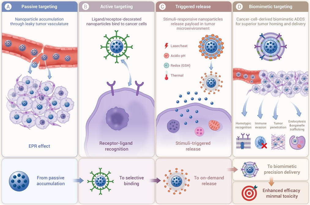
Figure 1.
The development of ADDS
-
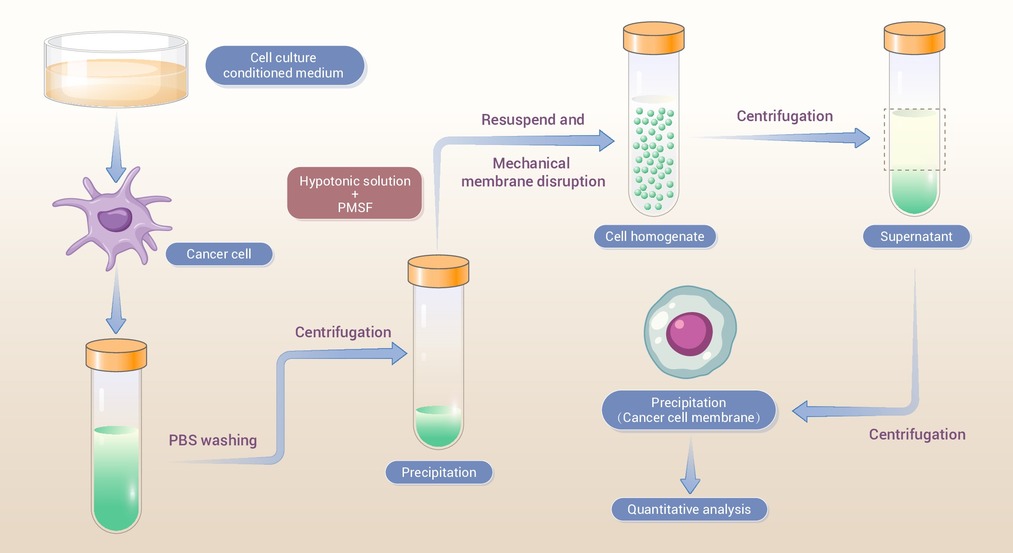
Figure 2.
The brief process of CCM isolation
-
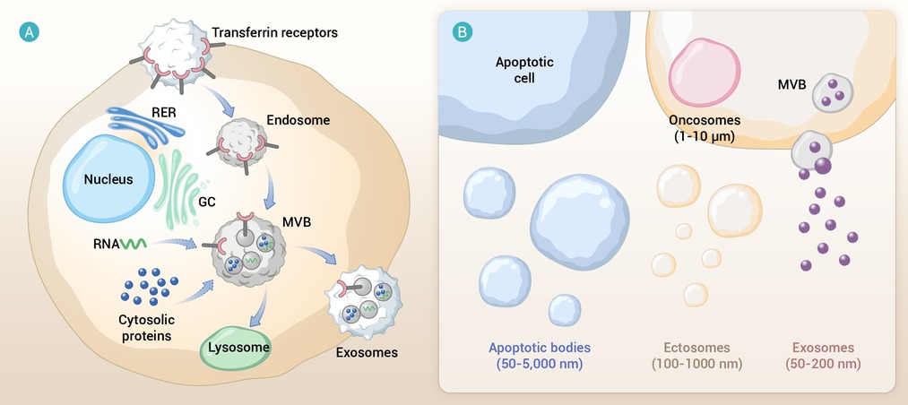
Figure 3.
Biogenesis of TEVs and TEV sub-types
-
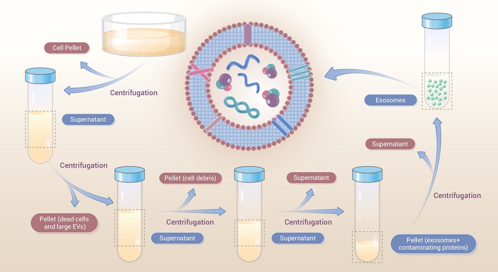
Figure 4.
The brief process of TEV isolation
-
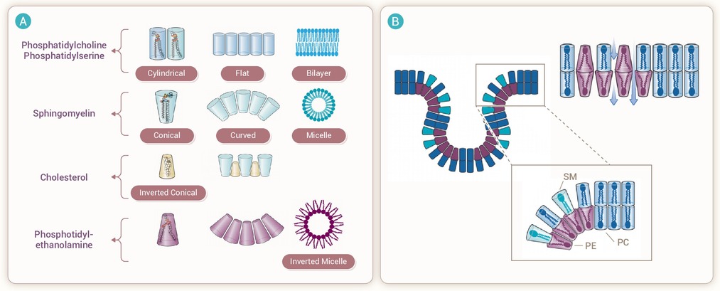
Figure 5.
Lipid polymorphism and its compositional effects on membrane integrity, endocytosis, and intracellular transport
