
The evolution of in-silico physiological models of bone remodelling: From cell population models to micro-MPA approaches
-
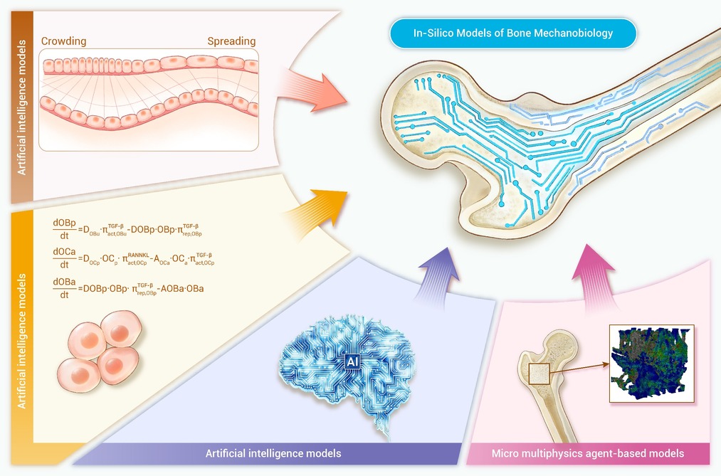
GRAPHICAL ABSTRACT
 DownLoad:
Full size image
DownLoad:
Full size image
-
PUBLIC SUMMARY
-
In-silico models of bone are crucial for understanding the behaviour of bone under certain conditions.
There are five types of in-silico models: bone cell population, bone tissue dynamics, FE, AI, and micro MPA models.
Micro MPA models can include bone mediators, cells, as well as a response to 3D stress.
-
-
ABSTRACT
Bone remodelling is a mechanically regulated, multiscale process that maintains skeletal integrity and is disrupted in common metabolic bone diseases. Experiments, however, cannot easily resolve the coupled cellular, biochemical, and mechanical feedback that drive long-term outcomes. In this Review, we examine five in-silico modelling classes used to study bone remodelling: bone cell population dynamics models, bone tissue dynamics models, finite element models, AI models, and three-dimensional micro-multiphysics agent-based models. We compare these approaches through biological fidelity, computational tractability, and translational readiness. FEM excels at macro-scale mechanics but depends on explicit material laws. Cell population models remain dominant for long-horizon and pharmacological simulations because they are interpretable and efficient, although their non-spatial structure limits prediction of microarchitecture. Tissue dynamics models capture curvature-controlled growth efficiently but often rely on simplified mechanobiology. Micro-multiphysics agent-based models provide the strongest link between cell behaviour, mechanics, and microarchitecture, but face major challenges in parameter identifiability, scalable computation, and validation. AI models can bypass explicit material laws and accelerate other methods through surrogate modelling, but risk non-physical predictions without appropriate constraints. We conclude that the field’s next step is to develop integrated, testable workflows that improve calibration and validation while making assumptions explicit. In this context, AI is best viewed as an enabling tool for surrogate modelling, inverse problems, and data-assimilation workflows, rather than as a replacement for mechanistic understanding. -

-
REFERENCES
[1] Bartl R., Bartl C. (2019). Structure and architecture of bone. The Osteoporosis Manual. (Springer, Cham), pp:11-19. DOI: 10.1007/978-3-032-21885-8_2 [2] Kameo Y., Miya Y., Hayashi M., et al. (2020). In silico experiments of bone remodeling explore metabolic diseases and their drug treatment. Sci. Adv. 6:eaax0938. DOI:10.1126/sciadv.aax0938 [3] Scheiner S., Pivonka P. and Hellmich C. (2013). Coupling systems biology with multiscale mechanics, for computer simulations of bone remodeling. Comput. Methods Appl. Mech. Eng. 254:181−196. DOI:10.1016/j.cma.2012.10.015 [4] Pivonka P., Zimak J., Smith D. W., et al. (2008). Model structure and control of bone remodeling: A theoretical study. Bone (New York, N.Y.) 43:249−263. DOI:10.1016/j.bone.2008.03.025 [5] Lemaire V., Tobin F. L., Greller L. D., et al. (2004). Modeling the interactions between osteoblast and osteoclast activities in bone remodeling. J. Theor. Biol. 229:293−309. DOI:10.1016/j.jtbi.2004.03.023 [6] Pastrama M.-I., Scheiner S., Pivonka P., et al. (2018). A mathematical multiscale model of bone remodeling, accounting for pore space-specific mechanosensation. Bone (New York, N.Y.) 107:208−221. DOI:10.1016/j.bone.2017.11.009 [7] Martínez-Reina J. and Pivonka P. (2019). Effects of long-term treatment of denosumab on bone mineral density: Insights from an in-silico model of bone mineralization. Bone (New York, N.Y.) 125:87−95. DOI:10.1016/j.bone.2019.04.022 [8] Martínez-Reina J., Calvo-Gallego J. L., Martin M., et al. (2022). Assessment of strategies for safe drug discontinuation and transition of denosumab treatment in PMO—Insights from a mechanistic PK/PD model of bone turnover. Front. Bioeng. Biotechnol. 10:886579. DOI:10.3389/fbioe.2022.886579 [9] Martínez-Reina J., Calvo-Gallego J. L. and Pivonka P. (2021). Combined effects of exercise and denosumab treatment on local failure in post-menopausal osteoporosis–Insights from bone remodelling simulations accounting for mineralisation and damage. Front. Bioeng. Biotechnol. 9:635056. DOI:10.3389/fbioe.2021.635056 [10] Martínez-Reina J., Calvo-Gallego J. L. and Pivonka P. (2021). Are drug holidays a safe option in treatment of osteoporosis.— Insights from an in silico mechanistic PK–PD model of denosumab treatment of postmenopausal osteoporosis. J. Mech. Behav. Biomed. Mater. 113:104140. DOI:10.1016/j.jmbbm.2020.104140 [11] Boaretti D., Marques F. C., Ledoux C., et al. (2023). Trabecular bone remodeling in the aging mouse: A micro-multiphysics agent-based in silico model using single-cell mechanomics. Front. Bioeng. Biotechnol. 11:1091294. DOI:10.3389/fbioe.2023.1091294 [12] Kendall J. J., Ledoux C., Marques F. C., et al. (2023). An in silico micro-multiphysics agent-based approach for simulating bone regeneration in a mouse femur defect model. Front. Bioeng. Biotechnol. 11:1289127. DOI:10.3389/fbioe.2023.1289127 [13] Tourolle D. C., Dempster D. W., Ledoux C., et al. (2021). Ten‐year simulation of the effects of denosumab on bone remodeling in human biopsies. JBMR Plus 5:e10494−n/a. DOI:10.1002/jbm4.10494 [14] Tourolle, D.C. (2019). A micro-scale multiphysics framework for fracture healing and bone remodelling. PhD thesis (ETH Zurich). https://doi.org/10.3929/ethz-b-000364637 [15] Khosla S., Oursler M. J. and Monroe D. G. (2012). Estrogen and the skeleton. Trends Endocrinol. Metab. 23:576−581. DOI:10.1016/j.tem.2012.03.008 [16] Mundy G. R. (1999). Cellular and molecular regulation of bone turnover. Bone (New York, N.Y.) 24:35S−38S. DOI:10.1016/S8756-3282(99)00044-7 [17] Burr D. B. (2002). Targeted and nontargeted remodeling. Bone (New York, N.Y.) 30:2−4. DOI:10.1016/S8756-3282(01)00619-6 [18] Martin R. B. (2002). Is all cortical bone remodeling initiated by microdamage. Bone (New York, N.Y.) 30:8−13. DOI:10.1016/S8756-3282(01)00620-2 [19] Parfitt A. M. (2002). Targeted and nontargeted bone remodeling: Relationship to basic multicellular unit origination and progression. Bone (New York, N.Y.) 30:5−7. DOI:10.1016/s8756-3282(01)00642-1 [20] Martin R.B., Burr D.B., Sharkey N.A., et al. (2015). Skeletal Tissue Mechanics, Second Edition (Springer). DOI: 10.1007/978-1-4939-3002-9 [21] Ledoux C., Boaretti D., Sachan A., et al. (2022). Clinical data for parametrization of in silico bone models incorporating cell-cytokine dynamics: A systematic review of literature. Front. Bioeng. Biotechnol. 10:901720. DOI:10.3389/fbioe.2022.901720 [22] Mundy G. R. (1995). Bone Remodelling. Martin Dunitz (ed). Bone remodeling and its disorders (CRC Press), pp: 1- 12. DOI: 10.1201/9781003076944 [23] Silbermann, R., and Roodman, G.D. (2013). Mechanisms of Bone Destruction in Myeloma. R. Marcus, D. Feldman, D.W. Dempster, M. Luckey, and J.A. Cauley (eds). Osteoporosis (Fourth Edition) (Elsevier), pp: 1465–1478. DOI: 10.1016/B978-0-12-415853-5.00062-5 [24] Bell N. H. (2003). RANKL and the regulation of skeletal remodeling. J. Clin. Inves. 111:1120−1122. DOI:10.1172/JCI18358 [25] Martin T. J. (2004). Paracrine regulation of osteoclast formation and activity: Milestones in discovery. J. Musculoskelet. Neuronal. Interact. 4:243–253. https://europepmc.org/article/MED/15615492 [26] Hofbauer L. C., Neubauer A. and Heufelder A. E. (2001). Receptor activator of nuclear factor kappa B ligand and osteoprotegrin. Cancer 92:460−470. DOI:10.1002/1097-0142(20010801 [27] Hofbauer L. C. and Schoppet M. (2004). Clinical implications of the osteoprotegerin/rankl/rank system for bone and vascular diseases. JAMA 292:490−495. DOI:10.1001/jama.292.4.490 [28] Hofbauer L. C., Kühne C. A. and Viereck V. (2004). The OPG/RANKL/RANK system in metabolic bone diseases. J. Musculoskelet. Neuronal. Interact. 4:268–275. https://europepmc.org/article/MED/15615494 [29] Boyle W. J., Simonet W. S. and Lacey D. L. (2003). Osteoclast differentiation and activation. Nature (London) 423:337−342. DOI:10.1038/nature01658 [30] Rodan G. A. and Martin T. J. (2000). Therapeutic approaches to bone diseases. Science 289:1508−1514. DOI:10.1126/science.289.5484.1508 [31] Scheiner S., Pivonka P. and Hellmich C. (2016). Poromicromechanics reveals that physiological bone strains induce osteocyte-stimulating lacunar pressure. Biomech. model. mechanobiol. 15:9−28. DOI:10.1007/s10237-015-0704-y [32] Martin M., Sansalone V., Cooper D. M. L., et al. (2020). Assessment of romosozumab efficacy in the treatment of postmenopausal osteoporosis: Results from a mechanistic pk-pd mechanostat model of bone remodeling. Bone (New York, N.Y.) 133:115223. DOI:10.1016/j.bone.2020.115223 [33] Martin M., Sansalone V., Cooper D. M. L., et al. (2019). Mechanobiological osteocyte feedback drives mechanostat regulation of bone in a multiscale computational model. Biomech. Model Mechanobiol. 18:1475−1496. DOI:10.1007/s10237-019-01158-w [34] Pivonka P., Zimak J., Smith D. W., et al. (2010). Theoretical investigation of the role of the RANK–RANKL–OPG system in bone remodeling. J. Theor. Biol. 262:306−316. DOI:10.1016/j.jtbi.2009.09.021 [35] Ruiz-Lozano R., Calvo-Gallego J. L., Pivonka P., et al. (2025). Optimisation of romosozumab plus denosumab sequential treatments against postmenopausal osteoporosis. Insights from in silico simulations. Biomech. Model. Mechanobiol. 24:383−404. DOI:10.1007/s10237-024-01900-z [36] Aubin J. E. (1998). Advances in the osteoblast lineage. Biochem. Cell Biol. 76:899−910. DOI:10.1139/o99-005 [37] Aubin J. E. (1998). Bone stem cells. J. Cell. Biochem. 30-31:73–82. DOI:10.1002/(sici)1097-4644(1998)72:30/31+%3C73::aid-jcb11%3E3.0.co;2-l [38] Alliston T., Choy L., Ducy P., et al. (2001). Tgf-β-induced repression of cbfa1 by smad3 decreases cbfa1 and osteocalcin expression and inhibits osteoblast differentiation. EMBO J. 20:2254−2272. DOI:10.1093/emboj/20.9.2254 [39] Ducy P., Schinke T. and Karsenty G. (2000). The osteoblast: A sophisticated fibroblast under central surveillance. Science 289:1501−1504. DOI:10.1126/science.289.5484.1501 [40] Roodman G. D. (1999). Cell biology of the osteoclast. Exp. hematol. 27:1229−41. DOI:10.1016/s0301-472x(99)00061-2 [41] Greenfield E. M., Bi Y. and Miyauchi A. (1999). Regulation of osteoclast activity. Life Sci. 65:1087−1102. DOI:10.1016/s0024-3205(99)00156-3 [42] Siegel P. M. and Massagué J. (2003). Cytostatic and apoptotic actions of TGF-β in homeostasis and cancer. Nat. Rev. Cancer 3:807−820. DOI:10.1038/nrc1208 [43] Fuller K., Lean J. M., Bayley K. E., et al. (2000). A role for TGFbeta(1) in osteoclast differentiation and survival. J. Cell Sci. 113:2445−2453. DOI:10.1242/jcs.113.13.2445 [44] Teitelbaum S. L. (2000). Bone resorption by osteoclasts. Science 289:1504−1508. DOI:10.1126/science.289.5484.1504 [45] Jilka R. L., Weinstein R. S., Bellido T., et al. (1998). Osteoblast programmed cell death (apoptosis): Modulation by growth factors and cytokines. JBMR 13:793−802. DOI:10.1359/jbmr.1998.13.5.793 [46] Aubin J. E. and Bonnelye E. (2000). Osteoprotegerin and its ligand: A new paradigm for regulation of osteoclastogenesis and bone resorption. Osteoporos. Int. 11:905−913. DOI:10.1007/s001980070028 [47] Parfitt A. M., Mundy G. R., Roodman G. D., et al. (1996). A new model for the regulation of bone resorption, with particular reference to the effects of bisphosphonates. JBMR 11:150−159. DOI:10.1002/jbmr.5650110203 [48] Bland R. (2000). Steroid hormone receptor expression and action in bone. Clin Sci. 98:217−240. DOI:10.1042/cs0980217 [49] Huang W. H. and Zheng M. H. (1999). Steroid hormones and bone. Histol Histopathol. 14:1257−1268. DOI:10.14670/HH-14.1257 [50] Goltzman D. (1999). Interactions of pth and pthrp with the pth/pthrp receptor and with downstream signaling pathways: exceptions that provide the rules. JBMR. 14:173−177. DOI:10.1359/jbmr.1999.14.2.173 [51] Halladay D. L., Miles R. R., Thirunavukkarasu K., et al. (2002). Identification of signal transduction pathways and promoter sequences that mediate parathyroid hormone 1-38 inhibition of osteoprotegerin gene expression. J. cell. biochem. 84:1−11. DOI:10.1002/jcb.1273 [52] Ma Y. L., Cain R. L., Halladay D. L., et al. (2001). Catabolic effects of continuous human PTH (1–38) in vivo is associated with sustained stimulation of RANKL and inhibition of osteoprotegerin and gene-associated bone formation. Endocrin. (Philadelphia) 142:4047−4054. DOI:10.1210/en.142.9.4047 [53] Kroll M. H. (2000). Parathyroid hormone temporal effects on bone formation and resorption. Bull. of math. biol. 62:163−188. DOI:10.1006/bulm.1999.0146 [54] Rattanakul C., Lenbury Y., Krishnamara N., et al. (2003). Modeling of bone formation and resorption mediated by parathyroid hormone: Response to estrogen/PTH therapy. BioSystems 70:55−72. DOI:10.1016/S0303-2647(03)00040-6 [55] Komarova S. V., Smith R. J., Dixon S. J., et al. (2003). Mathematical model predicts a critical role for osteoclast autocrine regulation in the control of bone remodeling. Bone (New York, N.Y.) 33:206−215. DOI:10.1016/S8756-3282(03)00157-1 [56] Udagawa N., Takahashi N., Jimi E., et al. (1999). Osteoblasts/stromal cells stimulate osteoclast activation through expression of osteoclast differentiation factor/RANKL but not macrophage colony-stimulating factor. Bone (New York, N.Y.) 25:517−523. DOI:10.1016/S8756-3282(99)00210-0 [57] Hofbauer L. C., Dunstan C. R., Spelsberg T. C., et al. (1998). Osteoprotegerin production by human osteoblast lineage cells is stimulated by vitamin d, bone morphogenetic protein-2, and cytokines. Biochem. Biophys. Res. Commun. 250:776−781. DOI:10.1006/bbrc.1998.9394 [58] Gori F., Hofbauer L. C., Dunstan C. R., et al. (2000). The expression of osteoprotegerin and RANK ligand and the support of osteoclast formation by stromal-osteoblast lineage cells is developmentally regulated. Endocrinology (Philadelphia) 141:4768. DOI:10.1210/endo.141.12.7840 [59] Huang J. C., Sakata T., Pfleger L. L., et al. (2004). PTH differentially regulates expression of RANKL and OPG. JBMR 19:235−244. DOI:10.1359/jbmr.0301226 [60] Thomas G. P., Baker S. U., Eisman J. A., et al. (2001). Changing RANKL/OPG mRNA expression in differentiating murine primary osteoblasts. J. Endocrinol. 170:451−460. DOI:10.1677/joe.0.1700451 [61] Glass D. A., Bialek P., Ahn J. D., et al. (2005). Canonical wnt signaling in differentiated osteoblasts controls osteoclast differentiation. Dev. Cell 8:751−764. DOI:10.1016/j.devcel.2005.02.017 [62] Zaoui A. (2002). Continuum micromechanics: Survey. J. eng. mech. 128:808−816. DOI:10.1061/(ASCE)0733-9399(2002)128:8(808 [63] Zaoui A. (1997). Structural morphology and constitutive behaviour of microheterogeneous materials. Suquet, P. (eds). Continuum Micromechanics. International Centre for Mechanical Sciences (Springer, Vienna), pp: 291–347. DOI: 10.1007/978-3-7091-2662-2_6 [64] Hill R. (1963). Elastic properties of reinforced solids: Some theoretical principles. J. Mech. Phys. Solids. 11:357−372. DOI:10.1016/0022-5096(63)90036-X [65] Hill R. (1965). Continuum micro-mechanics of elastoplastic polycrystals. J. Mech. Phys. Solids. 13:89−101. DOI:10.1016/0022-5096(65)90023-2 [66] Hellmich C., Kober C. and Erdmann B. (2008). Micromechanics-based conversion of ct data into anisotropic elasticity tensors, applied to fe simulations of a mandible. Ann. Biomed. Eng. 36:108−122. DOI:10.1007/s10439-007-9393-8 [67] Fritsch A. and Hellmich C. (2007). ‘Universal’ microstructural patterns in cortical and trabecular, extracellular and extravascular bone materials: Micromechanics-based prediction of anisotropic elasticity. J. Theor. Biol. 244:597−620. DOI:10.1016/j.jtbi.2006.09.013 [68] Hellmich C. and Ulm F.-J. (2005). Drained and undrained poroelastic properties of healthy and pathological bone: A poro-micromechanical investigation. Transp. Porous Media. 58:243−268. DOI:10.1007/s11242-004-6298-y [69] Hellmich C. and Ulm F. J. (2008). Micromechanical model for ultra-structural stiffness of mineralized tissues. J. Eng. Mech 8:898−908. DOI:10.1061/(ASCE)0733-9399(2002)128:8(898 [70] Winkler D. G., Sutherland M. K., Geoghegan J. C., et al. (2003). Osteocyte control of bone formation via sclerostin, a novel bmp antagonist. EMBO J. 22:6267−6276. DOI:10.1093/emboj/cdg599 [71] van Bezooijen R. L., Roelen B. A. J., Visser A., et al. (2004). Sclerostin is an osteocyte-expressed negative regulator of bone formation, but not a classical bmp antagonist. J. Exp. Med. 199:805−814. DOI:10.1084/jem.20031454 [72] Robling A. G., Niziolek P. J., Baldridge L. A., et al. (2008). Mechanical stimulation of bone in vivo reduces osteocyte expression of sost/sclerostin. J. Biol. Chem. 283:5866−5875. DOI:10.1074/jbc.M705092200 [73] Robling A. G., Castillo A. B. and Turner C. H. (2006). Biomechanical and molecular regulation of bone remodeling. Annu. Rev. Biomed. Eng. 8:455−498. DOI:10.1146/annurev.bioeng.8.061505.095721 [74] Weyts F. A. A., Bosmans B., Niesing R., et al. (2003). Mechanical control of human osteoblast apoptosis and proliferation in relation to differentiation. Calcif. Tissue Int. 72:505−512. DOI:10.1007/s00223-002-2027-0 [75] Owan I., Burr D. B., Turner C. H., et al. (1997). Mechanotransduction in bone: Osteoblasts are more responsive to fluid forces than mechanical strain. Am. J. Physiol. Cell Physiol. 42:C810−C815. DOI:10.1152/ajpcell.1997.273.3.C810 [76] Jones D. B., Nolte H., Scholübbers J. G., et al. (1991). Biochemical signal transduction of mechanical strain in osteoblast-like cells. Biomaterials 12:101−110. DOI:10.1016/0142-9612(91)90186-E [77] Kaspar D., Seidl W., Neidlinger-Wilke C., et al. (2002). Proliferation of human-derived osteoblast-like cells depends on the cycle number and frequency of uniaxial strain. J. Biomech. 35:873−880. DOI:10.1016/S0021-9290(02)00058-1 [78] Martínez G., García Aznar J. M., Doblaré M., et al. (2006). External bone remodeling through boundary elements and damage mechanics. Math. Comput. Simul. 73:183−199. DOI:10.1016/j.matcom.2006.06.014 [79] Huiskes R., Weinans H., Grootenboer H. J., et al. (1987). Adaptive bone-remodeling theory applied to prosthetic-design analysis. J. Biomech. 20:1135−1150. DOI:10.1016/0021-9290(87)90030-3 [80] Fyhrie D. P. and Carter D. R. (1986). A unifying principle relating stress to trabecular bone morphology. J. Orthop. Res. 4:304−317. DOI:10.1002/jor.1100040307 [81] Henstock J. R., Rotherham M., Rose J. B., et al. (2013). Cyclic hydrostatic pressure stimulates enhanced bone development in the foetal chick femur in vitro. Bone (New York, N.Y.) 53:468−477. DOI:10.1016/j.bone.2013.01.010 [82] Rottmar M., Ackerknecht S., Wick P., et al. (2011). A high throughput system for long term application of intermittent cyclic hydrostatic pressure on cells in culture. J. Biomech. Eng. 133:024502. DOI:10.1115/1.4003313 [83] Liu C., Zhao Y., Cheung W.-Y., et al. (2010). Effects of cyclic hydraulic pressure on osteocytes. Bone (New York, N.Y.) 46:1449−1456. DOI:10.1016/j.bone.2010.02.006 [84] Liu J., Zhao Z., Li J., et al. (2009). Hydrostatic pressures promote initial osteodifferentiation with ERK1/2 not p38 MAPK signaling involved. J. Cell Biochem. 107:224−232. DOI:10.1002/jcb.22118 [85] Takai E., Mauck R. L., Hung C. T., et al. (2004). Osteocyte viability and regulation of osteoblast function in a 3d trabecular bone explant under dynamic hydrostatic pressure. JBMR 19:1403−1410. DOI:10.1359/jbmr.040516 [86] Imamura K., Ozawa H., Hiraide T., et al. (1990). Continuously applied compressive pressure induces bone resorption by a mechanism involving prostaglandin E2 synthesis. J. Cell. Physiol. 144:222−228. DOI:10.1002/jcp.1041440207 [87] Ozawa H., Imamura K., Abe E., et al. (1990). Effect of a continuously applied compressive pressure on mouse osteoblast-like cells (MC3T3-E1) in vitro. J. Cell. Physiol. 142:177−185. DOI:10.1002/jcp.1041420122 [88] Nagatomi J., Arulanandam B. P., Metzger D. W., et al. (2001). Frequency- and duration-dependent effects of cyclic pressure on select bone cell functions. Tissue eng. 7:717−728. DOI:10.1089/107632701753337672 [89] Nagatomi J., Arulanandam B. P., Metzger D. W., et al. (2002). Effects of cyclic pressure on bone marrow cell cultures. J Biomech. Eng. 124:308−314. DOI:10.1115/1.1468867 [90] Nagatomi J., Arulanandam B. P., Metzger D. W., et al. (2003). Cyclic pressure affects osteoblast functions pertinent to osteogenesis. Ann. Biomed. Eng. 31:917−923. DOI:10.1114/1.1590663 [91] Hellmich C. (2005). Microelasticity of Bone. Dormieux, L., Ulm, FJ. (eds). Applied micromechanics of porous materials (Springer Vienna), pp. 289–331. DOI:10.1007/3-211-38046-9_8 [92] Hellmich C., Ulm F.-J. and Dormieux L. (2004). Can the diverse elastic properties of trabecular and cortical bone be attributed to only a few tissue-independent phase properties and their interactions. Arguments from a multiscale approach. Biomech. Model. Mechanobiol. 2:219−238. DOI:10.1007/s10237-004-0040-0 [93] Turner C. H., Rho J., Takano Y., et al. (1999). The elastic properties of trabecular and cortical bone tissues are similar: Results from two microscopic measurement techniques. J. Biomech.s 32:437−441. DOI:10.1016/S0021-9290(98)00177-8 [94] Lees S., Ahern J. M. and Leonard M. (1983). Parameters influencing the sonic velocity in compact calcified tissues of various species. J. Acoust. Soc. Am. 74:28−33. DOI:10.1121/1.389723 [95] Lees S., Tao N. J. and Lindsay S. M. (1990). Studies of compact hard tissues and collagen by means of brillouin light scattering. Connect. Tissue Res. 24:187−205. DOI:10.3109/03008209009152148 [96] Bonfoh N., Novinyo E. and Lipinski P. (2011). Modeling of bone adaptative behavior based on cells activities. Biomech. Model. Mechanobiol. 10:789−798. DOI:10.1007/s10237-010-0274-y [97] Rapisarda A. C., Almasi M., Almasi N., et al. (2020). Bone mechanics and cell populations: Mathematical description and parametric study of the model. B.E. Abali, and I. Giorgio (eds). Developments and novel approaches in biomechanics and metamaterials. (Springer International Publishing). pp:107–126. DOI:10.1007/978-3-030-50464-9_7 [98] Rapisarda A. C., Della Corte A., Drobnicki R., et al. (2018). A model for bone mechanics and remodeling including cell populations dynamics. Z. Angew. Math. Phys. 70:9. DOI:10.1007/s00033-018-1055-1 [99] Mertiya A. S., Tiwari A. K., Mishra A., et al. (2023). Computational modeling for osteogenic potential assessment of physical exercises based on loading-induced mechanobiological environments in cortical bone remodeling. Biomech. Model. Mechanobiol. 22:281−295. DOI:10.1007/s10237-022-01647-5 [100] Peyroteo M. M. A., Belinha J. and Natal Jorge R. M. (2021). Load adaptation through bone remodeling: A mechanobiological model coupled with the finite element method. Biomech. Model. Mechanobiol. 20:1495−1507. DOI:10.1007/s10237-021-01458-0 [101] Peyroteo M. M. A., Belinha J. and Natal Jorge R. M. (2021). Predicting bone remodeling using a mechano-biological mathematical model combined with a natural neighbor meshless method. Eng. Anal. Bound. Elem. 132:437−445. DOI:10.1016/j.enganabound.2021.08.004 [102] Ramtani S., Sánchez J. F., Boucetta A., et al. (2023). A coupled mathematical model between bone remodeling and tumors: A study of different scenarios using Komarova’s model. Biomech. Model. Mechanobiol. 22:925−945. DOI:10.1007/s10237-023-01689-3 [103] Vemparala B., Ji M., Mageswaran P., et al. (2025). A computational multiscale framework for bone remodeling: coupling apparent density evolution and microscale shape optimization. Int. J. Numer. Method. Biomed. Eng. 41:e70097. DOI:10.1002/cnm.70097 [104] Tatsumi S., Ishii K., Amizuka N., et al. (2007). Targeted ablation of osteocytes induces osteoporosis with defective mechanotransduction. Cell Metab. 5:464−475. DOI:10.1016/j.cmet.2007.05.001 [105] Gaur T., Lengner C. J., Hovhannisyan H., et al. (2005). Canonical wnt signaling promotes osteogenesis by directly stimulating runx2 gene expression. J. Bio. Chem. 280:33132−33140. DOI:10.1074/jbc.M500608200 [106] Westendorf J. J., Kahler R. A. and Schroeder T. M. (2004). Wnt signaling in osteoblasts and bone diseases. Gene 341:19−39. DOI:10.1016/j.gene.2004.06.044 [107] Bidan C. M., Wang F. M. and Dunlop J. W. C. (2013). A three-dimensional model for tissue deposition on complex surfaces. Comput. Methods Biomech. Biomed. Eng. 16:1056−1070. DOI:10.1080/10255842.2013.774384 [108] Bidan C. M., Kommareddy K. P., Rumpler M., et al. (2013). Geometry as a factor for tissue growth: Towards shape optimization of tissue engineering scaffolds. Adv. Healthc. Mater. 2:186−194. DOI:10.1002/adhm.201200159 [109] Guyot Y., Luyten F. P., Schrooten J., et al. (2015). A three-dimensional computational fluid dynamics model of shear stress distribution during neotissue growth in a perfusion bioreactor. Biotech. Bioengin. 112:2591−2600. DOI:10.1002/bit.25672 [110] Guyot Y., Papantoniou I., Chai Y. C., et al. (2014). A computational model for cell/ECM growth on 3D surfaces using the level set method: A bone tissue engineering case study. Biomech. Model. Mechanobiol. 13:1361−1371. DOI:10.1007/s10237-014-0577-5 [111] Bidan C. M., Kommareddy K. P., Rumpler M., et al. (2012). How linear tension converts to curvature: Geometric control of bone tissue growth. PLoS ONE 7:e36336−e36336. DOI:10.1371/journal.pone.0036336 [112] Buenzli P. R., Pivonka P. and Smith D. W. (2011). Spatio-temporal structure of cell distribution in cortical bone multicellular units: A mathematical model. Bone 48:918−926. DOI:10.1016/j.bone.2010.12.009 [113] Buenzli P. (2015). Governing equations of tissue modelling and remodelling: A unified generalised description of surface and bulk balance. PLOS ONE 11. DOI:10.1371/journal.pone.0152582. [114] Buenzli P. R. (2015). Osteocytes as a record of bone formation dynamics: A mathematical model of osteocyte generation in bone matrix. J. Theo. Biol. 364:418−427. DOI:10.1016/j.jtbi.2014.09.028 [115] Buenzli P. R., Pivonka P. and Smith D. W. (2014). Bone refilling in cortical basic multicellular units: Insights into tetracycline double labelling from a computational model. Biomech. Model. Mechanobiol. 13:185−203. DOI:10.1007/s10237-013-0495-y [116] Alias M. A. and Buenzli P. R. (2020). A level‐set method for the evolution of cells and tissue during curvature‐controlled growth. Int. J. Numer. Methods Biomed. Eng. 36:e3279−n/a. DOI:10.1002/cnm.3279 [117] Alias M. A. and Buenzli P. R. (2018). Osteoblasts infill irregular pores under curvature and porosity controls: A hypothesis-testing analysis of cell behaviours. Biomech. Model. Mechanobiol. 17:1357−1371. DOI:10.1007/s10237-018-1031-x [118] Alias M. A. and Buenzli P. R. (2017). Modeling the effect of curvature on the collective behavior of cells growing new tissue. Biophys. J. 112:193−204. DOI:10.1016/j.bpj.2016.11.3203 [119] Callens S. J. P., Uyttendaele R. J. C., Fratila-Apachitei L. E., et al. (2020). Substrate curvature as a cue to guide spatiotemporal cell and tissue organization. Biomaterials 232:119739. DOI:10.1016/j.biomaterials.2019.119739 [120] Hon S. Y., Leung S. and Zhao H. (2014). A cell based particle method for modeling dynamic interfaces. J. Comp.l Phys. 272:279−306. DOI:10.1016/j.jcp.2014.04.032 [121] Rolli C. G., Nakayama H., Yamaguchi K., et al. (2012). Switchable adhesive substrates: Revealing geometry dependence in collective cell behavior. Biomaterials 33:2409−2418. DOI:10.1016/j.biomaterials.2011.12.012 [122] Paris M., Götz A., Hettrich I., et al. (2017). Scaffold curvature-mediated novel biomineralization process originates a continuous soft tissue-to-bone interface. Acta biomaterialia 60:64−80. DOI:10.1016/j.actbio.2017.07.029 [123] Chen C. S., Mrksich M., Huang S., et al. (1997). Geometric control of cell life and death. Science 276:1425−1428. DOI:10.1126/science.276.5317.1425 [124] Hegarty-Cremer S. G. D., Simpson M. J., Andersen T. L., et al. (2021). Modelling cell guidance and curvature control in evolving biological tissues. J. Theor. Biol. 520:110658. DOI:10.1016/j.jtbi.2021.110658 [125] Ambrosi D., Ben Amar M., Cyron C. J., et al. (2019). Growth and remodelling of living tissues: Perspectives, challenges and opportunities. J. R. Soc. Interface 16:20190233. DOI:10.1098/rsif.2019.0233 [126] Martin R. B. (2000). Does osteocyte formation cause the nonlinear refilling of osteons. Bone 26:71−78. DOI:10.1016/S8756-3282(99)00242-2 [127] Poujade M., Grasland-Mongrain E., Hertzog A., et al. (2007). Collective migration of an epithelial monolayer in response to a model wound. From the Cover 104:15988−15993. DOI:10.1073/pnas.0705062104 [128] Lowengrub J. S., Frieboes H. B., Jin F., et al. (2010). Nonlinear modelling of cancer: Bridging the gap between cells and tumours. Nonlinearity 23:R1−R91. DOI:10.1088/0951-7715/23/1/R01 [129] Ripamonti U. and Roden L. (2010). Biomimetics for the induction of bone formation. Expert Rev. Med. Devices 7:469−479. DOI:10.1586/erd.10.17 [130] Bidan C. M., Kollmannsberger P., Gering V., et al. (2016). Gradual conversion of cellular stress patterns into pre-stressed matrix architecture during in vitro tissue growth. J. R. Soc. Interface 13:20160136. DOI:10.1098/rsif.2016.0136 [131] Leung S., Lowengrub J. and Zhao H. (2011). A grid based particle method for solving partial differential equations on evolving surfaces and modeling high order geometrical motion. J. Comput. Phys. 230:2540−2561. DOI:10.1016/j.jcp.2010.12.029 [132] Leung S. and Zhao H. (2009). A grid based particle method for moving interface problems. J. Comput. Phys. 228:2993−3024. DOI:10.1016/j.jcp.2009.01.005 [133] Adachi T., Tsubota K.-i., Tomita Y., et al. (2001). Trabecular surface remodeling simulation for cancellous bone using microstructural voxel finite element models. J. Biomech. Eng. 123:403−409. DOI:10.1115/1.1392315 [134] Davies J. A. (2023). Cell Migration in Development. Mechanisms of morphogenesis, 3rd Edition. (Elsevier Science). pp:91-98. DOI: 10.1016/B978-0-323-99965-6.00021-X [135] Murray, J.D. (1993). Reaction diffusion, chemotaxis, and nonlocal mechanisms. Murray, J.D. (ed) Mathematical Biology. Interdisciplinary Applied Mathematics. (Springer). pp:395-417. DOI: 10.1007/978-0-387-22437-4_11 [136] Hegarty-Cremer S. G. D., Borggaard X. G., Andreasen C. M., et al. (2024). How osteons form: A quantitative hypothesis-testing analysis of cortical pore filling and wall asymmetry. Bone 180:116998. DOI:10.1016/j.bone.2023.116998 [137] Fernández J. R., García-Aznar J. M., Martínez R., et al. (2010). Numerical analysis of a strain-adaptive bone remodelling problem. Comp. Methods Appl. Mech. Eng. 199:1549−1557. DOI:10.1016/j.cma.2010.01.005 [138] Doblaré M. and Garcı́a J. M. (2002). Anisotropic bone remodelling model based on a continuum damage-repair theory. J. Biomech. 35:1−17. DOI:10.1016/S0021-9290(01)00178-6 [139] Giorgio I., dell’Isola F., Andreaus U., et al. (2023). An orthotropic continuum model with substructure evolution for describing bone remodeling: an interpretation of the primary mechanism behind Wolff’s law. Biomech. Model. Mechanobiol. 22:2135−2152. DOI:10.1007/s10237-023-01755-w [140] Vercher-Martínez A., Giner E., Fuenmayor F. J., et al. (2024). The role of the interfaces and cross-links on the mechanical behavior of mineralized collagen fibrils. A numerical approach. Eng. Fracture Mech. 309:110440. DOI:10.1016/j.engfracmech.2024.110440 [141] Atthapreyangkul A., Hoffman M. and Pearce G. (2021). Effect of geometrical structure variations on the viscoelastic and anisotropic behaviour of cortical bone using multi-scale finite element modelling. J. Mech. Behav. Biomed. Mater. 113:104153. DOI:10.1016/j.jmbbm.2020.104153 [142] Orlova D. and Berinskii I. (2024). Multiscale analysis of a 3D fibrous collagen tissue. Int. J. Eng. Sci. 195:104003. DOI:10.1016/j.ijengsci.2023.104003 [143] Gaziano P., Monaldo E., Falcinelli C., et al. (2022). Elasto-damage mechanics of osteons: A bottom-up multiscale approach. J. Mech. Phys. Solids. 167:104962. DOI:10.1016/j.jmps.2022.104962 [144] Atthapreyangkul A., Hoffman M., Pearce G., et al. (2023). Effect of geometrical structure variations on strength and damage onset of cortical bone using multi-scale cohesive zone based finite element method. J. Mech. Behav. Biomed. Mater. 138:105578. DOI:10.1016/j.jmbbm.2022.105578 [145] You T., Kim Y.-R. and Park T. (2017). Two-way coupled multiscale model for predicting mechanical behavior of bone subjected to viscoelastic deformation and fracture damage. J. Eng. Mater. Technol. 139:021016. DOI:10.1115/1.4035618 [146] Witt C., Kaiser T. and Menzel A. (2023). Modelling and numerical simulation of remodelling processes in cortical bone: An IGA approach to flexoelectricity-induced osteocyte apoptosis and subsequent bone cell diffusion. J. Mech. Phys. Solids 173:105194. DOI:10.1016/j.jmps.2022.105194 [147] Mohammadkhah M., Marinkovic D., Zehn M., et al. (2019). A review on computer modeling of bone piezoelectricity and its application to bone adaptation and regeneration. Bone 127:544−555. DOI:10.1016/j.bone.2019.07.024 [148] Mohammadkhah M., Slavkovic V. and Klinge S. (2025). Flexoelectricity in biological materials and its potential applications in biomedical research. Bioeng. 12:579. DOI:10.3390/bioengineering12060579 [149] Witt C., Kaiser T. and Menzel A. (2024). An IGA-FEA model for flexoelectricity-induced healing of microcracks in cortical bone. Comput. Methods Appl. Mech. Eng. 425:116919. DOI:10.1016/j.cma.2024.116919 [150] Titlbach A., Papastavrou A., McBride A., et al. (2025). Modelling the flexoelectric effect in human bone—a micromorphic approach. Comput. Methods Appl. Mech. Eng. 446:118234. DOI:10.1016/j.cma.2025.118234 [151] Blaszczyk M. and Hackl K. (2024). On the influence of the microstructure on multiscale bone simulations. PAMM 24:e202400040. DOI:10.1002/pamm.202400040 [152] Blaszczyk M. and Hackl K. (2022). Multiscale modeling of cancellous bone considering full coupling of mechanical, electric and magnetic effects. Biomech. Model. Mechanobiol. 21:163−187. DOI:10.1007/s10237-021-01525-6 [153] Ju C., Yang K., Yang Q., et al. (2025). Multiscale dynamics analysis of lumbar vertebral cortical bone based on the Abaqus submodel finite element method. Sci. Rep. 15:6861. DOI:10.1038/s41598-025-91918-9 [154] Johnson J. E. and Troy K. L. (2018). Simplified boundary conditions alter cortical-trabecular load sharing at the distal radius; A multiscale finite element analysis. J. Biomech. 66:180−185. DOI:10.1016/j.jbiomech.2017.10.036 [155] Levrero-Florencio F., Manda K., Margetts L., et al. (2017). Nonlinear homogenisation of trabecular bone: Effect of solid phase constitutive model. Proc. Inst. Mech. Eng. H 231:405−414. DOI:10.1177/0954411916676220 [156] Chakraborty A., Sahare K. D., Majumder S., et al. (2024). Understanding the biomechanical response of progressive thread dental implants using multi-scale finite element analysis. Int. J. Multiscale Comput. Eng. 22:31−44. DOI:10.1615/IntJMultCompEng.2023049024 [157] Jung G. S. and Buehler M. J. (2017). Multiscale modeling of muscular-skeletal systems. Annu. Rev. Biomed. Eng. 19:435−457. DOI:10.1146/annurev-bioeng-071516-044555 [158] Sabet F. A., Raeisi Najafi A., Hamed E., et al. (2016). Modelling of bone fracture and strength at different length scales: A review. Interface focus 6:20150055. DOI:10.1098/rsfs.2015.0055 [159] Estermann S.-J. and Scheiner S. (2018). Multiscale modeling provides differentiated insights to fluid flow-driven stimulation of bone cellular activities. Front. Phys. 6:76. DOI:10.3389/fphy.2018.00076 [160] Kumbolder V., Morin C., Scheiner S., et al. (2024). Hierarchical elastoplasticity of cortical bone: Observations, mathematical modeling, validation. Mech. Mat. 198:105140. DOI:10.1016/j.mechmat.2024.105140 [161] Gagliardi D., Naili S., Desceliers C., et al. (2017). Tissue mineral density measured at the sub-millimetre scale can provide reliable statistics of elastic properties of bone matrix. Biomech. Model. Mechanobiol. 16:1885−1910. DOI:10.1007/s10237-017-0926-2 [162] Giammarini A., Ramírez‐Torres A. and Grillo A. (2025). Effective elasto‐(visco) plastic coefficients of a bi‐phasic composite material with scale‐dependent size effects. Math. Methods Appl. Sci. 48:926−979. DOI:10.1002/mma.10367 [163] Colabella L., Naili S., Le Cann S., et al. (2024). Effect of collagen fibril orientation on the anisotropic properties of peri-implant bone. Biomech. Model. Mechanobiol. 23:879−891. DOI:10.1007/s10237-023-01811-5 [164] Hage I. S., Seif C. Y. and Hamade R. F. (2021). Cortical osteon stiffness: A comparative micromechanics-based homogenization study. Int. J. Multiscale Comput. Eng. 19:55−72. DOI:10.1615/IntJMultCompEng.2021034412 [165] Huiskes R., Ruimerman R., van Lenthe G. H., et al. (2000). Effects of mechanical forces on maintenance and adaptation of form in trabecular bone. Nature (London) 405:704−706. DOI:10.1038/35015116 [166] Ruimerman R., Hilbers P., van Rietbergen B., et al. (2005). A theoretical framework for strain-related trabecular bone maintenance and adaptation. J. Biomech. 38:931−941. DOI:10.1016/j.jbiomech.2004.03.037 [167] Bagge M. (2000). A model of bone adaptation as an optimization process. J.Biomech. 33:1349−1357. DOI:10.1016/S0021-9290(00)00124-X [168] Jang I. G. and Kim I. Y. (2008). Computational study of Wolff's law with trabecular architecture in the human proximal femur using topology optimization. J. Biomech. 41:2353−2361. DOI:10.1016/j.jbiomech.2008.05.037 [169] Nowak M. (2010). On some properties of bone functional adaptation phenomenon useful in mechanical design. Acta Bioeng. Biomech. 12:49–54. https://pubmed.ncbi.nlm.nih.gov/20882941/ [170] Goda I., Ganghoffer J.-F., Czarnecki S., et al. (2019). Topology optimization of bone using cubic material design and evolutionary methods based on internal remodeling. Mech. Res. Comms. 95:52−60. DOI:10.1016/j.mechrescom.2018.12.003 [171] Goda I., Ganghoffer J.-F. and Maurice G. (2016). Combined bone internal and external remodeling based on Eshelby stress. Int. J. Solids Struct. 94:138−157. DOI:10.1016/j.ijsolstr.2016.04.036 [172] Park J., Sutradhar A., Shah J. J., et al. (2018). Design of complex bone internal structure using topology optimization with perimeter control. Comput. Biol. Med. 94:74−84. DOI:10.1016/j.compbiomed.2018.01.001 [173] Beaupré G. S., Orr T. E. and Carter D. R. (1990). An approach for time-dependent bone modeling and remodeling-application: A preliminary remodeling simulation. J Orthop Res 8:662−670. DOI:10.1002/jor.1100080507 [174] Weinans H., Huiskes R. and Grootenboer H. J. (1992). The behavior of adaptive bone-remodeling simulation models. J. Biomech. 25:1425−1441. DOI:10.1016/0021-9290(92)90056-7 [175] Gombolay G. Y., Gopalan N., Bernasconi A., et al. (2023). Review of machine learning and artificial intelligence (ML/AI) for the pediatric neurologist. Pediatr. Neurol. 141:42−51. DOI:10.1016/j.pediatrneurol.2023.01.004 [176] Oishi A. and Yagawa G. (2017). Computational mechanics enhanced by deep learning. Comput. Methods Appl. Mech. Eng. 327:327−351. DOI:10.1016/j.cma.2017.08.040 [177] Mohammadkhah M., Savari A. and Klinge S. (2026). Capturing the multiscale nature of bone behavior: Classical, data-driven and hybrid techniques. Annal. Biomed. Eng. DOI:10.1007/s10439-026-04043-7 [178] Xiao P., Zhang T., Haque E., et al. (2021). Prediction of elastic behavior of human trabecular bone using a DXA image-based deep learning model. Jom 73:2366−2376. DOI:10.1007/s11837-021-04704-z [179] Luan S. and Morgan E. F. (2025). A data-driven framework for developing a unified density–modulus relationship for the human lumbar vertebral body. J. Mech. Behav. Biomed. Mater. 163:106888. DOI:10.1016/j.jmbbm.2025.106888 [180] Minku and Ghosh R. (2024). A macro–micro FE and ANN framework to assess site-specific bone ingrowth around the porous beaded-coated implant: An example with BOX® tibial implant for total ankle replacement. Med. Biol. Eng. Comput. 62:1639−1654. DOI:10.1007/s11517-024-03034-x [181] Jiang L., Schmid F., Nassr M., et al. (2023). Fabrication of porous polymeric-based scaffold for dental tissue repair in fracture healing: RVE simulation and ANN optimization. Mater. Sci. Eng. B 297:116770. DOI:10.1016/j.mseb.2023.116770 [182] Barkaoui A., Tlili B., Vercher-Martínez A., et al. (2016). A multiscale modelling of bone ultrastructure elastic proprieties using finite elements simulation and neural network method. Comput. Methods Programs Biomed. 134:69−78. DOI:10.1016/j.cmpb.2016.07.005 [183] Stieve V., Blaszczyk M. and Hackl K. (2022). Inverse modeling of cancellous bone using artificial neural networks. J. Appl. Math. Mechs. 102:202100541. DOI:10.1002/zamm.202100541 [184] Li Z. (2021). Predicting bone regeneration from machine learning. Nat. Comput. Sci. 1:509−510. DOI:10.1038/s43588-021-00116-w [185] Pled F., Desceliers C. and Zhang T. (2021). A robust solution of a statistical inverse problem in multiscale computational mechanics using an artificial neural network. Comput. Methods Appl. Mech. Eng. 373:113540. DOI:10.1016/j.cma.2020.113540 [186] Mondal A., Nguyen C., Ma X., et al. (2019). Network models for characterization of trabecular bone. Phys. Rev. E. 99:042406. DOI:10.1103/PhysRevE.99.042406 [187] Mora-Macías J., Ayensa-Jiménez J., Reina-Romo E., et al. (2020). A multiscale data-driven approach for bone tissue biomechanics. Comput. Methods Appl. Mech. Eng. 368:113136. DOI:10.1016/j.cma.2020.113136 [188] Mouloodi S., Rahmanpanah H., Burvill C., et al. (2020). Prediction of displacement in the equine third metacarpal bone using a neural network prediction algorithm. Biocybern. Biomed. Eng. 40:849−863. DOI:10.1016/j.bbe.2019.09.001 [189] Mouloodi S., Rahmanpanah H., Gohery S., et al. (2022). Feedforward backpropagation artificial neural networks for predicting mechanical responses in complex nonlinear structures: A study on a long bone. J. Mech. Behav. Biomed. Mater. 128:105079. DOI:10.1016/j.jmbbm.2022.105079 [190] Yagawa G. and Okuda H. (1996). Neural networks in computational mechanics. Arch. Comput. Methods Eng. 3:435−512. DOI:10.1007/BF02818935 [191] Hambli R. (2010). Application of neural networks and finite element computation for multiscale simulation of bone remodeling. J. Biomech. Eng. 132. DOI:10.1115/1.4002536 [192] Hambli R. (2011). Numerical procedure for multiscale bone adaptation prediction based on neural networks and finite element simulation. Finite Elem. Anal. Des. 47:835−842. DOI:10.1016/j.finel.2011.02.014 [193] Zadpoor A. A., Campoli G. and Weinans H. (2013). Neural network prediction of load from the morphology of trabecular bone. Appl. Math. Model. 37:5260−5276. DOI:10.1016/j.apm.2012.10.049 [194] Campoli G., Weinans H. and Zadpoor A. A. (2012). Computational load estimation of the femur. J. Mech. Behav. Biomed. 10:108−119. DOI:10.1016/j.jmbbm.2012.02.011 [195] Garijo N., Martínez J., García-Aznar J. M., et al. (2014). Computational evaluation of different numerical tools for the prediction of proximal femur loads from bone morphology. Comput. Methods Appl. Mech. Eng. 268:437−450. DOI:10.1016/j.cma.2013.10.005 [196] Pais A., Alves J. L. and Belinha J. (2025). A neural network to surrogate computational bone remodelling in the calcaneus. Knowledge-based Syst. 330:114445. DOI:10.1016/j.knosys.2025.114445 [197] Pais A., Alves J. L. and Belinha J. (2023). Predicting trabecular arrangement in the proximal femur: An artificial neural network approach for varied geometries and load cases. J. Biomech. 161:111860. DOI:10.1016/j.jbiomech.2023.111860 [198] Kumar R. (2024). Recurrent context layered radial basis function neural network for the identification of nonlinear dynamical systems. Neurocomp. 580:127524. DOI:10.1016/j.neucom.2024.127524 [199] Dass A., Srivastava S. and Kumar R. (2023). A novel Lyapunov-stability-based recurrent-fuzzy system for the Identification and adaptive control of nonlinear systems. Appl. Soft Comp. 137:110161. DOI:10.1016/j.asoc.2023.110161 [200] Shobana R., Kumar R. and Jaint B. (2026). A recurrent neural network-based identification of complex nonlinear dynamical systems: A novel structure, stability analysis and a comparative study. Soft Comp. 30:1245−1261. DOI:10.1007/s00500-023-09390-4 [201] Dong Y., Liu T., Li Z., et al. (2023). DeepFEM: A novel element-based deep learning approach for solving nonlinear partial differential equations in computational solid mechanics. J. Eng. Mech. 149:04022102. DOI:10.1061/JENMDT.EMENG-6643 [202] Truskey G. A. (2023). The potential of deep learning to advance clinical applications of computational biomechanics. Bioengineering 10:1066. DOI:10.3390/bioengineering10091066 [203] Ma S., Zhang J., Shi C., et al. (2024). Physics-informed deep learning for muscle force prediction with unlabeled semg signals. IEEE Trans. Neural Syst. Rehabil. Eng. 32:1246−1256. DOI:10.1109/TNSRE.2024.3375320 [204] Hambli R., Katerchi H. and Benhamou C.-L. (2011). Multiscale methodology for bone remodelling simulation using coupled finite element and neural network computation. Biomech.Model. Mechanobiol. 10:133−145. DOI:10.1007/s10237-010-0222-x [205] Khalid S., Yazdani M. H., Azad M. M., et al. (2025). Advancements in physics-informed neural networks for laminated composites: A comprehensive review. Mathematics 13:17. DOI:10.3390/math13010017 [206] Liu X., Tian S., Tao F., et al. (2021). A review of artificial neural networks in the constitutive modeling of composite materials. Compos. B Eng. 224:109152. DOI:10.1016/j.compositesb.2021.109152 [207] Wu C., Entezari A., Zheng K., et al. (2021). A machine learning-based multiscale model to predict bone formation in scaffolds. Nat. Comp. Sci. 1:532−541. DOI:10.1038/s43588-021-00115-x [208] Khadijeh M., Cerqueglini V., Kasbergen C., et al. (2025). Multistage physics informed neural network for solving coupled multiphysics problems in material degradation and fluid dynamics. Eng. Comput. 41:3491−3521. DOI:10.1007/s00366-025-02174-4 [209] Khaterchi H., Chamekh A. and BelHadjSalah H. (2015). Artificial neural network analysis for modeling fibril structure in bone. Int. J. Precis. Eng. Manuf. 16:581−587. DOI:10.1007/s12541-015-0078-1 [210] Kim Y. K., Kameo Y., Tanaka S., et al. (2017). Capturing microscopic features of bone remodeling into a macroscopic model based on biological rationales of bone adaptation. Biomech. Model. Mechanobiol. 16:1697−1708. DOI:10.1007/s10237-017-0914-6 [211] Wolff J. (2010). The classic: on the inner architecture of bones and its importance for bone growth. Clin. Orthop. Relat. Res. 468:1056−1065. DOI:10.1007/s11999-010-1239-2 [212] Tsubota K.-i., Suzuki Y., Yamada T., et al. (2009). Computer simulation of trabecular remodeling in human proximal femur using large-scale voxel FE models: Approach to understanding Wolff's law. J. biomech. 42:1088−1094. DOI:10.1016/j.jbiomech.2009.02.030 [213] Beno T., Yoon Y.-J., Cowin S. C., et al. (2006). Estimation of bone permeability using accurate microstructural measurements. J. Biomech. 39:2378−2387. DOI:10.1016/j.jbiomech.2005.08.005 [214] Xu R. (2014). Semaphorin 3A. Cell Adh. Migr. 8:5−10. DOI:10.4161/cam.27752 [215] Baron R. and Kneissel M. (2013). Wnt signaling in bone homeostasis and disease: From human mutations to treatments. Nat. Med. 19:179−192. DOI:10.1038/nm.3074 [216] Xie Y., Zhou J., Tian L., et al. (2023). Mir‐196b‐5p regulates osteoblast and osteoclast differentiation and bone homeostasis by targeting sema3a. JBMR. 38:1175−1191. DOI:10.1002/jbmr.4834 [217] Wu K., Huang D. and Huang X. (2023). The effects of semaphorin 3A in bone and cartilage metabolism: fundamental mechanism and clinical potential. Front. Cell Dev. Biol. 11:1321151. DOI:10.3389/fcell.2023.1321151 [218] Flaig C., Arbenz P. (2012). A scalable memory efficient multigrid solver for micro-finite element analyses based on CT images. Parallel Comp. 37:846-854.DOI: 10.1016/j.parco.2011.08.001 [219] Boyce B. F., Hughes D. E., Wright K. R., et al. (1999). Recent advances in bone biology provide insight into the pathogenesis of bone diseases. Lab. Invest. 79:83–94. https://europepmc.org/article/MED/10068197 -
ABOUT THIS ARTICLE
Cite this article:
Altaleb J., Hansen U., Abel R., et al. (2026). The evolution of in-silico physiological models of bone remodelling: From cell population models to micro-MPA approaches. The Innovation Life 4:100247. https://doi.org/10.59717/j.xinn-life.2026.100247
Welcome!
To request copyright permission to republish or share portions of our works, please visit Copyright Clearance Center's (CCC) Marketplace website at marketplace.copyright.com.
Export File
Citation
Altaleb J., Hansen U., Abel R., et al. (2026). The evolution of in-silico physiological models of bone remodelling: From cell population models to micro-MPA approaches. The Innovation Life 4:100247. https://doi.org/10.59717/j.xinn-life.2026.100247

Format
Content
-
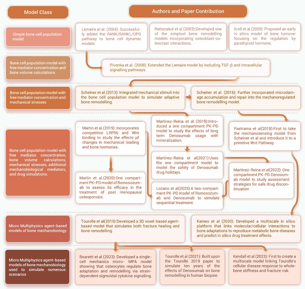
Figure 1.
Evolutionary hierarchy of physiological bone remodelling models
-
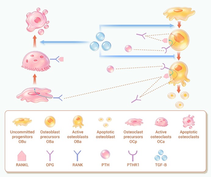
Figure 2.
Visual representation of model depicting the interaction between bone cells
-
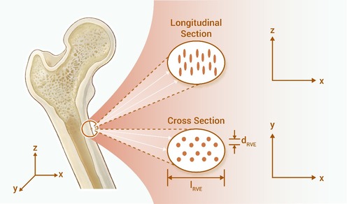
Figure 3.
Visual representation of the representative volume element
-
Figure 4.
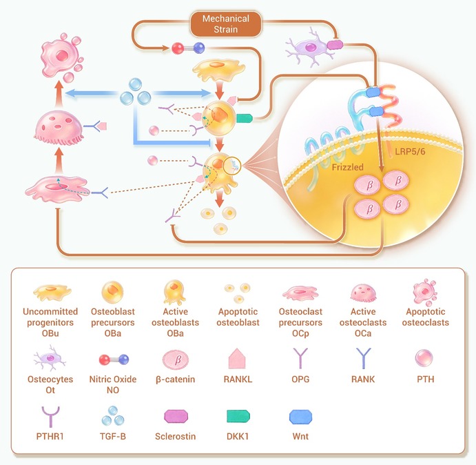
Schematic explaining how the Wnt pathway operates
-
Figure 5.
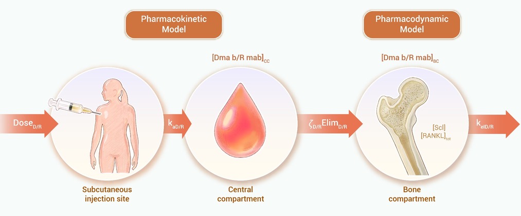
Schematic representation of how PK-PD modelling is implemented in cell population models
-
Figure 6.
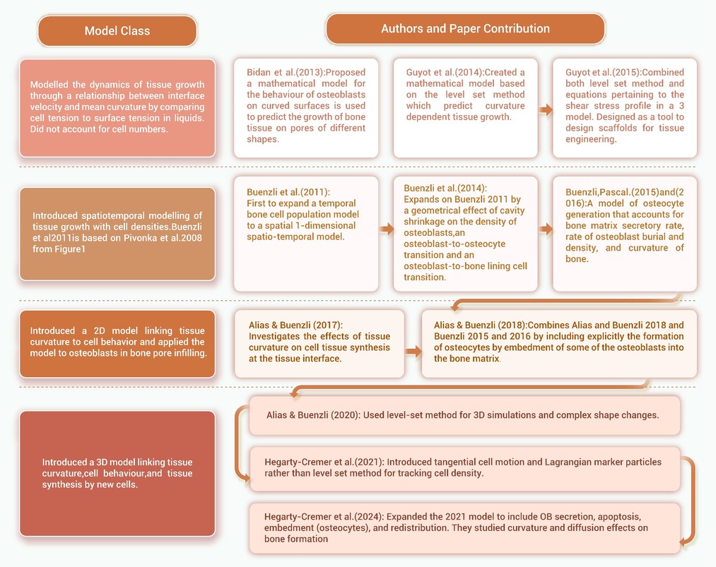
All relevant mathematical models of bone tissue growth in the literature and how they evolve from each other
-
Figure 7.
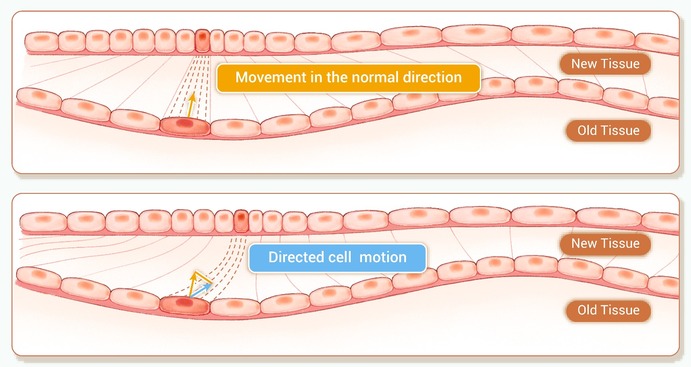
The influence of surface curvature on tissue growth mediated by surface-bound cells
-
Figure 8.
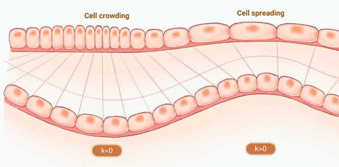
Curvature’s effect on cell distribution and tissue development
-
Figure 9.
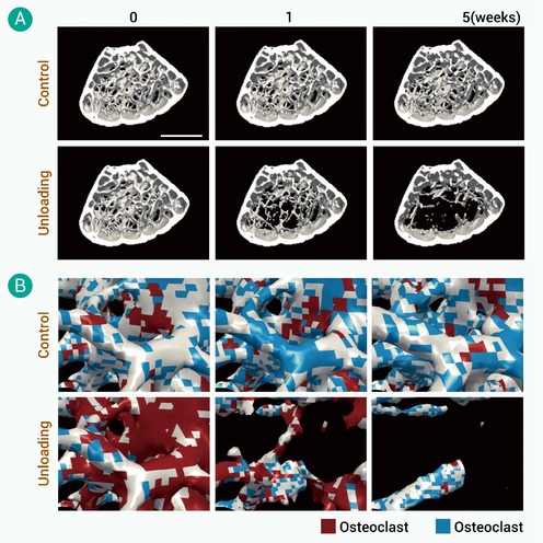
Figure depicting Kameo et al.’s 2020 model2
