
Topology-enabled long-acting interleukin-2 for advanced low-dose interleukin-2 therapy of autoimmune diseases
-
GRAPHICAL ABSTRACT
 DownLoad:
Full size image
-
PUBLIC SUMMARY
-
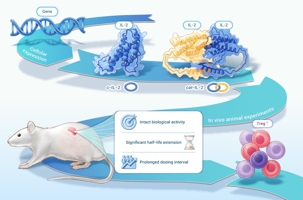
Cyclic and catenated interleukin-2 (IL-2) with intact bioactivity are directly synthesized in living cells.
These variants induce preferential Treg expansion to rebalance immunity.
Extended serum half-life enables weekly low-dose treatment for three autoimmune diseases.
Topology engineering overcomes protein therapeutic limits, offering a platform for next-generation biologics.
-
-
ABSTRACT
The therapeutic efficacy of protein biologics is often hampered by their rapid in vivo degradation and off-target effects. Interleukin-2 (IL-2), a cytokine promising for treating autoimmune diseases via regulatory T cell (Treg) expansion, exemplifies this challenge due to its short half-life and pleiotropic activity. Herein, we report a molecular-scale topology engineering strategy to overcome these limitations. By using split intein mediated ligation and genetically encoded entangling motif, p53dim (X), we constructed cyclic (c-IL-2) and catenated (cat-IL-2) variants of IL-2 via direct expression in E. coli. These topological proteins preserve the native conformation and receptor binding capacity of IL-2 with minimal to minor exogenous motifs, while exhibiting superior proteolytic stability and a dramatically extended serum half-life (approximately 4.5–5.0-fold longer than wt-IL-2). In murine models of three distinct autoimmune diseases (lupus, rheumatoid arthritis, and Sjögren’s syndrome), weekly administration of these topological IL-2 variants effectively rebalanced immunity, as evidenced by a marked expansion of Tregs and significant amelioration of tissue injury and clinical biomarkers. Transcriptomic profiling confirmed the induction of a tolerogenic program without systemic toxicity. This work establishes protein topology engineering as a minimalist, yet robust and generalizable platform to enhance the pharmacokinetics and functional specificity of therapeutic proteins, offering a powerful new modality for biologic drug development.-
Keywords:
- chemical topology /
- protein catenane /
- systemic lupus erythematosus /
- low-dose /
- half-life
-

-
REFERENCES
[1] Franklyn K., Hoi A., Nikpour M., et al. (2014). The need to define treatment goals for systemic lupus erythematosus. Nat. Rev. Rheumatol. 10:567−571. DOI:10.1038/nrrheum.2014.118 [2] Ugarte-Gil M. F., Wojdyla D., Pons-Estel G. J., et al. (2017). Remission and Low Disease Activity Status (LDAS) protect lupus patients from damage occurrence: Data from a multiethnic, multinational Latin American Lupus Cohort (GLADEL). Ann. Rheum. Dis. 76:2071−2074. DOI:10.1136/annrheumdis-2017-211814 [3] Morand E. F. and Mosca M. (2017). Treat to target, remission and low disease activity in SLE. Best Pract. Res. Clin. Rheumatol. 31:342−350. DOI:10.1016/j.berh.2017.09.009 [4] Boyman O. and Sprent J. (2012). The role of interleukin-2 during homeostasis and activation of the immune system. Nat. Rev. Immunol. 12:180−190. DOI:10.1038/nri3156 [5] Koreth J., Matsuoka K., Kim H. T., et al. (2011). Interleukin-2 and regulatory T cells in graft-versus-host disease. N. Engl. J. Med. 365:2055−2066. DOI:10.1056/NEJMoa1108188 [6] Saadoun D., Rosenzwajg M., Joly F., et al. (2011). Regulatory T-cell responses to low-dose interleukin-2 in HCV-induced vasculitis. N. Engl. J. Med. 365:2067−2077. DOI:10.1056/NEJMoa1105143 [7] Mizui M. and Tsokos G. C. (2016). Low-dose IL-2 in the treatment of lupus. Curr. Rheumatol. Rep. 18:68. DOI:10.1007/s11926-016-0617-5 [8] Kim N., Jeon Y. W., Nam Y. S., et al. (2016). Therapeutic potential of low-dose IL-2 in a chronic GVHD patient by in vivo expansion of regulatory T cells. Cytokine 78:22−26. DOI:10.1016/j.cyto.2015.11.020 [9] He J., Zhang X., Wei Y., et al. (2016). Low-dose interleukin-2 treatment selectively modulates CD4(+) T cell subsets in patients with systemic lupus erythematosus. Nat. Med. 22:991−993. DOI:10.1038/nm.4148 [10] Dwyer C. J., Ward N. C., Pugliese A., et al. (2016). Promoting immune regulation in type 1 diabetes using low-dose interleukin-2. Curr. Diab. Rep. 16:46. DOI:10.1007/s11892-016-0739-1 [11] Rosenzwajg M., Lorenzon R., Cacoub P., et al. (2019). Immunological and clinical effects of low-dose interleukin-2 across 11 autoimmune diseases in a single, open clinical trial. Ann. Rheum. Dis. 78:209−217. DOI:10.1136/annrheumdis-2018-214229 [12] He J., Zhang R., Shao M., et al. (2020). Efficacy and safety of low-dose IL-2 in the treatment of systemic lupus erythematosus: A randomised, double-blind, placebo-controlled trial. Ann. Rheum. Dis 79:141−149. DOI:10.1136/annrheumdis-2019-215396 [13] Li Z. G., He J., Miao M. (2023). Low-dose interleukin-2 therapy in autoimmune and inflammatory diseases: Facts and hopes. Sci. Bull. 68:10−13. DOI:10.1016/j.scib.2022.12.024 [14] von Spee-Mayer C., Siegert E., Abdirama D., et al. (2016). Low-dose interleukin-2 selectively corrects regulatory T cell defects in patients with systemic lupus erythematosus. Ann. Rheum. Dis. 75:1407−1415. DOI:10.1136/annrheumdis-2015-207776 [15] Miao M., Xiao X., Tian J., et al. (2021). Therapeutic potential of targeting Tfr/Tfh cell balance by low-dose-IL-2 in active SLE: A post hoc analysis from a double-blind RCT study. Arthritis Res. Ther. 23:167. DOI:10.1186/s13075-021-02535-6 [16] Shah N. R., Declouette B., Ansari-Gilani K., et al. (2021). High-dose interleukin-2 therapy related adverse events and implications on imaging. Diagn. Interv. Radiol. 27:684−689. DOI:10.5152/dir.2021.20503 [17] Zhang B., Sun J., Wang Y., et al. (2021). Site-specific PEGylation of interleukin-2 enhances immunosuppression via the sustained activation of regulatory T cells. Nat. Biomed. Eng. 5:1288−1305. DOI:10.1038/s41551-021-00797-8 [18] Tannir N. M., Formiga M. N., Penkov K., et al. (2024). Bempegaldesleukin plus nivolumab versus sunitinib or cabozantinib in previously untreated advanced clear cell renal cell carcinoma: A phase III randomized study (PIVOT-09). J. Clin. Oncol. 42:2800−2811. DOI:10.1200/JCO.23.02082 [19] Trotta E., Bessette P. H., Silveria S. L., et al. (2018). A human anti-IL-2 antibody that potentiates regulatory T cells by a structure-based mechanism. Nat. Med. 24:1005−1014. DOI:10.1038/s41591-018-0070-2 [20] Spangler J. B., Trotta E., Tomala J., et al. (2018). Engineering a single-agent cytokine/antibody fusion that selectively expands regulatory T cells for autoimmune disease therapy. J. Immunol. 201:2094−2106. DOI:10.4049/jimmunol.1800578 [21] Lee J. Y., Lee E., Hong S. W., et al. (2020). TCB2, a new anti-human interleukin-2 antibody, facilitates heterodimeric IL-2 receptor signaling and improves anti-tumor immunity. Oncoimmunology 9:1681869. DOI:10.1080/2162402x.2019.1681869 [22] Karakus U., Sahin D., Mittl P. R. E., et al. (2020). Receptor-gated IL-2 delivery by an anti-human IL-2 antibody activates regulatory T cells in three different species. Sci. Transl. Med. 12:eabb9283. DOI:10.1126/scitranslmed.abb9283 [23] Liu D. V., Maier L. M., Hafler D. A., et al. (2009). Engineered interleukin-2 antagonists for the inhibition of regulatory T cells. J. Immunother. 32:887−894. DOI:10.1097/CJI.0b013e3181b528da [24] Levin A. M., Bates D. L., Ring A. M., et al. (2012). Exploiting a natural conformational switch to engineer an interleukin-2 'superkine'. Nature 484:529−533. DOI:10.1038/nature10975 [25] Carmenate T., Pacios A., Enamorado M., et al. (2013). Human IL-2 mutein with higher antitumor efficacy than wild type IL-2. J. Immunol. 190:6230−6238. DOI:10.4049/jimmunol.1201895 [26] Mitra S., Ring A. M., Amarnath S., et al. (2015). Interleukin-2 activity can be fine tuned with engineered receptor signaling clamps. Immunity 42:826−838. DOI:10.1016/j.immuni.2015.04.018 [27] Sockolosky J. T., Trotta E., Parisi G., et al. (2018). Selective targeting of engineered T cells using orthogonal IL-2 cytokine-receptor complexes. Science 359:1037−1042. DOI:10.1126/science.aar3246 [28] Carmenate T., Ortíz Y., Enamorado M., et al. (2018). Blocking IL-2 signal in vivo with an IL-2 antagonist reduces tumor growth through the control of regulatory T cells. J. Immunol. 200:3475−3484. DOI:10.4049/jimmunol.1700433 [29] Ward N. C., Yu A., Moro A., et al. (2018). IL-2/CD25: A long-acting fusion protein that promotes immune tolerance by selectively targeting the IL-2 receptor on regulatory T cells. J. Immunol. 201:2579−2592. DOI:10.4049/jimmunol.1800907 [30] Lopes J. E., Fisher J. L., Flick H. L., et al. (2020). ALKS 4230: A novel engineered IL-2 fusion protein with an improved cellular selectivity profile for cancer immunotherapy. J. Immunother. Cancer 8:e000673. DOI:10.1136/jitc-2020-000673 [31] Zhang B., Sun J., Yuan Y., et al. (2023). Proximity-enabled covalent binding of IL-2 to IL-2Rα selectively activates regulatory T cells and suppresses autoimmunity. Signal Transduct. Target. Ther. 8:28. DOI:10.1038/s41392-022-01208-3 [32] Vaishampayan U. N., Muzaffar J., Winer I., et al. (2024). Nemvaleukin alfa, a modified interleukin-2 cytokine, as monotherapy and with pembrolizumab in patients with advanced solid tumors (ARTISTRY-1). J. Immunother. Cancer 12:e010143. DOI:10.1136/jitc-2024-010143 [33] Kobayashi M., Kojima K., Murayama K., et al. (2021). MK-6, a novel not-alpha IL-2, elicits a potent antitumor activity by improving the effector to regulatory T cell balance. Cancer Sci. 112:4478−4489. DOI:10.1111/cas.15127 [34] Moynihan K. D., Kumar M. P., Sultan H., et al. (2024). IL2 targeted to CD8+ T cells promotes robust effector T-cell responses and potent antitumor immunity. Cancer Discov. 14:1206−1225. DOI:10.1158/2159-8290.CD-23-1266 [35] Sun J., Guo L., Ji D., et al. (2025). Reshape the fates of Treg and CD8+ T cells through IL-2Ralpha by synergizing divergent receptor-biased IL-2 PEGylates. Adv. Sci. 12:e2414931. DOI:10.1002/advs.202414931 [36] Francl M. (2009). Stretching topology. Nat. Chem. 1:334−335. DOI:10.1038/nchem.302 [37] Wang X.-W. and Zhang W.-B. (2018). Chemical topology and complexity of protein architectures. Trends Biochem. Sci. 43:806−817. DOI:10.1016/j.tibs.2018.07.001 [38] Qu Z., Cheng S. Z. D., Zhang W.-B. (2021). Macromolecular topology engineering. Trends Chem. 3:402−415. DOI:10.1016/j.trechm.2021.02.002 [39] Li T., Zhang F., Fang J., et al. (2023). Rational design and cellular synthesis of proteins with unconventional chemical topology. Chin. J. Chem. 41:2873−2880. DOI:10.1002/cjoc.202300190 [40] Wang Y.-X., Wu W.-H., Jiang F.-Y., et al. (2024). Mechanically interlocked biomacromolecules. Supramol. Mater. 3:100059. DOI:10.1016/j.supmat.2023.100059 [41] Yan L. Z. and Dawson P. E. (2001). Design and synthesis of a protein catenane. Angew. Chem. Int. Ed. 40:3625−3627. DOI:3.0.CO;2-Q">10.1002/1521-3773(20011001)40:19<3625::AID-ANIE3625>3.0.CO;2-Q [42] King N. P., Jacobitz A. W., Sawaya M. R., et al. (2010). Structure and folding of a designed knotted protein. Proc. Natl. Acad. Sci. USA 107:20732−20737. DOI:10.1073/pnas.1007602107 [43] Wang X.-W. and Zhang W.-B. (2016). Cellular synthesis of protein catenanes. Angew. Chem. Int. Ed. 55:3442−3446. DOI:10.1002/anie.201511640 [44] Liu Y., Wu W.-H., Hong S., et al. (2020). Lasso proteins: Modular design, cellular synthesis, and topological transformation. Angew. Chem. Int. Ed. 59:19153−19161. DOI:10.1002/anie.202006727 [45] Liu Y., Duan Z., Fang J., et al. (2020). Cellular synthesis and X-ray crystal structure of a designed protein heterocatenane. Angew. Chem. Int. Ed. 59:16122−16127. DOI:10.1002/anie.202005490 [46] Doyle L. A., Takushi B., Kibler R. D., et al. (2023). De novo design of knotted tandem repeat proteins. Nat. Commun. 14:6746. DOI:10.1038/s41467-023-42388-y [47] Zhang F., Liu Y., Da X.-D., et al. (2024). Toward selective synthesis of protein olympiadanes via orthogonal active templates in one step. CCS Chem. 6:1047−1059. DOI:10.31635/ccschem.023.202303071 [48] Qu Z., Xu L., Jiang F., et al. (2024). Folds from fold: Exploring topological isoforms of a single-domain protein. Proc. Natl. Acad. Sci. USA 121:e2407355121. DOI:10.1073/pnas.2407355121 [49] Xu L., Song X., Xu H., et al. (2026). Computational design and cellular synthesis of two protein topological isomers: Solomon link vs. three-twist knot. Proc. Natl. Acad. Sci. USA 123:e2537891123. DOI:10.1073/pnas.2537891123 [50] Wang X.-W. and Zhang W.-B. (2017). Protein catenation enhances both the stability and activity of folded structural domains. Angew. Chem. Int. Ed. 56:13985−13989. DOI:10.1002/anie.201705194 [51] Wu W.-H., Bai X., Shao Y., et al. (2021). Higher order protein catenation leads to an artificial antibody with enhanced affinity and in vivo stability. J. Am. Chem. Soc. 143:18029−18040. DOI:10.1021/jacs.1c06169 [52] Liu Y., Bai X., Lyu C., et al. (2022). Mechano-bioconjugation strategy empowering fusion protein therapeutics with aggregation resistance, prolonged circulation, and enhanced antitumor efficacy. J. Am. Chem. Soc. 144:18387−18396. DOI:10.1021/jacs.2c06532 [53] Liu Y., Tian X., Zhang F., et al. (2024). Probing the topological effects on stability enhancement and therapeutic performance of protein bioconjugates: Tadpole, macrocycle versus figure-of-eight. Adv. Healthc. Mater. 13:e2400466. DOI:10.1002/adhm.202400466 [54] Qu Z., Fang J., Wang Y.-X., et al. (2023). A single-domain green fluorescent protein catenane. Nat. Commun. 14:3480. DOI:10.1038/s41467-023-39233-7 [55] Fang J., Li T., Lee J., et al. (2023). A single-domain protein catenane of dihydrofolate reductase. Natl. Sci. Rev. 10:nwad304. DOI:10.1093/nsr/nwad304 [56] Shah N. H., Eryilmaz E., Cowburn D., et al. (2013). Naturally split inteins assemble through a "capture and collapse" mechanism. J. Am. Chem. Soc. 135:18673−18681. DOI:10.1021/ja4104364 [57] Moro A., Gao Z., Wang L., et al. (2022). Dynamic transcriptional activity and chromatin remodeling of regulatory T cells after varied duration of interleukin-2 receptor signaling. Nat. Immunol. 23:802−813. DOI:10.1038/s41590-022-01179-1 [58] Raeber M. E., Rosalia R. A., Schmid D., et al. (2020). Interleukin-2 signals converge in a lymphoid-dendritic cell pathway that promotes anticancer immunity. Sci. Transl. Med. 12:eaba5464. DOI:10.1126/scitranslmed.aba5464 [59] Zhang X., Miao M., Zhang R., et al. (2022). Efficacy and safety of low-dose interleukin-2 in combination with methotrexate in patients with active rheumatoid arthritis: A randomized, double-blind, placebo-controlled phase 2 trial. Signal Transduct. Target. Ther. 7:67. DOI:10.1038/s41392-022-00887-2 [60] He J., Chen J., Miao M., et al. (2022). Efficacy and safety of low-dose interleukin 2 for primary Sjögren Syndrome: A randomized clinical trial. JAMA Netw. Open 5:e2241451. DOI:10.1001/jamanetworkopen.2022.41451 [61] Zhou H., Zhao X., Zhang R., et al. (2023). Low-dose IL-2 mitigates glucocorticoid-induced Treg impairment and promotes improvement of SLE. Signal Transduct. Target. Ther. 8:141. DOI:10.1038/s41392-023-01350-6 [62] Xie Y., Wei C., Fu D., et al. (2025). Large-scale multicenter study reveals anticitrullinated SR-A peptide antibody as a biomarker and exacerbator for rheumatoid arthritis. Sci. Adv. 11:eadr8078. DOI:10.1126/sciadv.adr8078 [63] Odegard J. M., Marks B. R., DiPlacido L. D., et al. (2008). ICOS-dependent extrafollicular helper T cells elicit IgG production via IL-21 in systemic autoimmunity. J. Exp. Med. 205:2873−2886. DOI:10.1084/jem.20080840 [64] Wang D., Fu B., Shen X., et al. (2021). Restoration of HBV-specific CD8(+) T-cell responses by sequential low-dose IL-2 treatment in non-responder patients after IFN-alpha therapy. Signal Transduct. Target. Ther. 6:376. DOI:10.1038/s41392-021-00776-0 [65] Zhou P., Chen J., He J., et al. (2021). Low-dose IL-2 therapy invigorates CD8+ T cells for viral control in systemic lupus erythematosus. PLoS Pathog. 17:e1009858. DOI:10.1371/journal.ppat.1009858 [66] Rajendiran A. and Tenbrock K. (2021). Regulatory T cell function in autoimmune disease. J. Transl. Autoimmun. 4:100130. DOI:10.1016/j.jtauto.2021.100130 -
ABOUT THIS ARTICLE
Cite this article:
Fang J., Feng R., Tian X., et al. (2026). Topology-enabled long-acting interleukin-2 for advanced low-dose interleukin-2 therapy of autoimmune diseases. The Innovation Life 4:100259. https://doi.org/10.59717/j.xinn-life.2026.100259
Welcome!
To request copyright permission to republish or share portions of our works, please visit Copyright Clearance Center's (CCC) Marketplace website at marketplace.copyright.com.
Export File
Citation
Fang J., Feng R., Tian X., et al. (2026). Topology-enabled long-acting interleukin-2 for advanced low-dose interleukin-2 therapy of autoimmune diseases. The Innovation Life 4:100259. https://doi.org/10.59717/j.xinn-life.2026.100259

Format
Content
-
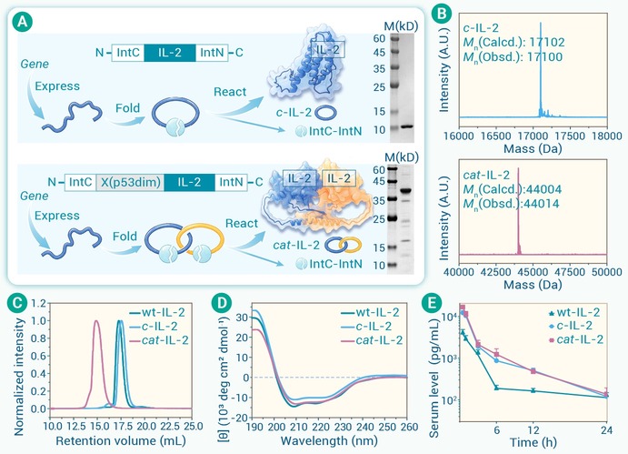
Figure 1.
Cellular synthesis and biophysical characterizations of topological IL-2 variants
-
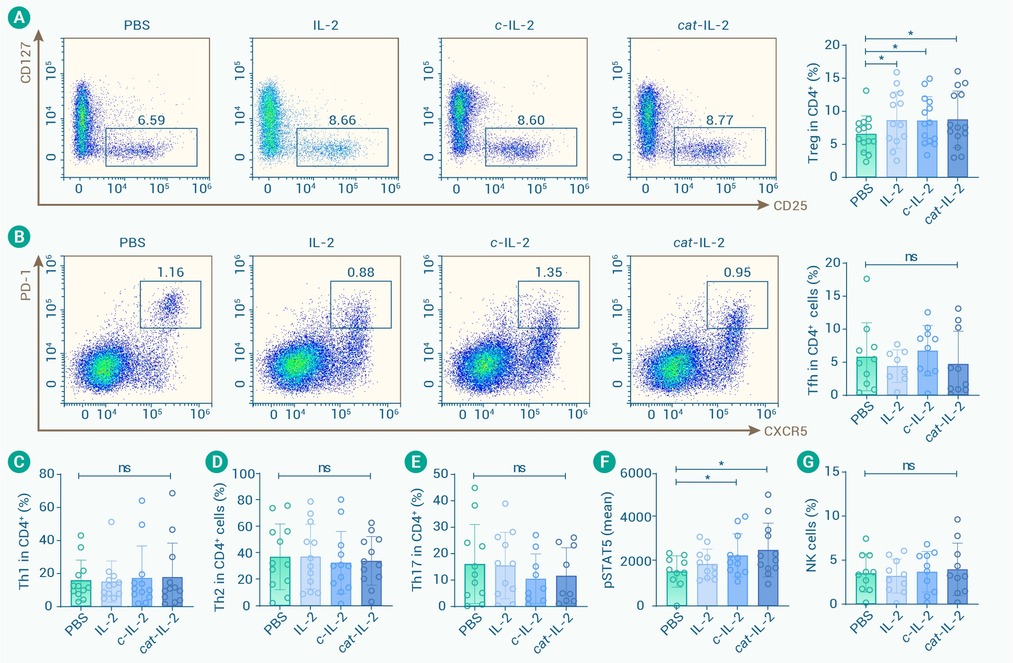
Figure 2.
Immunomodulatory effects of topological IL-2 variants on Treg and Teff cell balance in SLE
-
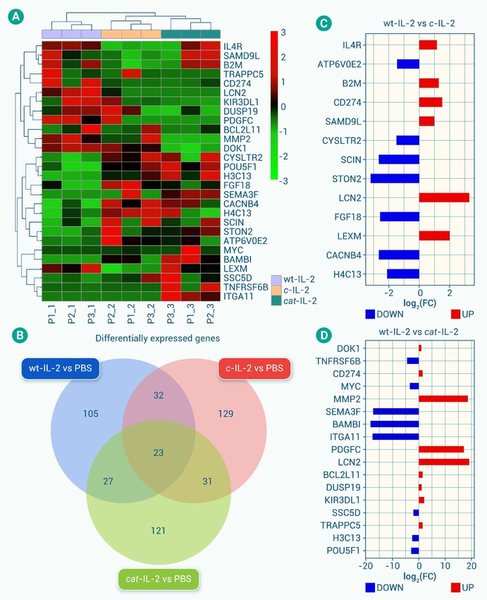
Figure 3.
Transcriptomic analysis of CD4+T cells in response to IL-2 treatment
-
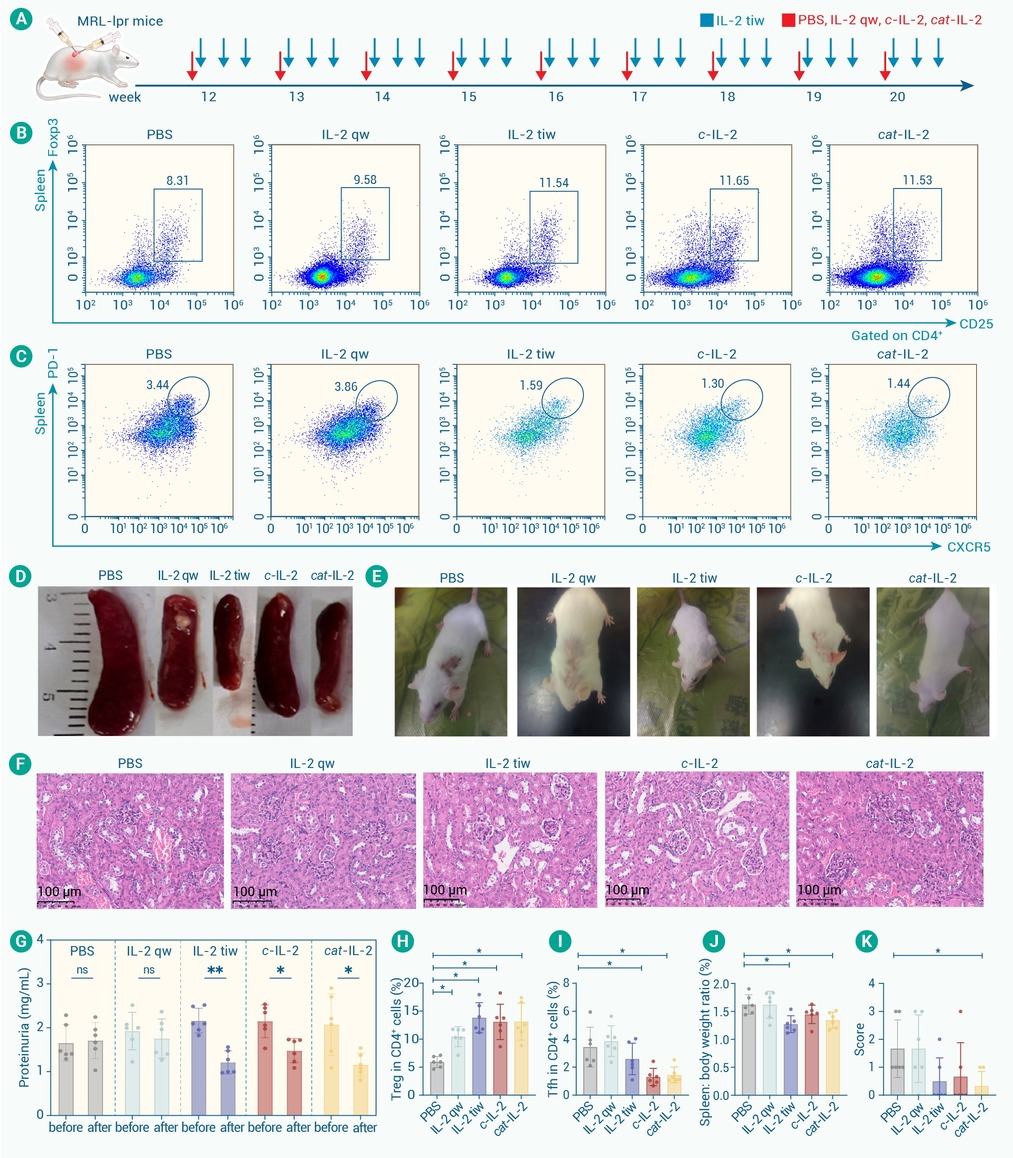
Figure 4.
Therapeutic efficacy of half-life-extended IL-2 variants in MRL/lpr lupus-prone mice
-
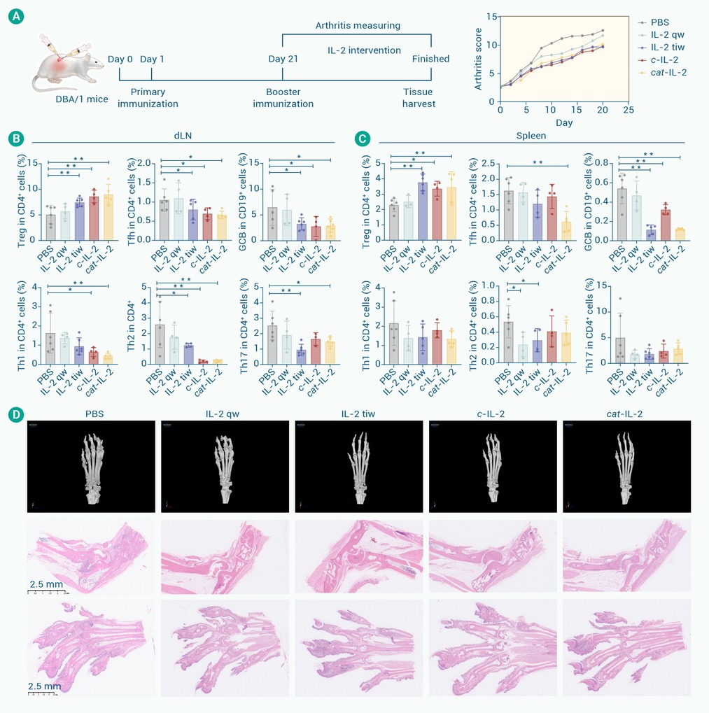
Figure 5.
Efficacy of topological IL-2 variants therapy in CIA mouse
-
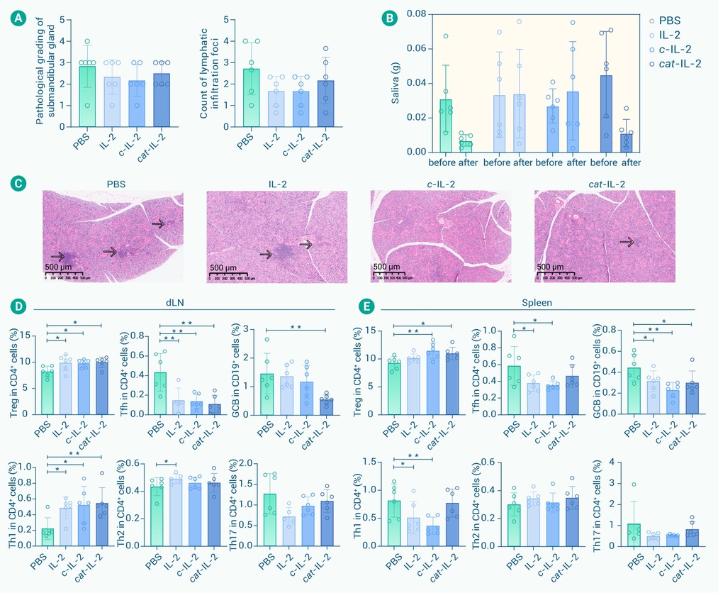
Figure 6.
Efficacy of topological IL-2 variants therapy in SS mouse model-NOD
