
PD-L1-targeted dual-modal MR/NIR-II probe for imaging and image-guided surgery of non-small cell lung cancer
-
GRAPHICAL ABSTRACT
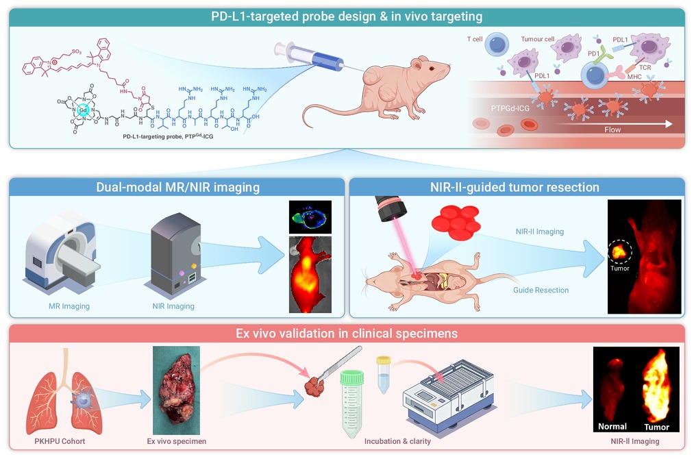
 DownLoad:
Full size image
-
PUBLIC SUMMARY
-
A PD-L1-targeted MR/NIR-II dual-modal probe integrates preoperative imaging and intraoperative navigation.
The probe shows high tumor-to-background ratios in xenograft, orthotopic, and metastatic tumor models.
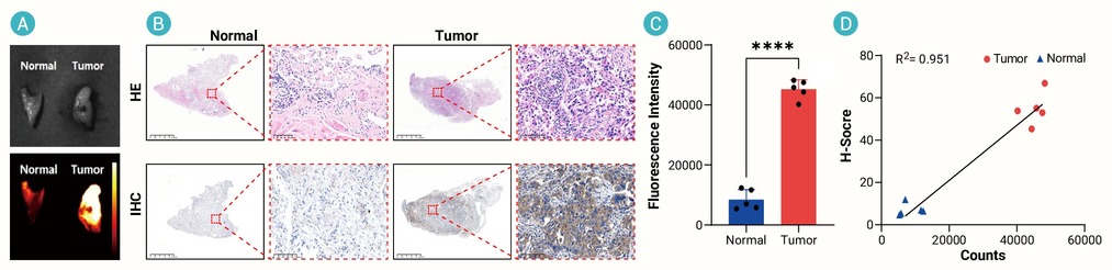
Rapid ex vivo NIR-II imaging accurately maps surgical margins and correlates with PD-L1 IHC in patients.
-
-
ABSTRACT
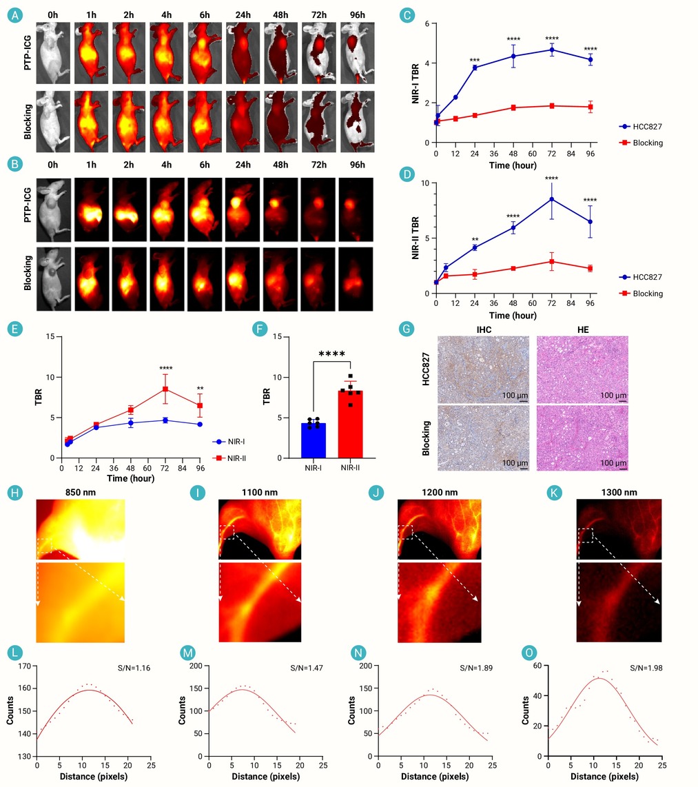
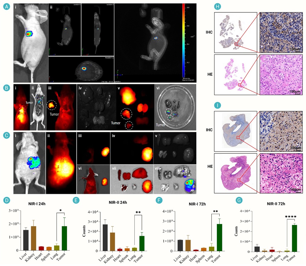
Recurrence of non-small cell lung cancer remains a critical clinical challenge. Primarily due to undetectable micrometastatic lesions and incomplete tumor resection. Current preoperative imaging modalities lack sufficient resolution to identify small lesions, while intraoperative guidance relies on visual inspection or conventional fluorescence. Accurate preoperative diagnosis and precise excision of malignant tissue remain critical factors for improving patient prognosis. Considering the differential expression of PD-L1 in NSCLC and normal lung tissue, we developed a novel imaging probe (PTPGd-ICG). This probe enabled successful MRI-based tumor targeting with high contrast in tumor-bearing mice, with the experimental group showing a 2.39-fold higher signal-to-noise ratio (SNR) than the blocking group at 0.5 h post-injection. NIR-II fluorescence imaging guided tumor removal, with the probe exhibiting sustained tumor enrichment from 4-96 h post-injection and reaching a peak tumor-to-background ratio (TBR) of 8.77 at 72 h, enabling precise tumor resection in animal models. In addition, the probe exhibited significantly higher fluorescence intensity in human NSCLC tissues than in adjacent normal tissues, facilitating the assessment of positive surgical margins ex vivo. These findings demonstrate that PD-L1 is a promising imaging target for NSCLC. This strategy may enable preoperative identification of patients with high PD-L1 expression while facilitating rapid pathological assessment and intraoperative margin delineation. -

-
REFERENCES
[1] Ren F., Fei Q., Qiu K., et al. (2024). Liquid biopsy techniques and lung cancer: Diagnosis, monitoring and evaluation. J. Exp. Clin. Cancer Res. 43:96. DOI:10.1186/s13046-024-03026-7 [2] Rakaee M., Tafavvoghi M., Ricciuti B., et al. (2025). Deep learning model for predicting immunotherapy response in advanced non-small cell lung cancer. JAMA Oncol. 11:109−118. DOI:10.1001/jamaoncol.2024.5356 [3] Xu J.Y., Zhang C., Wang X., et al. (2020). Integrative proteomic characterization of human lung adenocarcinoma. Cell 182:245−261.e217. DOI:10.1016/j.cell.2020.05.043 [4] Mariathasan S., Turley S.J., Nickles D., et al. (2018). TGFβ attenuates tumour response to PD-L1 blockade by contributing to exclusion of T cells. Nature 554:544−548. DOI:10.1038/nature25501 [5] Yang Z., Liu X., Zhu J., et al. (2025). Inhibiting intracellular CD28 in cancer cells enhances antitumor immunity and overcomes anti-PD-1 resistance via targeting PD-L1. Cancer Cell 43:86−102.e110. DOI:10.1016/j.ccell.2024.11.008 [6] Yi M., Niu M., Xu L., et al. (2021). Regulation of PD-L1 expression in the tumor microenvironment. J. Hematol. Oncol. 14:10. DOI:10.1186/s13045-020-01027-5 [7] Yi M., Jiao D., Xu H., et al. (2018). Biomarkers for predicting efficacy of PD-1/PD-L1 inhibitors. Mol. Cancer 17:129. DOI:10.1186/s12943-018-0864-3 [8] Lin E., Song M., Wang B., et al. (2025). Fibroblast activation protein peptide-targeted NIR-I/II fluorescence imaging for stable and functional detection of hepatocellular carcinoma. Eur. J. Nucl. Med. Mol. Imaging 52:2157−2170. DOI:10.1007/s00259-025-07093-6 [9] Zhou K.I., Peterson B., Serritella A., et al. (2020). Spatial and temporal heterogeneity of PD-L1 expression and tumor mutational burden in gastroesophageal adenocarcinoma at baseline diagnosis and after chemotherapy. Clin. Cancer Res. 26:6453−6463. DOI:10.1158/1078-0432.CCR-20-2085 [10] Wang Y., Chen Y., Ji D.K., et al. (2024). Bio-orthogonal click chemistry strategy for PD-L1-targeted imaging and pyroptosis-mediated chemo-immunotherapy of triple-negative breast cancer. J. Nanobiotechnol. 22:461. DOI:10.1186/s12951-024-02727-7 [11] Kim H.S., Lee K.S., Ohno Y., et al. (2015). PET/CT versus MRI for diagnosis, staging, and follow-up of lung cancer. J. Magn. Reson. Imaging 42:247−260. DOI:10.1002/jmri.24776 [12] Biederer J., Ohno Y., Hatabu H., et al. (2017). Screening for lung cancer: Does MRI have a role. Eur. J. Radiol. 86:353−360. DOI:10.1016/j.ejrad.2016.09.016 [13] Tian Y., Shen H., Li L., et al. (2024). Enhancing surgical outcomes: Accurate identification and removal of prostate cancer with B7-H3-targeted NIR-II molecular imaging. Eur. J. Nucl. Med. Mol. Imaging 51:2569−2582. DOI:10.1007/s00259-024-06714-w [14] Li C., Mi J., Wang Y., et al. (2023). New and effective EGFR-targeted fluorescence imaging technology for intraoperative rapid determination of lung cancer in freshly isolated tissue. Eur. J. Nucl. Med. Mol. Imaging 50:494−507. DOI:10.1007/s00259-022-05975-7 [15] Guo X., Li C., Jia X., et al. (2023). NIR-II fluorescence imaging-guided colorectal cancer surgery targeting CEACAM5 by a nanobody. EBioMedicine 89:104476. DOI:10.1016/j.ebiom.2023.104476 [16] Predina J.D., Newton A., Corbett C., et al. (2018). Localization of pulmonary ground-glass opacities with folate receptor-targeted intraoperative molecular imaging. J. Thorac. Oncol. 13:1028−1036. DOI:10.1016/j.jtho.2018.03.023 [17] Li J., Ma J., Wang L., et al. (2025). Application of non-invasive imaging on cell tracking of adoptive cell therapy: A systemic review. Innov. Med. 3:100137. DOI:10.59717/j.xinn-med.2025.100137 [18] Hu L., Pei Y., Wang X., et al. (2025). The frequency of core MMR gene alterations in lung cancer and their clinical characterization: A comprehensive study of over 26,000 cases. Innov. Med. 3:100163. DOI:10.59717/j.xinn-med.2025.100163 [19] Heskamp S., Hobo W., Molkenboer-Kuenen J.D., et al. (2015). Noninvasive imaging of tumor PD-L1 expression using radiolabeled anti-PD-L1 antibodies. Cancer Res. 75:2928−2936. DOI:10.1158/0008-5472.CAN-14-3477 [20] Wang L., Tang G., Hu K., et al. (2022). Comparison of (68)Ga-FAPI and (18)F-FDG PET/CT in the evaluation of advanced lung cancer. Radiology 303:191−199. DOI:10.1148/radiol.211424 [21] Herlemann A., Wenter V., Kretschmer A., et al. (2016). (68)Ga-PSMA positron emission tomography/computed tomography provides accurate staging of lymph node regions prior to lymph node dissection in patients with prostate cancer. Eur. Urol. 70:553−557. DOI:10.1016/j.eururo.2015.12.051 [22] Xia L., Liu Y., and Wang Y. (2019). PD-1/PD-L1 blockade therapy in advanced non-small-cell lung cancer: Current status and future directions. Oncologist 24:S31−S41. DOI:10.1634/theoncologist.2019-IO-S1-s05 [23] Brahmer J.R., Govindan R., Anders R.A., et al. (2018). The Society for Immunotherapy of Cancer consensus statement on immunotherapy for the treatment of non-small cell lung cancer (NSCLC). J. Immunother. Cancer 6:75. DOI:10.1186/s40425-018-0382-2 [24] Doroshow D.B., Bhalla S., Beasley M.B., et al. (2021). PD-L1 as a biomarker of response to immune-checkpoint inhibitors. Nat. Rev. Clin. Oncol. 18:345−362. DOI:10.1038/s41571-021-00473-5 [25] Kok I.C., Hooiveld J.S., van de Donk P.P., et al. (2022). (89)Zr-pembrolizumab imaging as a non-invasive approach to assess clinical response to PD-1 blockade in cancer. Ann. Oncol. 33:80−88. DOI:10.1016/j.annonc.2021.10.213 [26] Christensen C., Kristensen L.K., Alfsen M.Z., et al. (2020). Quantitative PET imaging of PD-L1 expression in xenograft and syngeneic tumour models using a site-specifically labelled PD-L1 antibody. Eur. J. Nucl. Med. Mol. Imaging 47:1302−1313. DOI:10.1007/s00259-019-04646-4 [27] Truillet C., Oh H.L.J., Yeo S.P., et al. (2018). Imaging PD-L1 expression with ImmunoPET. Bioconjug. Chem. 29:96−103. DOI:10.1021/acs.bioconjchem.7b00631 [28] Zhou X., Jiang J., Yang X., et al. (2022). First-in-humans evaluation of a PD-L1-binding peptide PET radiotracer in non-small cell lung cancer patients. J. Nucl. Med. 63:536−542. DOI:10.2967/jnumed.121.262045 -
ABOUT THIS ARTICLE
Cite this article:
Liu Q., Jia X., Li Y., et al. (2026). PD-L1-targeted dual-modal MR/NIR-II probe for imaging and image-guided surgery of non-small cell lung cancer. The Innovation Medicine 4:100251. https://doi.org/10.59717/j.xinn-med.2026.100251
Welcome!
To request copyright permission to republish or share portions of our works, please visit Copyright Clearance Center's (CCC) Marketplace website at marketplace.copyright.com.
Export File
Citation
Liu Q., Jia X., Li Y., et al. (2026). PD-L1-targeted dual-modal MR/NIR-II probe for imaging and image-guided surgery of non-small cell lung cancer. The Innovation Medicine 4:100251. https://doi.org/10.59717/j.xinn-med.2026.100251

Format
Content
-
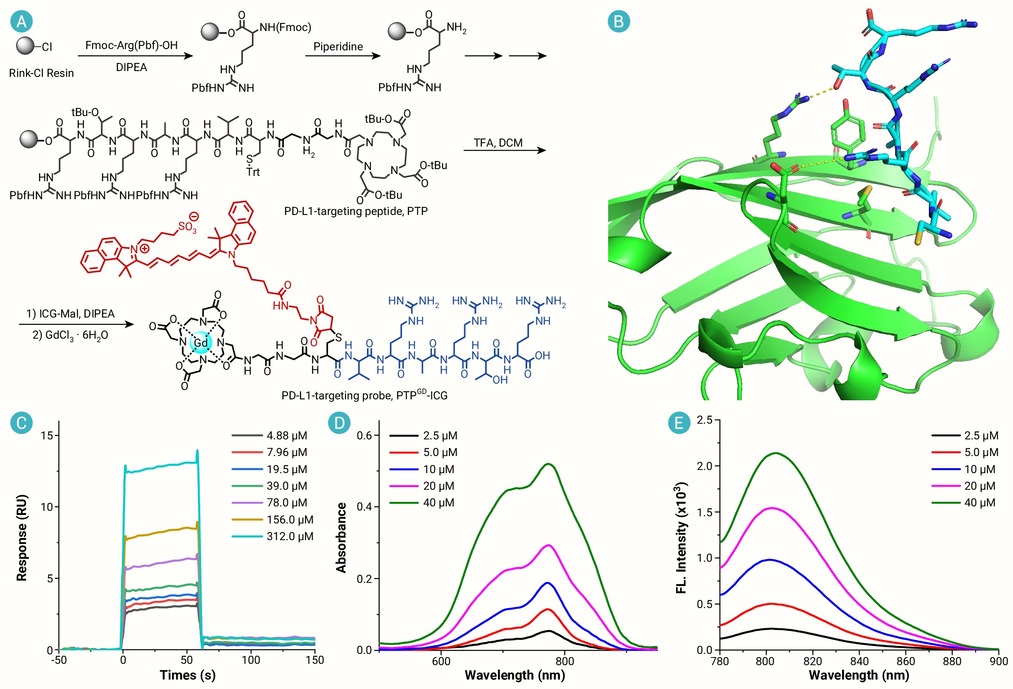
Figure 1.
Preparation and characterization of PTP and PTPGd-ICG
-
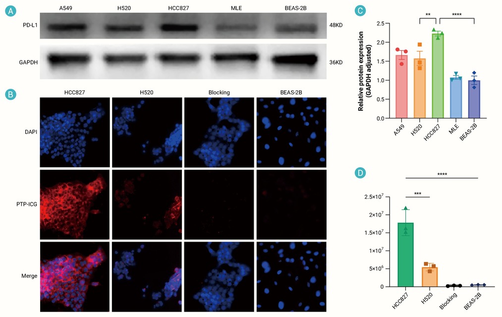
Figure 2.
Cell lines PD-L1 expression and cellular binding of PTP-ICG
-
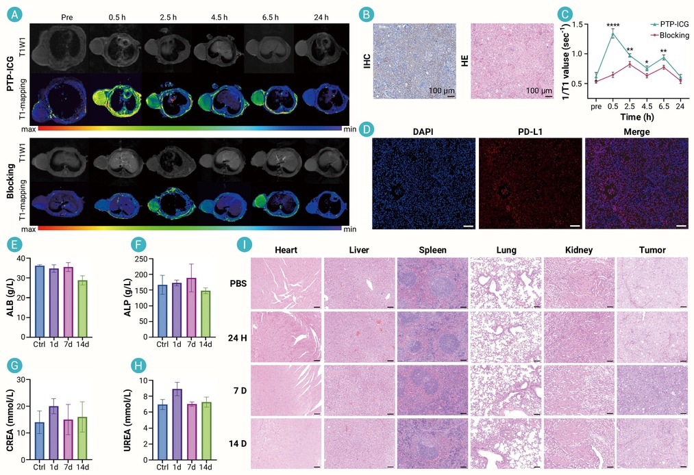
Figure 3.
In vivo MR imaging recognition of subcutaneous tumor in mice
-
Figure 4.
In vivo specificity and biodistribution of PTP-ICG
-
Figure 5.
NIR-II fluorescence imaging-guided orthotopic / metastasis lung cancer resection
-
Figure 6.
In vitro cultivation and imaging of fresh NSCLC specimens
