
Ionic-network dysregulation in the brain: Mechanisms, diseases, and therapeutic strategies
-
GRAPHICAL ABSTRACT
 DownLoad:
Full size image
-
PUBLIC SUMMARY
-
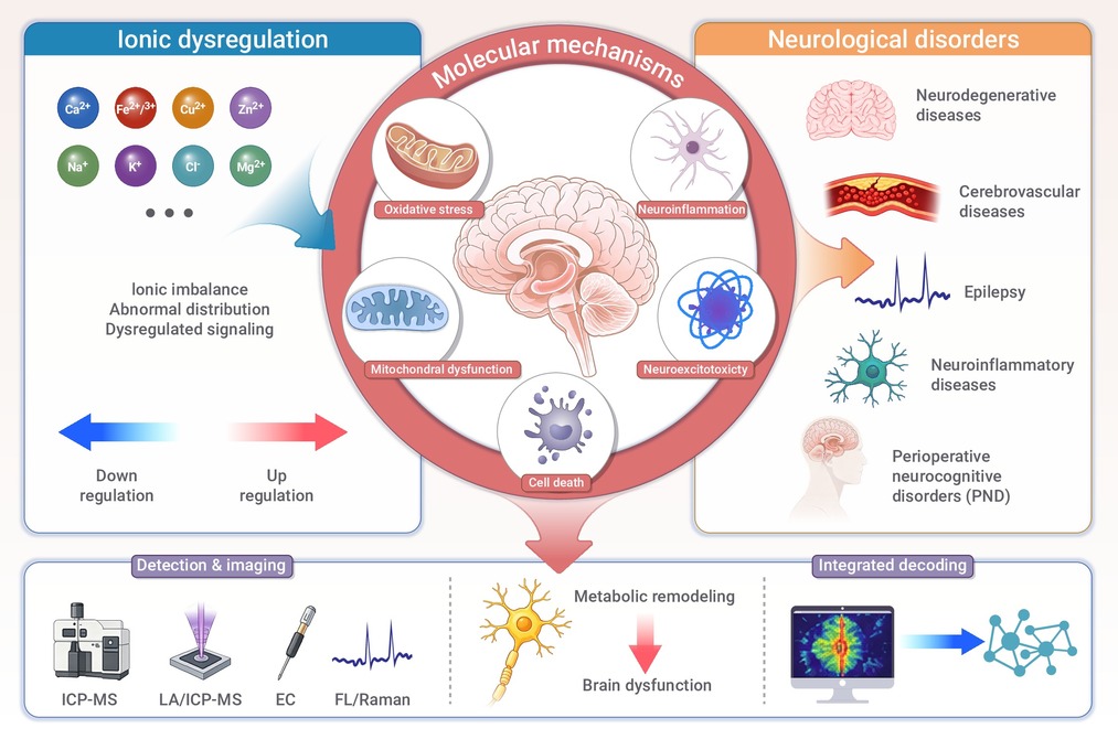
Ion imbalance is a key molecular event in many neurological disorders.
Altered Ca2+, Fe2+/3+, Cu+/2+, Zn2+, Na+, K+, Cl-, and Mg2+ disrupt brain function.
Shared ionic mechanisms link neurodegeneration, stroke, epilepsy and perioperative cognitive disorders.
Artificial intelligence may help decode multidimensional ion networks in brain diseases.
Existing therapies target parts of ionic network, but ionomics-guided strategies remain emerging frontiers.
-
-
ABSTRACT
Ionic homeostasis is a fundamental organizing principle of brain function, supporting electrical signaling, metabolic coupling, cellular communication, and neurovascular stability. Ionic-network dysregulation has emerged as an important mechanism of neurological diseases. Herein, we review the physiological architecture of brain ionic homeostasis and its dynamic organization across spatial and temporal scales. We then discuss the involvement of ionic network dysregulation in pathological cascades, including excitability imbalance, mitochondrial injury, redox stress, metal-dependent cell death, protein misfolding, neuroinflammation and barrier dysfunction and their involvement in representative neurological diseases. Furthermore, we discuss the potential role of multimodal chemical analysis and artificial intelligence in decoding ionic characteristics in neurological diseases. Finally, we review established and experimental therapies targeting ion channels, redox pathways, metal dyshomeostasis and chloride transport. This review provides an integrated framework for understanding ionic-network dysregulation as both a driver of neurological disease and a promising target for precision diagnosis, risk stratification, and mechanism-guided intervention. -

-
REFERENCES
[1] Nowak, L., Bregestovski, P., Ascher, P., et al. (1984). Magnesium gates glutamate-activated channels in mouse central neurones. Nature 307:462−465. DOI:10.1038/307462a0 [2] Hodgkin, A.L., Huxley, A.F. (1952). A quantitative description of membrane current and its application to conduction and excitation in nerve. J. Physiol. 117:500−544. DOI:10.1113/jphysiol.1952.sp004764 [3] Clapham, D.E. (2007). Calcium signaling. Cell 131:1047−1058. DOI:10.1016/j.cell.2007.11.028 [4] Kawahara, M., Kato-Negishi, M., Tanaka, K.-I. (2023). Dietary Trace Elements and the Pathogenesis of Neurodegenerative Diseases. Nutrients 15:2067. DOI:10.3390/nu15092067 [5] Abbott, N.J., Pizzo, M.E., Preston, J.E., et al. (2018). The role of brain barriers in fluid movement in the CNS: is there a ‘glymphatic’ system. Acta. Neuropathol. 135:387−407. DOI:10.1007/s00401-018-1812-4 [6] Calvo-Rodriguez, M., Hou, S.S., Snyder, A.C., et al. (2020). Increased mitochondrial calcium levels associated with neuronal death in a mouse model of Alzheimer’s disease. Nat. Commun. 11:2146. DOI:10.1038/s41467-020-16074-2 [7] Liu, W., Xue, Y., Cao, C., et al. (2026). Copper Homeostasis and Cuproptosis in Neurological Disorders. DDDT 20:1−32. DOI:10.2147/DDDT.S580005 [8] Benarroch, E. (2023). What Are the Functions of Zinc in the Nervous System. Neurology 101:714−720. DOI:10.1212/WNL.0000000000207912 [9] Hare, D.J., New, E.J., Jonge, M.D. de, et al. (2015). Imaging metals in biology: balancing sensitivity, selectivity and spatial resolution. Chem. Soc. Rev. 44:5941−5958. DOI:10.1039/c5cs00055f [10] Grover, K., Koblova, A., Pezacki, A.T., et al. (2024). Small-Molecule Fluorescent Probes for Binding- and Activity-Based Sensing of Redox-Active Biological Metals. Chem Rev 124:5846−5929. DOI:10.1021/acs.chemrev.3c00819 [11] Fang, T., Chen, W., Cheng, Y., et al. (2022). LA-ICP-MS bioimaging demonstrated disturbance of metal ions in the brain of Parkinson’s disease model mouse undergoing manganese-enhanced MRI. Anal. Bioanal. Chem. 414:5561−5571. DOI:10.1007/s00216-022-03994-9 [12] Kaila, K., Price, T.J., Payne, J.A., et al. (2014). Cation-chloride cotransporters in neuronal development, plasticity and disease. Nat. Rev. Neurosci. 15:637−654. DOI:10.1038/nrn3819 [13] Garza-Lombó, C., Posadas, Y., Quintanar, L., et al. (2018). Neurotoxicity Linked to Dysfunctional Metal Ion Homeostasis and Xenobiotic Metal Exposure: Redox Signaling and Oxidative Stress. Antioxid. Redox Signal. 28:1669−1703. DOI:10.1089/ars.2017.7272 [14] Belosludtsev, K.N., Dubinin, M.V., Belosludtseva, N.V. (2025). Ca2+-Dependent Mitochondrial Permeability Transition Pore: Structure, Properties, and Role in Cellular Pathophysiology. Biochem. Mosc. 90:1789−1810. DOI:10.1134/S0006297925602369 [15] Cheli, V.T., Correale, J., Paez, P.M., et al. (2020). Iron Metabolism in Oligodendrocytes and Astrocytes, Implications for Myelination and Remyelination. ASN Neuro. 12:1759091420962681 DOI:10.1177/1759091420962681. [16] Gale, J., Aizenman, E. (2024). The physiological and pathophysiological roles of copper in the nervous system. Eur J Neurosci 60:3505−3543. DOI:10.1111/ejn.16370 [17] Giorgi, C., Marchi, S., Pinton, P. (2018). The machineries, regulation and cellular functions of mitochondrial calcium. Nat. Rev. Mol. Cell. Biol. 19:713−730. DOI:10.1038/s41580-018-0052-8 [18] Boyman, L., Karbowski, M., Lederer, W.J. (2020). Regulation of Mitochondrial ATP Production: Ca2+ Signaling and Quality Control. Trends. Mol. Med. 26:21−39. DOI:10.1016/j.molmed.2019.10.007 [19] Markovinovic, A., Greig, J., Martín-Guerrero, S.M., et al. (2022). Endoplasmic reticulum-mitochondria signaling in neurons and neurodegenerative diseases. J. Cell Sci. 135:jcs248534. DOI:10.1242/jcs.248534 [20] Park, S.J., Lee, S.B., Suh, Y., et al. (2017). DISC1 Modulates Neuronal Stress Responses by Gate-Keeping ER-Mitochondria Ca2+ Transfer through the MAM. Cell Rep. 21:2748−2759. DOI:10.1016/j.celrep.2017.11.043 [21] Peng, W., Wong, Y.C., Krainc, D. (2020). Mitochondria-lysosome contacts regulate mitochondrial Ca2+ dynamics via lysosomal TRPML1. Proc. Natl. Acad. Sci. U.S.A. 117:19266−19275. DOI:10.1073/pnas.2003236117 [22] Lénárt, N., Cserép, C., Császár, E., et al. (2024). Microglia–neuron–vascular interactions in ischemia. Glia 72:833−856. DOI:10.1002/glia.24487 [23] Górska, A., Markiewicz-Gospodarek, A., Markiewicz, R., et al. (2023). Distribution of Iron, Copper, Zinc and Cadmium in Glia, Their Influence on Glial Cells and Relationship with Neurodegenerative Diseases. Brain Sciences 13:911. DOI:10.3390/brainsci13060911 [24] Catterall, W.A. (2023). Voltage gated sodium and calcium channels: Discovery, structure, function, and Pharmacology. Channels 17:2281714. DOI:10.1080/19336950.2023.2281714 [25] Chen, L., Shen, Q., Liu, Y., et al. (2025). Homeostasis and metabolism of iron and other metal ions in neurodegenerative diseases. Sig. Transduct. Target. Ther. 10:31. DOI:10.1038/s41392-024-02071-0 [26] Verkhratsky, A., Hansen, L.M.B., Staehr, C., et al. (2026). The astrocytic engine: Na+,K+-ATPase at the nexus of brain function and malfunction. Am. J. Physiol. Cell Physiol 330:C238−C251. DOI:10.1152/ajpcell.00814.2025 [27] Verkhratsky, A., Untiet, V., Rose, C.R. (2020). Ionic signalling in astroglia beyond calcium. J. Physiol. 598:1655−1670. DOI:10.1113/JP277478 [28] Murphy-Royal, C., Ching, S., Papouin, T. (2023). A conceptual framework for astrocyte function. Nat. Neurosci. 26:1848−1856. DOI:10.1038/s41593-023-01448-8 [29] Tang, M., Fleming, E., Gu, J., et al. (2026). Trace Elements Dyshomeostasis and Toxic Metals Neurotoxicity in Neurodegenerative Diseases. Biol. Trace. Elem. Res. 204:7264–7299 DOI:10.1007/s12011-026-05187-2. [30] Djukic, B., Casper, K.B., Philpot, B.D., et al. (2007). Conditional knock-out of Kir4.1 leads to glial membrane depolarization, inhibition of potassium and glutamate uptake, and enhanced short-term synaptic potentiation. J. Neurosci. 27:11354–11365 DOI:10.1523/JNEUROSCI.0723-07.2007. [31] Surmeier, D.J., Obeso, J.A., Halliday, G.M. (2017). Selective neuronal vulnerability in Parkinson disease. Nat. Rev. Neurosci. 18:101−113. DOI:10.1038/nrn.2016.178 [32] Segarra, M., Aburto, M.R., Acker-Palmer, A. (2021). Blood–Brain Barrier Dynamics to Maintain Brain Homeostasis. Trends Neurosci. 44:393−405. DOI:10.1016/j.tins.2020.12.002 [33] Institoris, A., Vandal, M., Peringod, G., et al. (2022). Astrocytes amplify neurovascular coupling to sustained activation of neocortex in awake mice. Nat. Commun. 13:7872. DOI:10.1038/s41467-022-35383-2 [34] Liu, R., Collier, J.M., Abdul-Rahman, N.-H., et al. (2024). Dysregulation of Ion Channels and Transporters and Blood-Brain Barrier Dysfunction in Alzheimer’s Disease and Vascular Dementia. Aging Dis. 15:1748−1770. DOI:10.14336/AD.2023.1201 [35] Huang, X., Qi, J., Su, Y., et al. (2024). Endothelial DR6 in blood-brain barrier malfunction in Alzheimer’s disease. Cell Death Dis. 15:258. DOI:10.1038/s41419-024-06639-0 [36] Korte, N., Barkaway, A., Wells, J., et al. (2024). Inhibiting Ca2+ channels in Alzheimer’s disease model mice relaxes pericytes, improves cerebral blood flow and reduces immune cell stalling and hypoxia. Nat. Neurosci. 27:2086−2100. DOI:10.1038/s41593-024-01753-w [37] Cressman, J.R., Ullah, G., Ziburkus, J., et al. (2009). The influence of sodium and potassium dynamics on excitability, seizures, and the stability of persistent states: I. Single neuron dynamics. J. Comput. Neurosci. 26:159−170. DOI:10.1007/s10827-008-0132-4 [38] Hadi, F., Mortaja, M., Hadi, Z. (2024). Calcium (Ca2+) hemostasis, mitochondria, autophagy, and mitophagy contribute to Alzheimer’s disease as early moderators. Cell Biochem. Funct. 42:e4085. DOI:10.1002/cbf.4085 [39] Jin, S., Tian, Y., Hacker, J., et al. (2024). Inflammatory cytokines disrupt astrocyte exosomal HepaCAM-mediated protection against neuronal excitotoxicity in the SOD1G93A ALS model. Sci. Adv. 10:eadq3350. DOI:10.1126/sciadv.adq3350 [40] Wu, W., Gong, X., Qin, Z., et al. (2025). Molecular mechanisms of excitotoxicity and their relevance to the pathogenesis of neurodegenerative diseases—an update. Acta. Pharmacol. Sin. 46:3129−3142. DOI:10.1038/s41401-025-01576-w [41] Zhu, M., Sun, H., Cao, L., et al. (2022). Role of Na+/K+-ATPase in ischemic stroke: in-depth perspectives from physiology to pharmacology. J. Mol. Med. (Berl) 100:395−410. DOI:10.1007/s00109-021-02143-6 [42] Zhao, J., Sun, J., Zheng, Y., et al. (2022). Activated astrocytes attenuate neocortical seizures in rodent models through driving Na+-K+-ATPase. Nat. Commun. 13:7136. DOI:10.1038/s41467-022-34662-2 [43] Côme, E., Marques, X., Poncer, J.C., et al. (2020). KCC2 membrane diffusion tunes neuronal chloride homeostasis. Neuropharmacology 169:107571. DOI:10.1016/j.neuropharm.2019.03.014 [44] Adrichem, J.J. van, Loo, R.J. van der, Ambrosini Defendi, R., et al. (2025). Progressive remote memory decline coincides with parvalbumin interneuron hyperexcitability and enhanced inhibition of cortical engram cells in a mouse model of Alzheimer’s disease. eLife 14:RP106866. DOI:10.7554/eLife.106866 [45] Best, F.V., Hartings, J.A., Ngwenya, L.B. (2025). Repetitive cortical spreading depolarizations are prolonged early after experimental traumatic brain injury. Exp. Neurol. 385:115120. DOI:10.1016/j.expneurol.2024.115120 [46] Zhang, W., Xiong, B.R., Zhang, L.Q., et al. (2020). Disruption of the GABAergic system contributes to the development of perioperative neurocognitive disorders after anesthesia and surgery in aged mice. CNS Neurosci. Ther. 26:913−924. DOI:10.1111/cns.13388 [47] Calvo-Rodriguez, M., Kharitonova, E.K., Snyder, A.C., et al. (2024). Real-time imaging of mitochondrial redox reveals increased mitochondrial oxidative stress associated with amyloid β aggregates in vivo in a mouse model of Alzheimer’s disease. Mol. Neurodegeneration 19:6. DOI:10.1186/s13024-024-00702-2 [48] Feofilaktova, T., Kushnireva, L., Segal, M., et al. (2025). Calcium signaling in postsynaptic mitochondria: mechanisms, dynamics, and role in ATP production. Front. Mol. Neurosci. 18:1621070. DOI:10.3389/fnmol.2025.1621070. [49] Jomova, K., Alomar, S.Y., Valko, R., et al. (2025). The role of redox-active iron, copper, manganese, and redox-inactive zinc in toxicity, oxidative stress, and human diseases. EXCLI J 24:880−954. DOI:10.17179/excli2025-8449 [50] Ji, S.G., Weiss, J.H. (2018). Zn2+-induced disruption of neuronal mitochondrial function: synergism with Ca2+, critical dependence upon cytosolic Zn2+ buffering, and contributions to neuronal injury. Exp. Neurol. 302:181−195. DOI:10.1016/j.expneurol.2018.01.012 [51] Ru, Q., Li, Y., Chen, L., et al. (2024). Iron homeostasis and ferroptosis in human diseases: mechanisms and therapeutic prospects. Signal Transduction Targeted Ther. 9:271. DOI:10.1038/s41392-024-01969-z [52] Feinsod, H., Stockwell, B.R. (2026). Fundamental mechanism of ferroptosis: Three unanswered questions. Ferroptosis Oxid Stress 2:202512. DOI:10.70401/fos.2026.0015 [53] Bersuker, K., Hendricks, J.M., Li, Z., et al. (2019). The CoQ oxidoreductase FSP1 acts parallel to GPX4 to inhibit ferroptosis. Nature 575:688−692. DOI:10.1038/s41586-019-1705-2 [54] Mao, C., Liu, X., Zhang, Y., et al. (2021). DHODH-mediated ferroptosis defense is a targetable vulnerability in cancer. Nature 593:586−590. DOI:10.1038/s41586-021-03539-7 [55] Li, Q., Han, X., Lan, X., et al. (2017). Inhibition of neuronal ferroptosis protects hemorrhagic brain. JCI Insight 2:e90777. DOI:10.1172/jci.insight.90777. [56] Levi, S., Ripamonti, M., Moro, A.S., et al. (2024). Iron imbalance in neurodegeneration. Mol. Psychiatry 29:1139−1152. DOI:10.1038/s41380-023-02399-z [57] Miao, M., Han, Y., Wang, Y., et al. (2024). Dysregulation of iron homeostasis and ferroptosis in sevoflurane and isoflurane associated perioperative neurocognitive disorders. CNS Neurosci. Ther. 30:e14553. DOI:10.1111/cns.14553 [58] Chen, L., Min, J., Wang, F. (2022). Copper homeostasis and cuproptosis in health and disease. Signal Transduct Target Ther 7:378. DOI:10.1038/s41392-022-01229-y [59] Tsvetkov, P., Coy, S., Petrova, B., et al. (2022). Copper induces cell death by targeting lipoylated TCA cycle proteins. Science 375:1254−1261. DOI:10.1126/science.abf0529 [60] Li, Q., Li, D., Li, Y., et al. (2025). Investigating cuproptosis and mitochondrial dysfunction in brain cells: uncovering novel mechanisms and biomarkers for Parkinson’s disease. Metab Brain Dis 40:144. DOI:10.1007/s11011-025-01574-1 [61] Hansson, O. (2021). Biomarkers for neurodegenerative diseases. Nat. Med. 27:954−963. DOI:10.1038/s41591-021-01382-x [62] Juárez-Romero, G.U., Sun, X., Gerez, J.A., et al. (2026). Structural insights into copper and zinc binding to tau protein and the impact of metal binding on amyloid aggregation. Chem. Sci. 17:7277−7296. DOI:10.1039/d5sc08604c [63] Hosseinpour Mashkani, S.M., Bishop, D.P., Raoufi-Rad, N., et al. (2023). Distribution of Copper, Iron, and Zinc in the Retina, Hippocampus, and Cortex of the Transgenic APP/PS1 Mouse Model of Alzheimer’s Disease. Cells 12:1144. DOI:10.3390/cells12081144 [64] Mao, Y., Zhang, L., Zhang, C., et al. (2025). The close relationship between trace elements (Cu, Fe, Zn, Se, Rb, Si, Cr, and V) and Alzheimer’s disease: Research progress and insights. J. Trace Elem. Med. Biol. 90:127692. DOI:10.1016/j.jtemb.2025.127692 [65] Fani, G., Mannini, B., Vecchi, G., et al. (2021). Aβ Oligomers Dysregulate Calcium Homeostasis by Mechanosensitive Activation of AMPA and NMDA Receptors. ACS Chem. Neurosci. 12:766−781. DOI:10.1021/acschemneuro.0c00811 [66] Cosson, T., Faller, P., Pountney, D.L. (2025). Toxic Tango in Parkinson’s Disease: A dance between αSyn and iron import, export and speciation. Neurochem. Int. 190:106053. DOI:10.1016/j.neuint.2025.106053 [67] Castillo-Casaña, Y., Arias, C., Coria, R. (2025). Tau–Mitochondria Interactions in Neurodegeneration: Mechanisms and Therapeutic Potential. Cell Mol Neurobiol 46:3. DOI:10.1007/s10571-025-01634-1 [68] Gonzalez-Garcia, M., Fusco, G., De Simone, A. (2023). Metal interactions of α-synuclein probed by NMR amide-proton exchange. Front. Chem. 11:1167766 DOI:10.3389/fchem.2023.1167766 [69] Eren-Koçak, E., Dalkara, T. (2021). Ion Channel Dysfunction and Neuroinflammation in Migraine and Depression. Front. Pharmacol. 12:777607. DOI:10.3389/fphar.2021.777607 [70] Zimmermann, S., Mathew, A., Bondareva, O., et al. (2024). Chronic kidney disease leads to microglial potassium efflux and inflammasome activation in the brain. Kidney Int. 106:1101−1116. DOI:10.1016/j.kint.2024.06.028 [71] Fomina, A.F., Nguyen, H.M., Wulff, H. (2021). Kv1.3 inhibition attenuates neuroinflammation through disruption of microglial calcium signaling. Channels 15:67–78 DOI:10.1080/19336950.2020.1853943. [72] Sarkar, S., Nguyen, H.M., Malovic, E., et al. (2020). Kv1.3 modulates neuroinflammation and neurodegeneration in Parkinson’s disease. J. Clin. Invest. 130:4195–4212 DOI:10.1172/JCI136174. [73] Lee, G.-S., Subramanian, N., Kim, A.I., et al. (2012). The calcium-sensing receptor regulates the NLRP3 inflammasome through Ca2+ and cAMP. Nature 492:123−127. DOI:10.1038/nature11588 [74] Galic, M.A., Riazi, K., Pittman, Q.J. (2012). Cytokines and brain excitability. Front. Neuroendocrinol. 33:116−125. DOI:10.1016/j.yfrne.2011.12.002 [75] Kim, H.Y., Kim, S., Akaydin, A.N., et al. (2026). The rise of astrocytes: are they guardians or troublemakers of the brain disorder. Exp. Mol. Med. 58:301−318. DOI:10.1038/s12276-025-01627-6 [76] Yauger, Y.J., Bermudez, S., Moritz, K.E., et al. (2019). Iron accentuated reactive oxygen species release by NADPH oxidase in activated microglia contributes to oxidative stress in vitro. J. Neuroinflammation 16:41. DOI:10.1186/s12974-019-1430-7 [77] Hou, Y., Dan, X., Babbar, M., et al. (2019). Ageing as a risk factor for neurodegenerative disease. Nat. Rev. Neurol. 15:565−581. DOI:10.1038/s41582-019-0244-7 [78] Mao, L., Wang, L., Huang, Z., et al. (2025). Perioperative neurocognitive disorders: Advances in molecular mechanisms and bioactive molecules. Ageing Res. Rev. 112:102885. DOI:10.1016/j.arr.2025.102885 [79] Badenes, R., Romero-Garcia, N., Marshall, T.J. (2026). The perioperative brain: navigating neurological risks in non-neurosurgical patients. Intensive Care Med:1–4 DOI:10.1007/s00134-026-08299-4. [80] Wu, D., Chen, Q., Chen, X., et al. (2023). The blood–brain barrier: Structure, regulation and drug delivery. Signal Transduct Target Ther. 8:217. DOI:10.1038/s41392-023-01481-w [81] Andjelkovic, A.V., Situ, M., Citalan-Madrid, A.F., et al. (2023). Blood-Brain Barrier Dysfunction in Normal Aging and Neurodegeneration: Mechanisms, Impact, and Treatments. Stroke 54:661−672. DOI:10.1161/STROKEAHA.122.040578 [82] Zille, M., Karuppagounder, S.S., Chen, Y., et al. (2017). Neuronal Death After Hemorrhagic Stroke In Vitro and In Vivo Shares Features of Ferroptosis and Necroptosis. Stroke 48:1033−1043. DOI:10.1161/STROKEAHA.116.015609 [83] Bayır, H., Anthonymuthu, T.S., Tyurina, Y.Y., et al. (2020). Achieving Life through Death: Redox Biology of Lipid Peroxidation in Ferroptosis. Cell Chem. Biol. 27:387−408. DOI:10.1016/j.chembiol.2020.03.014 [84] Gruenbaum, B.F., Zlotnik, A., Dubilet, M., et al. (2026). Role of Blood–Brain Barrier Permeability in Delirium Pathophysiology and Treatment. Anesthesiology 144:203. DOI:10.1097/ALN.0000000000005722 [85] Uchida, Y., Kan, H., Sakurai, K., et al. (2020). Iron leakage owing to blood-brain barrier disruption in small vessel disease CADASIL. Neurology 95:e1188−e1198. DOI:10.1212/WNL.0000000000010148 [86] Hu, S., Fei, Y., Jin, C., et al. (2024). Ginsenoside Rd enhances blood-brain barrier integrity after cerebral ischemia/reperfusion by alleviating endothelial cells ferroptosis via activation of NRG1/ErbB4-mediated PI3K/Akt/mTOR signaling pathway. Neuropharmacology 251:109929. DOI:10.1016/j.neuropharm.2024.109929 [87] Aron, L., Ngian, Z.K., Qiu, C., et al. (2025). Lithium deficiency and the onset of Alzheimer’s disease. Nature 645:712−721. DOI:10.1038/s41586-025-09335-x [88] Deng, P., Silva, M., Yang, N., et al. (2025). Artemisinin inhibits neuronal ferroptosis in Alzheimer’s disease models by targeting KEAP1. Acta Pharmacol. Sin. 46:326−337. DOI:10.1038/s41401-024-01378-6 [89] Guan, X., Lancione, M., Ayton, S., et al. (2024). Neuroimaging of Parkinson’s disease by quantitative susceptibility mapping. Neuroimage 289:120547. DOI:10.1016/j.neuroimage.2024.120547 [90] López-Aguirre, M., Balzano, T., Monje, M.H.G., et al. (2025). Nigrostriatal iron accumulation in the progression of Parkinson’s disease. npj Parkinsons Dis. 11:72. DOI:10.1038/s41531-025-00911-6 [91] Ryan, S.K., Zelic, M., Han, Y., et al. (2023). Microglia ferroptosis is regulated by SEC24B and contributes to neurodegeneration. Nat. Neurosci. 26:12−26. DOI:10.1038/s41593-022-01221-3 [92] Peikon, I., Andrews, N.C. (2026). Isn’t it ironic? Functional iron deficiency at the core of Parkinson’s disease pathobiology. J Clin Invest 136 :e202244. DOI:10.1172/JCI202244. [93] Yao, Z., Jiao, Q., Du, X., et al. (2024). Ferroptosis in Parkinson’s disease -- The iron-related degenerative disease. Ageing Res. Rev. 101:102477 DOI:10.1016/j.arr.2024.102477 [94] Sirabella, R., Valsecchi, V., Anzilotti, S., et al. (2018). Ionic Homeostasis Maintenance in ALS: Focus on New Therapeutic Targets. Front. Neurosci. 12:510. DOI:10.3389/fnins.2018.00510. [95] LoRusso, E., Hickman, J.J., Guo, X. (2019). Ion channel dysfunction and altered motoneuron excitability in ALS. Neurol. Disord. Epilepsy J. 3:124 [96] Wang, H., Sun, Q., Du, R., et al. (2026). Serum Trace Elements and Their Associations with Disease Progression and Survival in Sporadic Amyotrophic Lateral Sclerosis: Insights from a Chinese Cohort. Biomed. Environ. Sci. 39:183−191. DOI:10.3967/bes2025.094 [97] Benatar, M., Robertson, J., Andersen, P.M. (2025). Amyotrophic lateral sclerosis caused by SOD1 variants: from genetic discovery to disease prevention. The Lancet Neurology 24:77−86. DOI:10.1016/S1474-4422(24)00479-4 [98] Witte, M.E., Schumacher, A.-M., Mahler, C.F., et al. (2019). Calcium Influx through Plasma-Membrane Nanoruptures Drives Axon Degeneration in a Model of Multiple Sclerosis. Neuron 101:615−624.e5. DOI:10.1016/j.neuron.2018.12.023 [99] Hu, H.-J., Song, M. (2017). Disrupted Ionic Homeostasis in Ischemic Stroke and New Therapeutic Targets. J. Stroke Cerebrovasc. Dis. 26:2706−2719. DOI:10.1016/j.jstrokecerebrovasdis.2017.09.011 [100] Lindquist, B.E. (2024). Spreading depolarizations pose critical energy challenges in acute brain injury. J. Neurochem. 168:868−887. DOI:10.1111/jnc.15966 [101] Schoknecht, K., Baeza-Lehnert, F., Hirrlinger, J., et al. (2025). Spreading depolarizations exhaust neuronal ATP in a model of cerebral ischemia. Proc. Natl. Acad. Sci. U.S.A. 122:e2415358122. DOI:10.1073/pnas.2415358122 [102] Cao, W., Xu, Y. (2025). Nimodipine Blocks Histone-Induced Calcium Overload to Protect Neurons after Traumatic Brain Injury. Antioxid. Redox Sign. 43:869−885. DOI:10.1177/15230864251376030 [103] Zhang, X., Zhang, Y., Su, Q., et al. (2024). Ion Channel Dysregulation Following Intracerebral Hemorrhage. Neurosci. Bull. 40:401−414. DOI:10.1007/s12264-023-01118-6 [104] Liu, J., Pang, S.-Y., Zhou, S.-Y., et al. (2024). Lipocalin-2 aggravates blood-brain barrier dysfunction after intravenous thrombolysis by promoting endothelial cell ferroptosis via regulating the HMGB1/Nrf2/HO-1 pathway. Redox Biol. 76:103342. DOI:10.1016/j.redox.2024.103342 [105] Khanna, A., Kahle, K.T., Walcott, B.P., et al. (2014). Disruption of ion homeostasis in the neurogliovascular unit underlies the pathogenesis of ischemic cerebral edema. Transl. Stroke Res. 5:3−16. DOI:10.1007/s12975-013-0307-9 [106] Zhang, Y., Jiang, M., Wei, M., et al. (2025). MgSO4 as a novel hypothermia infusion solution promotes ischemic stroke recovery through Ca2+ regulation of neurovascular units. Theranostics 15:1896−1913. DOI:10.7150/thno.104879 [107] Yang, Q., Wei, X., Deng, B., et al. (2022). Cerebral small vessel disease alters neurovascular unit regulation of microcirculation integrity involved in vascular cognitive impairment. Neurobiol. Dis. 170:105750. DOI:10.1016/j.nbd.2022.105750 [108] Debanne, D., Mylonaki, K., Musella, M.L., et al. (2024). Voltage-gated ion channels in epilepsies: circuit dysfunctions and treatments. Trends Pharmacol. Sci 45:1018−1032. DOI:10.1016/j.tips.2024.09.004 [109] Oyrer, J., Maljevic, S., Scheffer, I.E., et al. (2018). Ion Channels in Genetic Epilepsy: From Genes and Mechanisms to Disease-Targeted Therapies. Pharmacol. Rev. 70:142−173. DOI:10.1124/pr.117.014456 [110] McMoneagle, E., Zhou, J., Zhang, S., et al. (2024). Neuronal K+-Cl- cotransporter KCC2 as a promising drug target for epilepsy treatment. Acta Pharmacol. Sin. 45:1−22. DOI:10.1038/s41401-023-01149-9 [111] Çarçak, N., Onat, F., Sitnikova, E. (2023). Astrocytes as a target for therapeutic strategies in epilepsy: current insights. Front. Mol. Neurosci. 16:1183775. DOI:10.3389/fnmol.2023.1183775 [112] Du, M., Li, J., Chen, L., et al. (2018). Astrocytic Kir4.1 channels and gap junctions account for spontaneous epileptic seizure. PLoS Comput. Biol. 14:e1005877 DOI:10.1371/journal.pcbi.1005877. [113] Tyurikova, O., Kopach, O., Zheng, K., et al. (2025). Astrocyte Kir4.1 expression level territorially controls excitatory transmission in the brain. Cell Rep. 44:115299 DOI:10.1016/j.celrep.2025.115299 [114] Chen, Z.-P., Zhao, X., Wang, S., et al. (2025). GABA-dependent microglial elimination of inhibitory synapses underlies neuronal hyperexcitability in epilepsy. Nat. Neurosci. 28:1404−1417. DOI:10.1038/s41593-025-01979-2 [115] Ji, S.G., Weiss, J.H. (2018). Zn2+-induced disruption of neuronal mitochondrial function: synergism with Ca2+, critical dependence upon cytosolic Zn2+ buffering, and contributions to neuronal injury. Exp. Neurol. 302:181−195. DOI:10.1016/j.expneurol.2018.01.012 [116] Reiffurth, C., Berndt, N., Gonzalez Lopez, A., et al. (2023). Deep Isoflurane Anesthesia Is Associated with Alterations in Ion Homeostasis and Specific Na+/K+-ATPase Impairment in the Rat Brain. Anesthesiology 138:611−623. DOI:10.1097/ALN.0000000000004553 [117] Mattson, M.P., Arumugam, T.V. (2018). Hallmarks of Brain Aging: Adaptive and Pathological Modification by Metabolic States. Cell Metab. 27:1176−1199. DOI:10.1016/j.cmet.2018.05.011 [118] Cong P., Chen Q., Wu Q., et al. (2025). Aging and perioperative brain health: Mechanisms, management, and future. Chin. Med. J. 138:2381. DOI:10.1097/CM9.0000000000003735 [119] Chen, M., Yan, R., Ding, L., et al. (2023). Research Advances of Mitochondrial Dysfunction in Perioperative Neurocognitive Disorders. Neurochem. Res. 48:2983−2995. DOI:10.1007/s11064-023-03962-4 [120] Huo, Q., Zhang, Y., Guo, J., et al. (2025). Dexmedetomidine protects against postoperative neurocognitive disorder by mitigating mitochondrial dysfunction through regulating the IP3R-GRP75-VDAC1 complex-mediated calcium transport. Immunol. Res. 73:156. DOI:10.1007/s12026-025-09705-7 [121] Zhang, Z., Yang, W., Wang, L., et al. (2024). Unraveling the role and mechanism of mitochondria in postoperative cognitive dysfunction: a narrative review. J. Neuroinflammation 21:293. DOI:10.1186/s12974-024-03285-3 [122] Wu, J., Yang, J.-J., Cao, Y., et al. (2020). Iron overload contributes to general anaesthesia-induced neurotoxicity and cognitive deficits. J. Neuroinflammation 17:110. DOI:10.1186/s12974-020-01777-6 [123] Ge, X., Zuo, Y., Xie, J., et al. (2021). A new mechanism of POCD caused by sevoflurane in mice: cognitive impairment induced by cross-dysfunction of iron and glucose metabolism. Aging (Albany NY) 13:22375−22389. DOI:10.18632/aging.203544 [124] Lin, L., Ling, X., Chen, T., et al. (2025). Inhibition of Hippocampal Neuronal Ferroptosis by Liproxstatin-1 Improves Learning and Memory Function in Aged Mice with Perioperative Neurocognitive Dysfunction. J. Inflamm. Res. 18:2991–3007. DOI:10.2147/JIR.S503784. [125] Zhang, Y., Xu, Y., Zheng, L. (2020). Disease Ionomics: Understanding the Role of Ions in Complex Disease. Int. J. Mol. Sci. 21:8646. DOI:10.3390/ijms21228646 [126] Ebner, B.A., Erdahl, S.A., Lundgreen, C.S., et al. (2025). Brain tissue metal concentrations and Alzheimer’s disease neuropathology in total joint arthroplasty patients versus controls. Acta Neuropathol. 149:18. DOI:10.1007/s00401-025-02856-9 [127] Ashraf, A., Michaelides, C., Walker, T.A., et al. (2019). Regional Distributions of Iron, Copper and Zinc and Their Relationships With Glia in a Normal Aging Mouse Model. Front. Aging Neurosci. 11 DOI:10.3389/fnagi.2019.00351 [128] Pushie, M.J., Sylvain, N.J., Hou, H., et al. (2024). X-ray fluorescence mapping of brain tissue reveals the profound extent of trace element dysregulation in stroke pathophysiology. Metallomics 16:mfae054. DOI:10.1093/mtomcs/mfae054 [129] Timoshenko, R.V., Gorelkin, P.V., Vaneev, A.N., et al. (2024). Electrochemical Nanopipette Sensor for In Vitro/In Vivo Detection of Cu2+ Ions. Anal. Chem. 96:127−136. DOI:10.1021/acs.analchem.3c03337 [130] Gu, C., Kong, F., Liang, S., et al. (2024). In vivo dynamic tracking of cerebral chloride regulation using molecularly tailored liquid/liquid interfacial ultramicro iontronics. Sci. Adv. 10:eadr7218. DOI:10.1126/sciadv.adr7218 [131] Guo, L., Xie, C., Miao, R., et al. (2024). DeepION: A Deep Learning-Based Low-Dimensional Representation Model of Ion Images for Mass Spectrometry Imaging. Anal. Chem. 96:3829−3836. DOI:10.1021/acs.analchem.3c05002 [132] Dürr, S.L., Levy, A., Rothlisberger, U. (2023). Metal3D: a general deep learning framework for accurate metal ion location prediction in proteins. Nat. Commun. 14:2713. DOI:10.1038/s41467-023-37870-6 [133] Lucas, A., Revell, A., Davis, K.A. (2024). Artificial intelligence in epilepsy — applications and pathways to the clinic. Nat. Rev. Neurol. 20:319−336. DOI:10.1038/s41582-024-00965-9 [134] Epilepsies in children, young people and adults (2025). National Institute for Health and Care Excellence (NICE). [135] Reuben, D.B., Kremen, S., Maust, D.T. (2024). Dementia Prevention and Treatment: A Narrative Review. JAMA Intern. Med. 184:563−572. DOI:10.1001/jamainternmed.2023.8522 [136] Hoh, B.L., Ko, N.U., Amin-Hanjani, S., et al. (2023). 2023 Guideline for the Management of Patients With Aneurysmal Subarachnoid Hemorrhage: A Guideline From the American Heart Association/American Stroke Association. Stroke 54:e314−e370. DOI:10.1161/STR.0000000000000436 [137] Abe, K., Aoki, M., Tsuji, S., et al. (2017). Safety and efficacy of edaravone in well defined patients with amyotrophic lateral sclerosis: a randomised, double-blind, placebo-controlled trial. Lancet Neurol. 16:505−512. DOI:10.1016/S1474-4422(17)30115-1 [138] Huang, S.-L., Shen, Y.-L., Peng, W.-Y., et al. (2024). Edaravone for patients with amyotrophic lateral sclerosis: a systematic review and meta-analysis. Acta Neurol. Belg. 124:895−904. DOI:10.1007/s13760-024-02476-2 [139] Ayton, S., Barton, D., Brew, B., et al. (2025). Deferiprone in Alzheimer Disease: A Randomized Clinical Trial. JAMA Neurol. 82:11−18. DOI:10.1001/jamaneurol.2024.3733 [140] Devos, D., Labreuche, J., Rascol, O., et al. (2022). Trial of Deferiprone in Parkinson’s Disease. N. Engl. J. Med. 387:2045−2055. DOI:10.1056/NEJMoa2209254 [141] Wei, C., Wang, J., Foster, L.D., et al. (2022). Effect of Deferoxamine on Outcome According to Baseline Hematoma Volume: A Post Hoc Analysis of the i-DEF Trial. Stroke 53:1149−1156. DOI:10.1161/STROKEAHA.121.035421 [142] Shi, W., Yuan, S., Cheng, G., et al. (2024). Blood brain barrier-targeted lipid nanoparticles improved the neuroprotection of Ferrostatin-1 against cerebral ischemic damage in an experimental stroke model. Exp. Neurol. 379:114849. DOI:10.1016/j.expneurol.2024.114849 [143] Geng, Y., Qiu, L., Cheng, Y., et al. (2024). Alleviating Recombinant Tissue Plasminogen Activator‐induced Hemorrhagic Transformation in Ischemic Stroke via Targeted Delivery of a Ferroptosis Inhibitor. Adv. Sci. 11:2309517. DOI:10.1002/advs.202309517 [144] Lam, P., Newland, J., Faull, R.L.M., et al. (2023). Cation-Chloride Cotransporters KCC2 and NKCC1 as Therapeutic Targets in Neurological and Neuropsychiatric Disorders. Molecules 28:1344. DOI:10.3390/molecules28031344 [145] Soul, J.S., Bergin, A.M., Stopp, C., et al. (2021). A Pilot Randomized, Controlled, Double‐Blind Trial of Bumetanide to Treat Neonatal Seizures. Ann. Neurol. 89:327−340. DOI:10.1002/ana.25959 [146] Boudries, R., Williams, H., Paquereau-Gaboreau, S., et al. (2024). Surface-Enhanced Raman Scattering Nanosensing and Imaging in Neuroscience. ACS Nano 18:22620–22647 DOI:10.1021/acsnano.4c05200 -
ABOUT THIS ARTICLE
Cite this article:
Han S., Feng E. and Xiong L. (2026). Ionic-network dysregulation in the brain: Mechanisms, diseases, and therapeutic strategies. The Innovation Neurology 1:100015. https://doi.org/10.59717/j.xinn-tine.2026.100015
Welcome!
To request copyright permission to republish or share portions of our works, please visit Copyright Clearance Center's (CCC) Marketplace website at marketplace.copyright.com.
Export File
Citation
Han S., Feng E. and Xiong L. (2026). Ionic-network dysregulation in the brain: Mechanisms, diseases, and therapeutic strategies. The Innovation Neurology 1:100015. https://doi.org/10.59717/j.xinn-tine.2026.100015

Format
Content
-
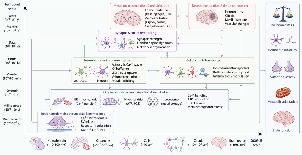
Figure 1.
Multiscale organization of ionic homeostasis in the brain
-
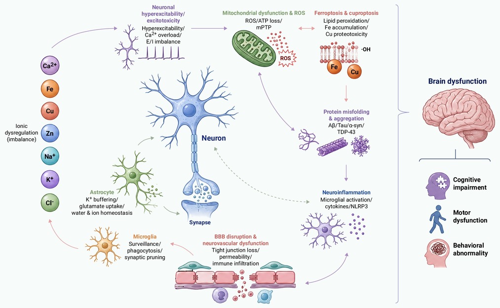
Figure 2.
Molecular and cellular mechanisms linking ionic dysregulation to brain dysfunction
-
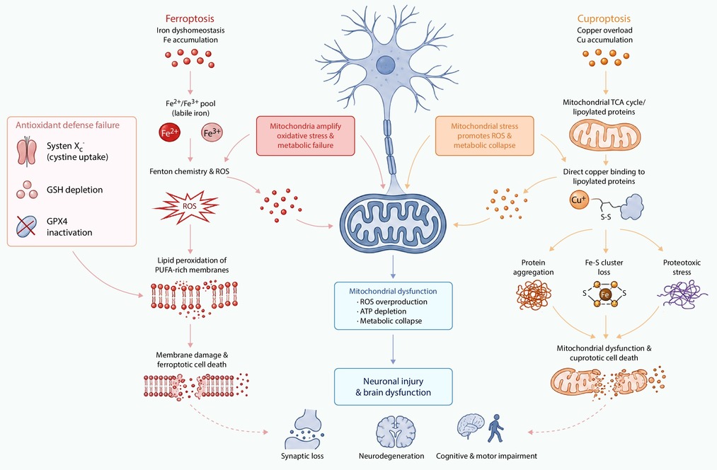
Figure 3.
Metal-dependent cell death pathways in neurological disorders
-
Figure 4.
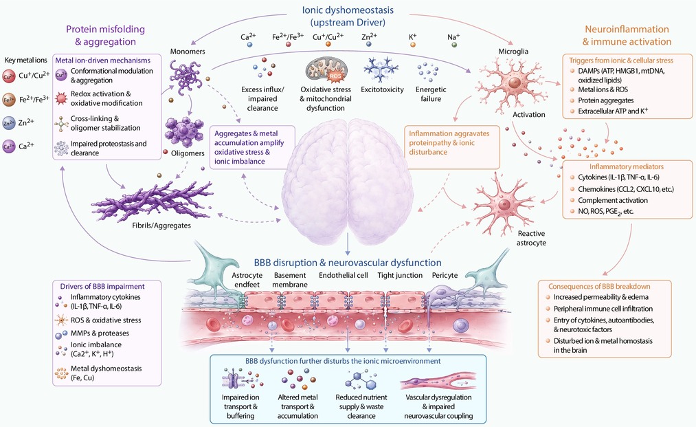
Protein misfolding, neuroinflammation and BBB dysfunction related to ionic-network dysregulation
-
Figure 5.
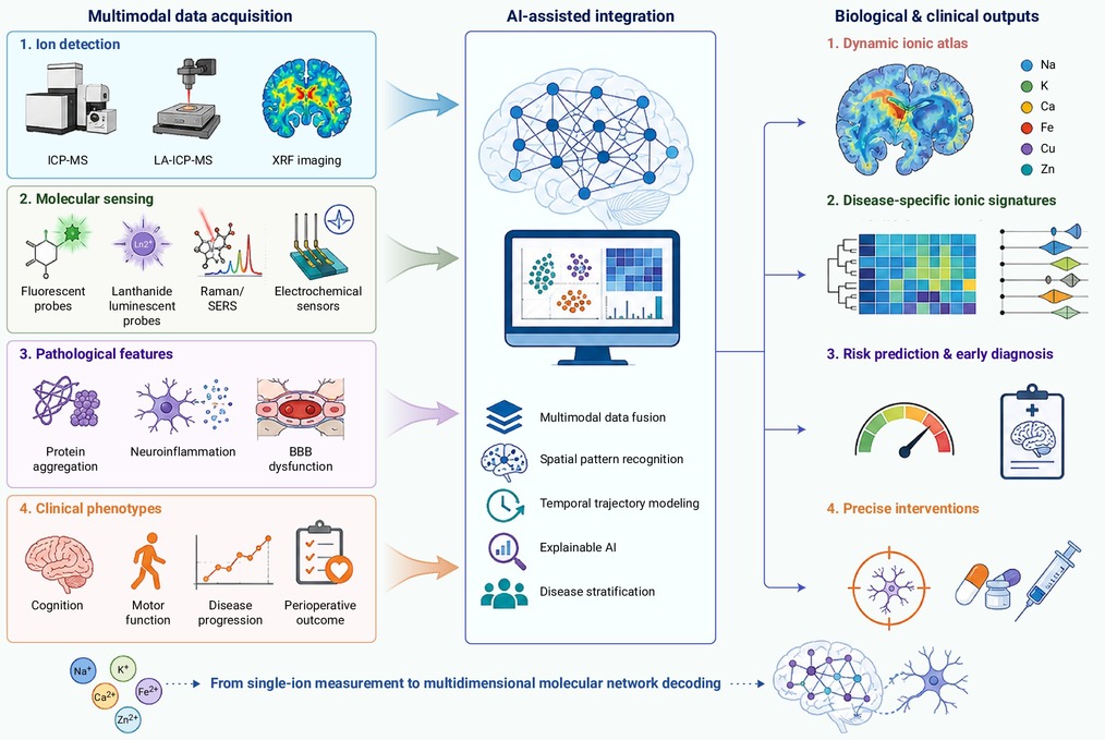
Ai-assisted multidimensional decoding of ionic network in neurological disorders
