
PAQC: A hybrid-intelligence framework for automated quality control of Raman spectroscopy via physics-aware and deep feature fusion
-
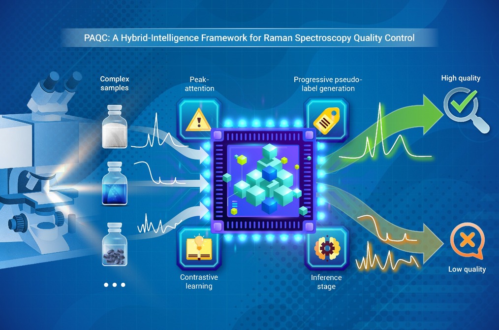
GRAPHICAL ABSTRACT
 DownLoad:
Full size image
DownLoad:
Full size image
-
PUBLIC SUMMARY
-
Raman spectroscopy reliability is often compromised by artifacts like noise and signal saturation.
We propose a hybrid framework synergizing explicit physical knowledge with implicit deep learning.
This approach generates high-confidence pseudo-labels to guide model training without manual input.
It achieves state-of-the-art accuracy and perfect recall, enabling robust automated quality control.
-
-
ABSTRACT
Raman spectroscopy is a critical analytical technique across numerous engineering disciplines; however, the reliability of its measurements is often compromised by artifacts stemming from complex instrumental and sample-specific variations. Automating quality assessment poses a significant challenge: conventional physics-based thresholds, though interpretable, lack the flexibility to accommodate diverse anomalies, while data-driven deep learning approaches typically overlook valuable domain knowledge. To address this gap, we propose PAQC—a hybrid-intelligence method that synergistically integrates explicit physical priors with implicit representation learning. Our approach processes unlabeled spectral data through dual parallel streams: an explicit knowledge stream that computes physically-informed quality metrics to produce high-confidence pseudo-labels, and an implicit knowledge stream that uses a peak-attention autoencoder, guided by known peak locations, to extract discriminative deep features. These two streams are cohesively fused through a progressive contrastive learning network, yielding a highly separable feature space tailored for anomaly detection. For practical deployment, a Mahalanobis distance-based classifier enables real-time quality diagnosis of individual spectra. Evaluated on a real-world dataset using a rigorous 5-fold cross-validation protocol, PAQC achieves state-of-the-art performance with an F1-score of 98.98% ± 0.67%, while critically maintaining a perfect 100.00% recall, underscoring its effectiveness as a robust and scalable solution for automated quality control in knowledge-sensitive engineering applications. -

-
REFERENCES
[1] Hutsebaut D., Vandenabeele P. and Moens L. (2005). Evaluation of an accurate calibration and spectral standardization procedure for Raman spectroscopy. Analyst 130:1204−1214. DOI:10.1039/B503624K [2] Nassif A.B., Talib M.A., Nasir Q., et al. (2021). Machine learning for anomaly detection: A systematic review. IEEE Access 9:78658−78700. DOI:10.1109/ACCESS.2021.3083060 [3] Nardecchia A., Motto-Ros V. and Duponchel L. (2021). Saturated signals in spectroscopic imaging: Why and how should we deal with this regularly observed phenomenon. Anal. Chim. Acta 1157:338389. DOI:10.1016/j.aca.2021.338389 [4] Vulchi R.T., Morgunov V., Junjuri R., et al. (2024). Artifacts and anomalies in raman spectroscopy: A review on origins and correction procedures. Molecules 29:4748. DOI:10.3390/molecules29194748 [5] Wei D., Chen S. and Liu Q. (2015). Review of fluorescence suppression techniques in raman spectroscopy. Appl. Spectrosc. Rev. 50:387−406. DOI:10.1080/05704928.2014.999936 [6] He S., Zhang W., Liu L., et al. (2014). Baseline correction for Raman spectra using an improved asymmetric least squares method. Anal. Methods 6:4402−4407. DOI:10.1039/C4AY00068D [7] Xu Y., Du P., Senger R., et al. (2021). ISREA: An efficient peak-preserving baseline correction algorithm for Raman spectra. Appl. Spectrosc. 75:34−45. DOI:10.1177/0003702820955245 [8] Guo S., Bocklitz T. and Popp J. (2016). Optimization of Raman-spectrum baseline correction in biological application. Analyst 141:2396−2404. DOI:10.1039/C6AN00041J [9] Smulko J.M., Dingari N.C., Soares J.S., et al. (2014). Anatomy of noise in quantitative biological Raman spectroscopy. Bioanalysis 6:411−421. DOI:10.4155/bio.13.337 [10] Moester M.J.B., Ariese F. and de Boer J.F. (2015). Optimized signal-to-noise ratio with shot noise limited detection in stimulated Raman scattering microscopy. J. Eur. Opt. Soc.-Rapid Publ. 10:15022. DOI:10.2971/jeos.2015.15022 [11] Ranjan R., Costa G., Ferrara M.A., et al. (2022). Noises investigations and image denoising in femtosecond stimulated Raman scattering microscopy. J. Biophotonics 15:e202100379. DOI:10.1002/jbio.202100379 [12] Pérez-Jiménez A.I., Lyu D., Lu Z., et al. (2020). Surface-enhanced Raman spectroscopy: Benefits, trade-offs and future developments. Chem. Sci. 11:4563−4577. DOI:10.1039/D0SC00809E [13] Fan X., Zeng Y., Zhi Y.-L., et al. (2021). Signal-to-noise ratio enhancement for Raman spectra based on optimized Raman spectrometer and convolutional denoising autoencoder. J. Raman Spectrosc. 52:890−900. DOI:10.1002/jrs.6065 [14] Smulko J., Wróbel M.S. and Barman I. (2015). Noise in biological Raman spectroscopy. Proc. ICNF 2015:1−6. DOI:10.1109/ICNF.2015.7288562 [15] Fang S., Wu S., Chen Z., et al. (2024). Recent progress and applications of Raman spectrum denoising algorithms in chemical and biological analyses: A review. TrAC Trends Anal. Chem. 172:117578. DOI:10.1016/j.trac.2024.117578 [16] Stöckel S., Kirchhoff J., Neugebauer U., et al. (2016). The application of Raman spectroscopy for the detection and identification of microorganisms. J. Raman Spectrosc. 47:89−109. DOI:10.1002/jrs.4844 [17] Liu M., Wang T., Zhang Q., et al. (2024). An outlier removal method based on PCA-DBSCAN for blood-SERS data analysis. Anal. Methods 16:846−855. DOI:10.1039/D3AY02037A [18] Li C., Guo L., Gao H., et al. (2021). Similarity-measured isolation forest: Anomaly detection method for machine monitoring data. IEEE Trans. Instrum. Meas. 70:1−12. DOI:10.1109/TIM.2021.3062684 [19] Strani L., Cocchi M., Tanzilli D., et al. (2025). One class classification (class modelling): State of the art and perspectives. TrAC Trends Anal. Chem. 183:118117. DOI:10.1016/j.trac.2024.118117 [20] Kelis Cardoso V.G. and Poppi R.J. (2021). Cleaner and faster method to detect adulteration in cassava starch using Raman spectroscopy and one-class support vector machine. Food Control 125:107917. DOI:10.1016/j.foodcont.2021.107917 [21] Zhang L., Huang X., Li P., et al. (2017). Multivariate adulteration detection for sesame oil. Chemometr. Intell. Lab. Syst. 161:147−150. DOI:10.1016/j.chemolab.2016.11.009 [22] Song S., Liu Z., Huang M., et al. (2020). Detection of fish bones in fillets by Raman hyperspectral imaging technology. J. Food Eng. 272:109808. DOI:10.1016/j.jfoodeng.2019.109808 [23] Feng Q., Zhang Y., Li C., et al. (2017). Anomaly detection of spectrum in wireless communication via deep auto-encoders. J. Supercomput. 73:3161−3178. DOI:10.1007/s11227-017-2017-7 [24] Mhamdi L., McLernon D., El-moussa F., et al. (2020). A deep learning approach combining autoencoder with one-class SVM for DDoS attack detection in SDNs. lEEE Int. Conf. Commun. 2020:1−6. DOI:10.1109/ComNet47917.2020.9306073 [25] Principi E., Rossetti D., Squartini S., et al. (2019). Unsupervised electric motor fault detection by using deep autoencoders. IEEE/CAA J. Autom. Sinica 6:441−451. DOI:10.1109/JAS.2019.1911393 [26] Zhou Y., Liang X., Zhang W., et al. (2021). VAE-based Deep SVDD for anomaly detection. Neurocomputing 453:131−140. DOI:10.1016/j.neucom.2021.04.089 [27] Zhang Z. and Deng X. (2021). Anomaly detection using improved deep SVDD model with data structure preservation. Pattern Recognit. Lett. 148:1−6. DOI:10.1016/j.patrec.2021.04.020 [28] Wei J., Wang Q., Zhang G., et al. (2025). Domain knowledge guided pseudo-label generation framework for semi-supervised domain generalization fault diagnosis. Adv. Eng. Inf. 67:103540. DOI:10.1016/j.aei.2025.103540 [29] Sun Y., Tao H. and Stojanovic V. (2025). Pseudo-label guided dual classifier domain adversarial network for unsupervised cross-domain fault diagnosis with small samples. Adv. Eng. Inf. 64:102986. DOI:10.1016/j.aei.2024.102986 [30] Li Y.-F., He W.-Y., Ren W.-X., et al. (2025). Moving load induced dynamic response analysis of bridge based on physics-informed neural network. Adv. Eng. Inf. 65:103215. DOI:10.1016/j.aei.2025.103215 [31] Zhu Y., Li S., Lang X., et al. (2025). Physics-informed CGAN and multi-scale attention CNN for pipeline leakage diagnosis under imbalanced data. Adv. Eng. Inf. 66:103471. DOI:10.1016/j.aei.2025.103471 [32] Kingma D.P. and Welling M. (2022). Auto-Encoding variational bayes. arXiv preprint. DOI:10.48550/arXiv.1312.6114 -
ABOUT THIS ARTICLE
Cite this article:
Chen X., Wang Z., Li Y., et al. (2026). PAQC: A hybrid-intelligence framework for automated quality control of Raman spectroscopy via physics-aware and deep feature fusion. The Innovation Informatics 2:100028. https://doi.org/10.59717/j.xinn-inform.2026.100028
Welcome!
To request copyright permission to republish or share portions of our works, please visit Copyright Clearance Center's (CCC) Marketplace website at marketplace.copyright.com.
Export File
Citation
Chen X., Wang Z., Li Y., et al. (2026). PAQC: A hybrid-intelligence framework for automated quality control of Raman spectroscopy via physics-aware and deep feature fusion. The Innovation Informatics 2:100028. https://doi.org/10.59717/j.xinn-inform.2026.100028

Format
Content
-
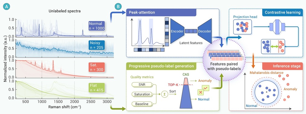
Figure 1.
Data Challenges and System Architecture of PAQC framework
-
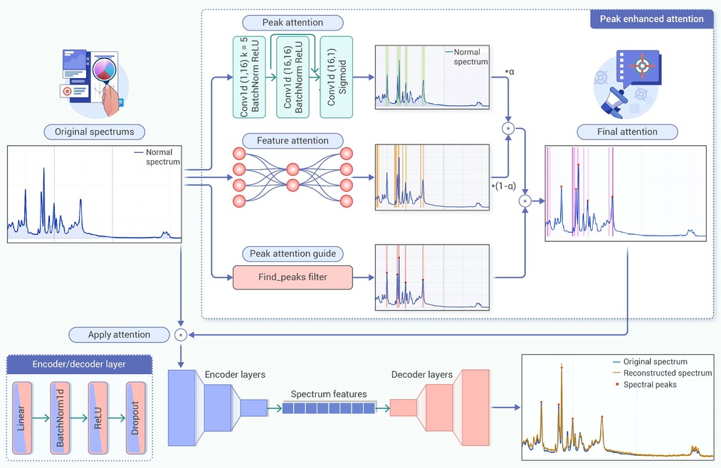
Figure 2.
Architecture of the Peak-attention enhanced Autoencoder.
-
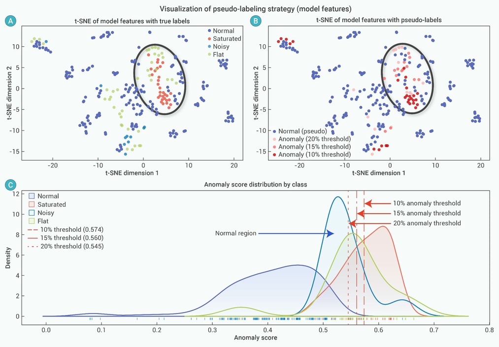
Figure 6.
Visualization of pseudo-label generation mechanism
-
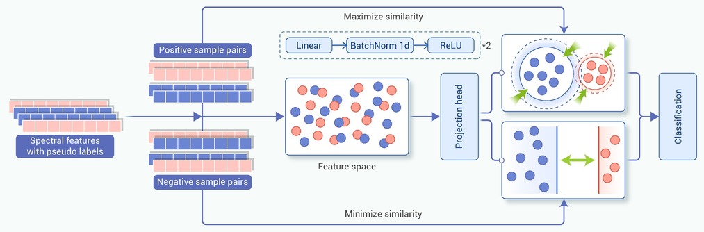
Figure 3.
Schematic diagram of the contrastive learning strategy.
-
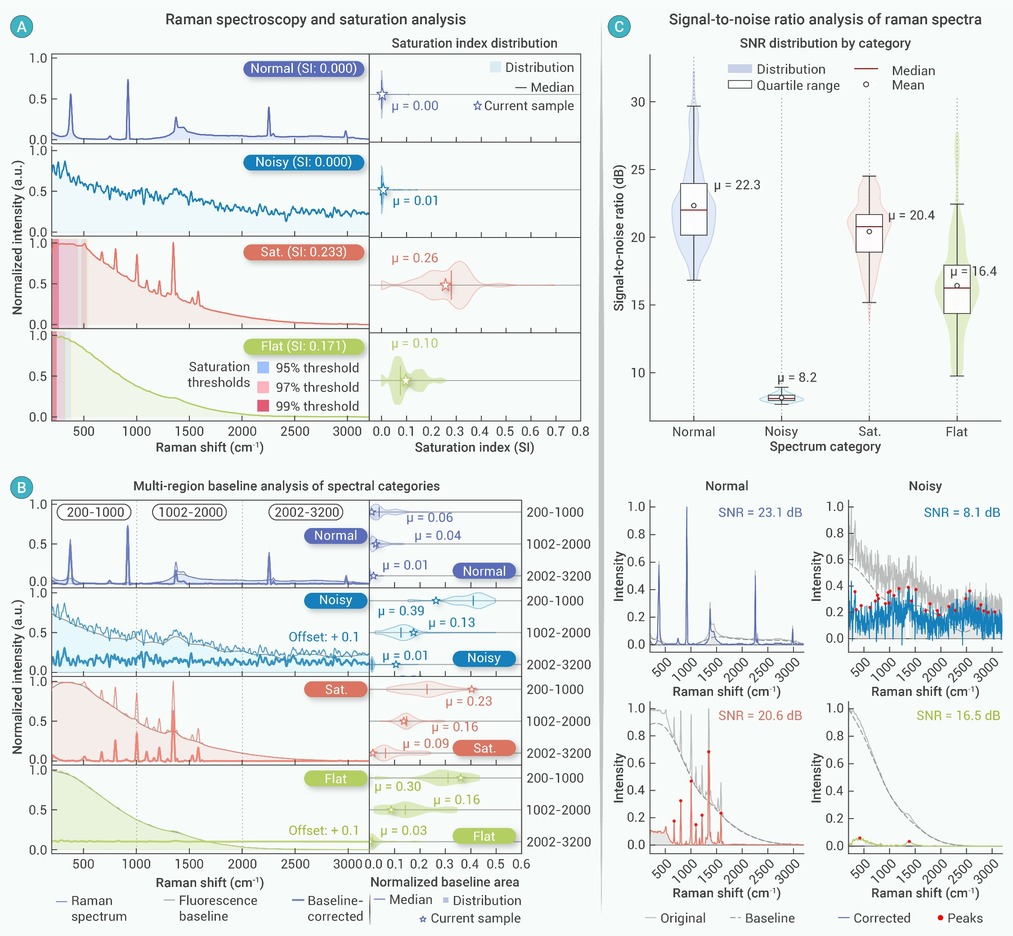
Figure 4.
Visualization and statistical analysis of physics-informed quality metrics
-
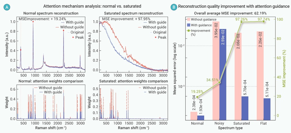
Figure 5.
Analysis of the impact of Peak-Guided attention mechanism on Autoencoder reconstruction quality
-
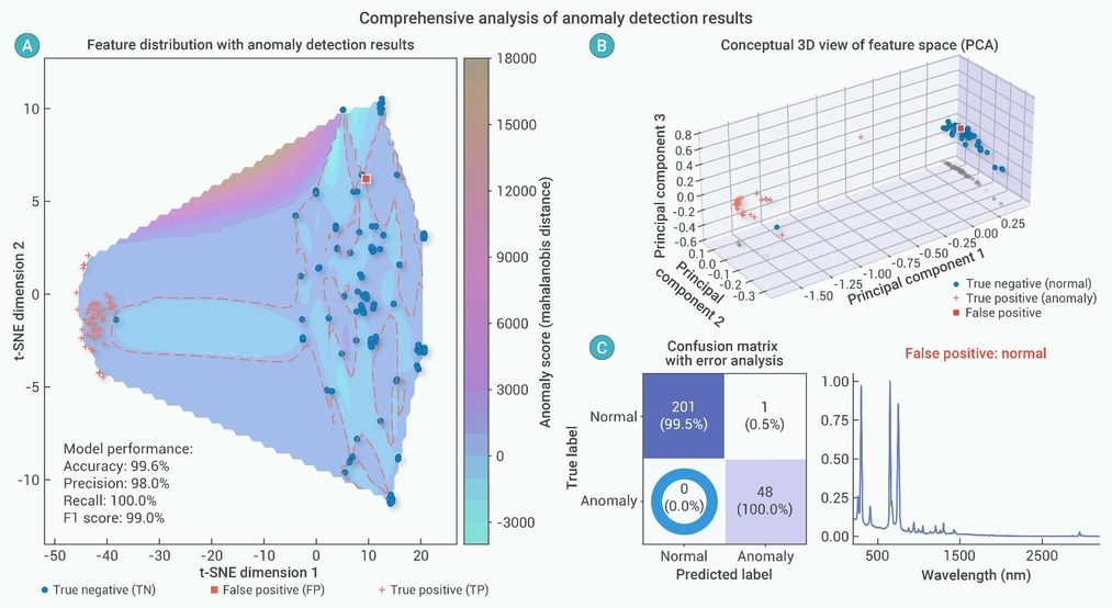
Figure 7.
Visualization of the final anomaly detection results and the real-time diagnostic mechanism
