
Rare-earth halide perovskite-like scintillators for X-ray detection and imaging
-
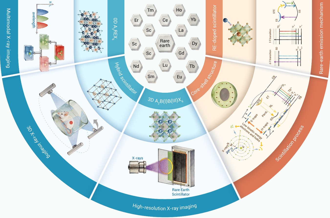
GRAPHICAL ABSTRACT
 DownLoad:
Full size image
DownLoad:
Full size image
-
PUBLIC SUMMARY
-
Rare-earth halide perovskite-like scintillators enable efficient and stable X-ray detection and imaging.
This review outlines scintillation mechanisms and recent advances in inorganic, hybrid, and doped systems.
These materials enable low-dose detection, high-resolution and dynamic imaging, and multimodal applications.
-
-
ABSTRACT
Scintillator-based X-ray detection technologies play an irreplaceable role in national security, medical diagnostics, and industrial non-destructive testing due to their broad dynamic response range, large effective imaging area, low maintenance requirements, and high operational reliability. In recent years, metal halide emitters with perovskite or perovskite-like structures have attracted extensive attention, among which rare-earth halides have shown remarkable scintillation characteristics, including high radiation conversion efficiency, tunable emission spectra, and excellent stability. These advantages arise from their structural diversity and rich photophysical properties, positioning them as strong candidates for next-generation scintillators. In this review, we provide a comprehensive overview of recent advances in rare-earth halide scintillators for X-ray detection and imaging. We first clarify the fundamental physical mechanisms governing the interaction between X-rays and rare-earth halide scintillators, along with the complete scintillation process. We then systematically summarize emerging classes of rare-earth halide scintillation materials, including fully inorganic compounds, organic-inorganic hybrids, and rare-earth-ion-doped systems, with emphasis on their design principles and strategies for modulating scintillation performance. In addition, we highlight the potential of these materials in high-resolution imaging, multimodal imaging, and X-ray photodynamic therapy. Finally, we identify the key challenges that currently constrain progress in this field and propose future research directions aimed at advancing rare-earth halide scintillator technologies.-
Keywords:
- scintillator /
- X-ray detection /
- X-ray imaging /
- rare-earth halide /
- perovskite-like
-

-
REFERENCES
[1] Zhou Y., Chen J., Bakr O. M., et al. (2021). Metal halide perovskites for X-ray imaging scintillators and detectors. ACS Energy Lett. 6:739−768. DOI:10.1021/acsenergylett.0c02430 [2] Wang T., Zeng G., Yang Y. M., et al. (2025). Advances in metal halide perovskite scintillators for X-ray detection. Nano-Micro Lett. 17:275. DOI:10.1007/s40820-025-01772-7 [3] Zhou Q., Li W., Xiao J., et al. (2024). Low‐dimensional metal halide for high performance scintillators. Adv. Funct. Mater. 34:2402902. DOI:10.1002/adfm.202402902 [4] Michail C., Liaparinos P., Kalyvas N., et al. (2024). Radiation detectors and sensors in medical imaging. Sensors 24:6251. DOI:10.3390/s24196251 [5] Guerra M., Manso M., Longelin S., et al. (2012). Performance of three different Si X-ray detectors for portable XRF spectrometers in cultural heritage applications. J. Instrum. 7:C10004. DOI:10.1088/1748-0221/7/10/c10004 [6] Ivanov Y. M., Kanevsky V. M., Dvoryankin V. F., et al. (2003). The possibilities of using semi‐insulating CdTe crystals as detecting material for X‐ray imaging radiography. Phys. Status Solidi C 0:840–844. DOI:10.1002/pssc.200306258 [7] Szeles C. (2004). CdZnTe and CdTe materials for X‐ray and gamma ray radiation detector applications. Phys. Status Solidi B 241:783−790. DOI:10.1002/pssb.200304296 [8] Chen J., Zhou Y., Fu Y., et al. (2021). Oriented halide perovskite nanostructures and thin films for optoelectronics. Chem. Rev. 121:12112−12180. DOI:10.1021/acs.chemrev.1c00181 [9] Pan W., Yang B., Niu G., et al. (2019). Hot‐Pressed CsPbBr3 quasi‐monocrystalline film for sensitive direct X‐ray detection. Adv. Mater. 31:1904405. DOI:10.1002/adma.201904405 [10] Jana A., Cho S., Patil S. A., et al. (2022). Perovskite: Scintillators, direct detectors, and X-ray imagers. Mater. Today 55:110−136. DOI:10.1016/j.mattod.2022.04.009 [11] Kim Y. C., Kim K. H., Son D.Y., et al. (2017). Printable organometallic perovskite enables large-area, low-dose X-ray imaging. Nature 550:87−91. DOI:10.1038/nature24032 [12] Zhou Y., Wang X., He T., et al. (2022). Large-area perovskite-related copper halide film for high-resolution flexible X-ray imaging scintillation screens. ACS Energy Lett. 7:844−846. DOI:10.1021/acsenergylett.2c00075 [13] Zhao Y., Zhou Y., Wang Y., et al. (2025). Copper iodide ink scintillator‐based X‐ray detector for high‐resolution computed tomography imaging. Laser Photonics Rev. 20:e02301. DOI:10.1002/lpor.202502301 [14] Yang B., Yin L., Niu G., et al. (2019). Lead‐free halide Rb2CuBr3 as sensitive X‐ray scintillator. Adv. Mater. 31:1904711. DOI:10.1002/adma.201904711 [15] Tsuda S. and Saito K. (2017). Spectrum–dose conversion operator of NaI(Tl) and CsI(Tl) scintillation detectors for air dose rate measurement in contaminated environments. J. Environ. Radioact. 166:419−426. DOI:10.1016/j.jenvrad.2016.02.008 [16] Santana G. C., de Mello A. C. S., Valerio M. E. G., et al. (2007). Scintillating properties of pure and doped BGO ceramics. J. Mater. Sci. 42:2231−2235. DOI:10.1007/s10853-006-1319-6 [17] Borghi G., Peet B. J., Tabacchini V., et al. (2016). A 32 mm× 32 mm × 22 mm monolithic LYSO:Ce detector with dual-sided digital photon counter readout for ultrahigh-performance TOF-PET and TOF-PET/MRI. Phys. Med. Biol. 61:4929−4949. DOI:10.1088/0031-9155/61/13/4929 [18] Fujimoto Y., Nakauchi D., Yanagida T., et al. (2021). Evaluation of Gd2SiO5:Ce (GSO), Lu2−xGdxSiO5:Ce (LGSO), and Lu2−xYxSiO5:Ce (LYSO) crystalline scintillators using a photoacoustic spectroscopy technique. Sens. Mater. 33:2147-2153. DOI:10.18494/sam.2021.3321 [19] Yoneyama A., Baba R. and Kawamoto M. (2021). Quantitative analysis of the physical properties of CsI, GAGG, LuAG, CWO, YAG, BGO, and GOS scintillators using 10-, 20- and 34-keV monochromated synchrotron radiation. Opt. Mater. Express 11:398−411. DOI:10.1364/ome.409161 [20] Tessitore G. A. M., Maurizio S. L., Kaur M., et al. (2023). The role of lanthanide luminescence in advancing technology. RSC Adv. 13:17787−17811. DOI:10.1039/D3RA00991B [21] Feng H., Jary V., Mihokova E., et al. (2010). Temperature dependence of luminescence characteristics of Lu2(1−x)Y2xSiO5:Ce3+ scintillator grown by the Czochralski method. J. Appl. Phys. 108:033519. DOI:10.1063/1.3457856 [22] Drozdowski K. J., Witkowski M. E., Makowski M., et al. (2025). Luminescent and scintillation properties of GOS and GYAGG ceramics activated with cerium and praseodymium ions. Ceram. Int. 51:21182−21190. DOI:10.1016/j.ceramint.2025.02.279 [23] Chen Q., Wu J., Ou X., et al. (2018). All-inorganic perovskite nanocrystal scintillators. Nature 561:88−93. DOI:10.1038/s41586-018-0451-1 [24] Zhou Y., Chen J., Bakr O. M., et al. (2018). Metal-doped lead halide perovskites: Synthesis, properties, and optoelectronic applications. Chem. Mater. 30:6589−6613. DOI:10.1021/acs.chemmater.8b02989 [25] Akkerman Q. A., Rainò G., Kovalenko M. V., et al. (2018). Genesis, challenges and opportunities for colloidal lead halide perovskite nanocrystals. Nat. Mater. 17:394−405. DOI:10.1038/s41563-018-0018-4 [26] Chen S., Zhang Y., Zhao J., et al. (2020). Transmission electron microscopy of organic-inorganic hybrid perovskites: myths and truths. Sci. Bull. 65:1643−1649. DOI:10.1016/j.scib.2020.05.020 [27] Ding Y., Liu D., Dang P., et al. (2025). Lead-free lanthanide-based Cs3LnCl6 metal halides. J. Mater. Chem. C 13:1557−1572. DOI:10.1039/d4tc03748k [28] Samanta T., Mukurala N., Viswanath N. S. M., et al. (2023). Recent progress in lanthanide-based metal halide perovskites: synthesis, properties, and applications. Opt. Mater. X 18:100238. DOI:10.1016/j.omx.2023.100238 [29] Lu H., Xu X., Feng G., et al. (2022). Terbium doped LiLuF4 nanocrystal scintillator-based flexible composite film for high resolution X-ray imaging. RSC Adv. 12:4615−4623. DOI:10.1039/d1ra08989g [30] Zeng S., Wang H., Lu W., et al. (2014). Dual-modal upconversion fluorescent/X-ray imaging using ligand-free hexagonal phase NaLuF4:Gd/Yb/Er nanorods for blood vessel visualization. Biomaterials 35:2934−2941. DOI:10.1016/j.biomaterials.2013.11.082 [31] Escudero A., Becerro A. I., Carrillo-Carrión C., et al. (2017). Rare earth based nanostructured materials: Synthesis, functionalization, properties and bioimaging and biosensing applications. Nanophotonics 6:881−921. DOI:10.1515/nanoph-2017-0007 [32] Ou X., Qin X., Huang B., et al. (2021). High-resolution X-ray luminescence extension imaging. Nature 590:410−415. DOI:10.1038/s41586-021-03251-6 [33] Yang H., Zhao C., Wang Y., et al. (2025). Sensitive and fast X‐ray scintillation with perovskite‐inspired cerium halide nanocrystals. Adv. Funct. Mater. 35:2422959. DOI:10.1002/adfm.202422959 [34] Yang Z., Cui J., Sun Y., et al. (2025). Na-alloy tailored 3D/0D metal halide heterostructures enabling efficient charge transfer for color-integrated white scintillators. Nat. Commun. 16:6909. DOI:10.1038/s41467-025-62195-x [35] Song N., Lv K., Zhang X., et al. (2025). High-entropy lanthanide metal-organic frameworks as multifunctional porous scintillators for radiation detection and dual-mode uranyl sensing. Angew. Chem. Int. Ed. 138:e19226. DOI:10.1002/anie.202519226 [36] Zi L., Song J., Wang N., et al. (2023). X‐ray quantum cutting scintillator based on CsPbClxBr3−x:Yb3+ single crystals. Laser Photonics Rev. 17:220852. DOI:10.1002/lpor.202200852 [37] Ran P., Yang L., Hui J., et al. (2025). Single-shot X-ray and near-infrared (NIR) dual-mode fusion imaging based on bifunctional NIR scintillators. Light: Sci. Appl. 14:315. DOI:10.1038/s41377-025-01898-8 [38] Lei L., Wang Y., Kuzmin A., et al. (2022). Next generation lanthanide doped nanoscintillators and photon converters. eLight 2:17. DOI:10.1186/s43593-022-00024-0 [39] Lusic H. and Grinstaff M. W. (2013). X-ray-computed tomography contrast agents. Chem. Rev. 113:1641−1666. DOI:10.1021/cr200358s [40] Simonenko I. O., Nazmitdinov R. G., Kinev V. A., et al. (2026). Halide perovskite scintillators for X-ray detection: From structure to engineering. Phys. Chem. Chem. Phys. 28:4992−5042. DOI:10.1039/d5cp03941j [41] Maddalena F., Tjahjana L., Xie A., et al. (2019). Inorganic, organic, and perovskite halides with nanotechnology for high–light yield X- and γ-ray scintillators. Crystals 9:88. DOI:10.3390/cryst9020088 [42] Samanta T., Viswanath N. S. M., Kim H. W., et al. (2024). Demystifying role of local distortion in emission colors tuning of lead-free zero-dimensional metal halide nanocrystals. Chem. Eng. J. 484:149697. DOI:10.1016/j.cej.2024.149697 [43] Wang Y., Dang P., Qiu L., et al. (2023). Multimode luminescence tailoring and improvement of Cs2NaHoCl6 cryolite crystals via Sb3+ /Yb3+ alloying for versatile photoelectric applications. Angew. Chem. Int. Ed. 62:e202311699. DOI:10.1002/anie.202311699 [44] Sun L., Dong B., Sun J., et al. (2024). Efficient and stable multicolor emissions of the coumarin-modified Cs3LnCl6 lead-free perovskite nanocrystals and LED application. Adv. Mater. 36:e2310065. DOI:10.1002/adma.202310065 [45] Jobe S., Siddig L. A., Najar A., et al. (2025). 0D cesium lanthanide chlorides: Spectroscopic insights and optical properties for emerging optoelectronic applications. Adv. Opt. Mater. 13:e02940. DOI:10.1002/adom.202502940 [46] Zhou W., Zhuang X., Xu X., et al. (2025). Expanding the toolbox of all‐inorganic perovskite‐like rare‐earth‐based metal halides for photonic applications. Adv. Opt. Mater. 13:e02690. DOI:10.1002/adom.202502690 [47] Li L., Li T., Hu Y., et al. (2022). Mechanism of the trivalent lanthanides' persistent luminescence in wide bandgap materials. Light Sci. Appl. 11:51. DOI:10.1038/s41377-022-00736-5 [48] Richard C. and Viana B. (2022). Persistent X-ray-activated phosphors: Mechanisms and applications. Light Sci. Appl. 11:123. DOI:10.1038/s41377-022-00808-6 [49] Kumar V., Tiwari S. P., Ntwaeaborwa O. M., et al. (2020). Luminescence properties of rare-earth doped oxide materials. Spectroscopy of lanthanide-doped oxide materials, pp:345–364. DOI: 10.1016/B978-0-08-102935-0.00010-1 [50] Dieke G. H. and Satten R. A. (1970). Spectra and energy Levels of rare earth ions in crystals. Am. J. Phys. 38:399−400. DOI:10.1119/1.1976350 [51] Wang Y., Wang C., Men L., et al. (2025). Rare earth double perovskites for underwater X-ray imaging applications. Inorg. Chem. Front. 12:3469−3477. DOI:10.1039/d4qi03325f [52] Wang T., Zhou D., Wang Y., et al. (2025). Lanthanide‐based lead‐free perovskite materials for visible‐range and infrared light‐emitting diodes. Adv. Funct. Mater. 35:2504436. DOI:10.1002/adfm.202504436 [53] Peacock R. D. (2007). The intensities of lanthanide f↔ f transitions. Rare Earths, pp:83–122. DOI:10.1007/BFb0116556 [54] Ofelt G. (1962). Intensities of crystal spectra of rare‐earth ions. J. Chem. Phys. 37:511−520. DOI:10.1063/1.1701366 [55] Ferhi M., Bouzidi C., Horchani-Naifer K., et al. (2015). Judd–Ofelt analysis of spectroscopic properties of Eu3+ doped KLa (PO3) 4. J. Lumin. 157:21−27. DOI:10.1016/j.jlumin.2014.08.017 [56] Dunina E. B. and Kornienko A. A. (2014). Influence of excited configurations on the intensities of electric-dipole transitions of rare-earth ions. Opt. Spectrosc. 116:706−711. DOI:10.1134/S0030400X14050087 [57] Tan J., Li D., Zhu J., et al. (2022). Self-trapped excitons in soft semiconductors. Nanoscale 14:16394−16414. DOI:10.1039/d2nr03935d [58] Li X., Gao X., Zhang X., et al. (2021). Lead-free halide perovskites for light emission: Recent advances and perspectives. Adv. Sci. 8:2003334. DOI:10.1002/advs.202003334 [59] Li S., Luo J., Liu J., et al. (2019). Self-trapped excitons in all-inorganic halide perovskites: fundamentals, status, and potential applications. J. Phys. Chem. Lett. 10:1999−2007. DOI:10.1021/acs.jpclett.8b03604 [60] Jiang F., Wu Z., Lu M., et al. (2023). Broadband emission origin in metal halide perovskites: Are self-trapped excitons or ions. Adv. Mater. 35:e2211088. DOI:10.1002/adma.202211088 [61] Li J., Zhu H., Tong G., et al. (2025). Structural transformation‐engineered yttrium‐based lead‐free metal halides with smart tunable luminescence via self‐trapped exciton and lanthanide ion intrinsic combined emissions. Adv. Funct. Mater. 35:2508778. DOI:10.1002/adfm.202508778 [62] Shiran N., Gektin A., Neicheva S., et al. (2005). Energy transfer in pure and Ce-doped LiCaAlF6 and LiSrAlF6 crystals. Nucl. Instrum. Methods Phys. Res., Sect. A 537:266−270. DOI:10.1016/j.nima.2004.08.023 [63] Li S., Hu Q., Luo J., et al. (2019). Self‐trapped exciton to dopant energy transfer in rare earth doped lead‐free double perovskite. Adv. Opt. Mater. 7:1901098. DOI:10.1002/adom.201901098 [64] Zhou B., Liu Z., Fang S., et al. (2022). Emission mechanism of self-trapped excitons in Sb3+-doped all-inorganic metal-halide perovskites. J. Phys. Chem. Lett. 13:9140−9147. DOI:10.1021/acs.jpclett.2c02759 [65] Han Y., Cheng X. and Cui B.-B. (2023). Factors influencing self-trapped exciton emission of low-dimensional metal halides. Mater. Adv. 4:355−373. DOI:10.1039/d2ma00676f [66] Stroyuk O., Raievska O., Zahn D. R. T., et al. (2024). Exploring highly efficient broadband self-trapped-exciton luminophors: from 0D to 3D materials. Chem. Rec. 24:e202300241. DOI:10.1002/tcr.202300241 [67] Yanagida T., Koshimizu M., Kurashima S., et al. (2015). Linear energy transfer effects on time profiles of scintillation of Ce-doped LiCaAlF6 crystals. Nucl. Instrum. Methods Phys. Res., Sect. B 365:529−532. DOI:10.1016/j.nimb.2015.07.065 [68] Chen G., Qiu H., Prasad P. N., et al. (2014). Upconversion nanoparticles: Design, nanochemistry, and applications in theranostics. Chem. Rev. 114:5161−5214. DOI:10.1021/cr400425h [69] Geng S., Li H., Lv Z., et al. (2025). Challenges and opportunities of upconversion nanoparticles for emerging NIR optoelectronic devices. Adv. Mater. 37:e2419678. DOI:10.1002/adma.202419678 [70] Chen X., Li X., Chen X., et al. (2022). Flexible X-ray luminescence imaging enabled by cerium-sensitized nanoscintillators. J. Lumin. 242:118589. DOI:10.1016/j.jlumin.2021.118589 [71] Wei G., Wang C., Huang J., et al. (2025). Boosting X‐ray‐activated persistent luminescence in nanoparticles toward highly accurate flexible X‐ray imaging and information security. Laser Photonics Rev. 19:2401261. DOI:10.1002/lpor.202401261 [72] Xu W., Zhou M., Deng R., et al. (2024). Single activator doped fluoride nanoparticles for X-ray excited high-resolution and delayed flexible nonplanar imaging. Chem. Eng. J. 499:156406. DOI:10.1016/j.cej.2024.156406 [73] Liu S., Yan L., Huang J., et al. (2022). Controlling upconversion in emerging multilayer core-shell nanostructures: From fundamentals to frontier applications. Chem. Soc. Rev. 51:1729−1765. DOI:10.1039/d1cs00753j [74] Wu Q., Xu X., Li X., et al. (2024). Probing energy-funneling kinetics in nanocrystal sublattices for superior X-ray imaging. Angew. Chem. Int. Ed. 63:e202404177. DOI:10.1002/anie.202404177 [75] Yang Z., Zhang P., Chen X., et al. (2023). High-confidentiality X-ray imaging encryption using prolonged imperceptible radioluminescence memory scintillators. Adv. Mater. 35:e2309413. DOI:10.1002/adma.202309413 [76] Zhu Q., Song C., Tong W., et al. (2020). Controllable phase/morphology tailoring of REF3 and NaREF4 (RE = La-Lu, Y), and insights into the up-conversion luminescence of GdF3:Yb3+/Tm3+ spheres. Adv. Powder Technol. 31:2235−2243. DOI:10.1016/j.apt.2020.03.016 [77] Seitz M., Oliver A. G. and Raymond K. N. (2007). The lanthanide contraction revisited. J. Am. Chem. Soc. 129:11153−11160. DOI:10.1021/ja072750f [78] Hou B., Yi L., Hu D., et al. (2023). A swallowable X-ray dosimeter for the real-time monitoring of radiotherapy. Nat. Biomed. Eng. 7:1242−1251. DOI:10.1038/s41551-023-01024-2 [79] Zheng B., Fan J., Chen B., et al. (2022). Rare-earth doping in nanostructured inorganic materials. Chem. Rev. 122:5519−5603. DOI:10.1021/acs.chemrev.1c00644 [80] Li F., Wang Z., Li Y., et al. (2025). Engineering full-spectrum upconversion through coherent energy recycling in NaYF4:Yb,Tm/Cs4PbBr6 heterostructure. Angew. Chem. Int. Ed.:e21996. DOI:10.1002/anie.202521996 [81] Chen X., Lu H., Gu Q., et al. (2022). Terbium-doped LiYbF4 nanomaterial-based scintillator responding to X-rays with high-resolution imaging applications. CrystEngComm 24:2551−2557. DOI:10.1039/d2ce00075j [82] Lu H., Xu X., Li Y., et al. (2024). Heterovalent co‐doped LiLuF4 composite flexible films as persistent luminescence scintillator for X‐ray high‐resolution extension imaging. Adv. Opt. Mater. 12:2303134. DOI:10.1002/adom.202303134 [83] Guan S., Zhang W., Chen X., et al. (2025). Broadband excitation from X-ray to UV for afterglow emission in monodisperse lanthanide-doped nanoengineered molecular nanotransducers. Adv. Mater. 37:e2508039. DOI:10.1002/adma.202508039 [84] Wang C., Liu D., Wei G., et al. (2024). Enabling multimodal luminescence in a single nanoparticle for X-ray imaging encryption and anticounterfeiting. Nano Lett. 24:9691−9699. DOI:10.1021/acs.nanolett.4c02468 [85] Zhang X., Shi Y., Wang X., et al. (2022). Flexible and transparent ceramic nanocomposite for laboratory X-ray imaging of micrometer resolution. ACS Nano 16:21576−21582. DOI:10.1021/acsnano.2c10531 [86] Lu L., Peng S., Zhao L., et al. (2023). Visualized X-ray dosimetry for multienvironment applications. Nano Lett. 23:8753−8760. DOI:10.1021/acs.nanolett.3c02826 [87] Yang X., Waterhouse G. I. N., Lu S., et al. (2023). Recent advances in the design of afterglow materials: Mechanisms, structural regulation strategies and applications. Chem. Soc. Rev. 52:8005−8058. DOI:10.1039/d2cs00993e [88] Chen X., Gong Z., Peng C., et al. (2025). Programmable afterglow tuning in monodisperse SiO2 microparticles through spatially confined emitter doping. Nat. Commun. 16:8900. DOI:10.1038/s41467-025-63901-5 [89] Lei L., Yi M., Wang Y., et al. (2024). Dual heterogeneous interfaces enhance X-ray excited persistent luminescence for low-dose 3D imaging. Nat. Commun. 15:1140. DOI:10.1038/s41467-024-45390-0 [90] Wang H., Yao J. and Zeng R. (2024). The luminescence modulation of rare earth-doped/containing lead-free double perovskites toward multifunctional applications: a review. Nanoscale 16:6837−6852. DOI:10.1039/d3nr06472g [91] Samanta T., Yadav A. N., Han J. H., et al. (2024). Cerium‐sensitized highly emissive 0D cesium cerium terbium chloride alloy nanocrystals for white light emission. Adv. Opt. Mater. 12:2400909. DOI:10.1002/adom.202400909 [92] Han J. H., Samanta T., Park Y. M., et al. (2022). Highly stable zero-dimensional lead-free metal halides for X-ray imaging. ACS Energy Lett. 8:545−552. DOI:10.1021/acsenergylett.2c02469 [93] Chai Z., Wang Z., Ke D., et al. (2025). Thermally robust 0D Cs3EuCl6 microcrystals for X-ray imaging. Small 21:e05594. DOI:10.1002/smll.202505594 [94] Samanta T., Han J. H., Lee H. U., et al. (2024). Large-scale mechanochemical synthesis of cesium lanthanide chloride for radioluminescence. Inorg. Chem. 63:16483−16490. DOI:10.1021/acs.inorgchem.4c02766 [95] Li H., Li K., Li Z., et al. (2025). Lanthanide-based metal halides prepared at room temperature by recrystallization method for X-ray imaging. Light Sci. Appl. 14:195. DOI:10.1038/s41377-025-01839-5 [96] Zhou W., Yu Y., Han P., et al. (2024). Sb-doped Cs3TbCl6 nanocrystals for highly efficient narrow-band green emission and X-ray imaging. Adv. Mater. 36:e2302140. DOI:10.1002/adma.202302140 [97] Ha J., Yeon S., Lee J., et al. (2024). Revealing the role of organic ligands in deep-blue-emitting colloidal europium bromide perovskite nanocrystals. ACS Nano 18:31891−31902. DOI:10.1021/acsnano.4c09018 [98] Slavney A. H., Hu T., Lindenberg A. M., et al. (2016). A bismuth-halide double perovskite with long carrier recombination lifetime for photovoltaic applications. J. Am. Chem. Soc. 138:2138−2141. DOI:10.1021/jacs.5b13294 [99] Giustino F. and Snaith H. J. (2016). Toward lead-free perovskite solar cells. ACS Energy Lett. 1:1233−1240. DOI:10.1021/acsenergylett.6b00499 [100] Goldschmidt V. M. (1926). Die gesetze der krystallochemie. Naturwissenschaften 14:477−485. DOI:10.1007/bf01507527 [101] Rao Z., Zhao X. and Gong X. (2024). Rare‐earth‐based lead‐free halide double perovskites for light emission: recent advances and applications. Adv. Funct. Mater. 34:2406424. DOI:10.1002/adfm.202406424 [102] Rooh G. H. K., Kim H. J., Park H., et al. (2009). The growth and scintillation properties of Cs2NaCeCl6 single crystal. J. Cryst. Growth 311:2470−2473. DOI:10.1016/j.jcrysgro.2009.01.091 [103] Hu Q., Deng Z., Hu M., et al. (2018). X-ray scintillation in lead-free double perovskite crystals. Sci. China Chem. 61:1581−1586. DOI:10.1007/s11426-018-9308-2 [104] Wang Z., Xu X., Wang S., et al. (2021). Cerium doping double perovskite scintillator for sensitive X-ray detection and imaging. Chem. Eur. J. 27:9071−9076. DOI:10.1002/chem.202100449 [105] Zhou S., Lin S., Zhao J., et al. (2025). Submicron double-perovskite structured terbium based lanthanide chloride crystals: Room temperature synthesis, near-unity quantum yield and high-resolution X-ray imaging. Chem. Eng. J. 522:167373. DOI:10.1016/j.cej.2025.167373 [106] Zhou W., Li C., Wu T., et al. (2023). Bright green-emitting all-inorganic terbium halide double perovskite nanocrystals for low-dose X-ray imaging. J. Phys. Chem. Lett. 14:8577−8583. DOI:10.1021/acs.jpclett.3c02070 [107] Mu D., Wang T., Yuan J., et al. (2024). X‐ray real‐time imaging and delayed imaging from lead‐free perovskite scintillators. Adv. Opt. Mater. 12:2401455. DOI:10.1002/adom.202401455 [108] Naresh V., Cha P. R. and Lee N. (2024). Cs2NaGdCl6:Tb3+ - a highly luminescent rare-earth double perovskite scintillator for low-dose X-ray detection and imaging. ACS Appl. Mater. Interfaces 16:19068−19080. DOI:10.1021/acsami.3c17301 [109] Zhang K., Li T., Cheng H., et al. (2024). Excitation power regulated up-conversion luminescence chroma of lanthanide double-perovskite Cs2NaLnCl6 (Ln = Ho3+, Er3+, Tm3+) microcrystals. J. Alloys Compd. 990:174410. DOI:10.1016/j.jallcom.2024.174410 [110] Huang R., Zhang X., Xu X., et al. (2025). Double perovskite scintillators enabled by Co‐doped trap regulation for thermally enhanced X‐ray imaging. Adv. Opt. Mater. 13:2403289. DOI:10.1002/adom.202403289 [111] Wang Z., Chen J., Xu X., et al. (2024). B(I)‐Site alkali metal engineering of lead‐free perovskite nanocrystals for efficient X‐ray detection and imaging. Adv. Opt. Mater. 12:2302617. DOI:10.1002/adom.202302617 [112] Liu Y., Tu D., Yang M., et al. (2025). Near-Infrared and visible dual-band self-trapped exciton emissions from Li+-doped Cs2NaScCl6 double perovskites. ACS Energy Lett. 10:2150−2159. DOI:10.1021/acsenergylett.5c00182 [113] Saghy P., Brown A. M., Chu C., et al. (2023). Lanthanide double perovskite nanocrystals with emissions covering the UV‐C to NIR spectral range. Adv. Opt. Mater. 11:2300277. DOI:10.1002/adom.202300277 [114] Wang X., Zhang X., Yan S., et al. (2022). Nearly-unity quantum yield and 12-hour afterglow from a transparent perovskite of Cs2NaScCl6:Tb. Angew. Chem. Int. Ed. 61:e202210853. DOI:10.1002/anie.202210853 [115] Zhang N., Zhang R., Xu X., et al. (2023). X‐ray‐activated long afterglow double‐perovskite scintillator for detection and extension imaging. Adv. Opt. Mater. 11:2300187. DOI:10.1002/adom.202300187 [116] Zhou F., Li Z., Lan W., et al. (2020). Halide perovskite, a potential scintillator for X‐ray detection. Small Methods 4:2000506. DOI:10.1002/smtd.202000506 [117] Wang X., Zheng W., Zhang X., et al. (2024). Multicolor luminescence and afterglow from Cs2NaScCl6:Sb3+,Mn2+ crystals. Inorg. Chem. Front. 11:8123−8129. DOI:10.1039/d4qi02019g [118] Suo H., Wang N., Zhang Y., et al. (2025). Excitation-mode-selective emission through multiexcitonic states in a double perovskite single crystal. Light Sci. Appl. 14:21. DOI:10.1038/s41377-024-01689-7 [119] Tong X., Yu H., Li X., et al. (2025). Cs2NaYCl6:Er@KBr composite for applications of NIR LED with low working temperature. Ceram. Int. 51:47974−47981. DOI:10.1016/j.ceramint.2025.08.057 [120] Chen H., Chen J., Li M., et al. (2022). Recent advances in metal-organic frameworks for X-ray detection. Sci. China Chem. 65:2338−2350. DOI:10.1007/s11426-022-1334-0 [121] Zhu X., Zhou R., Wang Z., et al. (2024). Lanthanide-metal-doped light-harvesting quantum dots for exceptional X-ray imaging scintillators. ACS Energy Lett. 9:5137−5144. DOI:10.1021/acsenergylett.4c02155 [122] Su P., Song F., Cao J., et al. (2025). Rare earth complex-based functional materials: From molecular design and performance regulation to unique applications. Acc. Chem. Res. 58:218−230. DOI:10.1021/acs.accounts.4c00649 [123] Pei H., He G., Jiang S., et al. (2025). Rare earth composite MOFs materials for energy, environmental and medical applications. RSC Adv. 15:21381−21400. DOI:10.1039/d5ra02759d [124] Zhu X., He T., Song X., et al. (2024). Large‐area metal–organic framework glasses for efficient X‐ray detection. Adv. Mater. 36:2412432. DOI:10.1002/adma.202412432 [125] Wang C., Volotskova O., Lu K., et al. (2014). Synergistic assembly of heavy metal clusters and luminescent organic bridging ligands in metal–organic frameworks for highly efficient X-ray scintillation. J. Am. Chem. Soc. 136:6171−6174. DOI:10.1021/ja500671h [126] Liu X., Li R., Xu X., et al. (2023). Lanthanide(III)-Cu4I4 organic framework scintillators sensitized by cluster-based antenna for high-resolution X-ray imaging. Adv. Mater. 35:e2206741. DOI:10.1002/adma.202206741 [127] Li H., Li Y., Zhang L., et al. (2024). A thermo‐responsive MOFs for X‐ray scintillator. Adv. Mater. 36:2405535. DOI:10.1002/adma.202405535 [128] Zhang X., Qiu H., Luo W., et al. (2023). High‐performance X‐ray imaging using lanthanide metal–organic frameworks. Adv. Sci. 10:2207004. DOI:10.1002/advs.202207004 [129] Xu J., Luo R., Luo Z., et al. (2024). Ultrabright molecular scintillators enabled by lanthanide-assisted near-unity triplet exciton recycling. Nat. Photonics 19:71−78. DOI:10.1038/s41566-024-01586-w [130] Qiu J., Gu C., Chen M., et al. (2025). Scattering-free lanthanide-ionic liquid scintillators for high-resolution and adaptive X-ray imaging. Nat. Commun. 16:11609. DOI:10.1038/s41467-025-66754-0 [131] Han K., Jin J., Wang Y., et al. (2024). Hybrid Eu(II)-bromide scintillators with efficient 5d-4f bandgap transition for X-ray imaging. Light Sci. Appl. 13:222. DOI:10.1038/s41377-024-01589-w [132] Zhao C., Wang Y., Bao S., et al. (2025). High‐performance hybrid organic‐inorganic lanthanide halide glass scintillators enabled by dehydration for efficient X‐ray imaging. Adv. Mater. 37:2500925. DOI:10.1002/adma.202500925 [133] Wang T. C., He Z. L., Luo J. B., et al. (2025). Organic–inorganic hybrid rare earth halide glasses for tunable multicolor X‐ray scintillation. Angew. Chem. Int. Ed. 64:e202504658. DOI:10.1002/anie.202504658 [134] Lian L., Xiong D., Fan Y., et al. (2025). Water‐assisted rapid evaporation synthesis of transparent and multicolor luminescent lanthanide‐based metal halide glasses for high‐resolution X‐ray imaging. Adv. Funct. Mater. 36:e23727. DOI:10.1002/adfm.202523727 [135] Wei J., Zhang X., Wang X., et al. (2025). New transparent rare‐earth‐based hybrid glasses: Synthesis, luminescence, and X‐Ray imaging application. Aggregate 6:e70021. DOI:10.1002/agt2.70021 [136] Gao T., Zan Y., Qian Z., et al. (2025). Development status of rare earth luminescence material. J. Alloys Compd. 1037:182399. DOI:10.1016/j.jallcom.2025.182399 [137] Ma W., Jiang T., Yang Z., et al. (2021). Highly resolved and robust dynamic X-ray imaging using perovskite glass-ceramic scintillator with reduced light scattering. Adv. Sci. 8:e2003728. DOI:10.1002/advs.202003728 [138] He T., Shao W., Yin J., et al. (2024). Multi-energy X-ray imaging enabled by ΔE-E telescope scintillator. Matter 7:2521−2535. DOI:10.1016/j.matt.2024.05.029 [139] Zhu Y., Ran P., Li C., et al. (2025). Near‐infrared emitting rare‐earth doped perovskite scintillator for X‐ray flexible imaging. Laser Photonics Rev. 20:e02227. DOI:10.1002/lpor.202502227 [140] Zhang H., Yang Z., Zhou M., et al. (2021). Reproducible X-ray imaging with a perovskite nanocrystal scintillator embedded in a transparent amorphous network structure. Adv. Mater. 33:e2102529. DOI:10.1002/adma.202102529 [141] Li H., Zhang Y., Zhou M., et al. (2022). A solar-blind perovskite scintillator realizing portable X-ray imaging. ACS Energy Lett. 7:2876−2883. DOI:10.1021/acsenergylett.2c01440 [142] Hui J., Ran P., Su Y., et al. (2025). Stacked scintillators based multispectral X-ray imaging featuring quantum-cutting perovskite scintillators with 570 nm absorption-emission shift. Adv. Mater. 37:e2416360. DOI:10.1002/adma.202416360 [143] Zhuang Y., Chen D., Chen W., et al. (2021). X-ray-charged bright persistent luminescence in NaYF4:Ln3+@NaYF4 nanoparticles for multidimensional optical information storage. Light Sci. Appl. 10:132. DOI:10.1038/s41377-021-00575-w [144] Lu H., Xu X., Zhang Z., et al. (2025). Trap-modulated fluoride scintillators for thermal mapping-assisted X-ray 3D tomographic imaging. Chem. Eng. J. 518:164480. DOI:10.1016/j.cej.2025.164480 [145] Sun B., Xie Y., Zhao Y., et al. (2021). A highly robust Ce3+-doped and Gd3+-mixed KLaF4 nano-glass composite scintillator. J. Mater. Chem. C 9:17504−17510. DOI:10.1039/d1tc04772h [146] Sun H., Yang X., Li P., et al. (2024). Solution synthesis and light-emitting applications of one-dimensional lead-free cerium(III) metal halides. Nano Lett. 24:10355−10361. DOI:10.1021/acs.nanolett.4c03019 [147] Wu Y., Du P., Liang Y., et al. (2025). Stable CsLu2F7:Tb/Eu nanocrystal scintillators for high‐resolution X‐ray imaging. Adv. Opt. Mater. 14:e03191. DOI:10.1002/adom.202503191 [148] Li Y., Luo Q., Huang X., et al. (2024). Red-emitting Cs2NaScCl6:Sm flexible films for high-resolution X-ray imaging. CrystEngComm 26:2404−2412. DOI:10.1039/d4ce00018h [149] Li L., Li Y., Wang Y., et al. (2026). Nd3+ doping induced anti‐thermal quenching in rare‐earth lead‐free halide double perovskite toward high‐resolution X‐ray imaging. Adv. Funct. Mater. 36:e20388. DOI:10.1002/adfm.202520388 [150] Wang Z., Zhang R., Mao X., et al. (2022). Boosting the self-trapped exciton emission in Cs2NaYCl6 double perovskite single crystals and nanocrystals. J. Phys. Chem. Lett. 13:8613−8619. DOI:10.1021/acs.jpclett.2c02351 [151] Fan W., Meng L., Wu Y., et al. (2025). Near-unity photoluminescence quantum yield in Cs2NaTbCl6 double perovskite for low-dose damp-heat resistant X-ray imaging. Mater. Today Chem. 49:103083. DOI:10.1016/j.mtchem.2025.103083 [152] Zhu J., Han L., Wang C., et al. (2025). Highly stable X-ray imaging enabled by rare earth based double perovskite scintillators. J. Colloid. Interface. Sci. 687:271−278. DOI:10.1016/j.jcis.2025.02.053 [153] Yang J., Lu H., Yang L., et al. (2024). Lanthanide organic-inorganic hybrids for x-ray scintillation and imaging. Inorg. Chem. 63:3642−3647. DOI:10.1021/acs.inorgchem.3c04061 [154] Liu X., Wang S., Xie W., et al. (2023). Rational design and synthesis of scintillating lanthanide coordination polymers for highly efficient X-ray imaging. J. Mater. Chem. C 11:7405−7410. DOI:10.1039/d3tc00761h [155] Zhang Y., Du T., Duan H., et al. (2025). Efficient and robust europium(III)‐based hybrid lanthanide scintillators for advanced X‐ray imaging. Angew. Chem. Int. Ed. 64:e202423155. DOI:10.1002/anie.202423155 [156] Liu Y., Cui Z., Li Y., et al. (2026). Reprocessable transparent rare-earth hybrid glasses for WLED and X-ray imaging. Mater. Today Chem. 52:103374. DOI:10.1016/j.mtchem.2026.103374 [157] Niu X., Lu H., Zhang B., et al. (2025). Chiral terbium halide for narrow-band X-ray scintillation. Nano Lett. 25:14420−14426. DOI:10.1021/acs.nanolett.5c03789 [158] Li L., Wang Y., Jin J., et al. (2025). Organic cation methylation design of hybrid Eu(II)-based halide scintillators for improved X-ray detection and imaging. Adv. Mater. 36:e10379. DOI:10.1002/adma.202510379 [159] Jin J., Han K., Wang Y., et al. (2024). Bandgap narrowing in europium(II)-based bromide hybrids toward improved X-ray scintillation and imaging. Chem. Mater. 36:4813−4820. DOI:10.1021/acs.chemmater.4c00664 [160] Wu X., Guo Z., Zhu S., et al. (2022). Ultrathin, transparent, and high density perovskite scintillator film for high resolution X-ray microscopic imaging. Adv. Sci. 9:e2200831. DOI:10.1002/advs.202200831 [161] Niu L., Wang L., Li W., et al. (2024). Enhanced luminescence and high stability in Gd3+-doped CsPbBr3 perovskite quantum dots glasses for X-ray detection. Ceram. Int. 50:1303−1308. DOI:10.1016/j.ceramint.2023.10.202 [162] Wang C., Xiao J., Ma S., et al. (2024). Efficient near‐Infrared emitting halide scintillators with mitigating external light crosstalk for portable X‐ray imaging. Adv. Funct. Mater. 34:2401995. DOI:10.1002/adfm.202401995 [163] Zhang Y., Li K., Niu L., et al. (2024). Eu2+ doped cesium alkaline earth chloride nanocrystals in glass for X-ray imaging. Ceram. Int. 50:31673−31679. DOI:10.1016/j.ceramint.2024.05.475 [164] Gao Q., Wang Q., Ran P., et al. (2025). Boosting scintillation yield in Cs3Cu2I5 single crystals via europium (II) incorporation toward high‐resolution and fast X‐ray imaging. Laser Photonics Rev. 20:e01596. DOI:10.1002/lpor.202501596 [165] Zhang X., Wang P., Zhou C., et al. (2025). Sc3+ doped CsPbI3 nanocrystals embedded in glass: Enhancing backlit display technologies and x-ray imaging applications. Chem. Eng. J. 521:166535. DOI:10.1016/j.cej.2025.166535 [166] Li H., Han K., Li Z., et al. (2024). Multiple energy transfer channels in rare earth doped multi-exciton emissive perovskites. Adv. Sci. 11:e2307354. DOI:10.1002/advs.202307354 [167] Hou B., Chen Q., Yi L., et al. (2024). Materials innovation and electrical engineering in X-ray detection. Nat. Rev. Electr. Eng. 1:639−655. DOI:10.1038/s44287-024-00086-x [168] Sun H., Li C., Li L., et al. (2025). The development of customized perovskite photodetectors. Nat. Electron. 8:1170−1181. DOI:10.1038/s41928-025-01517-9 [169] Xie H., Zheng C., Lei Y., et al. (2026). Single‐source evaporated pixelated Cs3CeBr6 crystals based scintillator film for high spatial resolution X‐ray imaging. Adv. Funct. Mater. 36:e29234. DOI:10.1002/adfm.202529234 [170] Wu Y., Han D., Chakoumakos B. C., et al. (2018). Zero-dimensional Cs4EuX6 (X = Br, I) all-inorganic perovskite single crystals for gamma-ray spectroscopy. J. Mater. Chem. C 6:6647−6655. DOI:10.1039/c8tc01458b [171] Fan W., Meng L., Sun S., et al. (2024). Achieving multi-excitation in lanthanide-based Cs2NaTbCl6:Ce/Yb double perovskite single crystals for anti-counterfeiting. Ceram. Int. 50:41802−41809. DOI:10.1016/j.ceramint.2024.08.033 [172] Shakeel S., Song P., Shah S. H., et al. (2024). DFT analysis of elastic and optoelectronic properties of Cs2NaXCl6 (X=In, La, Sc, Y) double perovskite compounds. Mater. Chem. Phys. 324:129683. DOI:10.1016/j.matchemphys.2024.129683 [173] Shakeel S., Wang X., Ali S., et al. (2025). Exploring Cs2NaXH6 (X = Sc, Y, La) perovskite hydrides as promising candidates for hydrogen storage: A DFT-simulation. Int. J. Hydrog. Energy 188:152154. DOI:10.1016/j.ijhydene.2025.152154 [174] Wang J., Dong C., Gou X., et al. (2026). Deep learning framework for atomic-level design and presynthesis prediction of coinage-Metal nanoclusters. ACS Cent. Sci. 12:88−99. DOI:10.1021/acscentsci.5c01610 [175] Wu L., Bak S., Shin Y., et al. (2023). Resolution-enhanced X-ray fluorescence microscopy via deep residual networks. npj Comput. Mater. 9:43. DOI:10.1038/s41524-023-00995-9 [176] Zhang B., Xue B., Xiao S., et al. (2024). Chemical stability of metal halide perovskite detectors. Inorganics 12:52. DOI:10.3390/inorganics12020052 [177] Chen C., Yang Y., Zhang L., et al. (2026). Multiple hydrogen bonding enables large-area doped phosphorescent glasses with robust stability and high-temperature afterglow. Nat. Commun. 17:1870. DOI:10.1038/s41467-026-68590-2 [178] Xu Z., Li N., Feng X., et al. (2025). Transparent organic glass with bright triplet excitons for high‐spatial‐resolution indirect X‐ray imaging via heavy metal halide incorporation. Adv. Opt. Mater. 13:2402961. DOI:10.1002/adom.202402961 [179] Wang X., Zhang X., Wei J., et al. (2025). Flexible hybrid glass fiber with low optical loss for X‐ray tracking. Laser Photonics Rev. 19:e01008. DOI:10.1002/lpor.202501008 [180] Wen Y., Xu Y., Zhang X., et al. (2025). Phase control via composite encapsulation for ultra-stable, high-resolution organic manganese halide scintillator array. Adv. Mater. 37:e07021. DOI:10.1002/adma.202507021 [181] Xiong D., Lian L., Zhang J., et al. (2026). In situ crystallization of zero-dimensional supramolecular cuprous iodide nanocrystalline glass-ceramics for high-resolution X-ray imaging. J. Colloid. Interface. Sci. 703:139245. DOI:10.1016/j.jcis.2025.139245 [182] Shao W., He T., Wang J.-X., et al. (2023). Transparent organic and metal halide tandem scintillators for high-resolution dual-energy X-ray imaging. ACS Energy Lett. 8:2505−2512. DOI:10.1021/acsenergylett.3c00784 [183] Fan P., Li M., Zhao X., et al. (2026). Energy-resolved color X-ray imaging via stacked ultra-narrowband scintillators for material discrimination. Mater. Today 92:339−346. DOI:10.1016/j.mattod.2025.12.014 [184] Li X., Hu X., Li C., et al. (2023). Are inorganic lead halide perovskite nanocrystals promising scintillators. ACS Energy Lett. 8:2996−3004. DOI:10.1021/acsenergylett.3c00920 [185] Zhang Y., Ma G., Wan P., et al. (2025). Light yield measurement technology and optimization methods of X-ray imaging scintillator. Chin. J. Lumin. 46:630−641. DOI:10.37188/cjl.20240310 [186] Zaffalon M. L., Gironi L., Nikl M., et al. (2026). From optoelectronics to radiation detection: light yield challenges in perovskite scintillators. ACS Energy Lett. 11:31−42. DOI:10.1021/acsenergylett.5c03188 [187] Sakhatskyi K., Bartosh V., Zhou Y., et al. (2025). Characterizing emerging detector materials for low-dose X-ray imaging. Adv. Mater. 37:e12795. DOI:10.1002/adma.202512795 [188] Pan L., Shrestha S., Taylor N., et al. (2021). Determination of X-ray detection limit and applications in perovskite X-ray detectors. Nat. Commun. 12:5258. DOI:10.1038/s41467-021-25648-7 [189] Zhou Y., He T., Shao W., et al. (2026). Exploration of copper halide linear-array detector prototype for security checks. Small 22:e14814. DOI:10.1002/smll.202514814 [190] Shao W., He T., Wang L., et al. (2024). Capillary manganese halide needle-like array scintillator with isolated light crosstalk for micro-X-ray imaging. Adv. Mater. 36:e2312053. DOI:10.1002/adma.202312053 [191] Tie S., Dong S., Yuan R., et al. (2023). Halide perovskites for sensitive, stable and scalable X-ray detection and imaging. Chem. Commun. 59:5016−5029. DOI:10.1039/d3cc00969f -
ABOUT THIS ARTICLE
Cite this article:
Wang N., Li B., Zhao Y., et al. (2026). Rare-earth halide perovskite-like scintillators for X-ray detection and imaging. The Innovation Materials 4:100227. https://doi.org/10.59717/j.xinn-mater.2026.100227
Welcome!
To request copyright permission to republish or share portions of our works, please visit Copyright Clearance Center's (CCC) Marketplace website at marketplace.copyright.com.
Export File
Citation
Wang N., Li B., Zhao Y., et al. (2026). Rare-earth halide perovskite-like scintillators for X-ray detection and imaging. The Innovation Materials 4:100227. https://doi.org/10.59717/j.xinn-mater.2026.100227

Format
Content
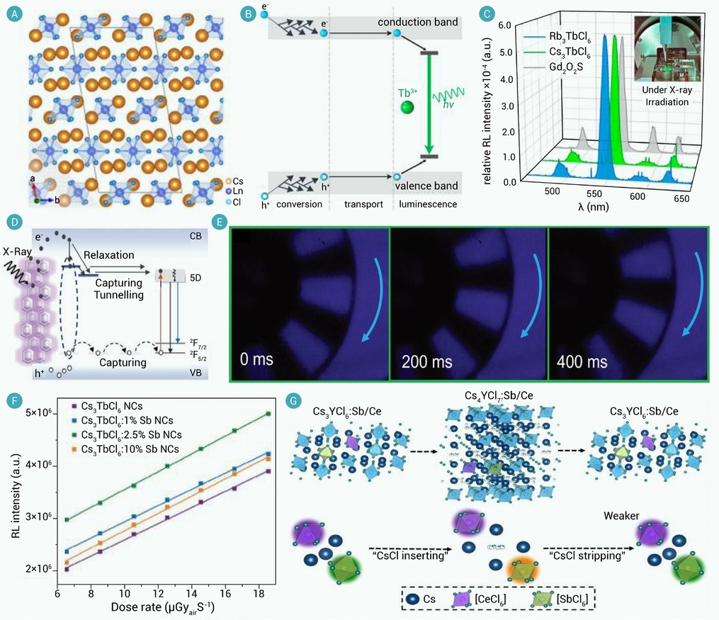
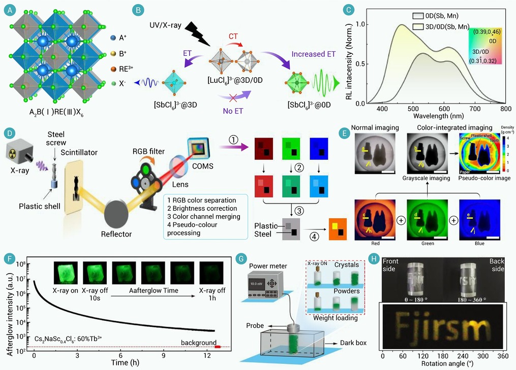
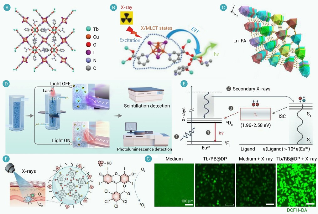
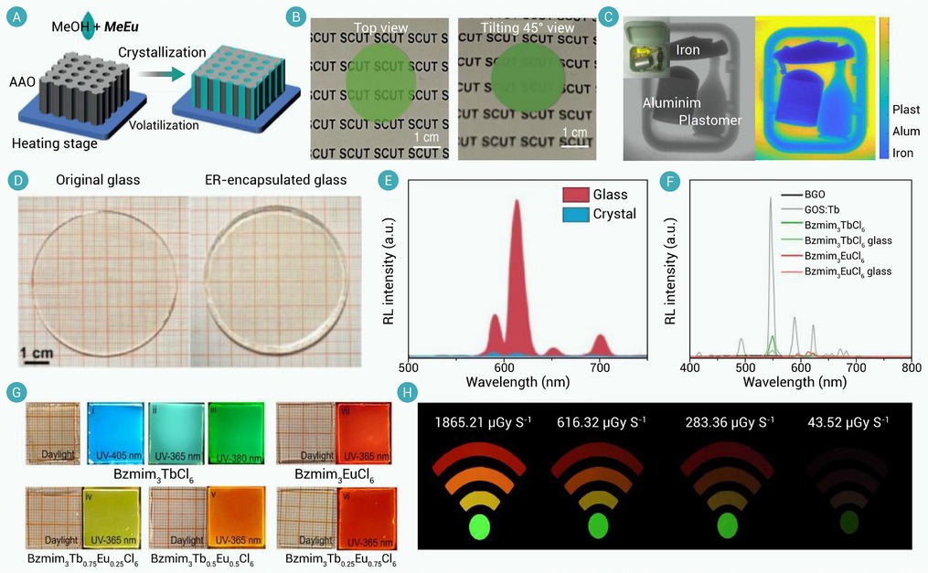
-
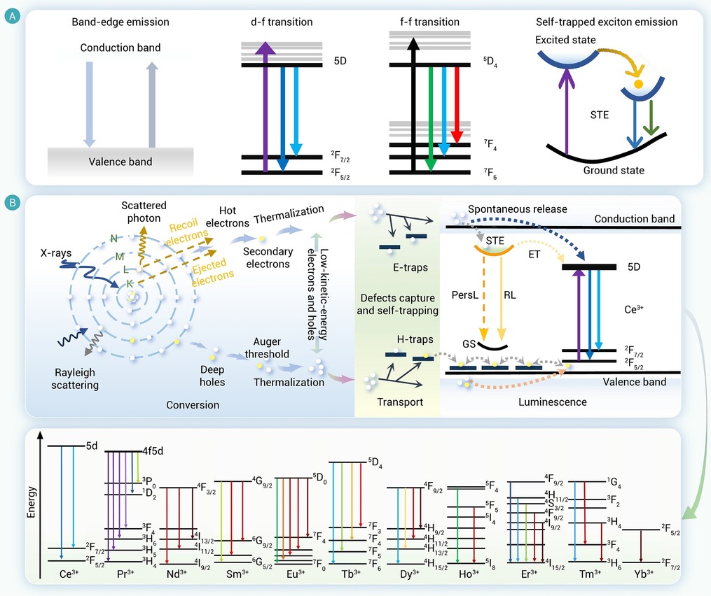
Figure 1.
Scintillation mechanisms of rare-earth halide perovskite-like scintillators
-
Figure 2.
Scintillation mechanisms of AREX4 rare-earth halides
-
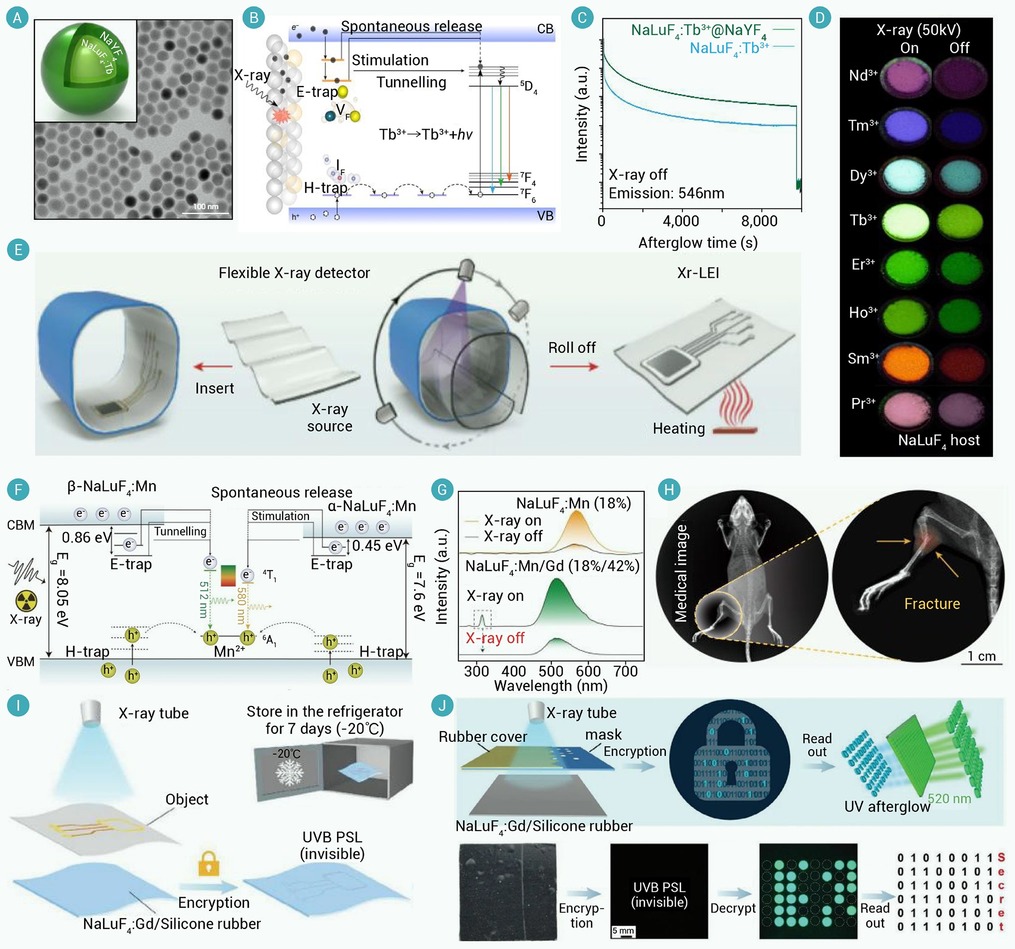
Figure 3.
AREX4 rare-earth halides for multi-scenario X-ray applications
-
Figure 4.
Scintillation mechanisms and properties of A3REX6 rare-earth halides
-
Figure 5.
Scintillation mechanisms and properties of A2BREX6 rare-earth halides
-
Figure 6.
Scintillation mechanism and properties of organic-inorganic hybrid rare-earth halides
-
Figure 7.
Organic-inorganic hybrid rare-earth halides for multi-scenario X-ray applications
-
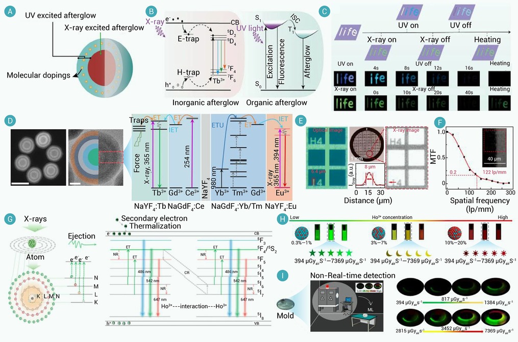
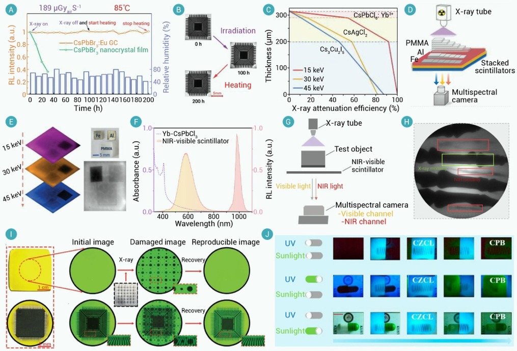
Figure 8.
Rare-earth-doped halides for multi-scenario X-ray applications
