
Single circulating tumor cell omics: Technologies and clinical applications
-
GRAPHICAL ABSTRACT
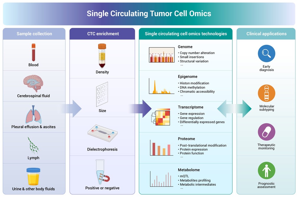
 DownLoad:
Full size image
DownLoad:
Full size image
-
PUBLIC SUMMARY
-
Circulating tumor cells (CTCs) carry tumor DNA, RNA, and proteins in blood for non-invasive diagnosis.
Single-cell omics technologies reveal CTC genomes, transcriptomes, proteomes, and metabolomes.
CTC single-cell omics aids early detection, guides therapy, tracks drug resistance, and predicts prognosis.
This review covers CTC enrichment, single-cell omics, and their clinical application in oncology.
Integrating multi-omics with artificial intelligence and CTC models will advance precision cancer medicine.
-
-
ABSTRACT
Disseminating from primary or metastatic lesions, circulating tumor cells (CTCs) enter various body fluids—including blood, cerebrospinal fluid, and pleural effusions—acting as a liquid biopsy that provides a comprehensive molecular repository. The rapid maturation of single-cell omics has redefined CTC analysis, shifting the focus from simple enumeration to high-resolution molecular characterization. The application of these single-cell technologies has become pivotal for the next generation of precision oncology. This review first delineates the landscape of CTC enrichment, critically evaluating mainstream strategies based on biophysical and biochemical properties alongside emerging innovations. We then analyze the workflow of CTC single-cell omics analysis across five core dimensions: isolation techniques, genomics, transcriptomics, proteomics, and metabolomics. Furthermore, we highlight the clinical translational value of these approaches across four critical axes: early cancer detection, molecular subtyping for companion diagnostics, dynamic monitoring of therapeutic resistance, and prognostic risk stratification. The analysis concludes by addressing current technical bottlenecks and outlining a roadmap for integrating CTC profiling into routine clinical practice. -

-
REFERENCES
[1] Xie W., Suryaprakash S., Wu C., et al. (2023). Trends in the use of liquid biopsy in oncology. Nat. Rev. Drug Discov. 22:612−613. DOI:10.1038/d41573-023-00111-y [2] Zhu Z., Hu E., Shen H., et al. (2023). The functional and clinical roles of liquid biopsy in patient-derived models. J. Hematol. Oncol. 16:36. DOI:10.1186/s13045-023-01433-5 [3] Yu W., Hurley J., Roberts D., et al. (2021). Exosome-based liquid biopsies in cancer: opportunities and challenges. Ann. Oncol. 32:466−477. DOI:10.1016/j.annonc.2021.01.074 [4] Lin D., Shen L., Luo M., et al. (2021). Circulating tumor cells: biology and clinical significance. Signal Transduct. Target. Ther. 6:404. DOI:10.1038/s41392-021-00817-8 [5] Ring A., Nguyen-Strauli B. D., Wicki A., et al. (2023). Biology, vulnerabilities and clinical applications of circulating tumour cells. Nat. Rev. Cancer 23:95−111. DOI:10.1038/s41568-022-00536-4 [6] Lawrence R., Watters M., Davies C. R., et al. (2023). Circulating tumour cells for early detection of clinically relevant cancer. Nat. Rev. Clin. Oncol. 20:487−500. DOI:10.1038/s41571-023-00781-y [7] Diamantopoulou Z., Castro-Giner F., Schwab F. D., et al. (2022). The metastatic spread of breast cancer accelerates during sleep. Nature 607:156−162. DOI:10.1038/s41586-022-04875-y [8] Sharifi M. N., Sperger J. M., Taylor A. K., et al. (2025). High-purity CTC RNA sequencing identifies prostate cancer lineage phenotypes prognostic for clinical outcomes. Cancer Discov. DOI:10.1158/2159-8290.CD-24-1509 %J Cancer Discovery. [9] Rawal S., Yang Y. P., Cote R., et al. (2017). Identification and Quantitation of Circulating Tumor Cells. Annu. Rev. Anal. Chem. (Palo Alto Calif.) 10:321−343. DOI:10.1146/annurev-anchem-061516-045405 [10] Li Y., Xu F., Qiao J., et al. (2023). Into the microscale: Low-input sequencing technologies and applications in medicine. Innov. Med. 1:100041. DOI:10.59717/j.xinn-med.2023.100041. [11] Zhang L., Li Z., Skrzypczynska K. M., et al. (2020). Single-Cell Analyses Inform Mechanisms of Myeloid-Targeted Therapies in Colon Cancer. Cell 181:442-459 e429. DOI:10.1016/j.cell.2020.03.048. [12] Lim B., Lin Y. and Navin N. (2020). Advancing Cancer Research and Medicine with Single-Cell Genomics. Cancer Cell 37:456−470. DOI:10.1016/j.ccell.2020.03.008 [13] Alix-Panabieres C. and Pantel K. (2025). Advances in liquid biopsy: From exploration to practical application. Cancer Cell 43:161−165. DOI:10.1016/j.ccell.2024.11.009 [14] Nikanjam M., Kato S. and Kurzrock R. (2022). Liquid biopsy: current technology and clinical applications. J. Hematol. Oncol. 15:131. DOI:10.1186/s13045-022-01351-y [15] Gao Y., Fan W.-H., Song Z., et al. (2021). Comparison of circulating tumor cell (CTC) detection rates with epithelial cell adhesion molecule (EpCAM) and cell surface vimentin (CSV) antibodies in different solid tumors: a retrospective study. PeerJ 9. DOI:10.7717/peerj.10777. [16] Descamps L., Le Roy D. and Deman A. L. (2022). Microfluidic-Based Technologies for CTC Isolation: A Review of 10 Years of Intense Efforts towards Liquid Biopsy. Int. J. Mol. Sci. 23. DOI:10.3390/ijms23041981. [17] Ding P., Wang Z., Wu Z., et al. (2020). Aptamer-based nanostructured interfaces for the detection and release of circulating tumor cells. J. Mater. Chem. B 8:3408−3422. DOI:10.1039/c9tb02457c [18] Gabriel M. T., Calleja L. R., Chalopin A., et al. (2016). Circulating Tumor Cells: A Review of Non-EpCAM-Based Approaches for Cell Enrichment and Isolation. Clin. Chem. 62:571−581. DOI:10.1373/clinchem.2015.249706 [19] Ahn J. C., Teng P. C., Chen P. J., et al. (2021). Detection of Circulating Tumor Cells and Their Implications as a Biomarker for Diagnosis, Prognostication, and Therapeutic Monitoring in Hepatocellular Carcinoma. Hepatology 73:422−436. DOI:10.1002/hep.31165 [20] Rahmanian M., Sartipzadeh Hematabad O., Askari E., et al. (2023). A micropillar array-based microfluidic chip for label-free separation of circulating tumor cells: The best micropillar geometry. J. Adv. Res. 47:105−121. DOI:10.1016/j.jare.2022.08.005 [21] Cheng S.-B., Chen M.-M., Wang Y.-K., et al. (2021). A Three-Dimensional Conductive Scaffold Microchip for Effective Capture and Recovery of Circulating Tumor Cells with High Purity. Anal. Chem. 93:7102−7109. DOI:10.1021/acs.analchem.1c00785 [22] Arora S., D'Souza A., Aland G., et al. (2022). Antibody mediated cotton-archetypal substrate for enumeration of circulating tumor cells and chemotherapy outcome in 3D tumors. Lab Chip 22:1519−1530. DOI:10.1039/d2lc00004k [23] Chen J., Lin Z., Wan X., et al. (2025). Magnetic Genetically Engineered Cells Constructed via Microfluidic Squeezing for Highly Efficient Capture of Circulating Tumor Cells. Small 21. DOI:10.1002/smll.202503795. [24] Zhang C., Zhang X., Zhang J., et al. (2026). Multivalent capture of circulating tumor cells using tetrahedral DNA framework-targeted nanomagnetic beads integrated microfluidic device. Colloids Surf. B. Biointerfaces 257. DOI:10.1016/j.colsurfb.2025.115142. [25] Pedrosa V. A., Chen K., George T. J., et al. (2023). Gold Nanoparticle-Based Microfluidic Chips for Capture and Detection of Circulating Tumor Cells. Biosensors 13. DOI:10.3390/bios13070706. [26] Cao Y., Xu Y., Luo T., et al. (2025). Nanotentacle-Armed Flexible Affinity Interface Enables Streamlined Capture, Labeling, and Release of Circulating Tumor Cells. ACS Nano 19:30115−30124. DOI:10.1021/acsnano.5c06198 [27] Xiang Y., Zhang H., Lu H., et al. (2023). Bioorthogonal Microbubbles with Antifouling Nanofilm for Instant and Suspended Enrichment of Circulating Tumor Cells. ACS Nano 17:9633−9646. DOI:10.1021/acsnano.3c03194 [28] Zhou X., Zhang Y., Kang K., et al. (2022). Controllable Environment Protein Corona-Disguised Immunomagnetic Beads for High-Performance Circulating Tumor Cell Enrichment. Anal. Chem. 94:4650−4657. DOI:10.1021/acs.analchem.1c04587 [29] Zhang Y., Han C., Li K., et al. (2025). Single circulating tumor cell sequencing based on improved high-porosity membranes and nanoporous microchambers. Biosens. Bioelectron. 276:117263. DOI:10.1016/j.bios.2025.117263 [30] Pan Y., Wang Z., Ma J., et al. (2022). Folic Acid-Modified Fluorescent-Magnetic Nanoparticles for Efficient Isolation and Identification of Circulating Tumor Cells in Ovarian Cancer. Biosensors 12. DOI:10.3390/bios12030184. [31] Woo H. J., Kim S.-H., Kang H. J., et al. (2022). Continuous centrifugal microfluidics (CCM) isolates heterogeneous circulating tumor cells via full automation. Theranostics 12:3676−3689. DOI:10.7150/thno.72511 [32] Liu Z., Huang Y., Liang W., et al. (2021). Cascaded filter deterministic lateral displacement microchips for isolation and molecular analysis of circulating tumor cells and fusion cells. Lab Chip 21:2881−2891. DOI:10.1039/d1lc00360g [33] Yu Y., Zhang Y., Chen Y., et al. (2023). Floating Immunomagnetic Microspheres for Highly Efficient Circulating Tumor Cell Isolation under Facile Magnetic Manipulation. ACS Sensors 8:1858−1866. DOI:10.1021/acssensors.3c00420 [34] Jiang W., Han L., Li G., et al. (2023). Bait-trap chip for accurate and ultrasensitive capture of living circulating tumor cells. Acta Biomater. 162:226−239. DOI:10.1016/j.actbio.2023.03.019 [35] Abdulla A., Zhang T., Li S., et al. (2022). Integrated microfluidic single-cell immunoblotting chip enables high-throughput isolation, enrichment and direct protein analysis of circulating tumor cells. Microsyst. Nanoeng. 8. DOI:10.1038/s41378-021-00342-2. [36] Wang Z., Wu Z., Sun N., et al. (2021). Antifouling hydrogel-coated magnetic nanoparticles for selective isolation and recovery of circulating tumor cells. J. Mater. Chem. B 9:677−682. DOI:10.1039/d0tb02380a [37] Jiang X., Zhang X., Guo C., et al. (2022). Protein corona-coated immunomagnetic nanoparticles with enhanced isolation of circulating tumor cells. Nanoscale 14:8474−8483. DOI:10.1039/d2nr01568d [38] Zhou X., Zhang Y., Kang K., et al. (2022). Artificial cell membrane camouflaged immunomagnetic nanoparticles for enhanced circulating tumor cell isolation. J. Mater. Chem. B 10:3119−3125. DOI:10.1039/d1tb02676c [39] Chu Q., Mu W., Lan C., et al. (2021). High-Specific Isolation and Instant Observation of Circulating Tumour Cell from HCC Patients via Glypican-3 Immunomagnetic Fluorescent Nanodevice. Int. J. Nanomed. Volume 16:4161−4173. DOI:10.2147/ijn.S307691 [40] Song J. W., Suh J., Lee S. W., et al. (2022). Isolation and Genomic Analysis of Single Circulating Tumor Cell Using Human Telomerase Reverse Transcriptase and Desmoglein-2. Small Methods 6:e2100938. DOI:10.1002/smtd.202100938 [41] Cha J., Cho H., Chung J.-S., et al. (2023). Effective Circulating Tumor Cell Isolation Using Epithelial and Mesenchymal Markers in Prostate and Pancreatic Cancer Patients. Cancers (Basel) 15. DOI:10.3390/cancers15102825. [42] Ding P., Wang Z., Wu Z., et al. (2021). Tannic Acid (TA)-Functionalized Magnetic Nanoparticles for EpCAM-Independent Circulating Tumor Cell (CTC) Isolation from Patients with Different Cancers. ACS Appl. Mater. Interfaces 13:3694−3700. DOI:10.1021/acsami.0c20916 [43] Stiefel J., Freese C., Sriram A., et al. (2022). Characterization of a novel microfluidic platform for the isolation of rare single cells to enable CTC analysis from head and neck squamous cell carcinoma patients. Eng. Life Sci. 22:391−406. DOI:10.1002/elsc.202100133 [44] Sun Y. and Sethu P. (2018). Low-stress Microfluidic Density-gradient Centrifugation for Blood Cell Sorting. Biomed. Microdevices 20. DOI:10.1007/s10544-018-0323-3. [45] Eslami S. Z., Cortes-Hernandez L. E., Thomas F., et al. (2022). Functional analysis of circulating tumour cells: the KEY to understand the biology of the metastatic cascade. Br. J. Cancer 127:800−810. DOI:10.1038/s41416-022-01819-1 [46] Danova M., Torchio M. and Mazzini G. (2011). Isolation of rare circulating tumor cells in cancer patients: technical aspects and clinical implications. Expert Rev. Mol. Diagn. 11:473−485. DOI:10.1586/erm.11.33 [47] Park J. M., Kim M. S., Moon H. S., et al. (2014). Fully automated circulating tumor cell isolation platform with large-volume capacity based on lab-on-a-disc. Anal. Chem. 86:3735−3742. DOI:10.1021/ac403456t [48] Huang Q., Wang F. B., Yuan C. H., et al. (2018). Gelatin Nanoparticle-Coated Silicon Beads for Density-Selective Capture and Release of Heterogeneous Circulating Tumor Cells with High Purity. Theranostics 8:1624−1635. DOI:10.7150/thno.23531 [49] Harouaka R. A., Nisic M. and Zheng S. Y. (2013). Circulating tumor cell enrichment based on physical properties. Journal of Laboratory Automation 18:455−468. DOI:10.1177/2211068213494391 [50] Ferreira M. M., Ramani V. C. and Jeffrey S. S. (2016). Circulating tumor cell technologies. Mol. Oncol. 10:374−394. DOI:10.1016/j.molonc.2016.01.007 [51] Ciccioli M., Kim K., Khazan N., et al. (2024). Identification of circulating tumor cells captured by the FDA-cleared Parsortix® PC1 system from the peripheral blood of metastatic breast cancer patients using immunofluorescence and cytopathological evaluations. J. Exp. Clin. Cancer Res. 43. DOI:10.1186/s13046-024-03149-x. [52] Miller M. C., Robinson P. S., Wagner C., et al. (2018). The Parsortix™ Cell Separation System—A versatile liquid biopsy platform. Cytom. A 93:1234−1239. DOI:10.1002/cyto.a.23571 [53] Zabaglo L., Ormerod M. G., Parton M., et al. (2003). Cell filtration‐laser scanning cytometry for the characterisation of circulating breast cancer cells. Cytom. A 55A:102-108. DOI:10.1002/cyto.a.10071. [54] Shirai K., Guan G., Meihui T., et al. (2022). Hybrid double-spiral microfluidic chip for RBC-lysis-free enrichment of rare cells from whole blood. Lab Chip 22:4418−4429. DOI:10.1039/d2lc00713d [55] Zhou J., Tu C., Liang Y., et al. (2018). Isolation of cells from whole blood using shear-induced diffusion. Sci. Rep. 8. DOI:10.1038/s41598-018-27779-2. [56] Zhao Y.-N., Zhang X., Bai J.-J., et al. (2024). Inertial and Deterministic Lateral Displacement Integrated Microfluidic Chips for Epithelial–Mesenchymal Transition Analysis. Anal. Chem. 96:18187−18194. DOI:10.1021/acs.analchem.4c04366 [57] Wang S., Xu Q., Zhang Z., et al. (2023). Reverse flow enhanced inertia pinched flow fractionation. Lab Chip 23:4324−4333. DOI:10.1039/d3lc00473b [58] Zhou J., Kulasinghe A., Bogseth A., et al. (2019). Isolation of circulating tumor cells in non-small-cell-lung-cancer patients using a multi-flow microfluidic channel. Microsyst. Nanoeng. 5. DOI:10.1038/s41378-019-0045-6. [59] Gascoyne P. R., Wang X. B., Huang Y., et al. (1997). Dielectrophoretic Separation of Cancer Cells from Blood. IEEE Transactions on Industry Applications 33:670−678. DOI:10.1109/28.585856 [60] Becker F. F., Wang X. B., Huang Y., et al. (1995). Separation of human breast cancer cells from blood by differential dielectric affinity. Proc. Natl. Acad. Sci. U. S. A. 92:860−864. DOI:10.1073/pnas.92.3.860 [61] Allard W. J., Matera J., Miller M. C., et al. (2004). Tumor Cells Circulate in the Peripheral Blood of All Major Carcinomas but not in Healthy Subjects or Patients With Nonmalignant Diseases. Clin. Cancer Res. 10:6897−6904. DOI:10.1158/1078-0432.CCR-04-0378 %J Clinical Cancer Research. DOI:10.1158/1078-0432.CCR-04-0378%JClinicalCancerResearch [62] Marsavela G., Aya-Bonilla C. A., Warkiani M. E., et al. (2018). Melanoma circulating tumor cells: Benefits and challenges required for clinical application. Cancer Lett. 424:1−8. DOI:10.1016/j.canlet.2018.03.013 [63] Liu X., Li J., Cadilha B. L., et al. (2019). Epithelial-type systemic breast carcinoma cells with a restricted mesenchymal transition are a major source of metastasis. Sci. Adv. 5:eaav4275. DOI. DOI:10.1126/sciadv.aav4275 [64] Hodan R., Gupta S., Weiss J. M., et al. (2024). Genetic/Familial High-Risk Assessment: Colorectal, Endometrial, and Gastric, Version 3.2024, NCCN Clinical Practice Guidelines In Oncology. J. Natl. Compr. Canc. Netw. 22:695-711. DOI:10.6004/jnccn.2024.0061. [65] Eibl R. H. and Schneemann M. (2021). Liquid Biopsy and Primary Brain Tumors. Cancers (Basel) 13. DOI:10.3390/cancers13215429. [66] Gorin M. A., Verdone J. E., van der Toom E., et al. (2017). Circulating tumour cells as biomarkers of prostate, bladder, and kidney cancer. Nat. Rev. Urol. 14:90−97. DOI:10.1038/nrurol.2016.224 [67] Loibl S., Poortmans P., Morrow M., et al. (2021). Breast cancer. Lancet 397:1750−1769. DOI:10.1016/S0140-6736(20)32381-3 [68] Yu M., Bardia A., Wittner B. S., et al. (2013). Circulating Breast Tumor Cells Exhibit Dynamic Changes in Epithelial and Mesenchymal Composition. Science 339:580−584. DOI. DOI:10.1126/science.1228522 [69] Markou A. and Lianidou E. S. (2011). Circulating Tumor Cells in Breast Cancer: Detection Systems, Molecular Characterization, and Future Challenges. Clin. Chem. 57:1242−1255. DOI:10.1373/clinchem.2011.165068. [70] Zhou J., Zhu X., Wu S., et al. (2020). Epithelial-mesenchymal transition status of circulating tumor cells in breast cancer and its clinical relevance. Cancer Biol. Med. 17:169−180. DOI:10.20892/j.issn.2095-3941.2019.0118 [71] Odintsov I. and Sholl L. M. (2024). Prognostic and predictive biomarkers in non-small cell lung carcinoma. Pathology 56:192−204. DOI:10.1016/j.pathol.2023.11.006 [72] Andrikou K., Rossi T., Verlicchi A., et al. (2023). Circulating Tumour Cells: Detection and Application in Advanced Non-Small Cell Lung Cancer. Int. J. Mol. Sci. 24. DOI:10.3390/ijms242216085. [73] Scharpenseel H., Hanssen A., Loges S., et al. (2019). EGFR and HER3 expression in circulating tumor cells and tumor tissue from non-small cell lung cancer patients. Sci. Rep. 9:7406. DOI:10.1038/s41598-019-43678-6 [74] Chen L., Peng M., Li N., et al. (2018). Combined use of EpCAM and FRα enables the high-efficiency capture of circulating tumor cells in non-small cell lung cancer. Sci. Rep. 8. DOI:10.1038/s41598-018-19391-1. [75] Welsch E., Holzer B., Schuster E., et al. (2024). Prognostic significance of circulating tumor cells and tumor related transcripts in small cell lung cancer: A step further to clinical implementation. Int. J. Cancer 154:2189−2199. DOI:10.1002/ijc.34886 [76] Hamilton G., Hochmair M., Rath B., et al. (2016). Small cell lung cancer: Circulating tumor cells of extended stage patients express a mesenchymal-epithelial transition phenotype. Cell Adh. Migr. 10:360−367. DOI:10.1080/19336918.2016.1155019 [77] Papakonstantinou D., Roumeliotou A., Pantazaka E., et al. (2024). Integrative analysis of circulating tumor cells (CTCs) and exosomes from small-cell lung cancer (SCLC) patients: a comprehensive approach. Mol. Oncol. DOI:10.1002/1878-0261.13765. [78] Hou J.-M., Krebs M. G., Lancashire L., et al. (2012). Clinical Significance and Molecular Characteristics of Circulating Tumor Cells and Circulating Tumor Microemboli in Patients With Small-Cell Lung Cancer. Signal Transduct. Target. Ther. 30:525−532. DOI:10.1200/jco.2010.33.3716 [79] Ricordel C., Chaillot L., Vlachavas E. I., et al. (2023). Genomic characteristics and clinical significance of CD56+ circulating tumor cells in small cell lung cancer. Sci. Rep. 13:3626. DOI:10.1038/s41598-023-30536-9 [80] Shou X., Li Y., Hu W., et al. (2019). Six-gene Assay as a new biomarker in the blood of patients with colorectal cancer: establishment and clinical validation. Mol. Oncol. 13:781−791. DOI:10.1002/1878-0261.12427 [81] Satelli A., Mitra A., Brownlee Z., et al. (2015). Epithelial-mesenchymal transitioned circulating tumor cells capture for detecting tumor progression. Clin. Cancer Res. 21:899−906. DOI:10.1158/1078-0432.CCR-14-0894 [82] Hinz S., Hendricks A., Wittig A., et al. (2017). Detection of circulating tumor cells with CK20 RT-PCR is an independent negative prognostic marker in colon cancer patients - a prospective study. BMC Cancer 17:53. DOI:10.1186/s12885-016-3035-1 [83] Neves M., Azevedo R., Lima L., et al. (2019). Exploring sialyl-Tn expression in microfluidic-isolated circulating tumour cells: A novel biomarker and an analytical tool for precision oncology applications. N. Biotechnol. 49:77−87. DOI:10.1016/j.nbt.2018.09.004 [84] Wei C., Yang C., Wang S., et al. (2019). Crosstalk between cancer cells and tumor associated macrophages is required for mesenchymal circulating tumor cell-mediated colorectal cancer metastasis. Molecular Cancer 18. DOI:10.1186/s12943-019-0976-4. [85] Liang M. X., Fei Y. J., Yang K., et al. (2022). Potential values of circulating tumor cell for detection of recurrence in patients of thyroid cancer: a diagnostic meta-analysis. BMC Cancer 22:954. DOI:10.1186/s12885-022-09976-5 [86] Yu H. W., Park E., Lee J. K., et al. (2024). Analyzing circulating tumor cells and epithelial-mesenchymal transition status of papillary thyroid carcinoma patients following thyroidectomy: a prospective cohort study. Int. J. Surg. 110:3357−3364. DOI:10.1097/JS9.0000000000001284 [87] Dai Y. J., Qiu Y. B., Jiang R., et al. (2017). Concomitant high expression of ERalpha36, EGFR and HER2 is associated with aggressive behaviors of papillary thyroid carcinomas. Sci. Rep. 7:12279. DOI:10.1038/s41598-017-12478-1 [88] Xu W., Cao L., Chen L., et al. (2011). Isolation of circulating tumor cells in patients with hepatocellular carcinoma using a novel cell separation strategy. Clin. Cancer Res. 17:3783−3793. DOI:10.1158/1078-0432.CCR-10-0498 [89] Li Y. M., Xu S. C., Li J., et al. (2013). Epithelial-mesenchymal transition markers expressed in circulating tumor cells in hepatocellular carcinoma patients with different stages of disease. Cell Death Dis. 4:e831. DOI:10.1038/cddis.2013.347 [90] Gerber T. S., Ridder D. A., Goeppert B., et al. (2024). N‐cadherin: A diagnostic marker to help discriminate primary liver carcinomas from extrahepatic carcinomas. Int. J. Cancer 154:1857−1868. DOI:10.1002/ijc.34836 [91] Li Y., Zhang X., Liu D., et al. (2018). Evolutionary Expression of HER2 Conferred by Chromosome Aneuploidy on Circulating Gastric Cancer Cells Contributes to Developing Targeted and Chemotherapeutic Resistance. Clin. Cancer Res. 24:5261−5271. DOI:10.1158/1078-0432.CCR-18-1205 %J Clinical Cancer Research. DOI:10.1158/1078-0432.CCR-18-1205%JClinicalCancerResearch [92] Zheng X., Fan L., Zhou P., et al. (2017). Detection of Circulating Tumor Cells and Circulating Tumor Microemboli in Gastric Cancer. Transl. Oncol. 10:431−441. DOI:10.1016/j.tranon.2017.02.007 [93] Mimori K., Fukagawa T., Kosaka Y., et al. (2008). A large-scale study of MT1-MMP as a marker for isolated tumor cells in peripheral blood and bone marrow in gastric cancer cases. Ann. Surg. Oncol. 15:2934−2942. DOI:10.1245/s10434-008-9916-z [94] Gopalan V. and Lam A. K. (2018). Circulatory Tumor Cells in Esophageal Adenocarcinoma. In Esophageal Adenocarcinoma: Methods and Protocols, A.K. Lam, ed. (Springer New York), pp. 177-186. 10.1007/978-1-4939-7734-5_16. [95] Joo D. C., Kim G. H., I H., et al. (2024). Clinical Implications of Circulating Tumor Cells in Patients with Esophageal Squamous Cell Carcinoma: Cancer-Draining Blood Versus Peripheral Blood. Cancers (Basel) 16. DOI:10.3390/cancers16162921. [96] Xi L., Nicastri D. G., El-Hefnawy T., et al. (2007). Optimal markers for real-time quantitative reverse transcription PCR detection of circulating tumor cells from melanoma, breast, colon, esophageal, head and neck, and lung cancers. Clin. Chem. 53:1206−1215. DOI:10.1373/clinchem.2006.081828 [97] Cho H., Byun S.-S., Son N.-H., et al. (2024). Impact of Circulating Tumor Cell–Expressed Prostate-Specific Membrane Antigen and Prostate-Specific Antigen Transcripts in Different Stages of Prostate Cancer. Clin. Cancer Res. 30:1788−1800. DOI:10.1158/1078-0432.CCR-23-3083 %J Clinical Cancer Research. DOI:10.1158/1078-0432.CCR-23-3083%JClinicalCancerResearch [98] Kilercik M., Ozgur E., Sahin S., et al. (2024). Detection of circulating tumor cells in non-metastatic prostate cancer through integration of a microfluidic CTC enrichment system and multiparametric flow cytometry. PLoS One 19:e0312296. DOI:10.1371/journal.pone.0312296 [99] Day K. C., Lorenzatti Hiles G., Kozminsky M., et al. (2017). HER2 and EGFR Overexpression Support Metastatic Progression of Prostate Cancer to Bone. Cancer Res. 77:74−85. DOI:10.1158/0008-5472.CAN-16-1656 [100] Nagrath S., Jack R. M., Sahai V., et al. (2016). Opportunities and Challenges for Pancreatic Circulating Tumor Cells. Gastroenterology 151:412−426. DOI:10.1053/j.gastro.2016.05.052 [101] Zhao X. H., Wang Z. R., Chen C. L., et al. (2019). Molecular detection of epithelial-mesenchymal transition markers in circulating tumor cells from pancreatic cancer patients: Potential role in clinical practice. World J. Gastroenterol. 25:138−150. DOI:10.3748/wjg.v25.i1.138 [102] Signorelli R., Giret T. M., Umland O., et al. (2022). ALCAM: A Novel Surface Marker on EpCAM(low) Circulating Tumor Cells. Biomedicines 10. DOI:10.3390/biomedicines10081983. [103] Rink M., Chun F. K., Dahlem R., et al. (2012). Prognostic Role and HER2 Expression of Circulating Tumor Cells in Peripheral Blood of Patients Prior to Radical Cystectomy: A Prospective Study. Eur. Urol. 61:810−817. DOI:10.1016/j.eururo.2012.01.017 [104] MacArthur K. M., Kao G. D., Chandrasekaran S., et al. (2014). Detection of Brain Tumor Cells in the Peripheral Blood by a Telomerase Promoter-Based Assay. Cancer Res. 74:2152−2159. DOI:10.1158/0008-5472.Can-13-0813 [105] Sullivan J. P., Nahed B. V., Madden M. W., et al. (2014). Brain Tumor Cells in Circulation Are Enriched for Mesenchymal Gene Expression. Cancer Discov. 4:1299−1309. DOI:10.1158/2159-8290.Cd-14-0471 [106] GRADILONE A., IACOVELLI R., CORTESI E., et al. (2011). Circulating Tumor Cells and “Suspicious Objects” Evaluated Through CellSearch® in Metastatic Renal Cell Carcinoma. Clin. Cancer Res. 31:4219−4221. [107] Bade R. M., Schehr J. L., Emamekhoo H., et al. (2021). Development and initial clinical testing of a multiplexed circulating tumor cell assay in patients with clear cell renal cell carcinoma. Mol. Oncol. 15:2330−2344. DOI:10.1002/1878-0261.12931 [108] Palmela Leitao T., Miranda M., Polido J., et al. (2021). Circulating tumor cell detection methods in renal cell carcinoma: A systematic review. Crit. Rev. Oncol. Hematol. 161:103331. DOI:10.1016/j.critrevonc.2021.103331 [109] Tinhofer I., Konschak R., Stromberger C., et al. (2014). Detection of circulating tumor cells for prediction of recurrence after adjuvant chemoradiation in locally advanced squamous cell carcinoma of the head and neck. Ann. Oncol. 25:2042−2047. DOI:10.1093/annonc/mdu271 [110] Chikamatsu K., Tada H., Takahashi H., et al. (2019). Expression of immune-regulatory molecules in circulating tumor cells derived from patients with head and neck squamous cell carcinoma. Oral Oncol. 89:34−39. DOI:10.1016/j.oraloncology.2018.12.002 [111] Strati A., Koutsodontis G., Papaxoinis G., et al. (2017). Prognostic significance of PD-L1 expression on circulating tumor cells in patients with head and neck squamous cell carcinoma. Ann. Oncol. 28:1923−1933. DOI:10.1093/annonc/mdx206 [112] Peeters D. J., Brouwer A., Van den Eynden G. G., et al. (2015). Circulating tumour cells and lung microvascular tumour cell retention in patients with metastatic breast and cervical cancer. Cancer Lett. 356:872−879. DOI:10.1016/j.canlet.2014.10.039 [113] Gies S., Melchior P., Molnar I., et al. (2025). PD-L1(+) CD49f(+) CD133(+) Circulating tumor cells predict outcome of patients with vulvar or cervical cancer after radio- and chemoradiotherapy. J. Transl. Med. 23:321. DOI:10.1186/s12967-025-06277-w [114] Yan C., Xiao Y., Zhang W., et al. (2023). Circulating Tumor Cells are an Independent Risk Factor for Poor Prognosis in Patients with Gallbladder Adenocarcinoma. Ann. Surg. Oncol. 30:7966−7975. DOI:10.1245/s10434-023-14231-7 [115] Mishra S., Kumari S. and Husain N. (2024). Liquid biopsy in gallbladder carcinoma: Current evidence and future prospective. J. Liq. Biopsy 6. DOI:10.1016/j.jlb.2024.100280. [116] Khoja L., Lorigan P., Zhou C., et al. (2013). Biomarker Utility of Circulating Tumor Cells in Metastatic Cutaneous Melanoma. J. Invest. Dermatol. 133:1582−1590. DOI:10.1038/jid.2012.468 [117] Fusi A., Reichelt U., Busse A., et al. (2011). Expression of the Stem Cell Markers Nestin and CD133 on Circulating Melanoma Cells. J. Invest. Dermatol. 131:487−494. DOI:10.1038/jid.2010.285 [118] Khoja L., Lorigan P., Dive C., et al. (2015). Circulating tumour cells as tumour biomarkers in melanoma: detection methods and clinical relevance. Ann. Oncol. 26:33−39. DOI:10.1093/annonc/mdu207 [119] Reza K. K., Dey S., Wuethrich A., et al. (2021). In Situ Single Cell Proteomics Reveals Circulating Tumor Cell Heterogeneity during Treatment. ACS Nano 15:11231−11243. DOI:10.1021/acsnano.0c10008 [120] Koyanagi K., O'Day S. J., Boasberg P., et al. (2010). Serial Monitoring of Circulating Tumor Cells Predicts Outcome of Induction Biochemotherapy plus Maintenance Biotherapy for Metastatic Melanoma. Clin. Cancer Res. 16:2402−2408. DOI:10.1158/1078-0432.Ccr-10-0037 [121] Serrano-Gomez S. J., Maziveyi M. and Alahari S. K. (2016). Regulation of epithelial-mesenchymal transition through epigenetic and post-translational modifications. Mol. Cancer 15. DOI:10.1186/s12943-016-0502-x. [122] Kuburich N. A., den Hollander P., Pietz J. T., et al. (2022). Vimentin and cytokeratin: Good alone, bad together. Semin. Cancer Biol. 86:816−826. DOI:10.1016/j.semcancer.2021.12.006 [123] Wei T., Zhang X., Zhang Q., et al. (2019). Vimentin-positive circulating tumor cells as a biomarker for diagnosis and treatment monitoring in patients with pancreatic cancer. Cancer Lett. 452:237−243. DOI:10.1016/j.canlet.2019.03.009 [124] Miettinen M., McCue P. A., Sarlomo-Rikala M., et al. (2014). GATA3: A Multispecific But Potentially Useful Marker in Surgical Pathology: A Systematic Analysis of 2500 Epithelial and Nonepithelial Tumors. Am. J. Surg. Pathol. 38:13−22. DOI:10.1097/PAS.0b013e3182a0218f [125] Werling R. W., Yaziji H., Bacchi C. E., et al. (2003). CDX2, a Highly Sensitive and Specific Marker of Adenocarcinomas of Intestinal Origin: An Immunohistochemical Survey of 476 Primary and Metastatic Carcinomas. Am. J. Surg. Pathol. 27:303−310. [126] Rossi T., Bandini S., Zanoni M., et al. (2025). Transcriptomic profiling of circulating tumor cells from metastatic breast cancer patients reveals new hints in their biological features and phenotypic heterogeneity. Exp. Hematol. Oncol. 14:67. DOI:10.1186/s40164-025-00659-y [127] Onstenk W., Sieuwerts A. M., Weekhout M., et al. (2015). Gene expression profiles of circulating tumor cells versus primary tumors in metastatic breast cancer. Cancer Lett. 362:36−44. DOI:10.1016/j.canlet.2015.03.020 [128] Stoecklein N. H., Fluegen G., Guglielmi R., et al. (2023). Ultra-sensitive CTC-based liquid biopsy for pancreatic cancer enabled by large blood volume analysis. Mol. Cancer 22. DOI:10.1186/s12943-023-01880-1. [129] Rieckmann L.-M., Spohn M., Ruff L., et al. (2024). Diagnostic leukapheresis reveals distinct phenotypes of NSCLC circulating tumor cells. Mol. Cancer 23. DOI:10.1186/s12943-024-01984-2. [130] Chauhan A., Pal A., Sachdeva M., et al. (2024). A FACS-based novel isolation technique identifies heterogeneous CTCs in oral squamous cell carcinoma. Front. Oncol. 14:1269211. DOI:10.3389/fonc.2024.1269211 [131] Lambros M. B., Seed G., Sumanasuriya S., et al. (2018). Single-Cell Analyses of Prostate Cancer Liquid Biopsies Acquired by Apheresis. Clin. Cancer Res. 24:5635−5644. DOI:10.1158/1078-0432.CCR-18-0862 [132] Pailler E., Faugeroux V., Oulhen M., et al. (2019). Acquired Resistance Mutations to ALK Inhibitors Identified by Single Circulating Tumor Cell Sequencing in ALK-Rearranged Non-Small-Cell Lung Cancer. Clin. Cancer Res. 25:6671−6682. DOI:10.1158/1078-0432.CCR-19-1176 [133] Li R., Jia F., Zhang W., et al. (2019). Device for whole genome sequencing single circulating tumor cells from whole blood. Lab Chip 19:3168−3178. DOI:10.1039/c9lc00473d [134] Li Y., Jiang X., Zhong M., et al. (2022). Whole Genome Sequencing of Single-Circulating Tumor Cell Ameliorates Unraveling Breast Cancer Heterogeneity. Breast Cancer (Dove Med Press) 14:505−513. DOI:10.2147/BCTT.S388653 [135] Kim O., Lee D., Chungwon Lee A., et al. (2019). Whole Genome Sequencing of Single Circulating Tumor Cells Isolated by Applying a Pulsed Laser to Cell-Capturing Microstructures. Small 15:e1902607. DOI:10.1002/smll.201902607 [136] Kojima M., Harada T., Fukazawa T., et al. (2021). Single-cell DNA and RNA sequencing of circulating tumor cells. Sci. Rep. 11:22864. DOI:10.1038/s41598-021-02165-7 [137] Liu H. E., Triboulet M., Zia A., et al. (2017). Workflow optimization of whole genome amplification and targeted panel sequencing for CTC mutation detection. NPJ Genom. Med. 2:34. DOI:10.1038/s41525-017-0034-3 [138] Daniel M., Knutson T. P., Sperger J. M., et al. (2021). AR gene rearrangement analysis in liquid biopsies reveals heterogeneity in lethal prostate cancer. Endocr. Relat. Cancer 28:645−655. DOI:10.1530/erc-21-0157 [139] Deger T., Mendelaar P. A. J., Kraan J., et al. (2022). A pipeline for copy number profiling of single circulating tumour cells to assess intrapatient tumour heterogeneity. Mol. Oncol. 16:2981−3000. DOI:10.1002/1878-0261.13174 [140] Yadav D., Patil-Takbhate B., Khandagale A., et al. (2023). Next-Generation sequencing transforming clinical practice and precision medicine. Clin. Chim. Acta 551. DOI:10.1016/j.cca.2023.117568. [141] Yu J., Gemenetzis G., Kinny-Koster B., et al. (2020). Pancreatic circulating tumor cell detection by targeted single-cell next-generation sequencing. Cancer Lett. 493:245−253. DOI:10.1016/j.canlet.2020.08.043 [142] Shen X., Dai J., Guo L., et al. (2024). Single-cell low-pass whole genome sequencing accurately detects circulating tumor cells for liquid biopsy-based multi-cancer diagnosis. NPJ Precis. Oncol. 8:30. DOI:10.1038/s41698-024-00520-1 [143] Xu X., Lin L., Yang J., et al. (2022). Simultaneous single-cell genome and transcriptome sequencing in nanoliter droplet with digital microfluidics identifying essential driving genes. Nano Today 46. DOI:10.1016/j.nantod.2022.101596. [144] Di L., Fu Y., Sun Y., et al. (2020). RNA sequencing by direct tagmentation of RNA/DNA hybrids. Proc. Natl. Acad. Sci. U. S. A. 117:2886−2893. DOI:10.1073/pnas.1919800117 [145] Hagemann-Jensen M., Ziegenhain C., Chen P., et al. (2020). Single-cell RNA counting at allele and isoform resolution using Smart-seq3. Nat. Biotechnol. 38:708−714. DOI:10.1038/s41587-020-0497-0 [146] Hahaut V., Pavlinic D., Carbone W., et al. (2022). Fast and highly sensitive full-length single-cell RNA sequencing using FLASH-seq. Nat. Biotechnol. 40:1447−1451. DOI:10.1038/s41587-022-01312-3 [147] He L., Dang K., Sun Q., et al. (2026). High-Resolution Multiplexed Sequencing of Single-Cell Full-length Transcriptome Via Combinational Barcoded Tn5 Transposon Insertion. Adv. Sci. 13:e16013. DOI:10.1002/advs.202516013 [148] Cheng Y. H., Chen Y. C., Lin E., et al. (2019). Hydro-Seq enables contamination-free high-throughput single-cell RNA-sequencing for circulating tumor cells. Nat. Commun. 10:2163. DOI:10.1038/s41467-019-10122-2 [149] Lightbody E. D., Sklavenitis-Pistofidis R., Wu T., et al. (2025). SWIFT-seq enables comprehensive single-cell transcriptomic profiling of circulating tumor cells in multiple myeloma and its precursors. Nat. Cancer 6:1595−1611. DOI:10.1038/s43018-025-01006-0 [150] Brechbuhl H. M., Vinod-Paul K., Gillen A. E., et al. (2020). Analysis of circulating breast cancer cell heterogeneity and interactions with peripheral blood mononuclear cells. Mol. Carcinog. 59:1129−1139. DOI:10.1002/mc.23242 [151] Sinkala E., Sollier-Christen E., Renier C., et al. (2017). Profiling protein expression in circulating tumour cells using microfluidic western blotting. Nat. Commun. 8:14622. DOI:10.1038/ncomms14622 [152] Shi Q., Qin L., Wei W., et al. (2012). Single-cell proteomic chip for profiling intracellular signaling pathways in single tumor cells. Proc. Natl. Acad. Sci. U. S. A. 109:419−424. DOI:10.1073/pnas.1110865109 [153] Yang L., Wang Z., Deng Y., et al. (2016). Single-Cell, Multiplexed Protein Detection of Rare Tumor Cells Based on a Beads-on-Barcode Antibody Microarray. Anal. Chem. 88:11077−11083. DOI:10.1021/acs.analchem.6b03086 [154] Deng Y., Zhang Y., Sun S., et al. (2014). An integrated microfluidic chip system for single-cell secretion profiling of rare circulating tumor cells. Sci. Rep. 4:7499. DOI:10.1038/srep07499 [155] Zhang Y., Tang Y., Sun S., et al. (2015). Single-Cell Codetection of Metabolic Activity, Intracellular Functional Proteins, and Genetic Mutations from Rare Circulating Tumor Cells. Anal. Chem. 87:9761−9768. DOI:10.1021/acs.analchem.5b01901 [156] Song Y., Tian T., Shi Y., et al. (2017). Enrichment and single-cell analysis of circulating tumor cells. Chem. Sci. 8:1736−1751. DOI:10.1039/c6sc04671a [157] Wang C., Yang L., Wang Z., et al. (2019). Highly multiplexed profiling of cell surface proteins on single circulating tumor cells based on antibody and cellular barcoding. Anal. Bioanal. Chem. 411:5373−5382. DOI:10.1007/s00216-019-01666-9 [158] Chai S., Ruiz-Velasco C., Naghdloo A., et al. (2022). Identification of epithelial and mesenchymal circulating tumor cells in clonal lineage of an aggressive prostate cancer case. NPJ Precis. Oncol. 6:41. DOI:10.1038/s41698-022-00289-1 [159] Karabacak N. M., Zheng Y., Dubash T. D., et al. (2022). Differential Kinase Activity Across Prostate Tumor Compartments Defines Sensitivity to Target Inhibition. Cancer Res. 82:1084−1097. DOI:10.1158/0008-5472.CAN-21-2609 [160] Payne K., Brooks J., Batis N., et al. (2023). Feasibility of mass cytometry proteomic characterisation of circulating tumour cells in head and neck squamous cell carcinoma for deep phenotyping. Br. J. Cancer 129:1590−1598. DOI:10.1038/s41416-023-02428-2 [161] Welter L., Zheng S., Setayesh S. M., et al. (2023). Cell State and Cell Type: Deconvoluting Circulating Tumor Cell Populations in Liquid Biopsies by Multi-Omics. Cancers (Basel) 15. DOI:10.3390/cancers15153949. [162] Guo T., Steen J. A. and Mann M. (2025). Mass-spectrometry-based proteomics: from single cells to clinical applications. Nature 638:901−911. DOI:10.1038/s41586-025-08584-0 [163] Bartkowiak K., Mohammadi P. M., Nissen P., et al. (2025). Discovery of a sushi domain-containing protein 2-positive phenotype in circulating tumor cells of metastatic breast cancer patients. Sci. Rep. 15:3913. DOI:10.1038/s41598-025-87122-4 [164] Horrmann A., Arafa A., Kamalanathan K., et al. (2025). A liquid biopsy-based proteomic assay to identify circulating tumor cell (CTC) –derived druggable targets in cancer patients. J. Clin. Oncol. 43:258−258. DOI:10.1200/JCO.2025.43.5_suppl.258 [165] Huang M. S., Fu L. H., Yan H. C., et al. (2022). Proteomics and liquid biopsy characterization of human EMT-related metastasis in colorectal cancer. Front. Oncol. 12:790096. DOI:10.3389/fonc.2022.790096 [166] Que Z., Xi Z., Qi D., et al. (2025). Src/FN1 pathway activation drives tumor cell cluster formation and metastasis in lung cancer: A promising therapeutic target. Sci. Adv. 11:eadv7377. DOI. DOI:10.1126/sciadv.adv7377 [167] Vuille J. A., Tanriover C., Antmen E., et al. (2025). The E3 ligase HECTD4 regulates COX-2-dependent tumor progression and metastasis. Proc. Natl. Acad. Sci. U. S. A. 122:e2425621122. DOI:10.1073/pnas.2425621122 [168] Wang L., Abdulla A., Wang A., et al. (2022). Sickle-like Inertial Microfluidic System for Online Rare Cell Separation and Tandem Label-Free Quantitative Proteomics (Orcs-Proteomics). Anal. Chem. 94:6026−6035. DOI:10.1021/acs.analchem.2c00679 [169] Tsai C. F., Zhang P., Scholten D., et al. (2021). Surfactant-assisted one-pot sample preparation for label-free single-cell proteomics. Commun. Biol. 4:265. DOI:10.1038/s42003-021-01797-9 [170] Budnik B., Levy E., Harmange G., et al. (2018). SCoPE-MS: mass spectrometry of single mammalian cells quantifies proteome heterogeneity during cell differentiation. Genome Biol. 19:161. DOI:10.1186/s13059-018-1547-5 [171] Gebreyesus S. T., Siyal A. A., Kitata R. B., et al. (2022). Streamlined single-cell proteomics by an integrated microfluidic chip and data-independent acquisition mass spectrometry. Nat. Commun. 13:37. DOI:10.1038/s41467-021-27778-4 [172] Derks J., Leduc A., Wallmann G., et al. (2023). Increasing the throughput of sensitive proteomics by plexDIA. Nat. Biotechnol. 41:50−59. DOI:10.1038/s41587-022-01389-w [173] Wang Y., Guan Z. Y., Shi S. W., et al. (2024). Pick-up single-cell proteomic analysis for quantifying up to 3000 proteins in a Mammalian cell. Nat. Commun. 15:1279. DOI:10.1038/s41467-024-45659-4 [174] Ye Z., Sabatier P., Martin-Gonzalez J., et al. (2024). One-Tip enables comprehensive proteome coverage in minimal cells and single zygotes. Nat. Commun. 15:2474. DOI:10.1038/s41467-024-46777-9 [175] Ye Z., Sabatier P., van der Hoeven L., et al. (2025). Enhanced sensitivity and scalability with a Chip-Tip workflow enables deep single-cell proteomics. Nat. Methods 22:499−509. DOI:10.1038/s41592-024-02558-2 [176] Liang L., Sun F., Wang H., et al. (2021). Metabolomics, metabolic flux analysis and cancer pharmacology. Pharmacol. Ther. 224:107827. DOI:10.1016/j.pharmthera.2021.107827 [177] Sun F., Li H., Sun D., et al. (2025). Single-cell omics: experimental workflow, data analyses and applications. Sci. China Life Sci. 68:5−102. DOI:10.1007/s11427-023-2561-0 [178] Zhang W., Xu F., Yao J., et al. (2023). Single-cell metabolic fingerprints discover a cluster of circulating tumor cells with distinct metastatic potential. Nat. Commun. 14:2485. DOI:10.1038/s41467-023-38009-3 [179] Hiyama E., Ali A., Amer S., et al. (2015). Direct Lipido-Metabolomics of Single Floating Cells for Analysis of Circulating Tumor Cells by Live Single-cell Mass Spectrometry. Anal. Sci. 31:1215−1217. DOI:10.2116/analsci.31.1215 [180] Abouleila Y., Onidani K., Ali A., et al. (2019). Live single cell mass spectrometry reveals cancer-specific metabolic profiles of circulating tumor cells. Cancer Sci. 110:697−706. DOI:10.1111/cas.13915 [181] Hu X., Xu T., Xia S., et al. (2025). Metabolic Adaptation Study of Tumor Cells during Lung Cancer Bone Metastasis in Mice Based on Single-Cell Metabolome Analysis. J. Am. Soc. Mass Spectrom. 36:1959−1969. DOI:10.1021/jasms.5c00177 [182] Xu Y., Hu X., Yuan Y., et al. (2025). Prediction of Lung Cancer Metastasis Risk Based on Single-Cell Metabolic Profiling of Circulating Tumor Cells. Adv. Sci. 12:e08878. DOI:10.1002/advs.202508878 [183] Luan H., Chen S., Lian J., et al. (2024). Biofluorescence imaging-guided spatial metabolic tracing: In vivo tracking of metabolic activity in circulating tumor cell-mediated multi-organ metastases. Talanta 280:126696. DOI:10.1016/j.talanta.2024.126696 [184] Radfar P., Ding L., de la Fuente L. R., et al. (2023). Rapid metabolomic screening of cancer cells via high-throughput static droplet microfluidics. Biosens. Bioelectron. 223:114966. DOI:10.1016/j.bios.2022.114966 [185] Dai C. S., Mishra A., Edd J., et al. (2025). Circulating tumor cells: Blood-based detection, molecular biology, and clinical applications. Cancer Cell 43:1399−1422. DOI:10.1016/j.ccell.2025.07.008 [186] Raza A., Khan A. Q., Inchakalody V. P., et al. (2022). Dynamic liquid biopsy components as predictive and prognostic biomarkers in colorectal cancer. J. Exp. Clin. Cancer Res. 41. DOI:10.1186/s13046-022-02318-0. [187] Lianidou E. S., Mavroudis D. and Georgoulias V. (2013). Clinical challenges in the molecular characterization of circulating tumour cells in breast cancer. Br. J. Cancer 108:2426−2432. DOI:10.1038/bjc.2013.265 [188] Crosby D., Bhatia S., Brindle K. M., et al. (2022). Early detection of cancer. Science 375. DOI:10.1126/science.aay9040. [189] Batool S. M., Yekula A., Khanna P., et al. (2023). The Liquid Biopsy Consortium: Challenges and opportunities for early cancer detection and monitoring. Cell Rep. Med. 4. DOI:10.1016/j.xcrm.2023.101198. [190] Rupp B., Nagpal N., Thanasiu B., et al. (2025). Multiplex characterization of circulating tumor cells from ductal carcinoma in situ patients suggests early tumor dissemination. Cancer Lett. 623:217703. DOI:10.1016/j.canlet.2025.217703 [191] Xu C., Xu X., Shao W., et al. (2023). CTCs Detection and Whole-exome Sequencing Might Be Used to Differentiate Benign and Malignant Pulmonary Nodules. Zhongguo Fei Ai Za Zhi 26:449−460. DOI:10.3779/j.issn.1009-3419.2023.106.12 [192] Wang S., Xu C., Xu X., et al. (2025). The Role of Circulating Tumor Cell as a Promising Biomarker in the Evaluation of Pulmonary Nodules: A Prospective Study. Cancer Res. Treat. DOI:10.4143/crt.2024.841. [193] Guo X., Lin F., Yi C., et al. (2022). Deep transfer learning enables lesion tracing of circulating tumor cells. Nat. Commun. 13:7687. DOI:10.1038/s41467-022-35296-0 [194] Kojima M., Harada T., Fukazawa T., et al. (2023). Single-cell next-generation sequencing of circulating tumor cells in patients with neuroblastoma. Cancer Sci. 114:1616−1624. DOI:10.1111/cas.15707 [195] Carter L., Rothwell D. G., Mesquita B., et al. (2016). Molecular analysis of circulating tumor cells identifies distinct copy-number profiles in patients with chemosensitive and chemorefractory small-cell lung cancer. Nat. Med. 23:114−119. DOI:10.1038/nm.4239 [196] Gasch C., Oldopp T., Mauermann O., et al. (2016). Frequent detection of PIK3CA mutations in single circulating tumor cells of patients suffering from HER2‐negative metastatic breast cancer. Mol. Oncol. 10:1330−1343. DOI:10.1016/j.molonc.2016.07.005 [197] Rimawi M. F., Schiff R. and Osborne C. K. (2015). Targeting HER2 for the Treatment of Breast Cancer. Annu. Rev. Med. 66:111−128. DOI:10.1146/annurev-med-042513-015127 [198] Van Poznak C., Somerfield M. R., Bast R. C., et al. (2015). Use of Biomarkers to Guide Decisions on Systemic Therapy for Women With Metastatic Breast Cancer: American Society of Clinical Oncology Clinical Practice Guideline. J. Clin. Oncol. 33:2695−2704. DOI:10.1200/jco.2015.61.1459 [199] Barnett E. S., Schultz N., Stopsack K. H., et al. (2023). Analysis of BRCA2 Copy Number Loss and Genomic Instability in Circulating Tumor Cells from Patients with Metastatic Castration-resistant Prostate Cancer. Eur. Urol. 83:112−120. DOI:10.1016/j.eururo.2022.08.010 [200] Mishra A., Huang S. B., Dubash T., et al. (2025). Tumor cell-based liquid biopsy using high-throughput microfluidic enrichment of entire leukapheresis product. Nat. Commun. 16:32. DOI:10.1038/s41467-024-55140-x [201] Lu X., Cheng L., Yang C., et al. (2024). Crosstalk between bladder cancer and the tumor microenvironment: Molecular mechanisms and targeted therapy. Innov. Med. 2. DOI:10.59717/j.xinn-med.2024.100094. [202] King J. D., Casavant B. P. and Lang J. M. (2014). Rapid translation of circulating tumor cell biomarkers into clinical practice: technology development, clinical needs and regulatory requirements. Lab Chip 14:24−31. DOI:10.1039/c3lc50741f [203] Maltoni R., Gallerani G., Fici P., et al. (2016). CTCs in early breast cancer: A path worth taking. Cancer Lett. 376:205−210. DOI:10.1016/j.canlet.2016.03.051 [204] Moon D. H., Lindsay D. P., Hong S., et al. (2018). Clinical indications for, and the future of, circulating tumor cells. Adv. Drug Del. Rev. 125:143−150. DOI:10.1016/j.addr.2018.04.002 [205] Lim M., Park J., Lowe A. C., et al. (2020). A lab-on-a-disc platform enables serial monitoring of individual CTCs associated with tumor progression during EGFR-targeted therapy for patients with NSCLC. Theranostics 10:5181−5194. DOI:10.7150/thno.44693 [206] Blau C. A., Ramirez A. B., Blau S., et al. (2016). A Distributed Network for Intensive Longitudinal Monitoring in Metastatic Triple-Negative Breast Cancer. J. Natl. Compr. Canc. Netw. 14:8−17. DOI:10.6004/jnccn.2016.0003 [207] Paoletti C., Cani A. K., Larios J. M., et al. (2018). Comprehensive Mutation and Copy Number Profiling in Archived Circulating Breast Cancer Tumor Cells Documents Heterogeneous Resistance Mechanisms. Cancer Res. 78:1110−1122. DOI:10.1158/0008-5472.Can-17-2686 [208] Franken A., Honisch E., Reinhardt F., et al. (2020). Detection of ESR1 Mutations in Single Circulating Tumor Cells on Estrogen Deprivation Therapy but Not in Primary Tumors from Metastatic Luminal Breast Cancer Patients. J. Mol. Diagn. 22:111−121. DOI:10.1016/j.jmoldx.2019.09.004 [209] Mezquita L., Oulhen M., Aberlenc A., et al. (2024). Resistance to BRAF inhibition explored through single circulating tumour cell molecular profiling in BRAF-mutant non-small-cell lung cancer. Br. J. Cancer 130:682−693. DOI:10.1038/s41416-023-02535-0 [210] Hong X., Roh W., Sullivan R. J., et al. (2021). The Lipogenic Regulator SREBP2 Induces Transferrin in Circulating Melanoma Cells and Suppresses Ferroptosis. Cancer Discov. 11:678−695. DOI:10.1158/2159-8290.Cd-19-1500 [211] Cani A. K., Dolce E. M., Darga E. P., et al. (2022). Serial monitoring of genomic alterations in circulating tumor cells of ER-positive/HER2-negative advanced breast cancer: feasibility of precision oncology biomarker detection. Mol. Oncol. 16:1969−1985. DOI:10.1002/1878-0261.13150 [212] Horwitz E., Dubash T. D., Szabolcs A., et al. (2025). CDKN1B (p27/kip1) enhances drug-tolerant persister CTCs by restricting polyploidy following mitotic inhibitors. Proc. Natl. Acad. Sci. U. S. A. 122:e2507203122. DOI:10.1073/pnas.2507203122 [213] Que Z., Luo B., Zhou Z., et al. (2019). Establishment and characterization of a patient-derived circulating lung tumor cell line in vitro and in vivo. Cancer Cell Int. 19. DOI:10.1186/s12935-019-0735-z. [214] Stewart C. A., Gay C. M., Xi Y., et al. (2020). Single-cell analyses reveal increased intratumoral heterogeneity after the onset of therapy resistance in small-cell lung cancer. Nat. Cancer 1:423−436. DOI:10.1038/s43018-019-0020-z [215] Suvilesh K. N., Nussbaum Y. I., Radhakrishnan V., et al. (2022). Tumorigenic circulating tumor cells from xenograft mouse models of non-metastatic NSCLC patients reveal distinct single cell heterogeneity and drug responses. Mol. Cancer 21. DOI:10.1186/s12943-022-01553-5. [216] Huebner H., Fasching P. A., Gumbrecht W., et al. (2018). Filtration based assessment of CTCs and CellSearch® based assessment are both powerful predictors of prognosis for metastatic breast cancer patients. BMC Cancer 18. DOI:10.1186/s12885-018-4115-1. [217] Pereira-Veiga T., Schneegans S., Pantel K., et al. (2022). Circulating tumor cell-blood cell crosstalk: Biology and clinical relevance. Cell Rep. 40. DOI:10.1016/j.celrep.2022.111298. [218] Deng Z., Wu S., Wang Y., et al. (2022). Circulating tumor cell isolation for cancer diagnosis and prognosis. eBioMedicine 83. DOI:10.1016/j.ebiom.2022.104237. [219] Ahmed A., Greene S. B., Dago A. E., et al. (2016). Chromosomal Instability Estimation Based on Next Generation Sequencing and Single Cell Genome Wide Copy Number Variation Analysis. PLoS One 11. DOI:10.1371/journal.pone.0165089. [220] Hoon D. S. B., Morton D. L., Irie R. F., et al. (2014). Genome-Wide Characterization of Circulating Tumor Cells Identifies Novel Prognostic Genomic Alterations in Systemic Melanoma Metastasis. Clin. Chem. 60:873−885. DOI:10.1373/clinchem.2013.213611 [221] Malihi P. D., Graf R. P., Rodriguez A., et al. (2020). Single-Cell Circulating Tumor Cell Analysis Reveals Genomic Instability as a Distinctive Feature of Aggressive Prostate Cancer. Clin. Cancer Res. 26:4143−4153. DOI:10.1158/1078-0432.CCR-19-4100 [222] Alves J. M., Estévez-Gómez N., Piñeiro R., et al. (2025). Genomic diversity and BCL9L mutational status in circulating tumor cells predict overall survival in metastatic colorectal cancer. Cell. Oncol. 48:1809−1820. DOI:10.1007/s13402-025-01109-x [223] Markou A., Farkona S., Schiza C., et al. (2014). PIK3CA Mutational Status in Circulating Tumor Cells Can Change During Disease Recurrence or Progression in Patients with Breast Cancer. Clin. Cancer Res. 20:5823−5834. DOI:10.1158/1078-0432.Ccr-14-0149 [224] Lohr J. G., Kim S., Gould J., et al. (2016). Genetic interrogation of circulating multiple myeloma cells at single-cell resolution. Sci. Transl. Med. 8:363ra147. DOI:10.1126/scitranslmed.aac7037 [225] Ebright R. Y., Lee S., Wittner B. S., et al. (2020). Deregulation of ribosomal protein expression and translation promotes breast cancer metastasis. Science 367:1468−1473. DOI:10.1126/science.aay0939 [226] Kozuka M., Battaglin F., Jayachandran P., et al. (2021). Clinical Significance of Circulating Tumor Cell Induced Epithelial-Mesenchymal Transition in Patients with Metastatic Colorectal Cancer by Single-Cell RNA-Sequencing. Cancers (Basel) 13. DOI:10.3390/cancers13194862. [227] Rossi T., Gallerani G., Angeli D., et al. (2020). Single-Cell NGS-Based Analysis of Copy Number Alterations Reveals New Insights in Circulating Tumor Cells Persistence in Early-Stage Breast Cancer. Cancers (Basel) 12. DOI:10.3390/cancers12092490. [228] Nitschke C., Markmann B., Tolle M., et al. (2022). Characterization of RARRES1 Expression on Circulating Tumor Cells as Unfavorable Prognostic Marker in Resected Pancreatic Ductal Adenocarcinoma Patients. Cancers (Basel) 14. DOI:10.3390/cancers14184405. [229] Szczerba B. M., Castro-Giner F., Vetter M., et al. (2019). Neutrophils escort circulating tumour cells to enable cell cycle progression. Nature 566:553−557. DOI:10.1038/s41586-019-0915-y [230] Hapach L. A., Carey S. P., Schwager S. C., et al. (2021). Phenotypic Heterogeneity and Metastasis of Breast Cancer Cells. Cancer Res. 81:3649−3663. DOI:10.1158/0008-5472.Can-20-1799 [231] Gao X., Li X., Xu W., et al. (2025). Identification of multiple genomic alterations and prediction of neoantigens from circulating tumor cells at the single-cell level. Nat. Commun. 16. DOI:10.1038/s41467-025-62215-w. [232] Peterson V. M., Zhang K. X., Kumar N., et al. (2017). Multiplexed quantification of proteins and transcripts in single cells. Nat. Biotechnol. 35:936−939. DOI:10.1038/nbt.3973 [233] Stoeckius M., Hafemeister C., Stephenson W., et al. (2017). Simultaneous epitope and transcriptome measurement in single cells. Nat. Methods 14:865−868. DOI:10.1038/nmeth.4380 [234] Fulcher J. M., Markillie L. M., Mitchell H. D., et al. (2024). Parallel measurement of transcriptomes and proteomes from same single cells using nanodroplet splitting. Nat. Commun. 15. DOI:10.1038/s41467-024-54099-z. [235] Jiang Y.-R., Zhu L., Cao L.-R., et al. (2023). Simultaneous deep transcriptome and proteome profiling in a single mouse oocyte. Cell Rep. 42. DOI:10.1016/j.celrep.2023.113455. [236] Wu J., Xu Q.-Q., Jiang Y.-R., et al. (2024). One-Shot Single-Cell Proteome and Metabolome Analysis Strategy for the Same Single Cell. Anal. Chem. 96:5499−5508. DOI:10.1021/acs.analchem.3c05659 [237] He Y., Yuan H., Liang Y., et al. (2023). On-capillary alkylation micro-reactor: a facile strategy for proteo-metabolome profiling in the same single cells. Chem. Sci. 14:13495−13502. DOI:10.1039/d3sc05047e [238] Hu Y., Wan S., Luo Y., et al. (2024). Benchmarking algorithms for single-cell multi-omics prediction and integration. Nat. Methods 21:2182−2194. DOI:10.1038/s41592-024-02429-w [239] Liu C., Ding S., Kim H. J., et al. (2025). Multitask benchmarking of single-cell multimodal omics integration methods. Nat. Methods 22:2449−2460. DOI:10.1038/s41592-025-02856-3 [240] Tien Y. W., Kuo H.-C., Ho B.-I., et al. (2016). A High Circulating Tumor Cell Count in Portal Vein Predicts Liver Metastasis From Periampullary or Pancreatic Cancer. Medicine 95. DOI:10.1097/md.0000000000003407. [241] Dong X., Ma Y., Zhao X., et al. (2020). Spatial heterogeneity in epithelial to mesenchymal transition properties of circulating tumor cells associated with distant recurrence in pancreatic cancer patients. Ann. Transl. Med. 8:676−676. DOI:10.21037/atm-20-782 [242] Sun Y.-F., Guo W., Xu Y., et al. (2018). Circulating Tumor Cells from Different Vascular Sites Exhibit Spatial Heterogeneity in Epithelial and Mesenchymal Composition and Distinct Clinical Significance in Hepatocellular Carcinoma. Clin. Cancer Res. 24:547−559. DOI:10.1158/1078-0432.CCR-17-1063 %J Clinical Cancer Research. DOI:10.1158/1078-0432.CCR-17-1063%JClinicalCancerResearch [243] Buscail E., Chiche L., Laurent C., et al. (2019). Tumor‐proximal liquid biopsy to improve diagnostic and prognostic performances of circulating tumor cells. Mol. Oncol. 13:1811−1826. DOI:10.1002/1878-0261.12534 [244] Sun Y. F., Wu L., Liu S. P., et al. (2021). Dissecting spatial heterogeneity and the immune-evasion mechanism of CTCs by single-cell RNA-seq in hepatocellular carcinoma. Nat. Commun. 12:4091. DOI:10.1038/s41467-021-24386-0 [245] Kahounová Z., Pícková M., Drápela S., et al. (2023). Circulating tumor cell-derived preclinical models: current status and future perspectives. Cell Death Dis. 14. DOI:10.1038/s41419-023-06059-6. [246] Lallo A., Schenk M. W., Frese K. K., et al. (2017). Circulating tumor cells and CDX models as a tool for preclinical drug development. Transl. Lung Cancer Res. 6:397−408. DOI:10.21037/tlcr.2017.08.01 [247] Qu S., Xu R., Yi G., et al. (2024). Patient-derived organoids in human cancer: a platform for fundamental research and precision medicine. Mol. Biomed. 5. DOI:10.1186/s43556-023-00165-9. [248] Li T., Deng B., Li S., et al. (2025). Circulating Tumor Cell‐Derived Organoids: Current Progress, Applications, and Future. MedComm – Future Medicine 4. DOI:10.1002/mef2.70030. [249] Wang P.-X., Zhong Y.-C., Duan B., et al. (2025). Exploring morphological heterogeneity of circulating tumor cells: machine learning-based approach for cell identification and prognostic implications. Sci. Bull. 70:2070−2074. DOI:10.1016/j.scib.2025.04.048 [250] Zhu S., Zhu Z., Ni C., et al. (2024). Liquid Biopsy Instrument for Ultra-Fast and Label-Free Detection of Circulating Tumor Cells. Research 7. DOI:10.34133/research.0431. [251] Zhang W., Lu Y., Su C., et al. (2023). Confinement-guided ultrasensitive optical assay with artificial intelligence for disease diagnostics. Innov. Med. 1. DOI:10.59717/j.xinn-med.2023.100023. [252] Mallery K., Bristow N. R., Heller N., et al. (2026). Circulating tumor cell detection in cancer patients using in-flow deep learning holography. NPJ Biosens. 3. DOI:10.1038/s44328-026-00084-z. [253] Theodoris C. V., Xiao L., Chopra A., et al. (2023). Transfer learning enables predictions in network biology. Nature 618:616−624. DOI:10.1038/s41586-023-06139-9 [254] Cui H., Wang C., Maan H., et al. (2024). scGPT: toward building a foundation model for single-cell multi-omics using generative AI. Nat. Methods 21:1470−1480. DOI:10.1038/s41592-024-02201-0 [255] Lin X., Tian T., Wei Z., et al. (2022). Clustering of single-cell multi-omics data with a multimodal deep learning method. Nat. Commun. 13. DOI:10.1038/s41467-022-35031-9. [256] Baek S., Song K. and Lee I. (2025). Single-cell foundation models: bringing artificial intelligence into cell biology. Exp. Mol. Med. 57:2169−2181. DOI:10.1038/s12276-025-01547-5 [257] Wang G., Zhao J., Lin Y., et al. (2025). scMODAL: a general deep learning framework for comprehensive single-cell multi-omics data alignment with feature links. Nat. Commun. 16. DOI:10.1038/s41467-025-60333-z. [258] Lopez R., Regier J., Cole M. B., et al. (2018). Deep generative modeling for single-cell transcriptomics. Nat. Methods 15:1053−1058. DOI:10.1038/s41592-018-0229-2 [259] Ergen C., Pour Amiri V. V., Kim M., et al. (2025). Scvi-hub: an actionable repository for model-driven single-cell analysis. Nat. Methods 22:1836−1845. DOI:10.1038/s41592-025-02799-9 [260] Cao C., Zhao W., Guo J., et al. (2025). Leveraging artificial intelligence and machine learning for unraveling pathogenesis and advancing precision medicine in autoimmune diseases. Innov. Med. 3. DOI:10.59717/j.xinn-med.2025.100154. -
ABOUT THIS ARTICLE
Cite this article:
Zhang W., He L., Merzougui C., et al. (2026). Single circulating tumor cell omics: Technologies and clinical applications. The Innovation Medicine 4:100227. https://doi.org/10.59717/j.xinn-med.2026.100227
Welcome!
To request copyright permission to republish or share portions of our works, please visit Copyright Clearance Center's (CCC) Marketplace website at marketplace.copyright.com.
Export File
Citation
Zhang W., He L., Merzougui C., et al. (2026). Single circulating tumor cell omics: Technologies and clinical applications. The Innovation Medicine 4:100227. https://doi.org/10.59717/j.xinn-med.2026.100227

Format
Content
-
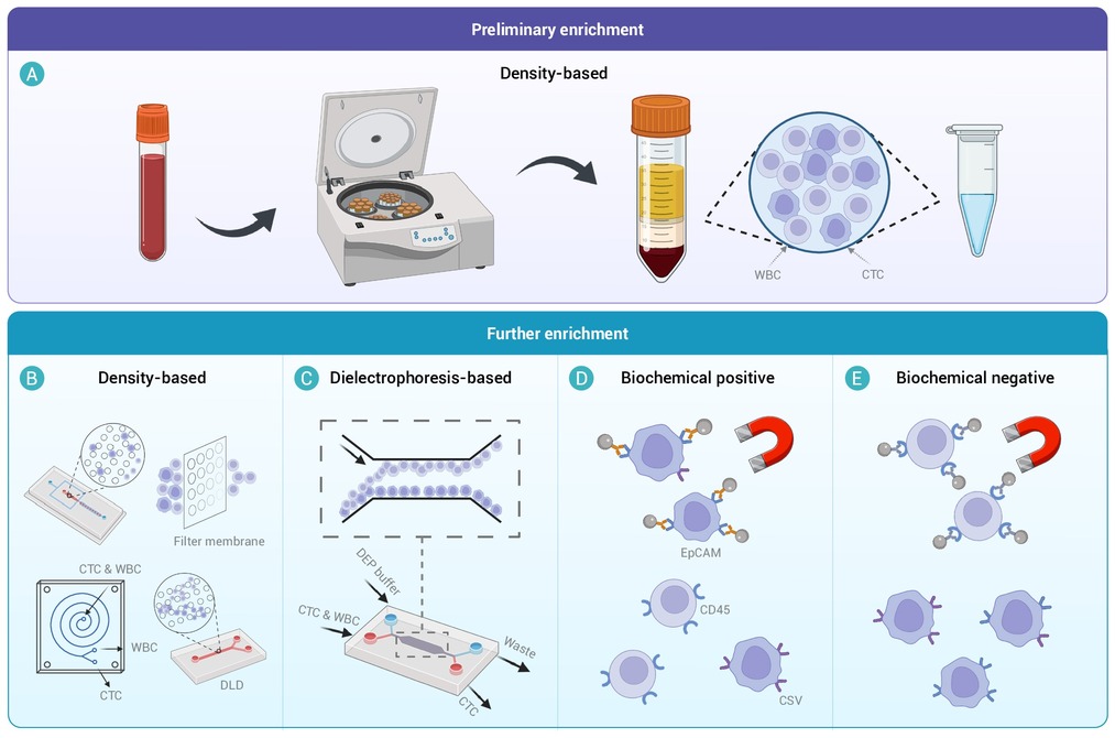
Figure 1.
Common CTC enrichment methods.
-
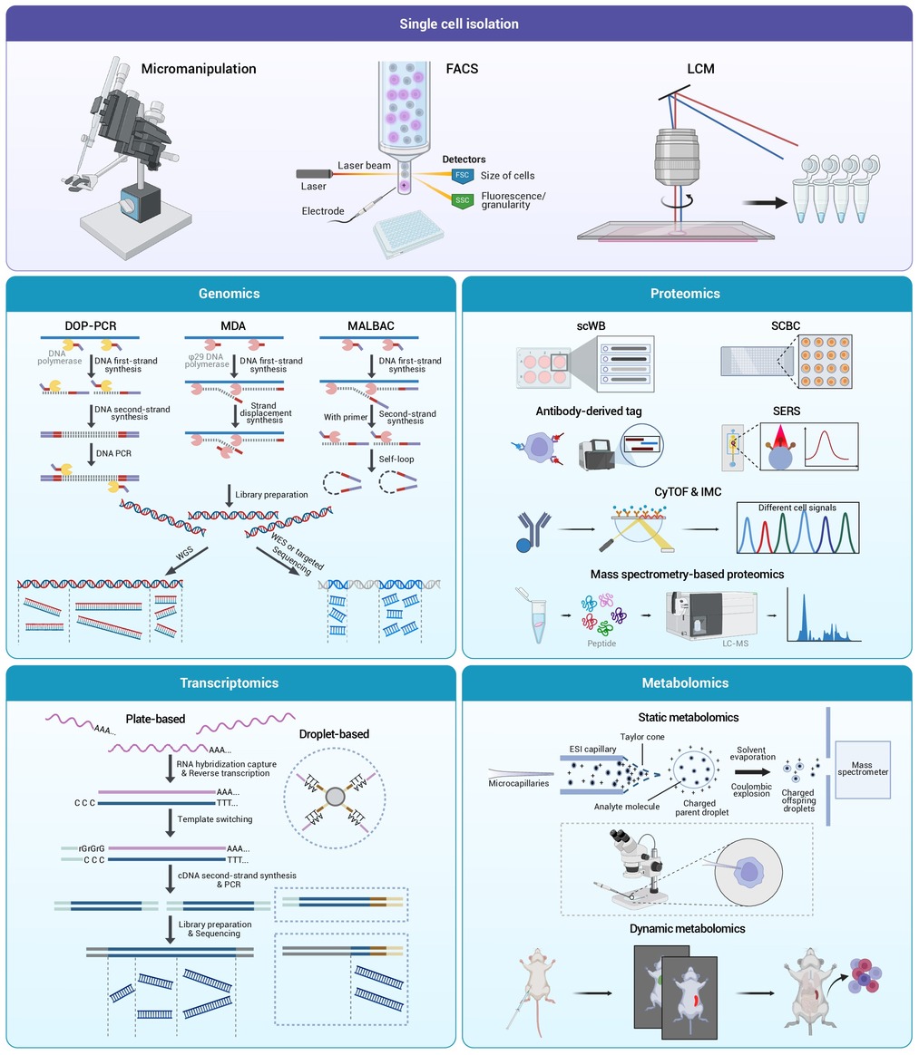
Figure 2.
Single circulating tumor cell omics analysis technologies.
-
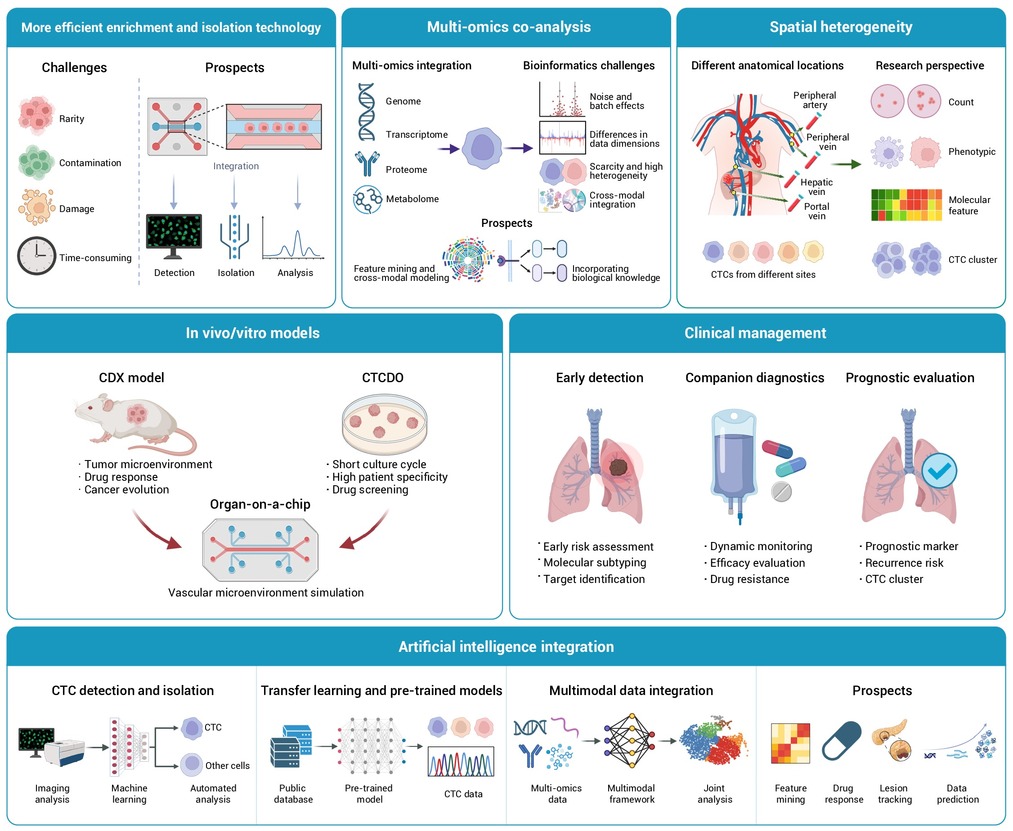
Figure 3.
Challenges and prospects of single CTC omics technologies and clinical applications.
