
Emerging contaminants and their impacts on atopic dermatitis: Current mechanisms and future perspectives
-
GRAPHICAL ABSTRACT
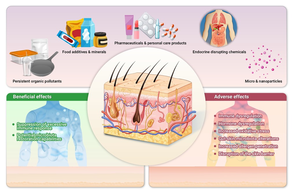
 DownLoad:
Full size image
DownLoad:
Full size image
-
PUBLIC SUMMARY
-
Definitions of emerging contaminants (ECs) are changing with the times.
ECs exhibit varied effects on atopic dermatitis, showing both exacerbating and protective potential.
Recommendations for the optimization of living environments and lifestyles are presented from the EC perspective.
Future research should focus on identifying high-priority ECs and their synergistic effects on AD.
-
-
ABSTRACT
With the progression of industrialization and urbanization, the diversity and concentration of emerging contaminants (ECs) in the environment have risen concomitantly. Atopic dermatitis (AD) is a chronic inflammatory skin condition driven by genetic and environmental factors. Research on the interactions between ECs and AD has expanded significantly in recent decades. This review examines the complex interplay between representative environmental ECs and AD, focusing on mechanisms of immune disruption, including Th2 polarization and aryl hydrocarbon receptor (AhR) activation. Drawing on recent evidence, we further identify critical research gaps to guide future investigations into the impacts of ECs on AD, highlight clinical implications and propose potential strategies for exposure reduction and lifestyle adjustments to prevent and manage AD. -

-
REFERENCES
[1] Melen E. and Garcia-Aymerich J. (2025). Predicting the future global burden of asthma and atopic dermatitis: identifying strategies for prevention. Lancet Respir. Med. 13:376−378. DOI:10.1016/S2213-2600(25)00041-4 [2] Laughter M. R., Maymone M. B. C., Mashayekhi S., et al. (2021). The global burden of atopic dermatitis: lessons from the Global Burden of Disease Study 1990-2017. Br. J. Dermatol. 184:304−309. DOI:10.1111/bjd.19580 [3] Schuler C. F. t., Billi A. C., Maverakis E., et al. (2023). Novel insights into atopic dermatitis. J. Allergy Clin. Immunol. 151:1145−1154. DOI:10.1016/j.jaci.2022.10.023 [4] Lobefaro F., Gualdi G., Di Nuzzo S., et al. (2022). Atopic dermatitis: Clinical aspects and unmet needs. Biomedicines 10. DOI:10.3390/biomedicines10112927 [5] Labib A., Ju T. and Yosipovitch G. (2023). Emerging treatments for itch in atopic dermatitis: A review. J. Am. Acad. Dermatol. 89:338−344. DOI:10.1016/j.jaad.2023.04.057 [6] Fang Z., Li L., Zhang H., et al. (2021). Gut microbiota, probiotics, and their interactions in prevention and preatment of atopic dermatitis: A Review. Front. Immunol. 12:720393. DOI:10.3389/fimmu.2021.720393 [7] Avena-Woods C. (2017). Overview of atopic dermatitis. Am. J. Manag. Care 23:S115−s123. [8] Luger T., Amagai M., Dreno B., et al. (2021). Atopic dermatitis: Role of the skin barrier, environment, microbiome, and therapeutic agents. J. Dermatol. Sci. 102:142−157. DOI:10.1016/j.jdermsci.2021.04.007 [9] Park S. K., Kim J. S. and Seo H. M. (2022). Exposure to air pollution and incidence of atopic dermatitis in the general population: A national population-based retrospective cohort study. J. Am. Acad. Dermatol. 87:1321−1327. DOI:10.1016/j.jaad.2021.05.061 [10] Lopez D. J., Lodge C. J., Bui D. S., et al. (2021). Association between ambient air pollution and development and persistence of atopic and non-atopic eczema in a cohort of adults. Allergy 76:2524−2534. DOI:10.1111/all.14783 [11] Murrison L. B., Brandt E. B., Myers J. B., et al. (2019). Environmental exposures and mechanisms in allergy and asthma development. J. Clin. Invest 129:1504−1515. DOI:10.1172/jci124612 [12] Fuller R., Landrigan P. J., Balakrishnan K., et al. (2022). Pollution and health: A progress update. Lancet Planet. Health 6:e535−e547. DOI:10.1016/S2542-5196(22)00090-0 [13] Viana M., de Leeuw F., Bartonova A., et al. (2020). Air quality mitigation in European cities: Status and challenges ahead. Environ. Int. 143:105907. DOI:10.1016/j.envint.2020.105907 [14] Wójcik-Gront E. and Gozdowski D. (2025). Air pollution monitoring and modeling: A comparative study of PM, NO2, and SO2 with meteorological correlations. Atmosphere 16:1199. DOI:10.3390/atmos16101199 [15] Colette Augustin R. L. (2021). ETC/ATNI report 2019/16: Air quality trends in Europe: 2000-2017. Assessment for surface SO2, NO2, Ozone, PM10 and PM2.5. [16] Puri M., Gandhi K. and Kumar M. S. (2023). Emerging environmental contaminants: A global perspective on policies and regulations. J. Environ. Manage 332:117344. DOI:10.1016/j.jenvman.2023.117344 [17] Ji X., Liang J., Wang Y., et al. (2023). Synthetic antioxidants as contaminants of emerging concern in Indoor environments: Knowns and unknowns. Environ. Sci. Technol. 57:21550−21557. DOI:10.1021/acs.est.3c06487 [18] Tao L., Tan H., Qiao X., et al. (2022). Emerging plasticizers in south China house dust and hand wipes: Calling for potential concern. Environ. Sci. Technol. 56:12190−12199. DOI:10.1021/acs.est.2c02106 [19] Sultan M. B., Anik A. H. and Rahman M. M. (2024). Emerging contaminants and their potential impacts on estuarine ecosystems: Are we aware of it. Mar. Pollut. Bull. 199:115982. DOI:10.1016/j.marpolbul.2023.115982 [20] Rudzanova B., Vlaanderen J., Kalina J., et al. (2023). Impact of PFAS exposure on prevalence of immune-mediated diseases in adults in the Czech Republic. Environ. Res. 229:115969. DOI:10.1016/j.envres.2023.115969 [21] Liaquat H., Imran M., Latif S., et al. (2022). Multifunctional nanomaterials and nanocomposites for sensing and monitoring of environmentally hazardous heavy metal contaminants. Environ. Res. 214:113795. DOI:10.1016/j.envres.2022.113795 [22] Huang J. W., Bai Y. Y., Wang D. S., et al. (2024). Positive association between chlorinated paraffins and the risk of allergic diseases in children and adolescents. J. Hazard Mater. 470:134226. DOI:10.1016/j.jhazmat.2024.134226 [23] Huang D., Liu X., Gao X., et al. (2025). Meteorin-like protein/METRNL/Interleukin-41 ameliorates atopic dermatitis-like inflammation. Allergy 80:474−488. DOI:10.1111/all.16150 [24] D'Erme A. M., Fidanzi C., Bevilacqua M., et al. (2024). Cord blood serum levels of IL-31 and CCL17, cutaneous markers, and development of atopic dermatitis. JAMA Dermatol. 160:1112-1115. DOI:10.1001/jamadermatol.2024.3178 [25] Fenton S. E., Ducatman A., Boobis A., et al. (2021). Per- and polyfluoroalkyl substance toxicity and human health review: Current state of knowledge and strategies for informing future research. Environ. Toxicol Chem. 40:606−630. DOI:10.1002/etc.4890 [26] Wen S., Zhao Y., Liu S., et al. (2022). Microplastics-perturbed gut microbiota triggered the testicular disorder in male mice: Via fecal microbiota transplantation. Environ. Pollut. 309:119789. DOI:10.1016/j.envpol.2022.119789 [27] Deng Y., Yan Z., Shen R., et al. (2020). Microplastics release phthalate esters and cause aggravated adverse effects in the mouse gut. Environ. Int. 143:105916. DOI:10.1016/j.envint.2020.105916 [28] Xiong Z., Zeng Y., Zhou J., et al. (2020). Exposure to dibutyl phthalate impairs lipid metabolism and causes inflammation via disturbing microbiota-related gut-liver axis. Acta. Biochim. Biophys. Sin. (Shanghai) 52:1382−1393. DOI:10.1093/abbs/gmaa128 [29] Valavanidis A., Vlachogianni T., Fiotakis K., et al. (2013). Pulmonary oxidative stress, inflammation and cancer: respirable particulate matter, fibrous dusts and ozone as major causes of lung carcinogenesis through reactive oxygen species mechanisms. Int. J. Environ. Res. Public Health 10:3886−3907. DOI:10.3390/ijerph10093886 [30] Chen X., Wang S., Mao X., et al. (2023). Adverse health effects of emerging contaminants on inflammatory bowel disease. Front. Public Health 11:1140786. DOI:10.3389/fpubh.2023.1140786 [31] Boraschi D. and Tagliabue A. (2023). Harnessing the power of inflammation in immunoprevention and immunotherapy. Innov. Life 1:100025. DOI:10.59717/j.xinn-life.2023.100025 [32] Xu R., Pan Y., Zheng K., et al. (2024). IL-33/ST2 induces macrophage-dependent ROS production and TRPA1 activation that mediate pain-like responses by skin incision in mice. Theranostics 14:5281−5302. DOI:10.7150/thno.97856 [33] Di A., Gao X. P., Qian F., et al. (2011). The redox-sensitive cation channel TRPM2 modulates phagocyte ROS production and inflammation. Nat. Immunol. 13:29−34. DOI:10.1038/ni.2171 [34] Mousavi Khaneghah A. and Mostashari P. (2024). Decoding food reactions: a detailed exploration of food allergies vs. intolerances and sensitivities. Crit. Rev. Food Sci. Nutr. 65:2669-2713. DOI:10.1080/10408398.2024.2349740 [35] Akdis C. A. (2021). Does the epithelial barrier hypothesis explain the increase in allergy, autoimmunity and other chronic conditions. Nat. Rev. Immunol. 21:739−751. DOI:10.1038/s41577-021-00538-7 [36] Mali S. S. and Bautista D. M. (2021). Basophils add fuel to the flame of eczema itch. Cell 184:294−296. DOI:10.1016/j.cell.2020.12.035 [37] Sroka-Tomaszewska J. and Trzeciak M. (2021). Molecular mechanisms of atopic dermatitis pathogenesis. Int. J. Mol. Sci. 22. DOI:10.3390/ijms22084130 [38] Merra G., Gualtieri P., La Placa G., et al. (2024). The relationship between exposome and microbiome. Microorganisms 12. DOI:10.3390/microorganisms12071386 [39] Hwangbo H., Kim E. J., Kim G. Y., et al. (2025). Polystyrene aaccelerates aging related-gut microbiome dysbiosis and -metabolites in old-aged mouse. J. Microbiol. Biotechnol. 35:e2504016. DOI:10.4014/jmb.2504.04016 [40] Liu X., Mao B., Gu J., et al. (2021). Blautia-a new functional genus with potential probiotic properties. Gut Microbes. 13:1−21. DOI:10.1080/19490976.2021.1875796 [41] Roslund M. I., Rantala S., Oikarinen S., et al. (2019). Endocrine disruption and commensal bacteria alteration associated with gaseous and soil PAH contamination among daycare children. Environ. Int. 130:104894. DOI:10.1016/j.envint.2019.06.004 [42] Chen Y. E., Fischbach M. A. and Belkaid Y. (2018). Skin microbiota-host interactions. Nature 553:427−436. DOI:10.1038/nature25177 [43] Boggio C. M. T., Veronese F., Armari M., et al. (2025). The western diet and atopic dermatitis: the potential role of nutrients, contaminants, and additives in dysbiosis and epithelial barrier dysfunction. Antioxidants (Basel) 14. DOI:10.3390/antiox14040386 [44] Pan S., Li Z., Rubbo B., et al. (2024). Applications of mixture methods in epidemiological studies investigating the health impact of persistent organic pollutants exposures: A scoping review. J. Expo. Sci Environ. Epidemiol. 35:522-534. DOI:10.1038/s41370-024-00717-3 [45] Wacławek S., Krawczyk K., Silvestri D., et al. (2022). Cyclodextrin-based strategies for removal of persistent organic pollutants. Adv. Colloid. Interface. Sci. 310:102807. DOI:10.1016/j.cis.2022.102807 [46] Mustieles V., Pérez-Carrascosa F. M., León J., et al. (2021). Adipose tissue redox microenvironment as a potential link between persistent organic pollutants and the 16-year incidence of non-hormone-dependent cancer. Environ. Sci. Technol. 55:9926−9937. DOI:10.1021/acs.est.0c08180 [47] Wang Q., Yuan H., Jin J., et al. (2018). Polychlorinated biphenyl concentrations in pooled serum from people in different age groups from five Chinese cities. Chemosphere 198:320−326. DOI:10.1016/j.chemosphere.2018.01.103 [48] Grandjean P., Poulsen L. K., Heilmann C., et al. (2010). Allergy and sensitization during childhood associated with prenatal and lactational exposure to marine pollutants. Environ. Health Perspect 118:1429−1433. DOI:10.1289/ehp.1002289 [49] Nakamoto M., Arisawa K., Uemura H., et al. (2013). Association between blood levels of PCDDs/PCDFs/dioxin-like PCBs and history of allergic and other diseases in the Japanese population. Int. Arch. Occup. Environ. Health 86:849−859. DOI:10.1007/s00420-012-0819-8 [50] Ochiai S., Shimojo N., Yuka I., et al. (2014). A pilot study for foetal exposure to multiple persistent organic pollutants and the development of infant atopic dermatitis in modern Japanese society. Chemosphere 94:48−52. DOI:10.1016/j.chemosphere.2013.09.009 [51] Berlin M., Flor-Hirsch H., Kohn E., et al. (2022). Maternal exposure to polychlorinated biphenyls and asthma, allergic rhinitis and atopic dermatitis in the offspring: The environmental health fund birth cohort. Front. Pharmacol. 13:802974. DOI:10.3389/fphar.2022.802974 [52] Deen L., Hougaard K. S., Meyer H. W., et al. (2025). Maternal exposure to polychlorinated biphenyls in indoor air and asthma, allergic rhinitis, atopic eczema, and respiratory tract infections in childhood. Int. J. Hyg. Environ. Health 266:114567. DOI:10.1016/j.ijheh.2025.114567 [53] Kim H. O., Kim J. H., Chung B. Y., et al. (2014). Increased expression of the aryl hydrocarbon receptor in patients with chronic inflammatory skin diseases. Exp. Dermatol. 23:278−281. DOI:10.1111/exd.12350 [54] Hatem G., Faria A. M., Pinto M. B., et al. (2025). Exposure to per-and poly-fluoroalkyl substances and respiratory and skin effects in children and adolescents: A systematic review and meta-analysis. J. Hazard Mater. 491:137978. DOI:10.1016/j.jhazmat.2025.137978 [55] Wang Z., DeWitt J. C., Higgins C. P., et al. (2017). A never-ending story of per- and polyfluoroalkyl substances (PFASs). Environ. Sci. Technol. 51:2508−2518. DOI:10.1021/acs.est.6b04806 [56] Chen Q., Huang R., Hua L., et al. (2018). Prenatal exposure to perfluoroalkyl and polyfluoroalkyl substances and childhood atopic dermatitis: a prospective birth cohort study. Environ. Health 17:8. DOI:10.1186/s12940-018-0352-7 [57] Wen H. J., Wang S. L., Chen P. C., et al. (2019). Prenatal perfluorooctanoic acid exposure and glutathione s-transferase T1/M1 genotypes and their association with atopic dermatitis at 2 years of age. PLoS. One 14:e0210708. DOI:10.1371/journal.pone.0210708 [58] Wen H. J., Wang S. L., Chuang Y. C., et al. (2019). Prenatal perfluorooctanoic acid exposure is associated with early onset atopic dermatitis in 5-year-old children. Chemosphere 231:25−31. DOI:10.1016/j.chemosphere.2019.05.100 [59] Luo Y., Deji Z. and Huang Z. (2020). Exposure to perfluoroalkyl substances and allergic outcomes in children: A systematic review and meta-analysis. Environ. Res. 191:110145. DOI:10.1016/j.envres.2020.110145 [60] Ding N., Harlow S. D., Randolph J. F., Jr., et al. (2020). Perfluoroalkyl and polyfluoroalkyl substances (PFAS) and their effects on the ovary. Hum. Reprod. Update 26:724−752. DOI:10.1093/humupd/dmaa018 [61] van Gerwen M., Colicino E., Guan H., et al. (2023). Per- and polyfluoroalkyl substances (PFAS) exposure and thyroid cancer risk. EBioMedicine 97:104831. DOI:10.1016/j.ebiom.2023.104831 [62] Sacks D., Baxter B., Campbell B. C. V., et al. (2018). Multisociety consensus quality improvement revised consensus statement for endovascular therapy of acute ischemic stroke. Int. J. Stroke 13:612−632. DOI:10.1177/1747493018778713 [63] Kvalem H. E., Nygaard U. C., Lødrup Carlsen K. C., et al. (2020). Perfluoroalkyl substances, airways infections, allergy and asthma related health outcomes - implications of gender, exposure period and study design. Environ. Int. 134:105259. DOI:10.1016/j.envint.2019.105259 [64] Dong G. H., Liu M. M., Wang D., et al. (2011). Sub-chronic effect of perfluorooctanesulfonate (PFOS) on the balance of type 1 and type 2 cytokine in adult C57BL6 mice. Arch. Toxicol 85:1235−1244. DOI:10.1007/s00204-011-0661-x [65] Midgett K., Peden-Adams M. M., Gilkeson G. S., et al. (2015). In vitro evaluation of the effects of perfluorooctanesulfonic acid (PFOS) and perfluorooctanoic acid (PFOA) on IL-2 production in human T-cells. J. Appl. Toxicol 35:459−465. DOI:10.1002/jat.3037 [66] Zhao M., Yin N., Yang R., et al. (2024). Understanding the effects of per- and polyfluoroalkyl substances on early skin development: Role of ciliogenesis inhibition and altered microtubule dynamics. Sci. Total Environ. 913:169702. DOI:10.1016/j.scitotenv.2023.169702 [67] Takacs M. L. and Abbott B. D. (2007). Activation of mouse and human peroxisome proliferator-activated receptors (alpha, beta/delta, gamma) by perfluorooctanoic acid and perfluorooctane sulfonate. Toxicol Sci. 95:108−117. DOI:10.1093/toxsci/kfl135 [68] Wielsoe M., Long M., Ghisari M., et al. (2015). Perfluoroalkylated substances (PFAS) affect oxidative stress biomarkers in vitro. Chemosphere 129:239−245. DOI:10.1016/j.chemosphere.2014.10.014 [69] Araki A., Saito I., Kanazawa A., et al. (2014). Phosphorus flame retardants in indoor dust and their relation to asthma and allergies of inhabitants. Indoor Air 24:3−15. DOI:10.1111/ina.12054 [70] Tang K. T., Chen P. A., Lee M. R., et al. (2023). The relationship between exposure to polycyclic aromatic hydrocarbons and adult atopic dermatitis. Asian Pac. J. Allergy Immunol. 41:311−317. DOI:10.12932/AP-210720-0926 [71] Tanaka M., Inoue K. I., Shimada A., et al. (2018). Physiological effects of brominated flame retardants on NC/Nga mice. Immunopharmacol. Immunotoxicol. 40:1−5. DOI:10.1080/08923973.2017.1405440 [72] Ait Bamai Y., Bastiaensen M., Araki A., et al. (2019). Multiple exposures to organophosphate flame retardants alter urinary oxidative stress biomarkers among children: The hokkaido study. Environ. Int. 131:105003. DOI:10.1016/j.envint.2019.105003 [73] Kim S., Carson K. A. and Chien A. L. (2022). The association between urinary polycyclic aromatic hydrocarbon metabolites and atopic triad by age and body weight in the US population. J. Dermatolog. Treat 33:2488−2494. DOI:10.1080/09546634.2021.1970705 [74] Jin H., Lin Z., Pang T., et al. (2024). Effects and mechanisms of polycyclic aromatic hydrocarbons in inflammatory skin diseases. Sci. Total. Environ. 925:171492. DOI:10.1016/j.scitotenv.2024.171492 [75] Yu Y. Y., Jin H. and Lu Q. (2022). Effect of polycyclic aromatic hydrocarbons on immunity. J. Transl. Autoimmun. 5:100177. DOI:10.1016/j.jtauto.2022.100177 [76] Ng W. G. G., Hon K. L., Kung J. S. C., et al. (2022). Effect of pine-tar bath on disease severity in moderate-to-severe childhood eczema: an investigator-blinded, crossover, randomized clinical trial. J. Dermatolog. Treat. 33:157−165. DOI:10.1080/09546634.2020.1732284 [77] Xiao Z. X., Hu X., Zhang X., et al. (2020). High salt diet accelerates the progression of murine lupus through dendritic cells via the p38 MAPK and STAT1 signaling pathways. Signal Transduct Target Ther. 5:34. DOI:10.1038/s41392-020-0139-5 [78] van den Bogaard E. H., Bergboer J. G., Vonk-Bergers M., et al. (2013). Coal tar induces AHR-dependent skin barrier repair in atopic dermatitis. J. Clin. Invest. 123:917−927. DOI:10.1172/JCI65642 [79] Galban-Malagon C., Gomez-Aburto V. A., Hirmas-Olivares A., et al. (2023). Dichlorodiphenyltrichloroethane (DDT) and Dichlorodiphenyldichloroethylene (DDE) levels in air and surface sea waters along the Antarctic Peninsula. Mar. Pollut. Bull. 197:115699. DOI:10.1016/j.marpolbul.2023.115699 [80] Karmaus W., Davis S., Chen Q., et al. (2003). Atopic manifestations, breast-feeding protection and the adverse effect of DDE. Paediatr. Perinat. Epidemiol. 17:212−220. DOI:10.1046/j.1365-3016.2003.00488.x [81] Darnerud P. O. and Bergman A. (2022). Critical review on disposition of chlorinated paraffins in animals and humans. Environ. Int. 163:107195. DOI:10.1016/j.envint.2022.107195 [82] Wang F., Zhang H., Geng N., et al. (2018). A metabolomics strategy to assess the combined toxicity of polycyclic aromatic hydrocarbons (PAHs) and short-chain chlorinated paraffins (SCCPs). Environ. Pollut. 234:572−580. DOI:10.1016/j.envpol.2017.11.073 [83] Hallgren S. and Darnerud P. O. (2002). Polybrominated diphenyl ethers (PBDEs), polychlorinated biphenyls (PCBs) and chlorinated paraffins (CPs) in rats-testing interactions and mechanisms for thyroid hormone effects. Toxicology 177:227−243. DOI:10.1016/s0300-483x(02)00222-6 [84] Jakkielska D., Topolska B., Baran K., et al. (2025). Nitrate and nitrite content in hot dogs and assessment of health risks to Polish consumers. Sci. Rep. 15:27000. DOI:10.1038/s41598-025-12891-x [85] Miyakawa M., Inomata N., Sagawa N., et al. (2017). Anaphylaxis due to carmine-containing foods induced by epicutaneous sensitization to red eye-liner. J. Dermatol. 44:96−97. DOI:10.1111/1346-8138.13434 [86] Devlin J. and David T. J. (1992). Tartrazine in atopic eczema. Arch. Dis. Child. 67:709−711. DOI:10.1136/adc.67.6.709 [87] Machler B. C. and Jacob S. E. (2018). Carmine red: A potentially overlooked allergen in children. Dermatitis 29:92−93. DOI:10.1097/DER.0000000000000354 [88] Fuglsang G., Madsen G., Halken S., et al. (1994). Adverse reactions to food additives in children with atopic symptoms. Allergy 49:31−37. DOI:10.1111/j.1398-9995.1994.tb00770.x [89] Anil H. and Harmanci K. (2020). Evaluation of contact sensitivity to food additives in children with atopic dermatitis. Postepy. Dermatol. Alergol. 37:390−395. DOI:10.5114/ada.2020.96112 [90] Sogaard R., Kursawe Larsen C., Johansen J. D., et al. (2025). Trends in contact allergy to preservatives from 2014 to 2023: Benzisothiazolinone on the rise. Contact Dermatitis 93:214−223. DOI:10.1111/cod.14818 [91] Worm M., Vieth W., Ehlers I., et al. (2001). Increased leukotriene production by food additives in patients with atopic dermatitis and proven food intolerance. Clin. Exp. Allergy 31:265−273. DOI:10.1046/j.1365-2222.2001.00979.x [92] Hoppin J. A., Umbach D. M., London S. J., et al. (2008). Pesticides and atopic and nonatopic asthma among farm women in the Agricultural Health Study. Am. J. Respir. Crit. Care Med. 177:11−18. DOI:10.1164/rccm.200706-821OC [93] Rodrigues M. B., Carvalho D. S., Chong-Silva D. C., et al. (2022). Association between exposure to pesticides and allergic diseases in children and adolescents: a systematic review with meta-analysis. J. Pediatr. (Rio J) 98:551−564. DOI:10.1016/j.jped.2021.10.007 [94] Zdanowski R., Krzyzowska M., Ujazdowska D., et al. (2015). Role of alpha7 nicotinic receptor in the immune system and intracellular signaling pathways. Cent. Eur. J. Immunol. 40:373−379. DOI:10.5114/ceji.2015.54602 [95] Ishida Y., Yonoichi S., Hara Y., et al. (2024). Effect of clothianidin exposure at the no-observed-adverse-effect level (NOAEL) in a mouse model of atopic dermatitis. J. Vet. Med. Sci. 86:333−339. DOI:10.1292/jvms.23-0515 [96] Hyland C., Bradman A., Gerona R., et al. (2019). Organic diet intervention significantly reduces urinary pesticide levels in U. S. children and adults. Environ. Res. 171:568−575. DOI:10.1016/j.envres.2019.01.024 [97] Filipoiu D. C., Bungau S. G., Endres L., et al. (2022). Characterization of the toxicological impact of heavy metals on human health in conjunction with modern analytical methods. Toxics 10. DOI:10.3390/toxics10120716 [98] Chen M., Zhang Y., Ji W., et al. (2024). Source identification and exposure risk management for soil arsenic in urban reclamation areas with high background levels: A case study in a coastal reclamation site from the Pearl River Delta, China. J. Hazard Mater. 465:133294. DOI:10.1016/j.jhazmat.2023.133294 [99] Rehman A., Liu G., Yousaf B., et al. (2023). Spectroscopic fingerprinting, pollution characterization, and health risk assessment of potentially toxic metals from urban particulate matter. Environ. Sci. Pollut. Res. Int. 30:92842−92858. DOI:10.1007/s11356-023-28834-w [100] Hoseinpour V. and Ghaemi N. (2018). Green synthesis of manganese nanoparticles: Applications and future perspective-A review. J. Photochem. Photobiol. B. 189:234−243. DOI:10.1016/j.jphotobiol.2018.10.022 [101] Wu Y., Zhou Z., Zhang M., et al. (2023). Hollow manganese dioxide-chitosan hydrogel for the treatment of atopic dermatitis through inflammation-suppression and ROS scavenging. J. Nanobiotechnol. 21:432. DOI:10.1186/s12951-023-02174-w [102] Papoiu A. D., Valdes-Rodriguez R., Nattkemper L. A., et al. (2013). A novel topical formulation containing strontium chloride significantly reduces the intensity and duration of cowhage-induced itch. Acta. Derm. Venereol. 93:520−526. DOI:10.2340/00015555-1564 [103] Pesce G., Sese L., Calciano L., et al. (2021). Foetal exposure to heavy metals and risk of atopic diseases in early childhood. Pediatr. Allergy Immunol. 32:242−250. DOI:10.1111/pai.13397 [104] Kampouri M., Gustin K., Stravik M., et al. (2023). Associations of gestational and early-life exposure to toxic metals and fluoride with a diagnosis of food allergy or atopic eczema at 1 year of age. Environ. Int. 178:108071. DOI:10.1016/j.envint.2023.108071 [105] Hon K. L., Wang S. S., Hung E. C., et al. (2010). Serum levels of heavy metals in childhood eczema and skin diseases: friends or foes. Pediatr. Allergy Immunol. 21:831−836. DOI:10.1111/j.1399-3038.2010.01022.x [106] Schena D., Fantuzzi F. and Girolomoni G. (2008). Contact allergy in chronic eczematous lip dermatitis. Eur. J. Dermatol. 18:688−692. DOI:10.1684/ejd.2008.0520 [107] Kim J., Kim S., Woo S. Y., et al. (2019). Prenatal exposure to lead and chromium is associated with IL-13 Levels in umbilical cord blood and severity of atopic dermatitis: COCOA study. Immune Netw. 19:e42. DOI:10.4110/in.2019.19.e42 [108] Inoue T., Inoue S. and Kubota K. (1999). Bactericidal activity of manganese and iodide ions against Staphylococcus aureus: a possible treatment for acute atopic dermatitis. Acta. Derm. Venereol. 79:360−362. DOI:10.1080/000155599750010265 [109] Shin J., Kim B. M., Ha M., et al. (2019). The association between mercury exposure and atopic dermatitis in early childhood: A mothers and children's environmental health study. Epidemiology 30 Suppl 1:S3-S8. DOI:10.1097/EDE.0000000000001002 [110] Wang J., Yin J., Hong X., et al. (2022). Exposure to heavy metals and allergic outcomes in children: a systematic review and meta-analysis. Biol. Trace. Elem. Res. 200:4615−4631. DOI:10.1007/s12011-021-03070-w [111] Park H. and Kim K. (2011). Association of blood mercury concentrations with atopic dermatitis in adults: a population-based study in Korea. Environ. Res. 111:573−578. DOI:10.1016/j.envres.2011.02.003 [112] Miyazaki J., Ikehara S., Tanigawa K., et al. (2023). Prenatal exposure to selenium, mercury, and manganese during pregnancy and allergic diseases in early childhood: The Japan Environment and Children's study. Environ. Int. 179:108123. DOI:10.1016/j.envint.2023.108123 [113] Liu K. L., Tsai T. L., Tsai W. C., et al. (2021). Prenatal heavy metal exposure, total immunoglobulin E, trajectory, and atopic diseases: A 15-year follow-up study of a Taiwanese birth cohort. J. Dermatol. 48:1542−1549. DOI:10.1111/1346-8138.16058 [114] Figueroa-Garduno I., Escamilla-Nunez C., Barraza-Villarreal A., et al. (2023). Docosahexaenoic acid effect on prenatal exposure to arsenic and atopic dermatitis in mexican preschoolers. Biol. Trace. Elem. Res. 201:3152−3161. DOI:10.1007/s12011-022-03411-3 [115] Delgado N., Bermeo L., Hoyos D. A., et al. (2020). Occurrence and removal of pharmaceutical and personal care products using subsurface horizontal flow constructed wetlands. Water Res. 187:116448. DOI:10.1016/j.watres.2020.116448 [116] Dubey D., Srivastav A. K., Singh J., et al. (2019). Photoexcited triclosan induced DNA damage and oxidative stress via p38 MAP kinase signaling involving type I radicals under sunlight/UVB exposure. Ecotoxicol. Environ. Saf. 174:270−282. DOI:10.1016/j.ecoenv.2019.02.065 [117] Kadry Taher M., Farhat N., Karyakina N. A., et al. (2019). Critical review of the association between perineal use of talc powder and risk of ovarian cancer. Reprod. Toxicol 90:88−101. DOI:10.1016/j.reprotox.2019.08.015 [118] Yang X., Huang H., Wang M., et al. (2017). Effect of nonylphenol on the regulation of cell growth in colorectal cancer cells. Mol. Med. Rep. 16:2211−2216. DOI:10.3892/mmr.2017.6817 [119] Kamijo Y., Hayashi I., Ide A., et al. (2009). Effects of inhaled monoethanolamine on bronchoconstriction. J. Appl. Toxicol 29:15−19. DOI:10.1002/jat.1373 [120] Peng F., Mu Z., He C., et al. (2018). Patch testing in facial dermatitis using Chinese baseline series (60 allergens) and cosmetic series (58 allergens). J. Eur. Acad. Dermatol. Venereol. 32:e288−e289. DOI:10.1111/jdv.14822 [121] Fernandez-Martin M. E. and Tarazona J. V. (2024). Market analysis of the presence of endocrine disrupting chemicals in cosmetic products intended for oncological patients and other vulnerable groups. Eur. J. Dermatol. 34:40−50. DOI:10.1684/ejd.2024.4615 [122] Virgens A. R., Goes H. F. O., de Carvalho G. C., et al. (2022). Perivascular clusters of Th2 cells and M2 macrophages in allergic contact dermatitis to methylchloroisothiazolinone and methylisothiazolinone. Exp. Dermatol. 31:191−201. DOI:10.1111/exd.14442 [123] Mahajan V. K., Sharma N., Sharma V., et al. (2024). Topical sunscreens: A narrative review for contact sensitivity, potential allergens, clinical evaluation, and management for their optimal use in clinical practice. Indian Dermatol. Online J. 15:920−929. DOI:10.4103/idoj.idoj_111_24 [124] Schwensen J. F., Lundov M. D., Bossi R., et al. (2015). Methylisothiazolinone and benzisothiazolinone are widely used in paint: a multicentre study of paints from five European countries. Contact Dermatitis 72:127−138. DOI:10.1111/cod.12322 [125] Sudarsan J. S., Dogra K., Kumar R., et al. (2024). Tricks and tracks of prevalence, occurrences, treatment technologies, and challenges of mixtures of emerging contaminants in the environment: With special emphasis on microplastic. J. Contam. Hydrol. 265:104389. DOI:10.1016/j.jconhyd.2024.104389 [126] Kim Y. S. and Kim H. S. (2024). Tetracyclines revisited: Tetracyclines in the field of dermatology. Dermatology 240:844−858. DOI:10.1159/000542006 [127] Wrzesniewska M., Woloszczak J., Swirkosz G., et al. (2024). The role of the microbiota in the pathogenesis and treatment of atopic dermatitis-a literature review. Int. J. Mol. Sci. 25. DOI:10.3390/ijms25126539 [128] Demessant-Flavigny A. L., Connetable S., Kerob D., et al. (2023). Skin microbiome dysbiosis and the role of Staphylococcus aureus in atopic dermatitis in adults and children: A narrative review. J. Eur. Acad. Dermatol. Venereol. 37 Suppl 5:3-17. DOI:10.1111/jdv.19125 [129] Leung D. Y. M. (2018). Can antibiotics be harmful in atopic dermatitis. Br. J. Dermatol. 179:807−808. DOI:10.1111/bjd.17023 [130] Cantarutti A., Amidei C. B., Bonaugurio A. S., et al. (2022). Early-life exposure to antibiotics and subsequent development of atopic dermatitis. Expert Rev. Clin. Pharmacol. 15:779−785. DOI:10.1080/17512433.2022.2092471 [131] Fuxench Z. C., Mitra N., Del Pozo D., et al. (2024). In utero or early-in-life exposure to antibiotics and the risk of childhood atopic dermatitis, a population-based cohort study. Br. J. Dermatol. 191:58−64. DOI:10.1093/bjd/ljad428 [132] Timm S., Schlunssen V., Olsen J., et al. (2017). Prenatal antibiotics and atopic dermatitis among 18-month-old children in the Danish National Birth Cohort. Clin. Exp. Allergy 47:929−936. DOI:10.1111/cea.12916 [133] Wan M. and Yang X. (2023). Maternal exposure to antibiotics and risk of atopic dermatitis in childhood: a systematic review and meta-analysis. Front. Pediatr. 11:1142069. DOI:10.3389/fped.2023.1142069 [134] Chu D. K., Koplin J. J., Ahmed T., et al. (2024). How to prevent atopic dermatitis (eczema) in 2024: theory and evidence. J. Allergy Clin. Immunol. Pract. 12:1695−1704. DOI:10.1016/j.jaip.2024.04.048 [135] McKeever T. M., Lewis S. A., Smith C., et al. (2002). The importance of prenatal exposures on the development of allergic disease: a birth cohort study using the West Midlands General Practice Database. Am. J. Respir. Crit. Care Med. 166:827−832. DOI:10.1164/rccm.200202-158OC [136] Wohl D. L., Curry W. J., Mauger D., et al. (2015). Intrapartum antibiotics and childhood atopic dermatitis. J. Am. Board. Fam. Med. 28:82−89. DOI:10.3122/jabfm.2015.01.140017 [137] Hoskinson C., Medeleanu M. V., Reyna M. E., et al. (2024). Antibiotics taken within the first year of life are linked to infant gut microbiome disruption and elevated atopic dermatitis risk. J. Allergy. Clin. Immunol. 154:131−142. DOI:10.1016/j.jaci.2024.03.025 [138] Kiecka A., Macura B. and Szczepanik M. (2023). Modulation of allergic contact dermatitis via gut microbiota modified by diet, vitamins, probiotics, prebiotics, and antibiotics. Pharmacol. Rep. 75:236−248. DOI:10.1007/s43440-023-00454-8 [139] Parrish M. and Kuperwasser C. (2025). Environmental endocrine disruptors: rethinking the origins of early-onset ER(+) breast cancer. Nat. Rev. Cancer. 25:819-820. DOI:10.1038/s41568-025-00854-3 [140] Pan K., Xu J., Li F., et al. (2025). The relationship between metabolic syndrome and environmental endocrine disruptors: A systematic review and meta-analysis. iScience 28:112907. DOI:10.1016/j.isci.2025.112907 [141] Mantovani A. (2016). Endocrine disrupters and the safety of food chains. Horm. Res. Paediatr. 86:279−288. DOI:10.1159/000441496 [142] Bellingham M., Fowler P. A., Amezaga M. R., et al. (2009). Exposure to a complex cocktail of environmental endocrine-disrupting compounds disturbs the kisspeptin/GPR54 system in ovine hypothalamus and pituitary gland. Environ. Health. Perspect. 117:1556−1562. DOI:10.1289/ehp.0900699 [143] Kim S. H., Yu S. Y., Choo J. H., et al. (2024). Epigenetic methylation changes in pregnant women: Bisphenol exposure and atopic dermatitis. Int. J. Mol. Sci. 25. DOI:10.3390/ijms25031579 [144] Teng Y., Fan Y., Ma J., et al. (2021). The PI3K/Akt pathway: Emerging roles in skin homeostasis and a group of non-malignant skin disorders. Cells 10. DOI:10.3390/cells10051219 [145] Li X. N., Wu D., Liu Y., et al. (2021). Prenatal exposure to bisphenols, immune responses in cord blood and infantile eczema: A nested prospective cohort study in China. Ecotoxicol. Environ. Saf. 228:112987. DOI:10.1016/j.ecoenv.2021.112987 [146] Weteska M., Zwolinska A., Pisarska-Troczynska K., et al. (2023). Relationship between prenatal and postnatal exposure to BPA and its analogues (BPS, BPF) and allergic diseases. Int. J. Occup. Med. Environ. Health 36:575−586. DOI:10.13075/ijomeh.1896.02184 [147] Tajiki-Nishino R., Makino E., Watanabe Y., et al. (2018). Oral administration of bisphenol a directly exacerbates allergic airway inflammation but not allergic skin inflammation in mice. Toxicol. Sci. 165:314−321. DOI:10.1093/toxsci/kfy132 [148] Callesen M., Beko G., Weschler C. J., et al. (2014). Phthalate metabolites in urine and asthma, allergic rhinoconjunctivitis and atopic dermatitis in preschool children. Int. J. Hyg. Environ. Health 217:645−652. DOI:10.1016/j.ijheh.2013.12.001 [149] Dye J. A., Venier M., Zhu L., et al. (2007). Elevated PBDE levels in pet cats: sentinels for humans. Environ. Sci. Technol. 41:6350−6356. DOI:10.1021/es0708159 [150] Callesen M., Beko G., Weschler C. J., et al. (2014). Associations between selected allergens, phthalates, nicotine, polycyclic aromatic hydrocarbons, and bedroom ventilation and clinically confirmed asthma, rhinoconjunctivitis, and atopic dermatitis in preschool children. Indoor Air 24:136−147. DOI:10.1111/ina.12060 [151] Zhang M. Z., Chu S. S., Xia Y. K., et al. (2021). Environmental exposure during pregnancy and the risk of childhood allergic diseases. World J. Pediatr. 17:467−475. DOI:10.1007/s12519-021-00448-7 [152] Just A. C., Whyatt R. M., Perzanowski M. S., et al. (2012). Prenatal exposure to butylbenzyl phthalate and early eczema in an urban cohort. Environ. Health Perspect. 120:1475−1480. DOI:10.1289/ehp.1104544 [153] Berger K., Eskenazi B., Balmes J., et al. (2018). Associations between prenatal maternal urinary concentrations of personal care product chemical biomarkers and childhood respiratory and allergic outcomes in the CHAMACOS study. Environ. Int. 121:538−549. DOI:10.1016/j.envint.2018.09.027 [154] Wang W. R., Chen N. T., Hsu N. Y., et al. (2021). Associations among phthalate exposure, DNA methylation of TSLP, and childhood allergy. Clin. Epigenetics. 13:76. DOI:10.1186/s13148-021-01061-1 [155] Wang Y. H., Angkasekwinai P., Lu N., et al. (2007). IL-25 augments type 2 immune responses by enhancing the expansion and functions of TSLP-DC-activated Th2 memory cells. J. Exp. Med. 204:1837−1847. DOI:10.1084/jem.20070406 [156] Kim E. H., Jeon B. H., Kim J., et al. (2017). Exposure to phthalates and bisphenol A are associated with atopic dermatitis symptoms in children: a time-series analysis. Environ. Health 16:24. DOI:10.1186/s12940-017-0225-5 [157] Balakirski G. and Novak N. (2022). Atopic dermatitis and pregnancy. J. Allergy Clin. Immunol. 149:1185−1194. DOI:10.1016/j.jaci.2022.01.010 [158] Mansfield J. A., Bergin S. W., Cooper J. R., et al. (2014). Comparative probiotic strain efficacy in the prevention of eczema in infants and children: a systematic review and meta-analysis. Mil. Med. 179:580−592. DOI:10.7205/milmed-d-13-00546 [159] Jungbauer F. H., Lensen G. J., Groothoff J. W., et al. (2005). Skin disease in paper mill workers. Occup. Med. (Lond) 55:109−112. DOI:10.1093/occmed/kqi022 [160] Bhabha F. K. and Nixon R. (2012). Occupational exposure to laboratory animals causing a severe exacerbation of atopic eczema. Australas. J. Dermatol. 53:155−156. DOI:10.1111/j.1440-0960.2011.00754.x [161] Stoeva I. (2021). Respiratory symptoms of exposure to substances in the workplace among dental laboratory technicians. Med. Pr. 72:105−111. DOI:10.13075/mp.5893.01033 [162] Stoeva I., Dencheva M., Mircheva K., et al. (2020). Respiratory symptoms of exposure to substances in the workplace among Bulgarian dental students: a self-report Questionnaire survey. Folia. Med. (Plovdiv) 62:141−146. DOI:10.3897/folmed.62.e48268 [163] Eom S. Y., Choi J., Bae S., et al. (2018). Health effects of environmental pollution in population living near industrial complex areas in Korea. Environ. Health. Toxicol. 33:e2018004. DOI:10.5620/eht.e2018004 [164] Lin L., Chen Y., Wei J., et al. (2022). The associations between residential greenness and allergic diseases in Chinese toddlers: A birth cohort study. Environ. Res. 214:114003. DOI:10.1016/j.envres.2022.114003 [165] Toledo M. F., Saraiva-Romanholo B. M., Oliveira R. C., et al. (2016). Changes over time in the prevalence of asthma, rhinitis and atopic eczema in adolescents from Taubate, Sao Paulo, Brazil (2005-2012): Relationship with living near a heavily travelled highway. Allergol. Immunopathol (Madr) 44:439−444. DOI:10.1016/j.aller.2016.02.006 [166] Nevid M. Z., Rabinovitch N., Crooks J., et al. (2023). The association of residential distance from highly trafficked roads with atopic dermatitis risk. J. Allergy. Clin. Immunol. Pract. 11:1554−1555. DOI:10.1016/j.jaip.2023.03.021 [167] Miyake Y., Tanaka K., Fujiwara H., et al. (2010). Residential proximity to main roads during pregnancy and the risk of allergic disorders in Japanese infants: the Osaka maternal and child health study. Pediatr. Allergy Immunol. 21:22−28. DOI:10.1111/j.1399-3038.2009.00951.x [168] Lee J. H., Lee H. S., Park M. R., et al. (2014). Relationship between indoor air pollutant levels and residential environment in children with atopic dermatitis. Allergy Asthma Immunol. Res. 6:517−524. DOI:10.4168/aair.2014.6.6.517 [169] Wei H., Yu Q., Chen D., et al. (2024). Residential energy transition and chronic respiratory diseases. The Innovation 5:100597. DOI:10.1016/j.xinn.2024.100597 [170] Liu Y., Sun S., Zhang D., et al. (2022). Effects of residential environment and lifestyle on atopic eczema among preschool children in Shenzhen, China. Front. Public Health 10:844832. DOI:10.3389/fpubh.2022.844832 [171] Xu F., Yan S., Zheng Q., et al. (2016). Residential risk factors for atopic dermatitis in 3- to 6-year old children: A cross-sectional study in Shanghai, China. Int. J. Environ. Res. Public Health 13. DOI:10.3390/ijerph13060537 [172] Kim M. J., Kang D., Lee G., et al. (2023). Interplays between cyanobacterial blooms and antibiotic resistance genes. Environ. Int. 181:108268. DOI:10.1016/j.envint.2023.108268 [173] Zha Y., Chen C., Jiao Q., et al. (2024). Comprehensive profiling of antibiotic resistance genes in diverse environments and novel function discovery. Innov. Life 2:100054. DOI:10.59717/j.xinn-life.2024.100054 [174] Ogonowska P., Szymczak K., Empel J., et al. (2023). Staphylococcus aureus from Atopic Dermatitis Patients: Its Genetic Structure and Susceptibility to Phototreatment. Microbiol. Spectr. 11:e0459822. DOI:10.1128/spectrum.04598-22 [175] Saheb Kashaf S., Harkins C. P., Deming C., et al. (2023). Staphylococcal diversity in atopic dermatitis from an individual to a global scale. Cell host. & microbe. 31:578-592 e576. DOI:10.1016/j.chom.2023.03.010 [176] Conte A. L., Brunetti F., Marazzato M., et al. (2023). Atopic dermatitis-derived Staphylococcus aureus strains: what makes them special in the interplay with the host. Front. Cell Infect Microbiol. 13:1194254. DOI:10.3389/fcimb.2023.1194254 [177] Chen C. and Hooper D. C. (2018). Effect of Staphylococcus aureus Tet38 native efflux pump on in vivo response to tetracycline in a murine subcutaneous abscess model. J. Antimicrob. Chemother. 73:720−723. DOI:10.1093/jac/dkx432 [178] Floyd J. L., Smith K. P., Kumar S. H., et al. (2010). LmrS is a multidrug efflux pump of the major facilitator superfamily from Staphylococcus aureus. Antimicrob. Agents. Chemother. 54:5406−5412. DOI:10.1128/AAC.00580-10 [179] Edslev S. M., Clausen M. L., Agner T., et al. (2018). Genomic analysis reveals different mechanisms of fusidic acid resistance in Staphylococcus aureus from Danish atopic dermatitis patients. J. Antimicrob. Chemother. 73:856−861. DOI:10.1093/jac/dkx481 [180] Harkins C. P., McAleer M. A., Bennett D., et al. (2018). The widespread use of topical antimicrobials enriches for resistance in Staphylococcus aureus isolated from patients with atopic dermatitis. Br. J. Dermatol. 179:951−958. DOI:10.1111/bjd.16722 [181] Carter G. P., Schultz M. B., Baines S. L., et al. (2018). Topical antibiotic use coselects for the carriage of mobile genetic elements conferring resistance to unrelated antimicrobials in Staphylococcus aureus. Antimicrob. Agents. Chemother. 62. DOI:10.1128/AAC.02000-17 [182] Yu T., Xu X., Liu Y., et al. (2024). Multi-omics signatures reveal genomic and functional heterogeneity of Cutibacterium acnes in normal and diseased skin. Cell host. & microbe. 32:1129-1146 e1128. DOI:10.1016/j.chom.2024.06.002 [183] Wang Z., Hulpusch C., Foesel B., et al. (2024). Genomic and functional divergence of Staphylococcus aureus strains from atopic dermatitis patients and healthy individuals: insights from global and local scales. Microbiol. Spectr. 12:e0057124. DOI:10.1128/spectrum.00571-24 [184] Dartora V. F. C., Passos J. S., Osorio B., et al. (2023). Chitosan hydrogels with MK2 inhibitor peptide-loaded nanoparticles to treat atopic dermatitis. J. Control. Release. 362:591−605. DOI:10.1016/j.jconrel.2023.08.061 [185] Rancan F., Gao Q., Graf C., et al. (2012). Skin penetration and cellular uptake of amorphous silica nanoparticles with variable size, surface functionalization, and colloidal stability. ACS. Nano. 6:6829−6842. DOI:10.1021/nn301622h [186] Souto E. B., Dias-Ferreira J., Oliveira J., et al. (2019). Trends in atopic dermatitis-from standard pharmacotherapy to novel drug delivery systems. Int. J. Mol. Sci. 20. DOI:10.3390/ijms20225659 [187] Singh S., Behl T., Sharma N., et al. (2022). Targeting therapeutic approaches and highlighting the potential role of nanotechnology in atopic dermatitis. Environ. Sci. Pollut. Res. Int. 29:32605−32630. DOI:10.1007/s11356-021-18429-8 [188] Hirai T., Yoshikawa T., Nabeshi H., et al. (2012). Amorphous silica nanoparticles size-dependently aggravate atopic dermatitis-like skin lesions following an intradermal injection. Part. Fibre. Toxicol. 9:3. DOI:10.1186/1743-8977-9-3 [189] Yanagisawa R., Takano H., Inoue K., et al. (2009). Titanium dioxide nanoparticles aggravate atopic dermatitis-like skin lesions in NC/Nga mice. Exp. Biol. Med. (Maywood) 234:314−322. DOI:10.3181/0810-RM-304 [190] Hoffmann S. S., Thiesson E. M., Johansen J. D., et al. (2024). Association between atopic disease and vaccination granulomas: A nested case-control study. Contact Dermatitis 90:411−419. DOI:10.1111/cod.14472 [191] Sokolovska A., Hem S. L. and HogenEsch H. (2007). Activation of dendritic cells and induction of CD4(+) T cell differentiation by aluminum-containing adjuvants. Vaccine 25:4575−4585. DOI:10.1016/j.vaccine.2007.03.045 [192] Cunningham A. F., Serre K., Toellner K. M., et al. (2004). Pinpointing IL-4-independent acquisition and IL-4-influenced maintenance of Th2 activity by CD4 T cells. Eur. J. Immunol. 34:686−694. DOI:10.1002/eji.200324510 [193] Netterlid E., Hindsen M., Siemund I., et al. (2013). Does allergen-specific immunotherapy induce contact allergy to aluminium. Acta. Derm. Venereol. 93:50−56. DOI:10.2340/00015555-1409 [194] Luo D., Chu X., Wu Y., et al. (2024). Micro- and nano-plastics in the atmosphere: A review of occurrence, properties and human health risks. J. Hazard Mater. 465:133412. DOI:10.1016/j.jhazmat.2023.133412 [195] Lu S., Wei Y., Xu R., et al. (2024). New insights: Discovery of microplastics in human bone and skeletal muscle. Innov. Med. 2:100100. DOI:10.59717/j.xinn-med.2024.100100 [196] Menichetti A., Mordini D. and Montalti M. (2024). Penetration of microplastics and nanoparticles through skin: Effects of size, shape, and surface chemistry. J. Xenobiot. 15. DOI:10.3390/jox15010006 [197] Li Y., Chen L., Zhou N., et al. (2024). Microplastics in the human body: A comprehensive review of exposure, distribution, migration mechanisms, and toxicity. Sci. Total Environ. 946:174215. DOI:10.1016/j.scitotenv.2024.174215 [198] Martin L., Simpson K., Brzezinski M., et al. (2024). Cellular response of keratinocytes to the entry and accumulation of nanoplastic particles. Part. Fibre. Toxicol. 21:22. DOI:10.1186/s12989-024-00583-9 [199] Aristizabal M., Jimenez-Orrego K. V., Caicedo-Leon M. D., et al. (2024). Microplastics in dermatology: Potential effects on skin homeostasis. J. Cosmet. Dermatol. 23:766−772. DOI:10.1111/jocd.16167 [200] Ramsperger A., Bergamaschi E., Panizzolo M., et al. (2023). Nano- and microplastics: a comprehensive review on their exposure routes, translocation, and fate in humans. NanoImpact 29:100441. DOI:10.1016/j.impact.2022.100441 [201] Ragusa A., Cristiano L., Di Vinci P., et al. (2025). Artificial plasticenta: how polystyrene nanoplastics affect in-vitro cultured human trophoblast cells. Front. Cell Dev. Biol. 13:1539600. DOI:10.3389/fcell.2025.1539600 [202] Jang W., Kim M., Ha E., et al. (2024). Association of maternal ultra-processed food consumption during pregnancy with atopic dermatitis in infancy: Korean Mothers and Children's Environmental Health (MOCEH) study. Nutr. J. 23:67. DOI:10.1186/s12937-024-00969-7 [203] Zhou N., Chen J., Ling Z., et al. (2023). Aryl hydrocarbon receptor sulfenylation promotes glycogenolysis and rescues cancer chemoresistance. J. Clin. Invest. 133. DOI:10.1172/JCI170753 [204] Lakhman S. S., Chen X., Gonzalez-Covarrubias V., et al. (2007). Functional characterization of the promoter of human carbonyl reductase 1 (CBR1). Role of XRE elements in mediating the induction of CBR1 by ligands of the aryl hydrocarbon receptor. Mol. Pharmacol. 72:734-743. DOI:10.1124/mol.107.035550 [205] Degrelle S. A., Ferecatu I. and Fournier T. (2022). Novel fluorescent and secreted transcriptional reporters for quantifying activity of the xenobiotic sensor aryl hydrocarbon receptor (AHR). Environ. Int. 169:107545. DOI:10.1016/j.envint.2022.107545 [206] Sulentic C. E. W., Kaplan B. L. F. and Lawrence B. P. (2025). Using the key characteristics framework to unlock the mysteries of aryl hydrocarbon receptor-mediated effects on the immune system. Annu. Rev. Immunol. 43:191−218. DOI:10.1146/annurev-immunol-083122-040107 [207] Huang W., Rui K., Wang X., et al. (2023). The aryl hydrocarbon receptor in immune regulation and autoimmune pathogenesis. J. Autoimmun. 138:103049. DOI:10.1016/j.jaut.2023.103049 [208] Jiang X., Wang J., Lin L., et al. (2024). Macrophages promote pre-metastatic niche formation of breast cancer through aryl hydrocarbon receptor activity. Signal. Transduct. Target Ther. 9:352. DOI:10.1038/s41392-024-02042-5 [209] Lee J. Y., Choi Y. H., Choi H. I., et al. (2024). Association between environmental mercury exposure and allergic disorders in Korean children: Korean National Environmental Health Survey (KoNEHS) cycles 3-4 (2015-2020). Sci. Rep. 14:1472. DOI:10.1038/s41598-024-51811-3 [210] Weidinger S., Krämer U., Dunemann L., et al. (2004). Body burden of mercury is associated with acute atopic eczema and total IgE in children from southern Germany. J. Allergy Clin. Immunol. 114:457−459. DOI:10.1016/j.jaci.2004.04.011 [211] Kim J. H., Jeong K. S., Ha E. H., et al. (2013). Association between prenatal exposure to cadmium and atopic dermatitis in infancy. J. Korean. Med. Sci. 28:516−521. DOI:10.3346/jkms.2013.28.4.516 [212] Tsai T. L., Wang S. L., Hsieh C. J., et al. (2021). Association between prenatal exposure to metals and atopic dermatitis among children aged 4 years in Taiwan. JAMA Netw. Open. 4:e2131327. DOI:10.1001/jamanetworkopen.2021.31327 [213] Lee S., Park S. K., Park H., et al. (2021). Prenatal heavy metal exposures and atopic dermatitis with gender difference in 6-month-old infants using multipollutant analysis. Environ. Res. 195:110865. DOI:10.1016/j.envres.2021.110865 [214] Choi H. S., Suh M. J., Hong S. C., et al. (2021). The association between the concentration of heavy metals in the indoor atmosphere and atopic dermatitis symptoms in children aged between 4 and 13 years: A pilot study. Children (Basel) 8. DOI:10.3390/children8111004 [215] Koh H. Y., Kim T. H., Sheen Y. H., et al. (2019). Serum heavy metal levels are associated with asthma, allergic rhinitis, atopic dermatitis, allergic multimorbidity, and airflow obstruction. J. Allergy. Clin. Immunol. Pract. 7:2912−2915.e2912. DOI:10.1016/j.jaip.2019.05.015 [216] Lejding T., Mowitz M., Isaksson M., et al. (2018). A retrospective investigation of hexavalent chromium allergy in southern Sweden. Contact Dermatitis 78:386−392. DOI:10.1111/cod.12969 [217] Udensi U. K., Graham-Evans B. E., Rogers C., et al. (2011). Cytotoxicity patterns of arsenic trioxide exposure on HaCaT keratinocytes. Clin. Cosmet. Investig. Dermatol. 4:183−190. DOI:10.2147/ccid.S24677 [218] Jin J., Li L., Wang Y., et al. (2023). Estrogen alleviates acute and chronic itch in mice. Exp. Ther. Med. 25:255. DOI:10.3892/etm.2023.11954 [219] Xu B., Pekkanen J., Husman T., et al. (2003). Maternal sex hormones in early pregnancy and asthma among offspring: a case-control study. J. Allergy Clin. Immunol. 112:1101−1104. DOI:10.1016/j.jaci.2003.09.027 [220] Hwang M., Choi K. and Park C. (2022). Urinary levels of phthalate, bisphenol, and paraben and allergic outcomes in children: Korean national environmental health survey 2015-2017. Sci. Total Environ. 818:151703. DOI:10.1016/j.scitotenv.2021.151703 [221] Wang I. J. and Karmaus W. J. (2015). The effect of phthalate exposure and filaggrin gene variants on atopic dermatitis. Environ. Res. 136:213−218. DOI:10.1016/j.envres.2014.09.032 [222] Sung M., Kim D. H., Jee H. M., et al. (2023). Urine phthalate levels were associated with skin barrier dysfunction and atopic sensitization in children. Eur. Rev. Med. Pharmacol. Sci. 27:2888−2898. DOI:10.26355/eurrev_202304_31920 [223] Ait Bamai Y., Shibata E., Saito I., et al. (2014). Exposure to house dust phthalates in relation to asthma and allergies in both children and adults. Sci. Total Environ. 485-486:153-163. DOI:10.1016/j.scitotenv.2014.03.059 [224] Bekö G., Callesen M., Weschler C. J., et al. (2015). Phthalate exposure through different pathways and allergic sensitization in preschool children with asthma, allergic rhinoconjunctivitis and atopic dermatitis. Environ. Res. 137:432−439. DOI:10.1016/j.envres.2015.01.012 [225] Stelmach I., Majak P., Jerzynska J., et al. (2015). The effect of prenatal exposure to phthalates on food allergy and early eczema in inner-city children. Allergy Asthma Proc. 36:72−78. DOI:10.2500/aap.2015.36.3867 [226] Lee J. Y., Lee J., Huh D. A., et al. (2021). Association between environmental exposure to phthalates and allergic disorders in Korean children: Korean national environmental health survey (KoNEHS) 2015-2017. Int. J. Hyg. Environ. Health 238:113857. DOI:10.1016/j.ijheh.2021.113857 [227] Lee S., Park S. K., Park H., et al. (2021). Joint association of prenatal bisphenol-A and phthalates exposure with risk of atopic dermatitis in 6-month-old infants. Sci. Total Environ. 789:147953. DOI:10.1016/j.scitotenv.2021.147953 [228] Podlecka D., Gromadzińska J., Mikołajewska K., et al. (2020). Longitudinal effect of phthalates exposure on allergic diseases in children. Ann. Allergy Asthma Immunol. 125:84−89. DOI:10.1016/j.anai.2020.03.022 [229] Li J., Li L., Zuo H., et al. (2014). T-helper type-2 contact hypersensitivity of Balb/c mice aggravated by dibutyl phthalate via long-term dermal exposure. PLoS One 9:e87887. DOI:10.1371/journal.pone.0087887 [230] Sadakane K., Ichinose T., Takano H., et al. (2014). Effects of oral administration of di-(2-ethylhexyl) and diisononyl phthalates on atopic dermatitis in NC/Nga mice. Immunopharmacol Immunotoxicol 36:61−69. DOI:10.3109/08923973.2013.866678 [231] Choi W. J., Kwon H. J., Hong S., et al. (2014). Potential nonmonotonous association between di(2-ethylhexyl) phthalate exposure and atopic dermatitis in Korean children. Br. J. Dermatol. 171:854−860. DOI:10.1111/bjd.12953 [232] Win-Shwe T. T., Yanagisawa R., Koike E., et al. (2013). Expression levels of neuroimmune biomarkers in hypothalamus of allergic mice after phthalate exposure. J. Appl. Toxicol. 33:1070−1078. DOI:10.1002/jat.2835 [233] Chuang Y. T., Yen C. Y., Liu W., et al. (2025). The protection of bisphenol A-modulated miRNAs and targets by natural products. Environ. Int. 196:109299. DOI:10.1016/j.envint.2025.109299 -
ABOUT THIS ARTICLE
Cite this article:
Huang L., Zhou Y., Xiao H., et al. (2026). Emerging contaminants and their impacts on atopic dermatitis: Current mechanisms and future perspectives. The Innovation Medicine 4:100233. https://doi.org/10.59717/j.xinn-med.2026.100233
Welcome!
To request copyright permission to republish or share portions of our works, please visit Copyright Clearance Center's (CCC) Marketplace website at marketplace.copyright.com.
Export File
Citation
Huang L., Zhou Y., Xiao H., et al. (2026). Emerging contaminants and their impacts on atopic dermatitis: Current mechanisms and future perspectives. The Innovation Medicine 4:100233. https://doi.org/10.59717/j.xinn-med.2026.100233

Format
Content
-
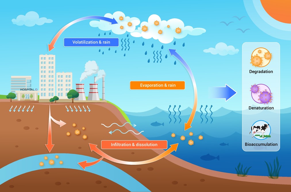
Figure 1.
Pathways through ECs and the environment
-
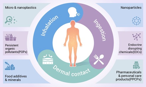
Figure 2.
The common ECs and exposure ways
-
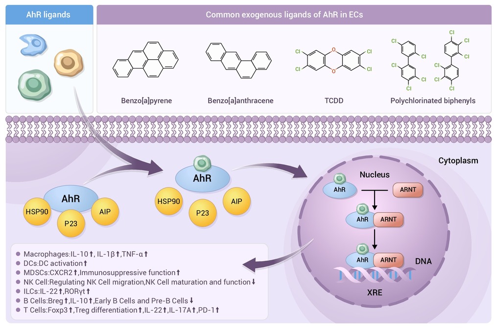
Figure 3.
Aryl Hydrocarbon Receptor (AhR) activation by ECs
-
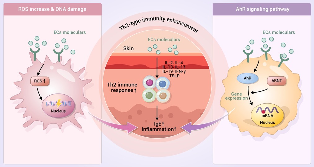
Figure 4.
Major pathways by which ECs affect AD
