
Artificial intelligence-driven virtual knockout in cancer research: Progress, challenges, and future perspectives
-
GRAPHICAL ABSTRACT
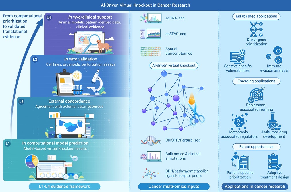
 DownLoad:
Full size image
DownLoad:
Full size image
-
PUBLIC SUMMARY
-
AI-driven virtual knockout simulates gene loss before experiments.
This review covers gene regulatory network, generative, metabolic, and foundation model tools.
An L1-L4 framework is established to assess the reliability of virtual knockout findings.
Virtual knockout prioritizes cancer targets and context-specific vulnerabilities.
Virtual knockout links computational prediction with experimental validation.
-
-
ABSTRACT
Artificial intelligence-driven virtual knockout is emerging as an intervention-oriented computational paradigm that helps move cancer research from descriptive association toward perturbation-informed hypothesis generation and model-derived counterfactual prediction. Virtual knockout simulates biologically meaningful loss-of-function perturbations in genes, pathways, or regulatory nodes. This approach facilitates target prioritization, model-based mechanistic interpretation, screening of combinatorial strategies, and optimization of downstream validation before experimental intervention. In this review, we define virtual knockout as a class of computational frameworks that model the consequences of loss-of-function perturbations and estimate model-derived post-perturbation biological states, while distinguishing strict virtual knockout models from related perturbation-inference paradigms. We summarize major modeling strategies, including gene regulatory network perturbation, trajectory and transcription factor program modeling, generative model-derived counterfactual prediction, constraint-based metabolic simulation, and communication-informed perturbation analysis, and review their applications in driver gene prioritization, context-specific vulnerability discovery, resistance-associated rewiring, tumor microenvironment analysis, metastasis-associated regulator prioritization, drug development, and patient-oriented therapeutic prioritization. We further propose a four-level evidence framework (L1-L4) and discuss major challenges, including data sparsity, tumor heterogeneity, context dependence, and cross-platform inconsistency. Overall, virtual knockout represents a promising bridge between computational inference, experimental validation, and precision oncology, although its current role is best viewed as one of prioritization and decision support rather than direct clinical replacement. -

-
REFERENCES
[1] Luo Q. and Smith D.P. (2025). Global cancer burden: Progress, projections, and challenges. Lancet 406:1536−1537. DOI:10.1016/S0140-6736(25)01570-3 [2] Hanahan D. and Weinberg R.A. (2011). Hallmarks of cancer: The next generation. Cell 144:646−674. DOI:10.1016/j.cell.2011.02.013 [3] Gaillard H., Garcia-Muse T. and Aguilera A. (2015). Replication stress and cancer. Nat. Rev. Cancer 15:276−289. DOI:10.1038/nrc3916 [4] Hanahan D. (2022). Hallmarks of cancer: New dimensions. Cancer Discov. 12:31−46. DOI:10.1158/2159-8290.CD-21-1059 [5] Togar T., Desai S., Mishra R., et al. (2020). Identifying cancer driver genes from functional genomics screens. Swiss Med. Wkly. 150:w20195. DOI:10.4414/smw.2020.20195 [6] Pinto B., Henriques A.C., Silva P.M.A., et al. (2020). Three-Dimensional spheroids as in vitro preclinical models for cancer research. Pharmaceutics 12. DOI:10.3390/pharmaceutics12121186. [7] Liu Y., Wu W., Cai C., et al. (2023). Patient-derived xenograft models in cancer therapy: Technologies and applications. Signal Transduct. Target Ther. 8:160. DOI:10.1038/s41392-023-01419-2 [8] DiMasi J.A., Feldman L., Seckler A., et al. (2010). Trends in risks associated with new drug development: Success rates for investigational drugs. Clin. Pharmacol. Ther. 87:272−277. DOI:10.1038/clpt.2009.295 [9] The ICGC/TCGA Pan-Cancer Analysis of Whole Genomes Consortium. (2020). Pan-cancer analysis of whole genomes. Nature 578:82−93. DOI:10.1038/s41586-020-1969-6 [10] An S., Cho J.W., Cao K., et al. (2025). scCausalVI disentangles single-cell perturbation responses with causality-aware generative model. Cell Syst. 16:101443. DOI:10.1016/j.cels.2025.101443 [11] Kamimoto K., Stringa B., Hoffmann C.M., et al. (2023). Dissecting cell identity via network inference and in silico gene perturbation. Nature 614:742−751. DOI:10.1038/s41586-022-05688-9 [12] Dong M., Wang B., Wei J., et al. (2023). Causal identification of single-cell experimental perturbation effects with CINEMA-OT. Nat. Methods 20:1769−1779. DOI:10.1038/s41592-023-02040-5 [13] Weng J., Ju F., Lyu Z., et al. (2025). Single-cell insights into tumor microenvironment heterogeneity and plasticity: Transforming precision therapy in gastrointestinal cancers. J. Exp. Clin. Cancer Res. 44:314. DOI:10.1186/s13046-025-03567-5 [14] Lotfollahi M., Wolf F.A. and Theis F.J. (2019). scGen predicts single-cell perturbation responses. Nat. Methods 16:715−721. DOI:10.1038/s41592-019-0494-8 [15] Lotfollahi M., Klimovskaia Susmelj A., De Donno C., et al. (2023). Predicting cellular responses to complex perturbations in high-throughput screens. Mol. Syst. Biol. 19:e11517. DOI:10.15252/msb.202211517 [16] Ma C., Zhang H., Rao Y., et al. (2025). AI-driven virtual cell models in preclinical research: Technical pathways, validation mechanisms, and clinical translation potential. npj Digit. Med. 9:25. DOI:10.1038/s41746-025-02198-6 [17] Theodoris C.V., Xiao L., Chopra A., et al. (2023). Transfer learning enables predictions in network biology. Nature 618:616−624. DOI:10.1038/s41586-023-06139-9 [18] Cui H., Wang C., Maan H., et al. (2024). scGPT: Toward building a foundation model for single-cell multi-omics using generative AI. Nat. Methods 21:1470−1480. DOI:10.1038/s41592-024-02201-0 [19] Hao M., Gong J., Zeng X., et al. (2024). Large-scale foundation model on single-cell transcriptomics. Nat. Methods 21:1481−1491. DOI:10.1038/s41592-024-02305-7 [20] Zhu O. and Li J. (2026). Scouter predicts transcriptional responses to genetic perturbations with large language model embeddings. Nat. Comput. Sci. 6:21−28. DOI:10.1038/s43588-025-00912-8 [21] Szappanos B., Kovacs K., Szamecz B., et al. (2011). An integrated approach to characterize genetic interaction networks in yeast metabolism. Nat. Genet. 43:656−662. DOI:10.1038/ng.846 [22] Osorio D., Zhong Y., Li G., et al. (2022). scTenifoldKnk: An efficient virtual knockout tool for gene function predictions via single-cell gene regulatory network perturbation. Patterns (N Y) 3:100434. DOI:10.1016/j.patter.2022.100434 [23] Roohani Y., Huang K. and Leskovec J. (2024). Predicting transcriptional outcomes of novel multigene perturbations with GEARS. Nat. Biotechnol. 42:927−935. DOI:10.1038/s41587-023-01905-6 [24] Valcarcel L.V., San Jose-Eneriz E., Ordonez R., et al. (2024). An automated network-based tool to search for metabolic vulnerabilities in cancer. Nat. Commun. 15:8685. DOI:10.1038/s41467-024-52725-4 [25] Marusyk A., Almendro V. and Polyak K. (2012). Intra-tumour heterogeneity: A looking glass for cancer. Nat. Rev. Cancer 12:323−334. DOI:10.1038/nrc3261 [26] Dagogo-Jack I. and Shaw A.T. (2018). Tumour heterogeneity and resistance to cancer therapies. Nat. Rev. Clin. Oncol. 15:81−94. DOI:10.1038/nrclinonc.2017.166 [27] Apaolaza I., San Jose-Eneriz E., Valcarcel L.V., et al. (2022). A network-based approach to integrate nutrient microenvironment in the prediction of synthetic lethality in cancer metabolism. PLoS Comput. Biol. 18:e1009395. DOI:10.1371/journal.pcbi.1009395 [28] O’Brien Edward J., Monk J.M. and Palsson B.O. (2015). Using genome-scale models to predict biological capabilities. Cell 161:971−987. DOI:10.1016/j.cell.2015.05.019 [29] Abbasian M.H., Sobhani N., Sisakht M.M., et al. (2025). Patient-derived organoids: A game-changer in personalized cancer medicine. Stem Cell Rev. Rep. 21:211−225. DOI:10.1007/s12015-024-10805-4 [30] Arora G., Banerjee M., Langthasa J., et al. (2023). Targeting metabolic fluxes reverts metastatic transitions in ovarian cancer. iScience 26:108081. DOI:10.1016/j.isci.2023.108081 [31] Heirendt L., Arreckx S., Pfau T., et al. (2019). Creation and analysis of biochemical constraint-based models using the COBRA Toolbox v.3.0. Nat. Protoc. 14:639-702. DOI:10.1038/s41596-018-0098-2. [32] TRIPOD+AI statement: Updated guidance for reporting clinical prediction models that use regression or machine learning methods. (2024). BMJ 385:q902. DOI:10.1136/bmj.q902. [33] Vogelstein B., Papadopoulos N., Velculescu V.E., et al. (2013). Cancer genome landscapes. Science 339:1546−1558. DOI:10.1126/science.1235122 [34] Collins G.S., Dhiman P., Ma J., et al. (2024). Evaluation of clinical prediction models (part 1): From development to external validation. BMJ 384:e074819. DOI:10.1136/bmj-2023-074819 [35] Riley R.D., Archer L., Snell K.I.E., et al. (2024). Evaluation of clinical prediction models (part 2): How to undertake an external validation study. BMJ 384:e074820. DOI:10.1136/bmj-2023-074820 [36] Riley R.D., Snell K.I.E., Archer L., et al. (2024). Evaluation of clinical prediction models (part 3): Calculating the sample size required for an external validation study. BMJ 384:e074821. DOI:10.1136/bmj-2023-074821 [37] Patterson E.A. and Whelan M.P. (2017). A framework to establish credibility of computational models in biology. Prog. Biophys. Mol. Biol. 129:13−19. DOI:10.1016/j.pbiomolbio.2016.08.007 [38] Tatka L.T., Smith L.P., Hellerstein J.L., et al. (2023). Adapting modeling and simulation credibility standards to computational systems biology. J. Transl. Med. 21:501. DOI:10.1186/s12967-023-04290-5 [39] Jafari M., Guan Y., Wedge D.C., et al. (2021). Re-evaluating experimental validation in the Big Data Era: A conceptual argument. Genome Biol. 22:71. DOI:10.1186/s13059-021-02292-4 [40] Dancik G.M. and Vlahopoulos S.A. (2025). Editorial for the special issue: Bioinformatics and computational biology for cancer prediction and prognosis. Genes (Basel) 16. DOI:10.3390/genes16020167 [41] Moons K.G.M., Wolff R.F., Riley R.D., et al. (2019). PROBAST: A tool to assess risk of bias and applicability of prediction model studies: Explanation and elaboration. Ann. Intern. Med. 170:W1−W33. DOI:10.7326/M18-1377 [42] Gross F. and MacLeod M. (2017). Prospects and problems for standardizing model validation in systems biology. Prog. Biophys. Mol. Biol. 129:3−12. DOI:10.1016/j.pbiomolbio.2017.01.003 [43] Steyerberg E.W., Harrell F.E.Jr., Borsboom G.J.J.M., et al. (2001). Internal validation of predictive models: Efficiency of some procedures for logistic regression analysis. J. Clin. Epidemiol. 54:774−781. DOI:10.1016/s0895-4356(01)00341-9 [44] Meinshausen N. and Bühlmann P. (2010). Stability selection. J. R. Stat. Soc. Ser. B Stat. Methodol. 72:417−473. DOI:10.1111/j.1467-9868.2010.00740.x [45] Golland P. and Fischl B. (2003). Permutation tests for classification: Towards statistical significance in image-based studies. Inf. Process Med. Imaging 18:330−341. DOI:10.1007/978-3-540-45087-0_28 [46] Rosenblatt M., Tejavibulya L., Jiang R., et al. (2024). Data leakage inflates prediction performance in connectome-based machine learning models. Nat. Commun. 15:1829. DOI:10.1038/s41467-024-46150-w [47] Liu Z., Xue Z., Song H., et al. (2026). Molecular mechanisms linking cadmium chloride exposure to ankylosing spondylitis: An integrative network-based study. Naunyn Schmiedebergs Arch. Pharmacol. DOI:10.1007/s00210-026-05394-7 [48] Qi L.S., Larson M.H., Gilbert L.A., et al. (2013). Repurposing CRISPR as an RNA-guided platform for sequence-specific control of gene expression. Cell 152:1173−1183. DOI:10.1016/j.cell.2013.02.022 [49] Bock C., Datlinger P., Chardon F., et al. (2022). High-content CRISPR screening. Nat. Rev. Methods Primers 2:8. DOI:10.1038/s43586-022-00098-7 [50] Joung J., Konermann S., Gootenberg J.S., et al. (2017). Genome-scale CRISPR-Cas9 knockout and transcriptional activation screening. Nat. Protoc. 12:828−863. DOI:10.1038/nprot.2017.016 [51] Wang Y., Zhai Y., Zhang M., et al. (2024). Escaping from CRISPR-Cas-mediated knockout: The facts, mechanisms, and applications. Cell Mol. Biol. Lett. 29:48. DOI:10.1186/s11658-024-00565-x [52] Tong L., Cui W., Zhang B., et al. (2024). Patient-derived organoids in precision cancer medicine. Med 5:1351−1377. DOI:10.1016/j.medj.2024.08.010 [53] Viceconti M., Pappalardo F., Rodriguez B., et al. (2021). In silico trials: Verification, validation and uncertainty quantification of predictive models used in the regulatory evaluation of biomedical products. Methods 185:120−127. DOI:10.1016/j.ymeth.2020.01.011 [54] Wang Y., Li J., Yan Y., et al. (2026). A metabolic-immune subtype of breast cancer defined by G6PD and SHMT2: From single-cell dissection to dual-targeted therapy. J. Steroid Biochem. Mol. Biol. 260:106983. DOI:10.1016/j.jsbmb.2026.106983 [55] Agren R., Mardinoglu A., Asplund A., et al. (2014). Identification of anticancer drugs for hepatocellular carcinoma through personalized genome-scale metabolic modeling. Mol. Syst. Biol. 10:721. DOI:10.1002/msb.145122 [56] Jolasun Y., Song K., Zheng Y., et al. (2025). SIDISH integrates single-cell and bulk transcriptomics to identify high-risk cells and guide precision therapeutics through in silico perturbation. Nat. Commun. 16:11271. DOI:10.1038/s41467-025-66162-4 [57] Lim J., Jung H. D., Park S. Y., et al. (2025). Genome-scale knockout simulation and clustering analysis of drug-resistant breast cancer cells reveal drug sensitization targets. Proc. Natl. Acad. Sci. USA 122:e2425384122. DOI:10.1073/pnas.2425384122 [58] Zhan X., Hu H., Liu Y., et al. (2025). TRIM28 drives immune evasion via PARP1 SUMOylation and NAD(+) depletion in clear cell renal cell carcinoma. J. Immunother. Cancer 13. DOI:10.1136/jitc-2025-013025 [59] Vinas T.R., Wiatrak M., Piran Z., et al. (2025). Systema: A framework for evaluating genetic perturbation response prediction beyond systematic variation. Nat. Biotechnol. DOI:10.1038/s41587-025-02777-8 [60] Yang Y., Li G., Zhong Y., et al. (2023). Gene knockout inference with variational graph autoencoder learning single-cell gene regulatory networks. Nucleic Acids Res. 51:6578−6592. DOI:10.1093/nar/gkad450 [61] Little D.R., Gerner-Mauro K.N., Flodby P., et al. (2019). Transcriptional control of lung alveolar type 1 cell development and maintenance by NK homeobox 2-1. Proc. Natl. Acad. Sci. USA 116:20545−20555. DOI:10.1073/pnas.1906663116 [62] Shiao S. L., Gouin K.H.III., Ing N., et al. (2024). Single-cell and spatial profiling identify three response trajectories to pembrolizumab and radiation therapy in triple negative breast cancer. Cancer Cell 42:70−84.e78. DOI:10.1016/j.ccell.2023.12.012 [63] Paul F., Arkin Y., Giladi A., et al. (2016). Transcriptional heterogeneity and lineage commitment in myeloid progenitors. Cell 164:325. DOI:10.1016/j.cell.2015.12.046 [64] Orkin S.H. and Zon L.I. (2008). Hematopoiesis: An evolving paradigm for stem cell biology. Cell 132:631−644. DOI:10.1016/j.cell.2008.01.025 [65] Yang K., Halima A. and Chan T.A. (2023). Antigen presentation in cancer - mechanisms and clinical implications for immunotherapy. Nat. Rev. Clin. Oncol. 20:604−623. DOI:10.1038/s41571-023-00789-4 [66] Garrido F., Ruiz-Cabello F. and Aptsiauri N. (2017). Rejection versus escape: The tumor MHC dilemma. Cancer Immunol. Immunother. 66:259−271. DOI:10.1007/s00262-016-1947-x [67] Maggs L., Sadagopan A., Moghaddam A.S., et al. (2021). HLA class I antigen processing machinery defects in antitumor immunity and immunotherapy. Trends in Cancer 7:1089−1101. DOI:10.1016/j.trecan.2021.07.006 [68] Kobayashi K.S. and van den Elsen P.J. (2012). NLRC5: A key regulator of MHC class I-dependent immune responses. Nat. Rev. Immunol. 12:813−820. DOI:10.1038/nri3339 [69] Kok V.C., Wang C.C.N., Liao S.H., et al. (2022). Cross-platform in-silico analyses exploring tumor immune microenvironment with prognostic value in triple-negative breast cancer. Breast Cancer (Dove Med Press) 14:85−99. DOI:10.2147/BCTT.S359346 [70] Jin S., Guerrero-Juarez C.F., Zhang L., et al. (2021). Inference and analysis of cell-cell communication using CellChat. Nat. Commun. 12:1088. DOI:10.1038/s41467-021-21246-9 [71] Aliazis K., Christofides A., Shah R., et al. (2025). The tumor microenvironment’s role in the response to immune checkpoint blockade. Nat. Cancer 6:924−937. DOI:10.1038/s43018-025-00986-3 [72] Allard B., Allard D., Buisseret L., et al. (2020). Publisher correction: The adenosine pathway in immuno-oncology. Nat. Rev. Clin. Oncol. 17:650. DOI:10.1038/s41571-020-0415-x [73] Mariathasan S., Turley S.J., Nickles D., et al. (2018). TGFbeta attenuates tumour response to PD-L1 blockade by contributing to exclusion of T cells. Nature 554:544−548. DOI:10.1038/nature25501 [74] Ghahremanifard P., Chanda A., Bonni S., et al. (2020). TGF-beta mediated immune evasion in cancer-spotlight on cancer-associated fibroblasts. Cancers (Basel) 12:3650. DOI:10.3390/cancers12123650 [75] Liu X., Ding Q., Zhang H., et al. (2025). The CD39-CD73-adenosine axis: Master regulator of immune evasion and therapeutic target in pancreatic ductal adenocarcinoma. Biochim. Biophys. Acta Rev. Cancer 1880:189443. DOI:10.1016/j.bbcan.2025.189443 [76] Zhang B. (2010). CD73: A novel target for cancer immunotherapy. Cancer Res. 70:6407−6411. DOI:10.1158/0008-5472.CAN-10-1544 [77] Chen S., Wainwright D. A., Wu J. D., et al. (2019). CD73: An emerging checkpoint for cancer immunotherapy. Immunotherapy 11:983−997. DOI:10.2217/imt-2018-0200 [78] Salgia R. and Kulkarni P. (2018). The genetic/non-genetic duality of drug 'Resistance' in cancer. Trends Cancer 4:110−118. DOI:10.1016/j.trecan.2018.01.001 [79] Kobayashi S., Boggon T.J., Dayaram T., et al. (2005). EGFR mutation and resistance of non-small-cell lung cancer to gefitinib. N. Engl. J. Med. 352:786−792. DOI:10.1056/NEJMoa044238 [80] Engelman J.A., Zejnullahu K., Mitsudomi T., et al. (2007). MET amplification leads to gefitinib resistance in lung cancer by activating ERBB3 signaling. Science 316:1039−1043. DOI:10.1126/science.1141478 [81] Asghari M., Abazari M.F., Bokharaei H., et al. (2018). Key genes and regulatory networks involved in the initiation, progression and invasion of colorectal cancer. Future Sci. OA 4:FSO278. DOI:10.4155/fsoa-2017-0108 [82] Bravo Gonzalez-Blas C., De Winter S., Hulselmans G., et al. (2023). SCENIC+: single-cell multiomic inference of enhancers and gene regulatory networks. Nat. Methods 20:1355−1367. DOI:10.1038/s41592-023-01938-4 [83] Tomasik B., Garbicz F., Braun M., et al. (2024). Heterogeneity in precision oncology. Camb. Prism. Precis. Med. 2:e2. DOI:10.1017/pcm.2023.23 [84] Roerden M. and Spranger S. (2025). Cancer immune evasion, immunoediting and intratumour heterogeneity. Nat. Rev. Immunol. 25:353−369. DOI:10.1038/s41577-024-01111-8 [85] Jie J., Wang Q., Chen Z., et al. (2026). Multi-omics analysis and preliminary experimental validation of acetyl tributyl citrate (ATBC) promoting bladder cancer progression via the AKR1B1/EMT axis. Environ. Pollut. 397:127970. DOI:10.1016/j.envpol.2026.127970 [86] Li R., Liu X., Huo C., et al. (2026). An endothelial-centered regulatory framework reveals context-dependent roles of MYLK in lung adenocarcinoma. Front. Immunol. 17:1719296. DOI:10.3389/fimmu.2026.1719296 [87] Frangieh C.J., Fan J.L., Melms J.C., et al. (2026). Single-cell and spatial profiling in cancer biology and clinical oncology. Nat. Cancer 7:597−607. DOI:10.1038/s43018-026-01142-1 [88] Wensink G.E., Elias S.G., Mullenders J., et al. (2021). Patient-derived organoids as a predictive biomarker for treatment response in cancer patients. npj Precis. Oncol. 5:30. DOI:10.1038/s41698-021-00168-1 [89] Thorel L., Perreard M., Florent R., et al. (2024). Patient-derived tumor organoids: A new avenue for preclinical research and precision medicine in oncology. Exp. Mol. Med. 56:1531−1551. DOI:10.1038/s12276-024-01272-5 [90] Zhang X. and Chen L. (2025). Quantifying interventional causality by knockoff operation. Sci. Adv. 11:eadu6464. DOI:10.1126/sciadv.adu6464 [91] Song F., Chan G.M.A. and Wei Y. (2020). Flexible experimental designs for valid single-cell RNA-sequencing experiments allowing batch effects correction. Nat. Commun. 11:3274. DOI:10.1038/s41467-020-16905-2 [92] Haghverdi L., Lun A.T.L., Morgan M.D., et al. (2018). Batch effects in single-cell RNA-sequencing data are corrected by matching mutual nearest neighbors. Nat. Biotechnol. 36:421−427. DOI:10.1038/nbt.4091 [93] Bouland G.A., Mahfouz A. and Reinders M.J.T. (2023). Consequences and opportunities arising due to sparser single-cell RNA-seq datasets. Genome Biol. 24:86. DOI:10.1186/s13059-023-02933-w [94] van Dijk D., Sharma R., Nainys J., et al. (2018). Recovering gene interactions from single-cell data using data diffusion. Cell 174:716-729 e727. DOI:10.1016/j.cell.2018.05.061 [95] Li W.V. and Li J.J. (2018). An accurate and robust imputation method scImpute for single-cell RNA-seq data. Nat. Commun. 9:997. DOI:10.1038/s41467-018-03405-7 [96] Huang M., Wang J., Torre E., et al. (2018). SAVER: Gene expression recovery for single-cell RNA sequencing. Nat. Methods 15:539−542. DOI:10.1038/s41592-018-0033-z [97] Lopez R., Regier J., Cole M.B., et al. (2018). Deep generative modeling for single-cell transcriptomics. Nat. Methods 15:1053−1058. DOI:10.1038/s41592-018-0229-2 [98] Hou W., Ji Z., Ji H., et al. (2020). A systematic evaluation of single-cell RNA-sequencing imputation methods. Genome Biol. 21:218. DOI:10.1186/s13059-020-02132-x [99] Zhang R., Atwal G.S. and Lim W.K. (2021). Noise regularization removes correlation artifacts in single-cell RNA-seq data preprocessing. Patterns (N Y) 2:100211. DOI:10.1016/j.patter.2021.100211 [100] Ahlmann-Eltze C., Huber W. and Anders S. (2025). Deep-learning-based gene perturbation effect prediction does not yet outperform simple linear baselines. Nat. Methods 22:1657−1661. DOI:10.1038/s41592-025-02772-6 [101] Xue Y., Friedl V., Ding H., et al. (2024). Single-cell signatures identify microenvironment factors in tumors associated with patient outcomes. Cell Rep. Methods 4. DOI:10.1016/j.crmeth.2024.100799. [102] Schmid K.T., Höllbacher B., Cruceanu C., et al. (2021). scPower accelerates and optimizes the design of multi-sample single cell transcriptomic studies. Nat. Commun. 12:6625. DOI:10.1038/s41467-021-26779-7 [103] Hegenbarth J.C., Lezzoche G., De Windt L.J., et al. (2022). Perspectives on bulk-tissue RNA sequencing and single-cell RNA sequencing for cardiac transcriptomics. Front. Mol. Med. 2:839338. DOI:10.3389/fmmed.2022.839338 [104] Vlachogiannis G., Hedayat S., Vatsiou A., et al. (2018). Patient-derived organoids model treatment response of metastatic gastrointestinal cancers. Science 359:920−926. DOI:10.1126/science.aao2774 [105] Wagner D.E. and Klein A.M. (2020). Lineage tracing meets single-cell omics: Opportunities and challenges. Nat. Rev. Genet. 21:410−427. DOI:10.1038/s41576-020-0223-2 [106] Binnewies M., Roberts E.W., Kersten K., et al. (2018). Understanding the tumor immune microenvironment (TIME) for effective therapy. Nat. Med. 24:541−550. DOI:10.1038/s41591-018-0014-x [107] Hu X., Li H., Chen M., et al. (2025). Reference-informed evaluation of batch correction for single-cell omics data with overcorrection awareness. Commun. Biol. 8:521. DOI:10.1038/s42003-025-07947-7 [108] Wei Z., Wang Y., Gao Y., et al. (2025). Benchmarking algorithms for generalizable single-cell perturbation response prediction. Nat. Methods 23:451–464. DOI:10.1038/s41592-025-02980-0 [109] Squair J.W., Gautier M., Kathe C., et al. (2021). Confronting false discoveries in single-cell differential expression. Nat. Commun. 12:5692. DOI:10.1038/s41467-021-25960-2 [110] Li W., Zhang Z., Xie B., et al. (2024). HiOmics: A cloud-based one-stop platform for the comprehensive analysis of large-scale omics data. Comput. Struct. Biotechnol. J. 23:659−668. DOI:10.1016/j.csbj.2024.01.002 [111] Huang Y., Luo J., Zhang Y., et al. (2023). Identification of MKNK1 and TOP3A as ovarian endometriosis risk-associated genes using integrative genomic analyses and functional experiments. Comput. Struct. Biotechnol. J. 21:1510−1522. DOI:10.1016/j.csbj.2023.02.001 [112] Yang T., Ma F., Qian H., et al. (2024). AI-driven construction of digital cell model. Innov. Life 2:100102. DOI:10.59717/j.xinn-life.2024.100102 [113] Wei R., Wang B., Yan B., et al. (2026). From equations to agents: The artificial intelligence virtual cell reshaping precision oncology. Innov. Oncol. 1:100002. DOI:10.59717/j.xinn-oncol.2026.100002 [114] Polak R., Zhang E.T. and Kuo C.J. (2024). Cancer organoids 2.0: Modelling the complexity of the tumour immune microenvironment. Nat. Rev. Cancer 24:523-539. DOI:10.1038/s41568-024-00706-6 [115] Badia I.M.P., Wessels L., Muller-Dott S., et al. (2023). Gene regulatory network inference in the era of single-cell multi-omics. Nat. Rev. Genet. 24:739−754. DOI:10.1038/s41576-023-00618-5 [116] Schäfer P.S.L., Dimitrov D., Villablanca E.J., et al. (2024). Integrating single-cell multi-omics and prior biological knowledge for a functional characterization of the immune system. Nat. Immunol. 25:405−417. DOI:10.1038/s41590-024-01768-2 [117] Walker C. and Angelo M. (2024). Toward clinical applications of spatial-omics in cancer research. Nat. Cancer 5:1771−1773. DOI:10.1038/s43018-024-00868-0 [118] Lambert A.W., Zhang Y. and Weinberg R.A. (2024). Cell-intrinsic and microenvironmental determinants of metastatic colonization. Nat. Cell Biol. 26:687−697. DOI:10.1038/s41556-024-01409-8 -
ABOUT THIS ARTICLE
Cite this article:
Wang X., Liu M., Jia M., et al. (2026). Artificial intelligence-driven virtual knockout in cancer research: Progress, challenges, and future perspectives. The Innovation Oncology 1:100027. https://doi.org/10.59717/j.xinn-oncol.2026.100027
Welcome!
To request copyright permission to republish or share portions of our works, please visit Copyright Clearance Center's (CCC) Marketplace website at marketplace.copyright.com.
Export File
Citation
Wang X., Liu M., Jia M., et al. (2026). Artificial intelligence-driven virtual knockout in cancer research: Progress, challenges, and future perspectives. The Innovation Oncology 1:100027. https://doi.org/10.59717/j.xinn-oncol.2026.100027

Format
Content
-
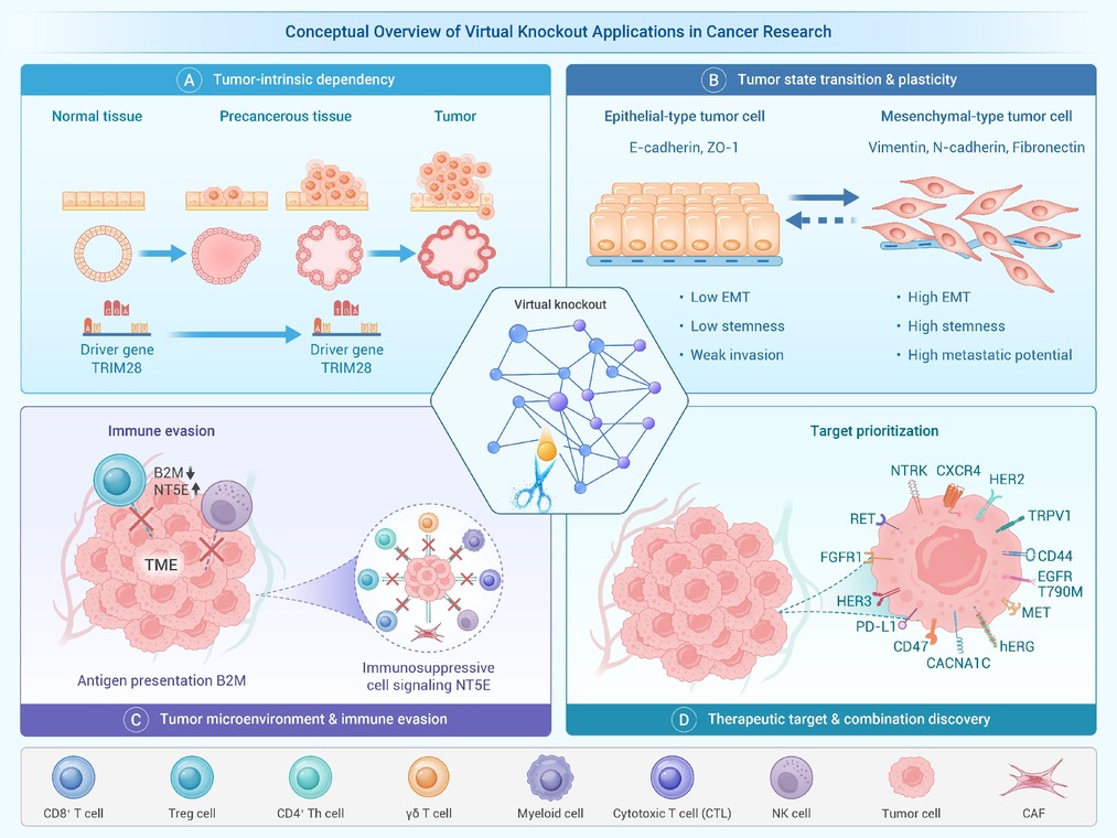
Figure 1.
Conceptual overview of virtual knockout applications in cancer research
-
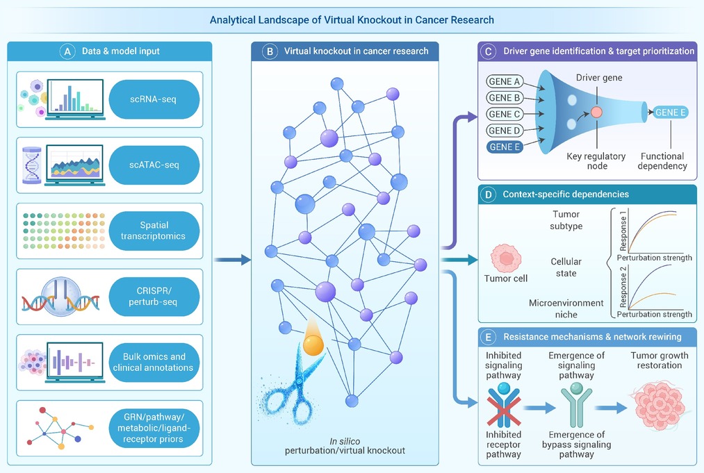
Figure 2.
Analytical landscape of virtual knockout in cancer research.
-
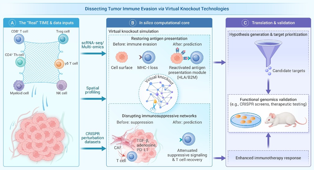
Figure 3.
Schematic overview of a virtual knockout-based closed-loop framework for dissecting immune evasion in the tumor immune microenvironment and guiding target prioritization and validation
-
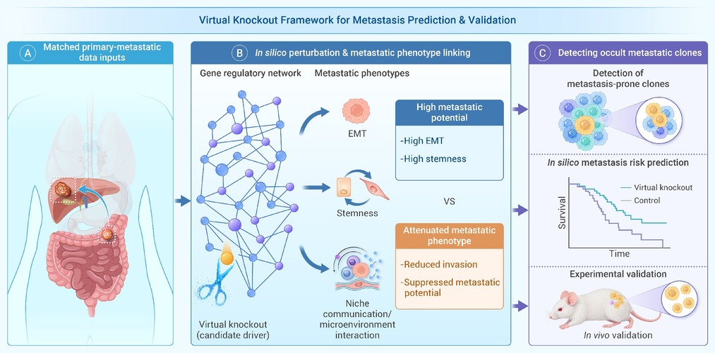
Figure 4.
Schematic overview of a virtual knockout-based framework for linking in silico perturbation to metastatic phenotypes and downstream validation
