
Advances in nanomaterial-enhanced ablation therapy for hepatocellular carcinoma
-
GRAPHICAL ABSTRACT
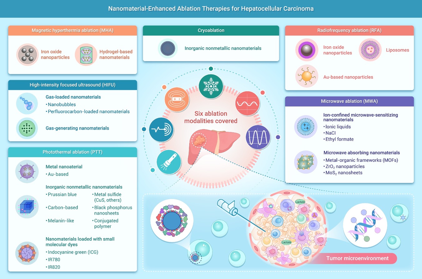
 DownLoad:
Full size image
DownLoad:
Full size image
-
PUBLIC SUMMARY
-
Ablation therapy is important for HCC.
Nanomaterials can be leveraged to potentiate ablation.
Nanomaterial-enhanced HCC nerve ablation is a novel direction for future studies.
We provide new insights and directions for improving anti-tumor therapy for HCC.
-
-
ABSTRACT
Ablation therapy is a cornerstone locoregional treatment for hepatocellular carcinoma (HCC), particularly in patients with early-stage disease or those who are not surgical candidates. Despite its widespread use, ablation is limited by incomplete tumor destruction, heat-sink effects, and a high risk of local recurrence. Nanomaterials can enhance ablation efficiency, expand the effective treatment zone, and improve the tumor microenvironment. Therefore, effective HCC management strategies that leverage nanomaterials to potentiate ablation must be developed. Nanomaterials can augment imaging preoperatively, boost ablation efficacy intraoperatively, and modulate the tumor microenvironment postoperatively to prevent recurrence. In addition, nanomaterials can serve as carriers for chemotherapeutic, targeted, or immunomodulatory agents, enabling synergistic combinations of ablation with systemic or transarterial therapies. Building on the role of nanomaterials in potentiating ablation, this review systematically examines the mechanisms by which different classes of nanomaterials—including iron oxide nanoparticles, Au-based nanoparticles, liposomes, and hydrogels—enhance radiofrequency ablation, microwave ablation, high-intensity focused ultrasound, photothermal ablation, magnetic hyperthermia, and cryoablation for HCC. We also discuss the clinical translation potential of these nanomaterials, providing new perspectives and research directions to advance ablation therapy for HCC. Our findings provide novel insights on developing effective anti-tumor therapy strategies based on ablation treatment for patients with HCC.-
Keywords:
- Hepatocellular carcinoma /
- Ablation /
- Nanomaterial /
- Microwave enhancement.
-

-
REFERENCES
[1] Mu Y., Ren X.H., Han D., et al. (2022). Codelivery of HBx-siRNA and plasmid encoding IL-12 for Inhibition of hepatitis B virus and reactivation of antiviral immunity. Pharmaceutics 14:1439. DOI:10.3390/pharmaceutics14071439 [2] Xue R., Zhang Q., Cao Q., et al. (2022). Liver tumour immune microenvironment subtypes and neutrophil heterogeneity. Nature 612:141−147. DOI:10.1038/s41586-022-05400-x [3] Hwang S.Y., Danpanichkul P., Agopian V., et al. (2025). Hepatocellular carcinoma: Updates on epidemiology, surveillance, diagnosis and treatment. Clin. Mol. Hepatol. 31:S228−S254. DOI:10.3350/cmh.2024.0824 [4] Zhao C., Zhang Y., Wang G., et al. (2025). Finotonlimab (PD-1 inhibitor) plus bevacizumab (bevacizumab biosimilar) as first-tier therapy for late-stage hepatocellular carcinoma: A randomized phase 2/3 trial. Signal Transduct. Target Ther. 10:249. DOI:10.1038/s41392-025-02333-5 [5] Finn R.S., Ryoo B.Y., Hsu C.H., et al. (2025). Tiragolumab in combination with atezolizumab and bevacizumab in patients with unresectable, locally advanced or metastatic hepatocellular carcinoma (MORPHEUS-Liver): a randomised, open-label, phase 1b-2, study. Lancet Oncol. 26:214−226. DOI:10.1016/S1470-2045(24)00679-X [6] Zhu H.D., Li H. L., Huang M.S., et al. (2023). Transarterial chemoembolization with PD-(L)1 inhibitors plus molecular targeted therapies for hepatocellular carcinoma (CHANCE001). Signal Transduct. Target Ther. 8:58. DOI:10.1038/s41392-022-01235-0 [7] Zhong B.Y., Fan W., Guan J.J., et al. (2025). Combination locoregional and systemic therapies in hepatocellular carcinoma. Lancet Gastroenterol. Hepatol. 10:369−386. DOI:10.1016/S2468-1253(24)00247-4 [8] Sugiura Y., Iwasaka T. and Tarumi N. (1983). Percutaneous ethanol injection therapy: A new treatment for hepatocellular carcinoma. Radiology 90:53−57. DOI:3.0.CO;2-O">10.1002/1097-0142(19911001)68:7<1524::AID-CNCR2820680711>3.0.CO;2-O [9] Ye F., Xie L., Liang L., et al. (2023). Mechanisms and therapeutic strategies to combat the recurrence and progression of hepatocellular carcinoma after thermal ablation. J. Interv. Med. 6:160−169. DOI:10.1016/j.jimed.2023.10.004 [10] Minami Y. and Kudo M. (2021). Image guidance in ablation for hepatocellular carcinoma: contrast-enhanced ultrasound and fusion imaging. Front. Oncol. 11:593636. DOI:10.3389/fonc.2021.593636 [11] Llovet J.M., De Baere T., Kulik L., et al. (2021). Locoregional therapies in the era of molecular and immune treatments for hepatocellular carcinoma. Nat. Rev. Gastroenterol. Hepatol. 18:293−313. DOI:10.1038/s41575-020-00395-0 [12] Kawaguchi Y., Hasegawa K., Kashiwabara K., et al. (2025). Surgery versus ablation for hepatocellular carcinoma: A randomized controlled trial (SURF-RCT Trial) and a nonrandomized prospective observational trial (SURF-Cohort Trial). J. Clin. Oncol. 43:2628−2638. DOI:10.1200/JCO-24-02030 [13] Song J., Cao L., Ma K., et al. (2024). Laparoscopic liver resection versus radiofrequency ablation for small hepatocellular carcinoma: randomized clinical trial. Br. J. Surg. 111:1442499. DOI:10.1093/bjs/znae099 [14] Yuan Y., Peng H., He W., et al. (2024). Partial hepatectomy versus interventional treatment in patients with hepatitis B virus-related hepatocellular carcinoma and clinically significant portal hypertension: a randomized comparative clinical trial. Cancer Commun. 44:1337−1349. DOI:10.1002/cac2.12614 [15] Zhang Y.J., Chen J., Zhou Z., et al. (2025). Transarterial chemoembolization with radiofrequency ablation versus surgical resection for small late-recurrence hepatocellular carcinoma. Radiology 314:e241096. DOI:10.1148/radiol.241096 [16] Chiang J., Rajendran P.S., Hao F., et al. (2023). Combination transarterial chemoembolization and microwave ablation vs. microwave ablation monotherapy for hepatocellular carcinomas greater than 3 cm: A comparative study. Diagn. Interv. Radiol. 29:805-812. DOI:10.4274/dir.2023.232159. [17] Zhou C., Li Y., Li J., et al. (2023). A phase 1/2 multicenter randomized trial of local ablation plus toripalimab versus toripalimab alone for previously treated unresectable hepatocellular carcinoma. Clin. Cancer. Res. 29:2816−2825. DOI:10.1158/1078-0432.CCR-23-0410 [18] Shi J., Votruba A.R., Farokhzad O.C., et al. (2010). Nanotechnology in drug delivery and tissue engineering: From discovery to applications. Nano Lett. 10:3223−3230. DOI:10.1021/nl102184c [19] Liu Y., Han Y., Chen S., et al. (2022). Liposome-based multifunctional nanoplatform as effective therapeutics for the treatment of retinoblastoma. Acta Pharm. Sin. B 12:2731−2739. DOI:10.1016/j.apsb.2021.10.009 [20] Kim M., Lee N.K., Wang C.J., et al. (2023). Reprogramming the tumor microenvironment with biotechnology. Biomater. Res. 27:5. DOI:10.1186/s40824-023-00343-4 [21] Wang Y., Wei Y., Wu Y., et al. (2023). Multifunctional nano-realgar hydrogel for enhanced glioblastoma synergistic chemotherapy and radiotherapy: A new paradigm of an old drug. Int. J. Nanomedicine 18:743−763. DOI:10.2147/IJN.S394377 [22] Skwierczynska M., Runowski M., Goderski S., et al. (2018). Luminescent-Magnetic Cellulose Fibers, Modified with Lanthanide-Doped Core/Shell Nanostructures. ACS Omega 3:10383−10390. DOI:10.1021/acsomega.8b00965 [23] Buzea C., Pacheco II. and Robbie K. (2007). Nanomaterials and nanoparticles: Sources and toxicity. Biointerphases 2:MR17−71. DOI:10.1116/1.2815690 [24] Sun W., Chu C., Li S., et al. (2023). Nanosensitizer-mediated unique dynamic therapy tactics for effective inhibition of deep tumors. Adv. Drug Deliv. Rev. 192:114643. DOI:10.1016/j.addr.2022.114643 [25] Mahmoudi M., Landry M.P., Moore A., et al. (2023). The protein corona from nanomedicine to environmental science. Nat. Rev. Mater. 8:1−17. DOI:10.1038/s41578-023-00552-2 [26] Sindhwani S., Syed A.M., Ngai J., et al. (2020). The entry of nanoparticles into solid tumours. Nat. Mater. 19:566−575. DOI:10.1038/s41563-019-0566-2 [27] Wilhelm S., Tavares A., Dai Q., et al. (2016). Analysis of nanoparticle delivery to tumours. Nat. Rev. Mater. 1:16014. DOI:10.1038/natrevmats.2016.14 [28] Nan Z., Shi L., Liu H., et al. (2025). Dual-frequency HIFU-activated multifunctional nanoparticles for trimodal synergistic therapy and immune responses with real-time US/MRI guidance. ACS Appl. Mater. Interfaces 17:53096−53109. DOI:10.1021/acsami.5c09753 [29] Priyadarshi N., Kaushal S., Garg P., et al. (2025). Advances in photothermal therapy for cancer and bacterial cells ablation using various nanomaterials. Adv. Colloid Interface Sci. 342:103541. DOI:10.1016/j.cis.2025.103541 [30] Kumbhakar D.V., Thakkar L., Akhand C., et al. (2025). Nanomaterials targeting cancer stem cells to overcome drug resistance and tumor recurrence. Front. Oncol. 15:1499283. DOI:10.3389/fonc.2025.1499283 [31] Glickstein B., Bismuth M., Gattegno R., et al. (2025). Volumetric Nanodroplet-enhanced ultrasound surgery combined with immune checkpoint inhibition as a cancer therapy platform. Small 21:e2411474. DOI:10.1002/smll.202411474 [32] Zhu H.-D., Liu R., Jia Z.-Z., et al. (2024). Transarterial chemoembolization for hepatocellular carcinoma: Treatment algorithm proposed by Chinese College of Interventionalists (CCI). EngMedicine 1:1−9. DOI:10.1016/j.engmed.2024.100037 [33] Chen L., Sun J. and Yang X. (2016). Radiofrequency ablation-combined multimodel therapies for hepatocellular carcinoma: Current status. Cancer Lett. 370:78−84. DOI:10.1016/j.canlet.2015.09.020 [34] Dumolard L., Ghelfi J., Roth G., et al. (2020). Percutaneous ablation-induced immunomodulation in hepatocellular carcinoma. Int. J. Mol. Sci. 21:4398. DOI:10.3390/ijms21124398 [35] Tanaka T., Takata K., Miyayama T., et al. (2023). Long-term outcome and eligibility of radiofrequency ablation for hepatocellular carcinoma over 3.0 cm in diameter. Sci. Rep. 13:16286. DOI:10.1038/s41598-023-43516-w. [36] Takayama T., Hasegawa K., Izumi N., et al. (2022). Surgery versus radiofrequency ablation for small hepatocellular carcinoma: A randomized controlled trial (SURF Trial). Liver Cancer 11:209−218. DOI:10.1159/000521665 [37] Majumdar A., Roccarina D., Thorburn D., et al. (2017). Management of people with early- or very early-stage hepatocellular carcinoma: An attempted network meta-analysis. Cochrane Database Syst. Rev. 3:CD011650. DOI:10.1002/14651858.CD011650.pub2 [38] Xu Z., Xie H., Zhou L., et al. (2019). The combination strategy of transarterial chemoembolization and radiofrequency ablation or microwave ablation against hepatocellular carcinoma. Anal. Cell Pathol. 2019:8619096. DOI:10.1155/2019/8619096 [39] Feng K. and Ma K.S. (2014). Value of radiofrequency ablation in the treatment of hepatocellular carcinoma. World J. Gastroenterol. 20:5987−5998. DOI:10.3748/wjg.v20.i20.5987 [40] Izzo F., Granata V., Grassi R., et al. (2019). Radiofrequency ablation and microwave ablation in liver tumors: An update. Oncologist 24:e990−e1005. DOI:10.1634/theoncologist.2018-0337 [41] Knavel E.M. and Brace C.L. (2013). Tumor ablation: Common modalities and general practices. Tech. Vasc. Interv. Radiol. 16:192−200. DOI:10.1053/j.tvir.2013.08.002 [42] Iannuccilli J.D. and Dupuy D.E. (2013). How to set up a successful tumor ablation practice. Tech. Vasc. Interv. Radiol. 16:201−208. DOI:10.1053/j.tvir.2013.08.003 [43] Shin S.W., Ahn K.S., Kim S.W., et al. (2021). Liver resection versus local ablation therapies for hepatocellular carcinoma within the milan criteria: A systematic review and meta-analysis. Ann. Surg. 273:656−666. DOI:10.1097/SLA.0000000000004350 [44] Yan F., Wang S., Yang W., et al. (2017). Tumor-penetrating peptide-integrated thermally sensitive liposomal doxorubicin enhances efficacy of radiofrequency ablation in liver tumors. Radiology 285:462−471. DOI:10.1148/radiol.2017162405 [45] Shao Y.L., Arjun B., Leo H.L., et al. (2017). Nano-assisted radiofrequency ablation of clinically extracted irregularly-shaped liver tumors. J. Therm. Biol. 66:101−113. DOI:10.1016/j.jtherbio.2017.04.005 [46] Fukuda K., Mori K., Hasegawa N., et al. (2019). Safety margin of radiofrequency ablation for hepatocellular carcinoma: a prospective study using magnetic resonance imaging with superparamagnetic iron oxide. Jpn. J. Radiol. 37:555−563. DOI:10.1007/s11604-019-00843-1 [47] Tang Y., Shu Z., Zhu M., et al. (2023). Size-tunable nanoregulator-based radiofrequency ablation suppresses MDSCs and their compensatory immune evasion in hepatocellular carcinoma. Adv. Healthc. Mater. 12:e2302013. DOI:10.1002/adhm.202302013 [48] Geppert M. and Himly M. (2021). Iron oxide nanoparticles in bioimaging - An immune perspective. Front. Immunol. 12:688927. DOI:10.3389/fimmu.2021.688927 [49] Canese R., Vurro F. and Marzola P. (2021). Iron oxide nanoparticles as theranostic agents in cancer immunotherapy. Nanomaterials 11:1950. DOI:10.3390/nano11081950 [50] Yang J., Wu J., Guo Z., et al. (2022). Iron oxide nanoparticles combined with static magnetic fields in bone remodeling. Cells 11:3298. DOI:10.3390/cells11203298 [51] Nakamura M., Kosuge H., Oyane A., et al. (2021). In vivo study of iron oxide-calcium phosphate composite nanoparticles for delivery to atherosclerosis. Nanotechnology 32:345101. DOI:10.1088/1361-6528/ac007d [52] Nagai M., Yamaguchi M., Mori K., et al. (2015). Magnetic resonance-based visualization of thermal ablative margins around hepatic tumors by means of systemic ferucarbotran administration before radiofrequency ablation: Animal study to reveal the connection between excess iron deposition and T2*-weighted hypointensity in ablative margins. Invest. Radiol. 50:376−383. DOI:10.1097/RLI.0000000000000137 [53] Koda M., Tokunaga S., Miyoshi K., et al. (2013). Ablative margin states by magnetic resonance imaging with ferucarbotran in radiofrequency ablation for hepatocellular carcinoma can predict local tumor progression. J. Gastroenterol. 48:1283−1292. DOI:10.1007/s00535-012-0747-0 [54] Tokunaga S., Koda M., Matono T., et al. (2012). Assessment of ablative margin by MRI with ferucarbotran in radiofrequency ablation for liver cancer: comparison with enhanced CT. Br. J. Radiol. 85:745−752. DOI:10.1259/bjr/64518148 [55] Ashokan A., Somasundaram V.H., Gowd G.S., et al. (2017). Biomineral nano-theranostic agent for magnetic resonance image guided, augmented radiofrequency ablation of liver tumor. Sci. Rep. 7:14481. DOI:10.1038/s41598-017-14976-8 [56] Chen Y. and Feng X. (2022). Gold nanoparticles for skin drug delivery. Int. J. Pharm. 625:122122. DOI:10.1016/j.ijpharm.2022.122122 [57] Nooranian S., Mohammadinejad A., Mohajeri T., et al. (2022). Biosensors based on aptamer-conjugated gold nanoparticles: A review. Biotechnol. Appl. Biochem. 69:1517−1534. DOI:10.1002/bab.2224 [58] Liu Y., Crawford B.M. and Vo-Dinh T. (2018). Gold nanoparticles-mediated photothermal therapy and immunotherapy. Immunotherapy 10:1175−1188. DOI:10.2217/imt-2018-0029 [59] Medici S., Peana M., Coradduzza D., et al. (2021). Gold nanoparticles and cancer: Detection, diagnosis and therapy. Semin. Cancer Biol. 76:27−37. DOI:10.1016/j.semcancer.2021.06.017 [60] Zhu Y., Li Q., Wang C., et al. (2023). Rational design of biomaterials to potentiate cancer thermal therapy. Chem. Rev. 123:7326−7378. DOI:10.1021/acs.chemrev.2c00822 [61] Mocan T., Stiufiuc R., Popa C., et al. (2021). Percutaneous ultrasound guided PEG-coated gold nanoparticles enhanced radiofrequency ablation in liver. Sci. Rep. 11:1316. DOI:10.1038/s41598-020-79917-4 [62] Li L., Guo X., Peng X., et al. (2020). Radiofrequency-responsive dual-valent gold nanoclusters for enhancing synergistic therapy of tumor ablation and artery embolization. Nano Today 35:100934. DOI:10.1016/j.nantod.2020.100934 [63] Tam A.L., Melancon M.P., Abdelsalam M., et al. (2016). Imaging intratumoral nanoparticle uptake after combining nanoembolization with various ablative therapies in Hepatic VX2 rabbit tumors. J. Biomed. Nanotechnol. 12:296−307. DOI:10.1166/jbn.2016.2174 [64] Jufri M., Yuwanda A., Surini S., et al. (2022). Study of valproic acid liposomes for delivery into the brain through an intranasal route. Heliyon 8:e09030. DOI:10.1016/j.heliyon.2022.e09030 [65] Gasselhuber A., Dreher M.R., Negussie A., et al. (2010). Mathematical spatio-temporal model of drug delivery from low temperature sensitive liposomes during radiofrequency tumour ablation. Int. J. Hyperthermia 26:499−513. DOI:10.3109/02656731003623590 [66] Wong A.D., Ye M., Ulmschneider M.B., et al. (2015). Quantitative analysis of the enhanced permeation and retention (EPR) effect. PLoS One 10:e0123461. DOI:10.1371/journal.pone.0123461 [67] Amin M., Lammers T. and Ten Hagen T.L.M. (2022). Temperature-sensitive polymers to promote heat-triggered drug release from liposomes: Towards bypassing EPR. Adv. Drug Deliv. Rev. 189:114503. DOI:10.1016/j.addr.2022.114503 [68] Lencioni R. and Cioni D. (2016). RFA plus lyso-thermosensitive liposomal doxorubicin: In search of the optimal approach to cure intermediate-size hepatocellular carcinoma. Hepat. Oncol. 3:193−200. DOI:10.2217/hep-2016-0005 [69] Regenold M., Bannigan P., Evans J.C., et al. (2022). Turning down the heat: The case for mild hyperthermia and thermosensitive liposomes. Nanomedicine 40:102484. DOI:10.1016/j.nano.2021.102484 [70] Adam L.C., Murali N., Chapiro J., et al. (2017). Science to practice: Molecular-targeted drug delivery in combination with radiofrequency ablation of liver cancer: A magic bullet. Radiology 285:333−335. DOI:10.1148/radiol.2017171527 [71] Rosenzweig R., Nillegoda N.B., Mayer M.P., et al. (2019). The Hsp70 chaperone network. Nat. Rev. Mol. Cell Biol. 20:665−680. DOI:10.1038/s41580-019-0133-3 [72] Albakova Z., Armeev G.A., Kanevskiy L.M., et al. (2020). HSP70 multi-functionality in cancer. Cells 9:587. DOI:10.3390/cells9030587 [73] Teng L.S., Jin K.T., Han N., et al. (2010). Radiofrequency ablation, heat shock protein 70 and potential anti-tumor immunity in hepatic and pancreatic cancers: A minireview. Hepatobiliary Pancreat. Dis. Int. 9:361−365. [74] Wang F., Xu C., Li G., et al. (2021). Incomplete radiofrequency ablation induced chemoresistance by up-regulating heat shock protein 70 in hepatocellular carcinoma. Exp. Cell Res. 409:112910. DOI:10.1016/j.yexcr.2021.112910 [75] Moussa M., Goldberg S.N., Kumar G., et al. (2014). Nanodrug-enhanced radiofrequency tumor ablation: Effect of micellar or liposomal carrier on drug delivery and treatment efficacy. PLoS One 9:e102727. DOI:10.1371/journal.pone.0102727 [76] Moussa M., Goldberg S.N., Kumar G., et al. (2016). Effect of thermal dose on heat shock protein expression after radio-frequency ablation with and without adjuvant nanoparticle chemotherapies. Int. J. Hyperthermia. 32:829−841. DOI:10.3109/02656736.2016.1164904 [77] Rossmann C., McCrackin M.A., Armeson K.E., et al. (2017). Temperature sensitive liposomes combined with thermal ablation: Effects of duration and timing of heating in mathematical models and in vivo. PLoS One 12:e0179131. DOI:10.1371/journal.pone.0179131 [78] Liu C., Zhou X., Zeng H., et al. (2021). HILPDA is a prognostic biomarker and correlates with macrophage infiltration in pan-cancer. Front. Oncol. 11:597860. DOI:10.3389/fonc.2021.597860 [79] Geng Y. and Schwabe R.F. (2025). Hepatic stellate cell heterogeneity: Functional aspects and therapeutic implications. Hepatology 106:1. DOI:10.1097/HEP.0000000000001386 [80] Cappuyns S., Pique-Gili M., Esteban-Fabro R., et al. (2025). Single-cell RNA sequencing-derived signatures define response patterns to atezolizumab + bevacizumab in advanced hepatocellular carcinoma. J. Hepatol. 82:1036−1049. DOI:10.1016/j.jhep.2024.12.016 [81] Yang L., Wei S., Liu Z., et al. (2025). Radiofrequency ablation and immunotherapy: orchestrating the immune microenvironment for improved hepatocellular carcinoma control. immunology 176:322−336. DOI:10.1111/imm.70004 [82] Mehta A., Oklu R. and Sheth R.A. (2016). Thermal ablative therapies and immune checkpoint modulation: Can locoregional approaches effect a systemic response. Gastroenterol. Res. Pract. 2016:9251375. DOI:10.1155/2016/9251375 [83] Liu Q., Zhang W., Jiao R., et al. (2022). Rational nanomedicine design enhances clinically physical treatment-inspired or combined immunotherapy. Adv. Sci. 9:e2203921. DOI:10.1002/advs.202203921 [84] Zou Y.W., Ren Z. G., Sun Y., et al. (2023). The latest research progress on minimally invasive treatments for hepatocellular carcinoma. Hepatobiliary Pancreat. Dis. Int. 22:54−63. DOI:10.1016/j.hbpd.2022.08.004 [85] Zhang Y., Xie M., Wen J., et al. (2025). Hepatic TM6SF2 activates antitumour immunity to suppress metabolic dysfunction-associated steatotic liver disease-related hepatocellular carcinoma and boosts immunotherapy. Gut 74:639−651. DOI:10.1136/gutjnl-2024-333154 [86] Liu Y., Tian W., Ge C., et al. (2025). SNX17 mediates STAT3 activation to promote hepatocellular carcinoma progression via a retromer dependent mechanism. Int. J. Biol. Sci. 21:2762−2779. DOI:10.7150/ijbs.110506 [87] Zhang Y., Li X., Chen H., et al. (2025). Cancer cell-derived exosomal miR-500a-3p modulates hepatic stellate cell activation and the immunosuppressive microenvironment. Adv. Sci. 12:e2404089. DOI:10.1002/advs.202404089 [88] Wang C., Zhang Y., Guo K., et al. (2016). Heat shock proteins in hepatocellular carcinoma: Molecular mechanism and therapeutic potential. Int. J. Cancer 138:1824−1834. DOI:10.1002/ijc.29723 [89] Shi Z.R., Duan Y.X., Cui F., et al. (2023). Integrated proteogenomic characterization reveals an imbalanced hepatocellular carcinoma microenvironment after incomplete radiofrequency ablation. J. Exp. Clin. Cancer. Res. 42:133. DOI:10.1186/s13046-023-02716-y [90] Zhu L., Ren Y., Dong M., et al. (2024). Ultrasmall metal TPZ complexes with deep tumor penetration for enhancing radiofrequency ablation therapy and inducing antitumor immune responses. Small:e2311244. DOI:10.1002/smll.202311244. [91] Zhang S., Huang Y., Pi S., et al. (2023). Autophagy-amplifying nanoparticles evoke immunogenic cell death combined with anti-PD-1/PD-L1 for residual tumors immunotherapy after RFA. J. Nanobiotechnol. 21:360. DOI:10.1186/s12951-023-02067-y [92] Chen X., Huang Y., Chen H., et al. (2022). Augmented EPR effect post IRFA to enhance the therapeutic efficacy of arsenic loaded ZIF-8 nanoparticles on residual HCC progression. J. Nanobiotechnol. 20:34. DOI:10.1186/s12951-021-01161-3 [93] Cao Y., Sun T., Sun B., et al. (2023). Injectable hydrogel loaded with lysed OK-432 and doxorubicin for residual liver cancer after incomplete radiofrequency ablation. J. Nanobiotechnol. 21:404. DOI:10.1186/s12951-023-02170-0 [94] Tian Z., Hu Q., Sun Z., et al. (2023). A booster for radiofrequency ablation: Advanced adjuvant therapy via in situ nanovaccine synergized with anti-programmed death ligand 1 immunotherapy for systemically constraining hepatocellular carcinoma. ACS Nano 17:19441−19458. DOI:10.1021/acsnano.3c08064 [95] Liu J., Li X., Chen J., et al. (2024). Targeting SUMOylation with an injectable nanocomposite hydrogel to optimize radiofrequency ablation therapy for hepatocellular carcinoma. J. Nanobiotechnol. 22:338. DOI:10.1186/s12951-024-02579-1 [96] Li M., Jiang A., Han H., et al. (2024). A trinity nano-vaccine system with spatiotemporal immune effect for the adjuvant cancer therapy after radiofrequency ablation. ACS Nano 18:4590−4612. DOI:10.1021/acsnano.3c03352 [97] Xie Y., Hou W., Song X., et al. (2016). Ferroptosis: Process and function. Cell Death Differ. 23:369−379. DOI:10.1038/cdd.2015.158 [98] Yu H., Guo P., Xie X., et al. (2017). Ferroptosis, a new form of cell death, and its relationships with tumourous diseases. J. Cell Mol. Med. 21:648−657. DOI:10.1111/jcmm.13008 [99] Tang X., Chen W., Liu H., et al. (2022). Research progress on SLC7A11 in the regulation of cystine/cysteine metabolism in tumors. Oncol. Lett. 23:47. DOI:10.3892/ol.2021.13165 [100] Barapatre A., Meena A.S., Mekala S., et al. (2016). In vitro evaluation of antioxidant and cytotoxic activities of lignin fractions extracted from Acacia nilotica. Int. J. Biol. Macromol. 86:443−453. DOI:10.1016/j.ijbiomac.2016.01.109 [101] Cai H.J., Zhuang Z.C., Wu Y., et al. (2021). Development and validation of a ferroptosis-related lncRNAs prognosis signature in colon cancer. Bosn. J. Basic Med. Sci. 21:569−576. DOI:10.17305/bjbms.2020.5617 [102] Xiao J., Guo S., Wang D., et al. (2024). Fenton-like reaction: Recent advances and new trends. Chemistry 30:e202304337. DOI:10.1002/chem.202304337 [103] Shin D., Lee J., You J.H., et al. (2020). Dihydrolipoamide dehydrogenase regulates cystine deprivation-induced ferroptosis in head and neck cancer. Redox Biol. 30:101418. DOI:10.1016/j.redox.2019.101418 [104] Fang Y., Luo X., Xu Y., et al. (2023). Sandwich-structured implants to obstruct multipath energy supply and trigger self-enhanced hypoxia-initiated chemotherapy against postsurgical tumor recurrence and metastasis. Adv. Sci. 10:e2300899. DOI:10.1002/advs.202300899 [105] Su T., Huang M., Liao J., et al. (2021). Insufficient radiofrequency ablation promotes hepatocellular carcinoma metastasis through N6-methyladenosine mRNA methylation-dependent mechanism. Hepatology 74:1339−1356. DOI:10.1002/hep.31766 [106] Cheng L., Wang H., Wang Z., et al. (2020). Leflunomide inhibits proliferation and induces apoptosis via suppressing autophagy and PI3K/Akt signaling pathway in human bladder cancer cells. Drug Des. Devel. Ther. 14:1897−1908. DOI:10.2147/DDDT.S252626 [107] Sommer A., Berndt S., Lerchen H.G., et al. (2022). Antibody-drug conjugates harboring a kinesin spindle protein inhibitor with immunostimulatory properties. Oncoimmunology 11:2037216. DOI:10.1080/2162402X.2022.2037216 [108] Galluzzi L., Vitale I., Warren S., et al. (2020). Consensus guidelines for the definition, detection and interpretation of immunogenic cell death. J. Immunother. Cancer 8:e000337. DOI:10.1136/jitc-2019-000337 [109] Cirone M., Gilardini Montani M.S., Granato M., et al. (2019). Autophagy manipulation as a strategy for efficient anticancer therapies: possible consequences. J. Exp. Clin. Canc. Res. 38:262. DOI:10.1186/s13046-019-1275-z [110] Kang W., Tian Y., Zhao Y., et al. (2022). Applications of nanocomposites based on zeolitic imidazolate framework-8 in photodynamic and synergistic anti-tumor therapy. RSC Adv. 12:16927−16941. DOI:10.1039/d2ra01102f [111] Wang D., Wu Q., Ren X., et al. (2024). Tunable zeolitic imidazolate framework-8 nanoparticles for biomedical applications. Small Methods 8:e2301270. DOI:10.1002/smtd.202301270 [112] Yang M., Guo J., Li J., et al. (2025). Platycodon grandiflorum-derived extracellular vesicles suppress triple-negative breast cancer growth by reversing the immunosuppressive tumor microenvironment and modulating the gut microbiota. J. Nanobiotechnol. 23:92. DOI:10.1186/s12951-025-03139-x [113] Liu R. J., Yu X.D., Yan S.S., et al. (2024). Ferroptosis, pyroptosis and necroptosis in hepatocellular carcinoma immunotherapy: Mechanisms and immunologic landscape (Review). Int. J. Oncol. 64:63. DOI:10.3892/ijo.2024.5651 [114] Petrovic M., Porcello A., Tankov S., et al. (2022). Synthesis, formulation and characterization of immunotherapeutic glycosylated dendrimer/cGAMP complexes for CD206 targeted delivery to M2 macrophages in cold tumors. Pharmaceutics 14:1883. DOI:10.3390/pharmaceutics14091883 [115] Chen B., Rao X., Wang X., et al. (2021). cGAS-STING signaling pathway and liver disease: From basic research to clinical practice. Front. Pharmacol. 12:719644. DOI:10.3389/fphar.2021.719644 [116] Chen R., Du J., Zhu H., et al. (2021). The role of cGAS-STING signalling in liver diseases. JHEP Rep. 3:100324. DOI:10.1016/j.jhepr.2021.100324 [117] Brace C.L. (2009). Radiofrequency and microwave ablation of the liver, lung, kidney, and bone: What are the differences. Curr. Probl. Diagn. Radiol. 38:135−143. DOI:10.1067/j.cpradiol.2007.10.001 [118] Ryoma Y., Moriya Y., Okamoto M., et al. (2004). Biological effect of OK-432 (picibanil) and possible application to dendritic cell therapy. Anticancer Res. 24:3295−3301. [119] Sun T., Sun B., Cao Y., et al. (2023). Synergistic effect of OK-432 in combination with an anti-PD-1 antibody for residual tumors after radiofrequency ablation of hepatocellular carcinoma. Biomed. Pharmacother. 166:115351. DOI:10.1016/j.biopha.2023.115351 [120] Nakamoto Y. and Kaneko S. (2010). [Dendritic cell-based immunotherapy for hepatocellular carcinoma]. Gan To Kagaku Ryoho. 37:413−416. [121] Langston S.P., Grossman S., England D., et al. (2021). Discovery of TAK-981, a first-in-class inhibitor of SUMO-activating enzyme for the treatment of cancer. J. Med. Chem. 64:2501−2520. DOI:10.1021/acs.jmedchem.0c01491 [122] Kumar S., Schoonderwoerd M.J.A., Kroonen J.S., et al. (2022). Targeting pancreatic cancer by TAK-981: A SUMOylation inhibitor that activates the immune system and blocks cancer cell cycle progression in a preclinical model. Gut 71:2266−2283. DOI:10.1136/gutjnl-2021-324834 [123] Kotani H., Yamano T., Boucher J.C., et al. (2024). Comprehensive antitumor immune response boosted by dual inhibition of SUMOylation and MEK in MYC-expressing KRAS-mutant cancers. Exp. Hematol. Oncol. 13:94. DOI:10.1186/s40164-024-00563-x [124] Mestrallet G., Sone K. and Bhardwaj N. (2022). Strategies to overcome DC dysregulation in the tumor microenvironment. Front. Immunol. 13:980709. DOI:10.3389/fimmu.2022.980709 [125] Fu C., Zhou L., Mi Q.S., et al. (2022). Plasmacytoid dendritic cells and cancer immunotherapy. Cells 11:222. DOI:10.3390/cells11020222 [126] Kvedaraite E. and Ginhoux F. (2022). Human dendritic cells in cancer. Sci. Immunol. 7:eabm9409. DOI:10.1126/sciimmunol.abm9409 [127] Li X.-Y., Shen Y., Zhang L., et al. (2022). Understanding initiation and progression of hepatocellular carcinoma through single cell sequencing. BBA Rev. Cancer 1877:188720. DOI:10.1016/j.bbcan.2022.188720 [128] Yang T., Liang N., Li J., et al. (2022). MDSCs might be "Achilles heel" for eradicating CSCs. Cytokine Growth Factor Rev. 65:39−50. DOI:10.1016/j.cytogfr.2022.04.006 [129] Zhang Z., Liu S., Zhang B., et al. (2020). T cell dysfunction and exhaustion in cancer. Front. Cell Dev. Biol. 8:17. DOI:10.3389/fcell.2020.00017 [130] Arihara F., Mizukoshi E., Kitahara M., et al. (2013). Increase in CD14+HLA-DR -/low myeloid-derived suppressor cells in hepatocellular carcinoma patients and its impact on prognosis. Cancer Immunol. Immunother. 62:1421−1430. DOI:10.1007/s00262-013-1447-1 [131] Zeng X., Liao G., Li S., et al. (2023). Eliminating METTL1-mediated accumulation of PMN-MDSCs prevents hepatocellular carcinoma recurrence after radiofrequency ablation. Hepatology 77:1122−1138. DOI:10.1002/hep.32585 [132] Mizukoshi E., Yamashita T., Arai K., et al. (2013). Enhancement of tumor-associated antigen-specific T cell responses by radiofrequency ablation of hepatocellular carcinoma. Hepatology 57:1448−1457. DOI:10.1002/hep.26153 [133] Facciorusso A., Di Maso M. and Muscatiello N. (2016). Microwave ablation versus radiofrequency ablation for the treatment of hepatocellular carcinoma: A systematic review and meta-analysis. Int. J. Hyperthermia 32:339−344. DOI:10.3109/02656736.2015.1127434 [134] Dong T.T., Wang L., Li M., et al. (2023). Clinical results, risk factors, and future directions of ultrasound-guided percutaneous microwave ablation for hepatocellular carcinoma. J. Hepatocell Carcino. 10:733−743. DOI:10.2147/JHC.S409011 [135] Wang Z., Liu M., Zhang D.-Z., et al. (2022). Microwave ablation versus laparoscopic resection as first-line therapy for solitary 3-5-cm HCC. Hepatology 76:66−77. DOI:10.1002/hep.32323 [136] Tan W., Deng Q., Lin S., et al. (2019). Comparison of microwave ablation and radiofrequency ablation for hepatocellular carcinoma: a systematic review and meta-analysis. Int. J. Hyperthermia 36:264−272. DOI:10.1080/02656736.2018.1562571 [137] Liang P., Yu J., Yu X.L., et al. (2012). Percutaneous cooled-tip microwave ablation under ultrasound guidance for primary liver cancer: a multicentre analysis of 1363 treatment-naive lesions in 1007 patients in China. Gut 61:1100−1101. DOI:10.1136/gutjnl-2011-300975 [138] Zhang N.N., Lu W., Cheng X.J., et al. (2015). High-powered microwave ablation of larger hepatocellular carcinoma: Evaluation of recurrence rate and factors related to recurrence. Clin. Radiol. 70:1237−1243. DOI:10.1016/j.crad.2015.06.092 [139] Heydarinasab H., Sadeghi F.H., Mohammadloo H.E., et al. (2025). Multi-metal/ligand MOFs: Transformative materials for energy storage, photocatalysis, and sensor technologies. Adv. Colloid. Interface Sci. 344:103592. DOI:10.1016/j.cis.2025.103592 [140] Lingala S.S. (2023). Ionic-liquid-based nanofluids and their heat-transfer applications: A comprehensive review. Chemphyschem 24:e202300191. DOI:10.1002/cphc.202300191 [141] Correia D.M., Fernandes L.C., Martins P.M., et al. (2020). Ionic liquid–polymer composites: A new platform for multifunctional applications. Adv. Funct. Mater. 30:1909736. DOI:10.1002/adfm.201909736 [142] Luczak J., Paszkiewicz M., Krukowska A., et al. (2016). Ionic liquids for nano- and microstructures preparation. Part 1: Properties and multifunctional role. Adv. Colloid Interface Sci. 230:13-28. DOI:10.1016/j.cis.2015.08.006. [143] Correia D.M., Fernandes L.C., Fernandes M.M., et al. (2021). Ionic liquid-based materials for biomedical applications. Nanomaterials 11:2401. DOI:10.3390/nano11092401 [144] Shi H., Liu T., Fu C., et al. (2015). Insights into a microwave susceptible agent for minimally invasive microwave tumor thermal therapy. Biomaterials 44:91−102. DOI:10.1016/j.biomaterials.2014.12.035 [145] Zhao Z., Li H. and Gao X. (2024). Microwave encounters ionic liquid: synergistic mechanism, synthesis and emerging applications. Chem. Rev. 124:2651−2698. DOI:10.1021/acs.chemrev.3c00794 [146] Tang S., Du Q., Liu T., et al. (2016). In vivo magnetic resonance imaging and microwave thermotherapy of cancer using novel chitosan microcapsules. Nanoscale Res. Lett. 11:334. DOI:10.1186/s11671-016-1536-0 [147] Du Q., Ma T., Fu C., et al. (2015). Encapsulating ionic liquid and Fe3O4 nanoparticles in gelatin microcapsules as microwave susceptible agent for mr imaging-guided tumor thermotherapy. ACS Appl. Mater. Interfaces 7:13612−13619. DOI:10.1021/acsami.5b03230 [148] Xu J., Cheng X., Tan L., et al. (2019). Microwave responsive nanoplatform via P-selectin mediated drug delivery for treatment of hepatocellular carcinoma with distant metastasis. Nano Lett. 19:2914−2927. DOI:10.1021/acs.nanolett.8b05202 [149] Wang H., Liang Y., Liu Z., et al. (2024). POSTN(+) cancer-associated fibroblasts determine the efficacy of immunotherapy in hepatocellular carcinoma. J. Immunother. Cancer 12:e008721. DOI:10.1136/jitc-2023-008721 [150] Zhang Q., Sun B., Guo M., et al. (2025). Lipoic acid/choline ionic liquid enhanced intratumoral heat/mass transfer for suppressing thermo-mediated tumor relapse and metastasis. Adv. Mater. 37:e2415157. DOI:10.1002/adma.202415157 [151] Tan L., Tang W., Liu T., et al. (2016). Biocompatible hollow polydopamine nanoparticles loaded ionic liquid enhanced tumor microwave thermal ablation in vivo. ACS Appl. Mater. Interfaces 8:11237−11245. DOI:10.1021/acsami.5b12329 [152] Mao J., Tang S., Hong D., et al. (2017). Therapeutic efficacy of novel microwave-sensitized mPEG-PLGA@ZrO2@(DOX + ILS) drug-loaded microspheres in rabbit VX2 liver tumours. Nanoscale 9:3429−3439. DOI:10.1039/c6nr09862b [153] Jin Y., Liang X., An Y., et al. (2016). Microwave-triggered smart drug release from liposomes co-encapsulating doxorubicin and salt for local combined hyperthermia and chemotherapy of cancer. Bioconjug. Chem. 27:2931−2942. DOI:10.1021/acs.bioconjchem.6b00603 [154] Zhou Q., Wu S., Gong N., et al. (2017). Liposomes loading sodium chloride as effective thermo-seeds for microwave ablation of hepatocellular carcinoma. Nanoscale 9:11068−11076. DOI:10.1039/c7nr02955a [155] Dou J.P., Wu Q., Fu C.H., et al. (2019). Amplified intracellular Ca(2+) for synergistic anti-tumor therapy of microwave ablation and chemotherapy. J. Nanobiotechnol. 17:118. DOI:10.1186/s12951-019-0549-0 [156] Park H., Otte A. and Park K. (2022). Evolution of drug delivery systems: From 1950 to 2020 and beyond. J. Control. Release 342:53−65. DOI:10.1016/j.jconrel.2021.12.030 [157] Yang W., Lee J.C., Chen M.H., et al. (2019). Thermosensitive liposomal doxorubicin plus radiofrequency ablation increased tumor destruction and improved survival in patients with medium and large hepatocellular carcinoma: A randomized, double-blinded, dummy-controlled clinical trial in a single center. J. Cancer Res. Ther. 15:773−783. DOI:10.4103/jcrt.JCRT_801_18 [158] Halwani A.A. (2022). Development of pharmaceutical nanomedicines: From the bench to the market. Pharmaceutics 14:106. DOI:10.3390/pharmaceutics14010106 [159] Hou Q., Zhang K., Chen S., et al. (2022). Physical & chemical microwave ablation (MWA) enabled by nonionic MWA nanosensitizers repress incomplete MWA-arised liver tumor recurrence. ACS Nano 16:5704−5718. DOI:10.1021/acsnano.1c10714 [160] Zabransky D.J., Danilova L., Leatherman J.M., et al. (2023). Profiling of syngeneic mouse HCC tumor models as a framework to understand anti-PD-1 sensitive tumor microenvironments. Hepatology 77:1566−1579. DOI:10.1002/hep.32707 [161] Xiao T., Xiao Y., Wang W., et al. (2020). Targeting EphA2 in cancer. J. Hematol. Oncol. 13:114. DOI:10.1186/s13045-020-00944-9 [162] Wilson K., Shiuan E. and Brantley-Sieders D.M. (2021). Oncogenic functions and therapeutic targeting of EphA2 in cancer. Oncogene 40:2483−2495. DOI:10.1038/s41388-021-01714-8 [163] Liang W., Wied P., Carraro F., et al. (2021). Metal-organic framework-based enzyme biocomposites. Chem. Rev. 121:1077−1129. DOI:10.1021/acs.chemrev.0c01029 [164] Luo X., Sun H.-Y., Lu S.-Y., et al. (2024). Fe-doped Cu-based bimetallic metal-organic frameworks as nanoscale microwave sensitizers for enhancing microwave thermal and dynamic therapy for hepatocellular carcinoma. Nanoscale 16:11069−11080. DOI:10.1039/d4nr00654b [165] Wu M.X. and Yang Y.W. (2017). Metal-organic framework (MOF)-based drug/cargo delivery and cancer therapy. Adv. Mater. 29:1606134. DOI:10.1002/adma.201606134 [166] Furukawa H., Cordova K.E., O'Keeffe M., et al. (2013). The chemistry and applications of metal-organic frameworks. Science 341:1230444. DOI:10.1126/science.1230444 [167] Qu H., Hang L., Diao Y., et al. (2025). Mn-doped MOF nanoparticles mitigating hypoxia via in-situ substitution strategy for dual-imaging guided combination treatment of microwave dynamic therapy and chemotherapy. J. Colloid Interface Sci. 685:912−926. DOI:10.1016/j.jcis.2025.01.202 [168] Li S., Chen Z., Tan L., et al. (2022). MOF@COF nanocapsule for the enhanced microwave thermal-dynamic therapy and anti-angiogenesis of colorectal cancer. Biomaterials 283:121472. DOI:10.1016/j.biomaterials.2022.121472 [169] Giliopoulos D., Zamboulis A., Giannakoudakis D., et al. (2020). Polymer/metal organic framework (MOF) nanocomposites for biomedical applications. Molecules 25:185. DOI:10.3390/molecules25010185 [170] Qin Q., Yang M., Shi Y., et al. (2023). Mn-doped Ti-based MOFs for magnetic resonance imaging-guided synergistic microwave thermal and microwave dynamic therapy of liver cancer. Bioact. Mater. 27:72−81. DOI:10.1016/j.bioactmat.2023.03.019 [171] Feng Y., Chen Q., Jin C., et al. (2023). Microwave-activated Cu-doped zirconium metal-organic framework for a highly effective combination of microwave dynamic and thermal therapy. J. Control. Release 361:102−114. DOI:10.1016/j.jconrel.2023.07.046 [172] Ursini F. and Maiorino M. (2020). Lipid peroxidation and ferroptosis: The role of GSH and GPx4. Free Radical Bio. Med. 152:175−185. DOI:10.1016/j.freeradbiomed.2020.02.027 [173] Zhang D., Zhang Y., Luo Y., et al. (2023). Perfluoropentane/apatinib-encapsulated metal-organic framework nanoparticles enhanced the microwave ablation of hepatocellular carcinoma. Nanoscale Adv. 5:4892−4900. DOI:10.1039/d2na00880g [174] Kobayashi H., Watanabe R. and Choyke P.L. (2013). Improving conventional enhanced permeability and retention (EPR) effects; what is the appropriate target. Theranostics 4:81−89. DOI:10.7150/thno.7193 [175] Ni K., Luo T., Culbert A., et al. (2020). Nanoscale metal-organic framework co-delivers TLR-7 agonists and anti-CD47 antibodies to modulate macrophages and orchestrate cancer immunotherapy. J. Am. Chem. Soc. 142:12579−12584. DOI:10.1021/jacs.0c05039 [176] Xianwei M. (2015). What potential is there for the use of ZrO2 nanostructures for image-guided thermotherapy. Nanomedicine 10:3311−3313. DOI:10.2217/nnm.15.147 [177] Jaque D., Martinez Maestro L., del Rosal B., et al. (2014). Nanoparticles for photothermal therapies. Nanoscale 6:9494−9530. DOI:10.1039/c4nr00708e [178] Shi H., Niu M., Tan L., et al. (2015). A smart all-in-one theranostic platform for CT imaging guided tumor microwave thermotherapy based on IL@ZrO2 nanoparticles. Chem. Sci. 6:5016−5026. DOI:10.1039/c5sc00781j [179] Mirrahimi M., Alamzadeh Z., Beik J., et al. (2022). A 2D nanotheranostic platform based on graphene oxide and phase-change materials for bimodal CT/MR imaging, NIR-activated drug release, and synergistic thermo-chemotherapy. Nanotheranostics 6:350−364. DOI:10.7150/ntno.64790 [180] Long D., Liu T., Tan L., et al. (2016). Multisynergistic platform for tumor therapy by mild microwave irradiation-activated chemotherapy and enhanced ablation. ACS Nano 10:9516−9528. DOI:10.1021/acsnano.6b04749 [181] Long D., Niu M., Tan L., et al. (2017). Ball-in-ball ZrO(2) nanostructure for simultaneous CT imaging and highly efficient synergic microwave ablation and tri-stimuli-responsive chemotherapy of tumors. Nanoscale 9:8834−8847. DOI:10.1039/c7nr02511d [182] Li Y., Zhi X., Lin J., et al. (2017). Preparation and characterization of DOX loaded keratin nanoparticles for pH/GSH dual responsive release. Mater. Sci. Eng. C. Mater. Biol. Appl. 73:189−197. DOI:10.1016/j.msec.2016.12.067 [183] Chen X., Fu C., Wang Y., et al. (2018). Mitochondria-targeting nanoparticles for enhanced microwave ablation of cancer. Nanoscale 10:15677−15685. DOI:10.1039/c8nr03927e [184] Wu Q., Xia N., Long D., et al. (2019). Dual-functional supernanoparticles with microwave dynamic therapy and microwave thermal therapy. Nano Lett. 19:5277−5286. DOI:10.1021/acs.nanolett.9b01735 [185] Lu Q., He D., Liu X., et al. (2023). 1-Butyl-3-methylimidazolium chloride affects anaerobic digestion through altering organics transformation, cell viability, and microbial community. Environ. Sci. Technol. 57:3145−3155. DOI:10.1021/acs.est.2c08004 [186] Karim Darboe A., Qi X., Gong X., et al. (2022). Constructing MoSe2/MoS2 and MoS2/MoSe2 inner and outer-interchangeable flower-like heterojunctions: A combined strategy of interface polarization and morphology configuration to optimize microwave absorption performance. J. Colloid Interface Sci. 624:204−218. DOI:10.1016/j.jcis.2022.05.078 [187] Ning M.Q., Lu M.M., Li J.B., et al. (2015). Two-dimensional nanosheets of MoS2: A promising material with high dielectric properties and microwave absorption performance. Nanoscale 7:15734−15740. DOI:10.1039/c5nr04670j [188] Yang L., Wang J., Yang S., et al. (2019). Rod-shape MSN@MoS2 nanoplatform for FL/MSOT/CT imaging-guided photothermal and photodynamic therapy. Theranostics 9:3992−4005. DOI:10.7150/thno.32715 [189] Shi J., Zhang H., Chen Z., et al. (2017). A multi-functional nanoplatform for efficacy tumor theranostic applications. Asian J. Pharm. Sci. 12:235−249. DOI:10.1016/j.ajps.2016.12.001 [190] Fu C., He F., Tan L., et al. (2017). MoS2 nanosheets encapsulated in sodium alginate microcapsules as microwave embolization agents for large orthotopic transplantation tumor therapy. Nanoscale 9:14846−14853. DOI:10.1039/c7nr04274d [191] Tang S., Fu C., Tan L., et al. (2017). Imaging-guided synergetic therapy of orthotopic transplantation tumor by superselectively arterial administration of microwave-induced microcapsules. Biomaterials 133:144−153. DOI:10.1016/j.biomaterials.2017.04.027 [192] Shen L., Yang Z., Zhong Y., et al. (2025). Cholesterol targeted catalytic hydrogel fueled by tumor debris can enhance microwave ablation therapy and anti-tumor immune response. Adv. Sci. 12:e2406975. DOI:10.1002/advs.202406975 [193] Duan Y., Ding L., Meng X., et al. (2025). A therapeutic strategy integrating ultrasound-guided microwave ablation with nanocomposite hydrogels to enhance autophagy and suppress tumor growth in hepatocellular carcinoma. Acta Biomater. 198:413−427. DOI:10.1016/j.actbio.2025.04.032 [194] Li Y., Wang X., Ye F., et al. (2025). Acid-responsive engineered bacteria with aberrant In-Situ anti-PD-1 expression for post-ablation immunotherapy of hepatocellular carcinoma. Biomed. Pharmacother. 186:118046. DOI:10.1016/j.biopha.2025.118046 [195] Leuchte K., Staib E., Thelen M., et al. (2021). Microwave ablation enhances tumor-specific immune response in patients with hepatocellular carcinoma. Cancer Immunol. Immunother. 70:893−907. DOI:10.1007/s00262-020-02734-1 [196] Zhou P., Liang P., Dong B., et al. (2011). Phase I clinical study of combination therapy with microwave ablation and cellular immunotherapy in hepatocellular carcinoma. Cancer Biol. Ther. 11:450−456. DOI:10.4161/cbt.11.5.14669 [197] Lyu N., Kong Y., Li X., et al. (2020). Ablation reboots the response in advanced hepatocellular carcinoma with stable or atypical response during PD-1 therapy: A proof-of-concept study. Front. Oncol. 10:580241. DOI:10.3389/fonc.2020.580241 [198] Wang L., Li X., Dong X. J., et al. (2024). Dendritic cell-cytokine killer combined with microwave ablation reduced recurrence for hepatocellular carcinoma compared to ablation alone. Technol. Health Care 32:1819−1834. DOI:10.3233/THC-230871 [199] Wang K., Wang C., Jiang H., et al. (2021). Combination of ablation and immunotherapy for hepatocellular carcinoma: Where we are and where to go. Front. Immunol. 12:792781. DOI:10.3389/fimmu.2021.792781 [200] Zhu Y., Yang Z., Pan Z., et al. (2022). Metallo-alginate hydrogel can potentiate microwave tumor ablation for synergistic cancer treatment. Sci. Adv. 8:eabo5285. DOI:10.1126/sciadv.abo5285 [201] Zhou Q., Gong N., Zhang D., et al. (2021). Mannose-derived carbon dots amplify microwave ablation-induced antitumor immune responses by capturing and transferring "Danger Signals" to dendritic cells. ACS Nano 15:2920−2932. DOI:10.1021/acsnano.0c09120 [202] Li T., Li B., Lin L., et al. (2024). Anti-CTLA-4 antibody self-presented dendritic cell nanovesicles boost the immunotherapy of hepatocellular carcinoma after microwave ablation. J. Control. Release 376:913−929. DOI:10.1016/j.jconrel.2024.10.069 [203] Jin J., Yan P., Wang D., et al. (2025). Targeting lactylation reinforces NK cell cytotoxicity within the tumor microenvironment. Nat. Immunol. 26:1099−1112. DOI:10.1038/s41590-025-02178-8 [204] Chen J., Huang Z., Chen Y., et al. (2025). Lactate and lactylation in cancer. Signal Transduct. Target Ther. 10:38. DOI:10.1038/s41392-024-02082-x [205] Wang W., Liu X., Yan Z., et al. (2026). Tumor microenvironment activatable immunomodulator for cancer immunotherapy. J. Control. Release. 395:114949. DOI:10.1016/j.jconrel.2026.114949 [206] Wu B., Shen W., Wang X., et al. (2023). Plasma lipid levels are associated with the CD8+ T-cell infiltration and prognosis of patients with pancreatic cancer. Cancer Med. 12:14138−14148. DOI:10.1002/cam4.6080 [207] Chen Y., Bei J., Chen M., et al. (2024). Intratumoral lactate depletion based on injectable nanoparticles-hydrogel composite system synergizes with immunotherapy against postablative hepatocellular carcinoma recurrence. Adv. Healthc. Mater. 13:e2303031. DOI:10.1002/adhm.202303031 [208] Qiu G., Long J., Lin F., et al. (2025). Mesoporous multimetallic PdPtBi nanozymes target redox imbalance and glutamine deprivation for immuno-chemodynamic therapy. J. Nanobiotechnol. 23:762. DOI:10.1186/s12951-025-03856-3 [209] Luo J., Huang K., Yi X., et al. (2026). Bimetallic nanozyme amplifier for synergistic ferroptosis-cuproptosis and metabolic reprogramming to reshape immunosuppressive tumor microenvironment. Adv. Sci. 13:e12764. DOI:10.1002/advs.202512764 [210] Wang W., Cai Y., Wang Z., et al. (2026). Synergistic chemodynamic and metabolic reprogramming-based cancer therapy by CuO@HA nanozymes with oxygen vacancy. Theranostics 16:1374−1385. DOI:10.7150/thno.119806 [211] Zhang M., Ran S., Yin X., et al. (2023). Mesoporous polydopamine nanoplatforms loaded with calcium ascorbate for amplified oxidation and photothermal combination cancer therapy. BMEMat 1:e12041. DOI:10.1002/bmm2.12041 [212] Li X., Lovell J.F., Yoon J., et al. (2020). Clinical development and potential of photothermal and photodynamic therapies for cancer. Nat. Rev. Clin. Oncol. 17:657−674. DOI:10.1038/s41571-020-0410-2 [213] Melamed J.R., Edelstein R.S. and Day E.S. (2015). Elucidating the fundamental mechanisms of cell death triggered by photothermal therapy. ACS Nano 9:6−11. DOI:10.1021/acsnano.5b00021 [214] Jia W., Han Y., Mao X., et al. (2022). Nanotechnology strategies for hepatocellular carcinoma diagnosis and treatment. RSC Adv. 12:31068−31082. DOI:10.1039/d2ra05127c [215] Zou L., Wang H., He B., et al. (2016). Current approaches of photothermal therapy in treating cancer metastasis with nanotherapeutics. Theranostics 6:762−772. DOI:10.7150/thno.14988 [216] Gupta N. and Malviya R. (2021). Understanding and advancement in gold nanoparticle targeted photothermal therapy of cancer. Biochim. Biophys. Acta Rev. Cancer 1875:188532. DOI:10.1016/j.bbcan.2021.188532 [217] Singh P., Pandit S., Mokkapati V., et al. (2018). Gold nanoparticles in diagnostics and therapeutics for human cancer. Int. J. Mol. Sci. 19:1979. DOI:10.3390/ijms19071979 [218] Dheyab M.A., Aziz A.A., Khaniabadi P.M., et al. (2023). Gold nanoparticles-based photothermal therapy for breast cancer. Photodiagn. Photodyn. Ther.Photodiagn. Photodyn. Ther. 42:103312. DOI:10.1016/j.pdpdt.2023.103312 [219] Jain P.K., Huang X., El-Sayed I.H., et al. (2008). Noble metals on the nanoscale: Optical and photothermal properties and some applications in imaging, sensing, biology, and medicine. Acc. Chem. Res. 41:1578−1586. DOI:10.1021/ar7002804 [220] Hosseinzadeh H., Atyabi F., Varnamkhasti B.S., et al. (2017). SN38 conjugated hyaluronic acid gold nanoparticles as a novel system against metastatic colon cancer cells. Int. J. Pharm. 526:339−352. DOI:10.1016/j.ijpharm.2017.04.060 [221] Shi X., Perry H.L. and Wilton-Ely J. (2021). Strategies for the functionalisation of gold nanorods to reduce toxicity and aid clinical translation. Nanotheranostics 5:155−165. DOI:10.7150/ntno.56432 [222] Huang X., Neretina S. and El-Sayed M.A. (2009). Gold nanorods: From synthesis and properties to biological and biomedical applications. Adv. Mater. 21:4880−4910. DOI:10.1002/adma.200802789 [223] Haine A.T. and Niidome T. (2017). Gold nanorods as nanodevices for bioimaging, photothermal therapeutics, and drug delivery. Chem. Pharm. Bull. 65:625−628. DOI:10.1248/cpb.c17-00102 [224] Chen H., Shao L., Li Q., et al. (2013). Gold nanorods and their plasmonic properties. Chem. Soc. Rev. 42:2679−2724. DOI:10.1039/c2cs35367a [225] Zheng J., Cheng X., Zhang H., et al. (2021). Gold nanorods: The most versatile plasmonic nanoparticles. Chem. Rev. 121:13342−13453. DOI:10.1021/acs.chemrev.1c00422 [226] Yang H., He H., Tong Z., et al. (2020). The impact of size and surface ligand of gold nanorods on liver cancer accumulation and photothermal therapy in the second near-infrared window. J. Colloid Interface Sci. 565:186−196. DOI:10.1016/j.jcis.2020.01.026 [227] Locatelli E., Li Y., Monaco I., et al. (2019). A novel theranostic gold nanorods- and Adriamycin-loaded micelle for EpCAM targeting, laser ablation, and photoacoustic imaging of cancer stem cells in hepatocellular carcinoma. Int. J. Nanomedicine 14:1877−1892. DOI:10.2147/IJN.S197265 [228] Cui X., Cao C., Hao W., et al. (2025). A nanoplatform of reversing tumor immunosuppressive microenvironment based on the nir-ii gold hollow nanorod for the treatment of hepatocellular carcinoma. Small 21:e2500144. DOI:10.1002/smll.202500144 [229] Xie L., Zhang X., Chu C., et al. (2021). Preparation, toxicity reduction and radiation therapy application of gold nanorods. J. Nanobiotechnol. 19:454. DOI:10.1186/s12951-021-01209-4 [230] Mao W., Son Y. J. and Yoo H.S. (2020). Gold nanospheres and nanorods for anti-cancer therapy: Comparative studies of fabrication, surface-decoration, and anti-cancer treatments. Nanoscale 12:14996−15020. DOI:10.1039/d0nr01690j [231] Li X., Yuan H.J., Tian X.M., et al. (2021). Biocompatible copper sulfide-based nanocomposites for artery interventional chemo-photothermal therapy of orthotropic hepatocellular carcinoma. Mater. Today Bio. 12:100128. DOI:10.1016/j.mtbio.2021.100128 [232] Huang D., Dai H., Tang K., et al. (2020). A versatile UCST-type composite microsphere for image-guided chemoembolization and photothermal therapy against liver cancer. Nanoscale 12:20002−20015. DOI:10.1039/d0nr04592f [233] Liu L., Zhuang J., Tan J., et al. (2022). Doxorubicin-loaded UiO-66/Bi2S3 nanocomposite-enhanced synergistic transarterial chemoembolization and photothermal therapy against hepatocellular carcinoma. ACS Appl. Mater. Interfaces 14:7579−7591. DOI:10.1021/acsami.1c19121 [234] Li J., Zhou M., Liu F., et al. (2016). Hepatocellular carcinoma: Intra-arterial delivery of doxorubicin-loaded hollow gold nanospheres for photothermal ablation-chemoembolization therapy in rats. Radiology 281:427−435. DOI:10.1148/radiol.2016152510 [235] Zhao T., Li L., Li S., et al. (2019). Gold nanorod-enhanced two-photon excitation fluorescence of conjugated oligomers for two-photon imaging guided photodynamic therapy. J. Mater. Chem. C 7:14693−14700. DOI:10.1039/c9tc04429a [236] Spedalieri C., Szekeres G.P., Werner S., et al. (2021). Intracellular optical probing with gold nanostars. Nanoscale 13:968−979. DOI:10.1039/d0nr07031a [237] Chirico G., Pallavicini P. and Collini M. (2014). Gold nanostars for superficial diseases: a promising tool for localized hyperthermia. Nanomedicine 9:1−3. DOI:10.2217/nnm.13.186 [238] Pallavicini P., Dona A., Casu A., et al. (2013). Triton X-100 for three-plasmon gold nanostars with two photothermally active NIR (near IR) and SWIR (short-wavelength IR) channels. Chem. Commun. 49:6265−6267. DOI:10.1039/c3cc42999g [239] Pallavicini P., Chirico G., Collini M., et al. (2011). Synthesis of branched Au nanoparticles with tunable near-infrared LSPR using a zwitterionic surfactant. Chem. Commun. 47:1315−1317. DOI:10.1039/c0cc02682d [240] Liu Y., Yuan H., Kersey F.R., et al. (2015). Plasmonic gold nanostars for multi-modality sensing and diagnostics. Sensors 15:3706−3720. DOI:10.3390/s150203706 [241] Khoury C.G. and Vo-Dinh T. (2008). Gold nanostars for surface-enhanced raman scattering: Synthesis, characterization and optimization. J. Phys. Chem. C 112:18849−18859. DOI:10.1021/jp8054747 [242] Yuan H., Khoury C.G., Hwang H., et al. (2012). Gold nanostars: Surfactant-free synthesis, 3D modelling, and two-photon photoluminescence imaging. Nanotechnology 23:075102. DOI:10.1088/0957-4484/23/7/075102 [243] Srinivasan E.S., Liu Y., Odion R.A., et al. (2023). Gold nanostars obviate limitations to laser interstitial thermal therapy (LITT) for the treatment of intracranial tumors. Clin. Cancer. Res. 29:3214−3224. DOI:10.1158/1078-0432.CCR-22-1871 [244] Li Y., Hu P., Wang X., et al. (2021). Integrin alpha(v)beta(3)-targeted polydopamine-coated gold nanostars for photothermal ablation therapy of hepatocellular carcinoma. Regen. Biomater. 8:rbab046. DOI:10.1093/rb/rbab046 [245] Zheng Z., Xu H. and Luo L. (2025). Autophagy-related gene SQSTM1 predicts the prognosis of hepatocellular carcinoma. Comput. Biol. Med. 192:110358. DOI:10.1016/j.compbiomed.2025.110358 [246] Qiu J., Liu Y. and Xia Y. (2021). Radiolabeling of gold nanocages for potential applications in tracking, diagnosis, and image-guided therapy. Adv. Healthc. Mater. 10:e2002031. DOI:10.1002/adhm.202002031 [247] Xia Y., Li W., Cobley C.M., et al. (2011). Gold nanocages: From synthesis to theranostic applications. Acc. Chem. Res. 44:914−924. DOI:10.1021/ar200061q [248] Li W., Brown P.K., Wang L.V., et al. (2011). Gold nanocages as contrast agents for photoacoustic imaging. Contrast Media Mol. Imaging 6:370−377. DOI:10.1002/cmmi.439 [249] Pang B., Yang X. and Xia Y. (2016). Putting gold nanocages to work for optical imaging, controlled release and cancer theranostics. Nanomedicine 11:1715−1728. DOI:10.2217/nnm-2016-0109 [250] Xu Q., Wan J., Bie N., et al. (2018). A Biomimetic gold nanocages-based nanoplatform for efficient tumor ablation and reduced inflammation. Theranostics 8:5362−5378. DOI:10.7150/thno.27631 [251] Wang J., Zhang Y., Liu L., et al. (2019). Combined chemo/photothermal therapy based on mesoporous silica-Au core-shell nanoparticles for hepatocellular carcinoma treatment. Drug Dev. Ind. Pharm. 45:1487−1495. DOI:10.1080/03639045.2019.1629688 [252] Mocan L., Matea C., Tabaran F. A., et al. (2017). Selective ex vivo photothermal nano-therapy of solid liver tumors mediated by albumin conjugated gold nanoparticles. Biomaterials 119:33−42. DOI:10.1016/j.biomaterials.2016.12.009 [253] Busquets M.A. and Estelrich J. (2020). Prussian blue nanoparticles: synthesis, surface modification, and biomedical applications. Drug Discov. Today 25:1431−1443. DOI:10.1016/j.drudis.2020.05.014 [254] Kong B., Selomulya C., Zheng G., et al. (2015). New faces of porous Prussian blue: interfacial assembly of integrated hetero-structures for sensing applications. Chem. Soc. Rev. 44:7997−8018. DOI:10.1039/c5cs00397k [255] Wang Y., Liang Z., Liang Z., et al. (2022). Advancements of Prussian blue-based nanoplatforms in biomedical fields: Progress and perspectives. J. Control. Release 351:752−778. DOI:10.1016/j.jconrel.2022.10.007 [256] Cai X., Gao W., Zhang L., et al. (2016). Enabling Prussian blue with tunable localized surface plasmon resonances: simultaneously enhanced dual-mode imaging and tumor photothermal therapy. ACS Nano 10:11115−11126. DOI:10.1021/acsnano.6b05990 [257] Feng T., Wan J., Li P., et al. (2019). A novel NIR-controlled NO release of sodium nitroprusside-doped Prussian blue nanoparticle for synergistic tumor treatment. Biomaterials 214:119213. DOI:10.1016/j.biomaterials.2019.05.024 [258] Tao Q., He G., Ye S., et al. (2022). Mn doped Prussian blue nanoparticles for T(1)/T(2) MR imaging, PA imaging and Fenton reaction enhanced mild temperature photothermal therapy of tumor. J. Nanobiotechnol. 20:18. DOI:10.1186/s12951-021-01235-2 [259] Lu L., Zhang C., Zou B., et al. (2020). Hollow Prussian blue nanospheres for photothermal/chemo-synergistic therapy. Int. J. Nanomedicine 15:5165−5177. DOI:10.2147/IJN.S252505 [260] Pang H., Tian C., He G., et al. (2021). NIR-absorbing Prussian blue nanoparticles for transarterial infusion photothermal therapy of VX2 tumors implanted in rabbits. Nanoscale 13:8490−8497. DOI:10.1039/d1nr01394g [261] Zhou T., Liang X., Wang P., et al. (2020). A hepatocellular carcinoma targeting nanostrategy with hypoxia-ameliorating and photothermal abilities that, combined with immunotherapy, inhibits metastasis and recurrence. ACS Nano 14:12679−12696. DOI:10.1021/acsnano.0c01453 [262] Shen W., Han G., Yu L., et al. (2022). Combined Prussian blue nanozyme carriers improve photodynamic therapy and effective interruption of tumor metastasis. Int. J. Nanomedicine 17:1397−1408. DOI:10.2147/IJN.S359156 [263] Qu H., Jin X., Cheng W., et al. (2023). Uncovering the fate and risks of intravenously injected prussian blue nanoparticles in mice by an integrated methodology of toxicology, pharmacokinetics, proteomics, and metabolomics. Part. Fibre Toxicol. 20:18. DOI:10.1186/s12989-023-00529-7 [264] Goel S., Chen F. and Cai W. (2014). Synthesis and biomedical applications of copper sulfide nanoparticles: From sensors to theranostics. Small 10:631−645. DOI:10.1002/smll.201301174 [265] Turnley J.W. and Agrawal R. (2024). Solution processed metal chalcogenide semiconductors for inorganic thin film photovoltaics. Chem. Commun. 60:5245−5269. DOI:10.1039/d4cc01057d [266] Jang B., Xu L., Moorthy M.S., et al. (2017). Lipopolysaccharide-coated CuS nanoparticles promoted anti-cancer and anti-metastatic effect by immuno-photothermal therapy. Oncotarget 8:105584−105595. DOI:10.18632/oncotarget.22331 [267] Dorfs D., Härtling T., Miszta K., et al. (2011). Reversible tunability of the near-infrared valence band plasmon resonance in Cu(2-x)Se nanocrystals. J. Am. Chem. Soc. 133:11175−11180. DOI:10.1021/ja2016284 [268] Kriegel I., Jiang C., Rodriguez-Fernandez J., et al. (2012). Tuning the excitonic and plasmonic properties of copper chalcogenide nanocrystals. J. Am. Chem. Soc. 134:1583−1590. DOI:10.1021/ja207798q [269] Xie Y., Riedinger A., Prato M., et al. (2013). Copper sulfide nanocrystals with tunable composition by reduction of covellite nanocrystals with Cu+ ions. J. Am. Chem. Soc. 135:17630−17637. DOI:10.1021/ja409754v [270] Liu Y., Ji M. and Wang P. (2019). Recent advances in small copper sulfide nanoparticles for molecular imaging and tumor therapy. Mol. Pharm. 16:3322−3332. DOI:10.1021/acs.molpharmaceut.9b00273 [271] Chan W.J., Urandur S., Li H., et al. (2023). Recent advances in copper sulfide nanoparticles for phototherapy of bacterial infections and cancer. Nanomedicine 18:2185−2204. DOI:10.2217/nnm-2023-0202 [272] Mutalik C., Okoro G., Krisnawati D.I., et al. (2022). Copper sulfide with morphology-dependent photodynamic and photothermal antibacterial activities. J. Colloid Interface Sci. 607:1825−1835. DOI:10.1016/j.jcis.2021.10.019 [273] Shen Y., Li C., Huang R., et al. (2016). Eco-friendly p-type Cu(2)SnS(3) thermoelectric material: Crystal structure and transport properties. Sci. Rep. 6:32501. DOI:10.1038/srep32501 [274] Liu Q., Qian Y., Li P., et al. (2018). 131I-Labeled copper sulfide-loaded microspheres to treat hepatic tumors via hepatic artery embolization. Theranostics 8:785−799. DOI:10.7150/thno.21491 [275] Fan Y.P., Liao J.Z., Lu Y.Q., et al. (2017). MiR-375 and Doxorubicin co-delivered by liposomes for combination therapy of hepatocellular carcinoma. Mol. Ther. Nucleic Acids 7:181−189. DOI:10.1016/j.omtn.2017.03.010 [276] Wu L., Wu M., Zeng Y., et al. (2015). Multifunctional PEG modified DOX loaded mesoporous silica nanoparticle@CuS nanohybrids as photo-thermal agent and thermal-triggered drug release vehicle for hepatocellular carcinoma treatment. Nanotechnology 26:025102. DOI:10.1088/0957-4484/26/2/025102 [277] Yang J., Wang Z., Mo C., et al. (2023). An inorganic-organic-polymeric nanovehicle for targeting delivery of doxorubicin: Rational assembly, pH-stimulus release, and dual hyperthermia/chemotherapy of hepatocellular carcinoma. J. Photochem. Photobiol. B 241:112682. DOI:10.1016/j.jphotobiol.2023.112682 [278] Lin X., Fang Y., Tao Z., et al. (2019). Tumor-microenvironment-induced all-in-one nanoplatform for multimodal imaging-guided chemical and photothermal therapy of cancer. ACS Appl. Mater. Interfaces 11:25043−25053. DOI:10.1021/acsami.9b07643 [279] Li J.F., Zheng X.R., Zhang H.Y., et al. (2022). Effects of sensitized sorafenib with a paeoniflorin and geniposide mixture on liver cancer via the NF-kappaB-HIF-2alpha-SerpinB3 pathway. Evid-Based Compl. Alt. Med. 2022:1911311. DOI:10.1155/2022/1911311 [280] Pascual S., Herrera I. and Irurzun J. (2016). New advances in hepatocellular carcinoma. World J. Hepatol. 8:421−438. DOI:10.4254/wjh.v8.i9.421 [281] Ji B., Cai H., Yang Y., et al. (2020). Hybrid membrane camouflaged copper sulfide nanoparticles for photothermal-chemotherapy of hepatocellular carcinoma. Acta Biomater. 111:363−372. DOI:10.1016/j.actbio.2020.04.046 [282] Ren Y., Gu Y.K., Li Z., et al. (2020). CXCR3 confers sorafenib resistance of HCC cells through regulating metabolic alteration and AMPK pathway. Am. J. Transl. Res. 12:825−836. [283] Shu J., Wang X., Yang X., et al. (2023). ATM inhibitor KU60019 synergistically sensitizes lung cancer cells to topoisomerase II poisons by multiple mechanisms. Sci. Rep. 13:882. DOI:10.1038/s41598-023-28185-z [284] Chang L., Liu X., Wang D., et al. (2015). Hypoxia-targeted drug Q6 induces G2-M arrest and apoptosis via poisoning topoisomerase II under hypoxia. PLoS One 10:e0144506. DOI:10.1371/journal.pone.0144506 [285] Cai H., Dai X., Guo X., et al. (2021). Ataxia telangiectasia mutated inhibitor-loaded copper sulfide nanoparticles for low-temperature photothermal therapy of hepatocellular carcinoma. Acta Biomater. 127:276−286. DOI:10.1016/j.actbio.2021.03.051 [286] Guo L., Panderi I., Yan D.D., et al. (2013). A comparative study of hollow copper sulfide nanoparticles and hollow gold nanospheres on degradability and toxicity. ACS Nano 7:8780−8793. DOI:10.1021/nn403202w [287] Shin M.H., Park E.Y., Han S., et al. (2019). Multimodal cancer theranosis using hyaluronate-conjugated molybdenum disulfide. Adv. Healthc. Mater. 8:e1801036. DOI:10.1002/adhm.201801036 [288] Wang J., Sui L., Huang J., et al. (2021). MoS2-based nanocomposites for cancer diagnosis and therapy. Bioact. Mater. 6:4209−4242. [289] Tan L., Wang S., Xu K., et al. (2016). Layered MoS2 hollow spheres for highly-efficient photothermal therapy of rabbit liver orthotopic transplantation tumors. Small 12:2046−2055. DOI:10.1002/smll.201600191 [290] Li S., Tan L., Xu W., et al. (2017). Doxorubicin-loaded layered MoS(2) hollow spheres and its photothermo-chemotherapy on hepatocellular carcinoma. J. Biomed. Nanotechnol. 13:1557−1564. DOI:10.1166/jbn.2017.2461 [291] Guo Z., Zhu S., Yong Y., et al. (2017). Synthesis of BSA-Coated BiOI@Bi(2) S(3) semiconductor heterojunction nanoparticles and their applications for radio/photodynamic/photothermal synergistic therapy of tumor. Adv. Mater. 29:1704136. DOI:10.1002/adma.201704136 [292] Xi D., Xiao M., Cao J., et al. (2020). NIR light-driving barrier-free group rotation in nanoparticles with an 88.3% photothermal conversion efficiency for photothermal therapy. Adv. Mater. 32:e1907855. DOI:10.1002/adma.201907855. [293] Liu L., Zhuang J., Tan J., et al. (2022). Doxorubicin-loaded UiO-66/Bi(2)S(3) nanocomposite-enhanced synergistic transarterial chemoembolization and photothermal therapy against hepatocellular carcinoma. ACS Appl. Mater. Interfaces 14:7579−7591. DOI:10.1021/acsami.1c19121 [294] Irmania N., Dehvari K. and Chang J.Y. (2022). Multifunctional MnCuInSe/ZnS quantum dots for bioimaging and photodynamic therapy. J. Biomater. Appl. 36:1617−1628. DOI:10.1177/08853282211068959 [295] Maji S. K. (2022). Luminescence-tunable ZnS-AgInS(2) nanocrystals for cancer cell imaging and photodynamic therapy. ACS Appl. Bio. Mater. 5:1230−1238. DOI:10.1021/acsabm.1c01247 [296] Lv G., Guo W., Zhang W., et al. (2016). Near-infrared emission CuInS/ZnS quantum dots: All-in-one theranostic nanomedicines with intrinsic fluorescence/photoacoustic imaging for tumor phototherapy. ACS Nano 10:9637−9645. DOI:10.1021/acsnano.6b05419 [297] Zeng X., Yuan Y., Wang T., et al. (2017). Targeted imaging and induction of apoptosis of drug-resistant hepatoma cells by miR-122-loaded graphene-InP nanocompounds. J. Nanobiotechnol. 15:9. DOI:10.1186/s12951-016-0237-2 [298] Kim M., Lee J.H. and Nam J.M. (2019). Plasmonic photothermal nanoparticles for biomedical applications. Adv. Sci. 6:1900471. DOI:10.1002/advs.201900471 [299] Lv Z., He S., Wang Y., et al. (2021). Noble metal nanomaterials for NIR-triggered photothermal therapy in cancer. Adv. Healthc. Mater. 10:e2001806. DOI:10.1002/adhm.202001806 [300] Melancon M.P., Zhou M. and Li C. (2011). Cancer theranostics with near-infrared light-activatable multimodal nanoparticles. Acc. Chem. Res. 44:947−956. DOI:10.1021/ar200022e [301] Panikkanvalappil S.R., Bhagavatula S.K., Deans K., et al. (2023). Enhanced tumor accumulation of multimodal magneto-plasmonic nanoparticles via an implanted micromagnet-assisted delivery strategy. Adv. Healthc. Mater. 12:e2201585. DOI:10.1002/adhm.202201585 [302] Chang M., Hou Z., Wang M., et al. (2021). Recent advances in hyperthermia therapy-based synergistic immunotherapy. Adv. Mater. 33:e2004788. DOI:10.1002/adma.202004788 [303] Jariwala D., Sangwan V.K., Lauhon L.J., et al. (2013). Carbon nanomaterials for electronics, optoelectronics, photovoltaics, and sensing. Chem. Soc. Rev. 42:2824−2860. DOI:10.1039/c2cs35335k [304] Pasciak A., Marin R., Abiven L., et al. (2022). Quantitative comparison of the light-to-heat conversion efficiency in nanomaterials suitable for photothermal therapy. ACS Appl. Mater. Interfaces 14:33555−33566. DOI:10.1021/acsami.2c08013 [305] Miao W., Shim G., Lee S., et al. (2014). Structure-dependent photothermal anticancer effects of carbon-based photoresponsive nanomaterials. Biomaterials 35:4058−4065. DOI:10.1016/j.biomaterials.2014.01.043 [306] Balandin A.A. (2011). Thermal properties of graphene and nanostructured carbon materials. Nat. Mater. 10:569−581. DOI:10.1038/nmat3064 [307] Cui X., Ruan Q., Zhuo X., et al. (2023). Photothermal nanomaterials: A powerful light-to-heat converter. Chem. Rev. 123:6891−6952. DOI:10.1021/acs.chemrev.3c00159 [308] Xiong R., Sauvage F., Fraire J.C., et al. (2023). Photothermal nanomaterial-mediated photoporation. Acc. Chem. Res. 56:631−643. DOI:10.1021/acs.accounts.2c00770 [309] Cha C., Shin S.R., Annabi N., et al. (2013). Carbon-based nanomaterials: multifunctional materials for biomedical engineering. ACS Nano 7:2891−2897. DOI:10.1021/nn401196a [310] Gong T., Wang X., Ma Q., et al. (2021). Triformyl cholic acid and folic acid functionalized magnetic graphene oxide nanocomposites: Multiple-targeted dual-modal synergistic chemotherapy/photothermal therapy for liver cancer. J. Inorg. Biochem. 223:111558. DOI:10.1016/j.jinorgbio.2021.111558 [311] Hu Z., Zhou X., Zhang W., et al. (2025). Photothermal amplified multizyme activity for synergistic photothermal-catalytic tumor therapy. J. Colloid Interface Sci. 679:375−383. DOI:10.1016/j.jcis.2024.09.223 [312] Bourquin J., Milosevic A., Hauser D., et al. (2018). Biodistribution, clearance, and long-term fate of clinically relevant nanomaterials. Adv. Mater. 30:e1704307. DOI:10.1002/adma.201704307 [313] Saleem J., Wang L. and Chen C. (2018). Carbon-based nanomaterials for cancer therapy via targeting tumor microenvironment. Adv. Healthc. Mater. 7:e1800525. DOI:10.1002/adhm.201800525 [314] Gawne P.J., Ferreira M., Papaluca M., et al. (2023). New opportunities and old challenges in the clinical translation of nanotheranostics. Nat. Rev. Mater. 8:783−798. DOI:10.1038/s41578-023-00581-x [315] Loh K.P., Ho D., Chiu G.N.C., et al. (2018). Clinical applications of carbon nanomaterials in diagnostics and therapy. Adv. Mater. 30:e1802368. DOI:10.1002/adma.201802368 [316] Qing Y., Li R., Li S., et al. (2020). Advanced black phosphorus nanomaterials for bone regeneration. Int. J. Nanomedicine 15:2045−2058. DOI:10.2147/IJN.S246336 [317] Choi J.R., Yong K.W., Choi J.Y., et al. (2018). Black phosphorus and its biomedical applications. Theranostics 8:1005−1026. DOI:10.7150/thno.22573 [318] Jing Y., Tang Q., He P., et al. (2015). Small molecules make big differences: Molecular doping effects on electronic and optical properties of phosphorene. Nanotechnology 26:095201. DOI:10.1088/0957-4484/26/9/095201 [319] Xu F., Ma H., Lei S., et al. (2016). In situ TEM visualization of superior nanomechanical flexibility of shear-exfoliated phosphorene. Nanoscale 8:13603−13610. DOI:10.1039/c6nr02487d [320] Lv H.Y., Lu W.J., Shao D.F., et al. (2014). Enhanced thermoelectric performance of phosphorene by strain-induced band convergence. Phys. Rev. B 90:085433. DOI:10.1103/PhysRevB.90.085433 [321] Xu Y., Chen S., Zhang Y., et al. (2023). Antibacterial black phosphorus nanosheets for biomedical applications. J. Mater. Chem. B 11:7069−7093. DOI:10.1039/d3tb00723e [322] Yang H., Mu W., Yuan S., et al. (2024). Self-delivery photothermal-boosted-nanobike multi-overcoming immune escape by photothermal/chemical/immune synergistic therapy against HCC. J. Nanobiotechnol. 22:137. DOI:10.1186/s12951-024-02399-3 [323] Jia C., Zhang F., Lin J., et al. (2022). Black phosphorus-Au-thiosugar nanosheets mediated photothermal induced anti-tumor effect enhancement by promoting infiltration of NK cells in hepatocellular carcinoma. J. Nanobiotechnol. 20:90. DOI:10.1186/s12951-022-01286-z [324] Zhang L., You J., Lv H., et al. (2023). Black phosphorus - A rising star in the antibacterial materials. Int. J. Nanomedicine 18:6563−6584. DOI:10.2147/IJN.S438448 [325] Huang L., Liu M., Huang H., et al. (2018). Recent advances and progress on melanin-like materials and their biomedical applications. Biomacromolecules 19:1858−1868. DOI:10.1021/acs.biomac.8b00437 [326] Yang P., Zhang S., Zhang N., et al. (2019). Tailoring synthetic melanin nanoparticles for enhanced photothermal therapy. ACS Appl. Mater. Interfaces 11:42671−42679. DOI:10.1021/acsami.9b16861 [327] Lv L., Cheng H., Wang Z., et al. (2022). "Carrier-drug" layer-by-layer hybrid assembly of biocompatible polydopamine nanoparticles to amplify photo-chemotherapy. Nanoscale 14:13740−13754. DOI:10.1039/d2nr03200g [328] Wu M., Wang Q., Zhang D., et al. (2016). Magnetite nanocluster@poly(dopamine)-PEG@ indocyanine green nanobead with magnetic field-targeting enhanced MR imaging and photothermal therapy in vivo. Colloid Surface B 141:467−475. DOI:10.1016/j.colsurfb.2016.02.022 [329] Yuan H., Li X., Tang J., et al. (2019). Local application of doxorubicin- loaded Iron oxid nanoparticles and the vascular disrupting agent via the hepatic artery: Chemoembolization-photothermal ablation treatment of hepatocellular carcinoma in rats. Cancer Imaging 19:71. DOI:10.1186/s40644-019-0257-x [330] Clinical Guidelines Committee of Chinese College of I. (2023). [Chinese clinical practice guidelines for transarterial chemoembolization of hepatocellular carcinoma (2023 edition)]. Zhonghua Yi Xue Za Zhi 103:2674−2694. DOI:10.3760/cma.j.cn112137-20230630-01114 [331] Hu Q., Xu L., Huang X., et al. (2023). Polydopamine-modified zeolite imidazole framework drug delivery system for photothermal chemotherapy of hepatocellular carcinoma. Biomacromolecules 24:5964−5976. DOI:10.1021/acs.biomac.3c00971 [332] Yu S., Shen H., Chen X., et al. (2024). A cascade nanosystem with "Triple-Linkage" effect for enhanced photothermal and activatable metal ion therapy for hepatocellular carcinoma. J. Nanobiotechnol. 22:334. DOI:10.1186/s12951-024-02551-z [333] Qi S., Zhang Y., Liu G., et al. (2021). Plasmonic-doped melanin-mimic for CXCR4-targeted NIR-II photoacoustic computed tomography-guided photothermal ablation of orthotopic hepatocellular carcinoma. Acta Biomater. 129:245−257. DOI:10.1016/j.actbio.2021.05.034 [334] Mrowczynski R., Bunge A. and Liebscher J. (2014). Polydopamine--an organocatalyst rather than an innocent polymer. Chemistry 20:8647-8653. DOI:10.1002/chem.201402532. [335] Song G., Lv F., Huang Y., et al. (2022). Conjugated polymers for gene delivery and photothermal gene expression. Chempluschem 87:e202200073. DOI:10.1002/cplu.202200073 [336] Korupalli C., Kalluru P., Nuthalapati K., et al. (2020). Recent advances of polyaniline-based biomaterials for phototherapeutic treatments of tumors and bacterial infections. Bioengineering 7:94. DOI:10.3390/bioengineering7030094 [337] Xu L., Cheng L., Wang C., et al. (2014). Conjugated polymers for photothermal therapy of cancer. Polym. Chem. 5:1573−1580. DOI:10.1039/C3PY01196H [338] Wang J. and Qiu J. (2016). A review of organic nanomaterials in photothermal cancer therapy. Cancer Res. Front. 2:67−84. DOI:10.17980/2016.67 [339] Jung H.S., Verwilst P., Sharma A., et al. (2018). Organic molecule-based photothermal agents: an expanding photothermal therapy universe. Chem. Soc. Rev. 47:2280−2297. DOI:10.1039/c7cs00522a [340] Geng J., Sun C., Liu J., et al. (2015). Biocompatible conjugated polymer nanoparticles for efficient photothermal tumor therapy. Small 11:1603−1610. DOI:10.1002/smll.201402092 [341] Ponzio R.A., Ibarra L.E., Achilli E.E., et al. (2022). Sweet light o' mine: Photothermal and photodynamic inactivation of tenacious pathogens using conjugated polymers. J Photoch. Photobio. B 234:112510. DOI:10.1016/j.jphotobiol.2022.112510 [342] Salimian R. and Nardin C. (2023). Conjugated polymers for aptasensing applications. Biomacromolecules 24:3411−3437. DOI:10.1021/acs.biomac.3c00050 [343] Guo B., Sheng Z., Hu D., et al. (2017). Molecular engineering of conjugated polymers for biocompatible organic nanoparticles with highly efficient photoacoustic and photothermal performance in cancer theranostics. ACS Nano 11:10124−10134. DOI:10.1021/acsnano.7b04685 [344] Pu K., Shuhendler A.J., Jokerst J.V., et al. (2014). Semiconducting polymer nanoparticles as photoacoustic molecular imaging probes in living mice. Nat. Nanotechnol. 9:233−239. DOI:10.1038/nnano.2013.302 [345] Chen X., Hussain S., Abbas A., et al. (2022). Conjugated polymer nanoparticles and their nanohybrids as smart photoluminescent and photoresponsive material for biosensing, imaging, and theranostics. Mikrochim. Acta 189:83. DOI:10.1007/s00604-021-05153-w [346] Xu Y.C., Ding L., Yao Z.F., et al. (2023). Conjugated polymers in solution: A physical perspective. J. Phys. Chem. Lett. 14:927−939. DOI:10.1021/acs.jpclett.2c03600 [347] Sevilla M., Carro-Rodriguez J., Diez N., et al. (2020). Straightforward synthesis of Sulfur/N,S-codoped carbon cathodes for Lithium-Sulfur batteries. Sci. Rep. 10:4866. DOI:10.1038/s41598-020-61583-1 [348] Uzieliene I., Popov A., Vaiciuleviciute R., et al. (2024). Polypyrrole-based structures for activation of cellular functions under electrical stimulation. Bioelectrochemistry 155:108585. DOI:10.1016/j.bioelechem.2023.108585 [349] Calbo J., Weston C.E., White A.J.P., et al. (2017). Tuning azoheteroarene photoswitch performance through heteroaryl design. J. Am. Chem. Soc. 139:1261−1274. DOI:10.1021/jacs.6b11626 [350] Fan Y., Wang Z., Ren W., et al. (2022). Space-confined synthesis of thin polypyrrole nanosheets in layered bismuth oxychloride for a photoresponse antibacterial within the near-infrared window and accelerated wound healing. ACS Appl. Mater. Interfaces 14:36966−36979. DOI:10.1021/acsami.2c11503 [351] Zhao Y., Wang Y., Wang X., et al. (2023). Recent progress of photothermal therapy based on conjugated nanomaterials in combating microbial infections. Nanomaterials 13:2269. DOI:10.3390/nano13152269 [352] Wu L., Xie W., Zan H.M., et al. (2020). Platelet membrane-coated nanoparticles for targeted drug delivery and local chemo-photothermal therapy of orthotopic hepatocellular carcinoma. J. Mater. Chem. B 8:4648−4659. DOI:10.1039/d0tb00735h [353] Wang F., Li J., Chen C., et al. (2022). Preparation and synergistic chemo-photothermal therapy of redox-responsive carboxymethyl cellulose/chitosan complex nanoparticles. Carbohydr. Polym. 275:118714. DOI:10.1016/j.carbpol.2021.118714 [354] Hao L., Dong C. and Yu D. (2024). Polypyrrole derivatives: Preparation, properties and application. Polymers 16:2233. DOI:10.3390/polym16162233 [355] Hao L., Dong C., Zhang L., et al. (2022). Polypyrrole nanomaterials: Structure, preparation and application. Polymers 14:5139. DOI:10.3390/polym14235139 [356] Ibrahim J.S., Hanafi N., El-Tayeb T.A., et al. (2022). Polypyrrole-Gold nanocomposites as a promising photothermal agent: Preparation, characterization and cytotoxicity study. Spectrochim. Acta A 264:120221. DOI:10.1016/j.saa.2021.120221 [357] Moriuchi T. and Hirao T. (2012). Design and redox function of conjugated complexes with polyanilines or quinonediimines. Acc. Chem. Res. 45:347−360. DOI:10.1021/ar2001205 [358] Yang J., Choi J., Bang D., et al. (2011). Convertible organic nanoparticles for near-infrared photothermal ablation of cancer cells. Angew. Chem. Int. Ed. 50:441−444. DOI:10.1002/anie.201005075 [359] Hsiao C.-W., Chen H.-L., Liao Z.-X., et al. (2015). Effective photothermal killing of pathogenic bacteria by using spatially tunable colloidal gels with nano-localized heating sources. Adv. Funct. Mater. 25:721−728. DOI:10.1002/adfm.201403478 [360] Pina C.D. and Falletta E. (2022). Advances in polyaniline for biomedical applications. Curr. Med. Chem. 29:329−357. DOI:10.2174/0929867328666210419135519 [361] Li J.W., Zhou Y., Xu J., et al. (2022). Water-soluble and degradable gelatin/polyaniline assemblies with a high photothermal conversion efficiency for pH-switchable precise photothermal therapy. ACS Appl. Mater. Interfaces 14:52670−52683. DOI:10.1021/acsami.2c16480 [362] Tian Q., Li Y., Jiang S., et al. (2019). Tumor pH-responsive albumin/polyaniline assemblies for amplified photoacoustic imaging and augmented photothermal therapy. Small 15:e1902926. DOI:10.1002/smll.201902926 [363] Tian C., Xue X., Chen Y., et al. (2022). Phosphotungstate acid doped polyanilines nanorods for in situ nir-ii photothermal therapy of orthotopic hepatocellular carcinoma in rabbit. Int. J. Nanomedicine 17:5565−5579. DOI:10.2147/IJN.S380370 [364] Wang Y., Chang L., Gao H., et al. (2024). Nanomaterials-based advanced systems for photothermal / photodynamic therapy of oral cancer. Eur. J. Med. Chem. 272:116508. DOI:10.1016/j.ejmech.2024.116508 [365] Marshall M.V., Rasmussen J.C., Tan I.C., et al. (2010). Near-Infrared fluorescence imaging in humans with indocyanine green: A review and update. Open Surg. Oncol. J. 2:12−25. DOI:10.2174/1876504101002010012 [366] Giraudeau C., Moussaron A., Stallivieri A., et al. (2014). Indocyanine green: Photosensitizer or chromophore. Still a debate. Curr. Med. Chem. 21:1871−1897. DOI:10.2174/0929867321666131218095802 [367] McUmber H., Dabek R.J., Bojovic B., et al. (2019). Burn depth analysis using indocyanine green fluorescence: A review. J. Burn Care Res. 40:513−516. DOI:10.1093/jbcr/irz054 [368] Scerrati A., Della Pepa G.M., Conforti G., et al. (2014). Indocyanine green video-angiography in neurosurgery: A glance beyond vascular applications. Clin. Neurol. Neurosurg. 124:106−113. DOI:10.1016/j.clineuro.2014.06.032 [369] Alotaibi H., Hatahet T. and Al-Jamal W.T. (2024). Indocyanine green J-aggregate (IJA) theranostics: Challenges and opportunities. Int. J. of Pharmaceut. 661:124456. DOI:10.1016/j.ijpharm.2024.124456 [370] Mahmut Z., Zhang C., Ruan F., et al. (2023). Medical applications and advancement of near infrared photosensitive indocyanine green molecules. Molecules 28:6085. DOI:10.3390/molecules28166085 [371] Zhang L., Qin Y., Zhang Z., et al. (2018). Dual pH/reduction-responsive hybrid polymeric micelles for targeted chemo-photothermal combination therapy. Acta Biomater. 75:371−385. DOI:10.1016/j.actbio.2018.05.026 [372] Huang M., Qi M., Yang H., et al. (2023). Noninvasive strategies for the treatment of tiny liver cancer: Integrating photothermal therapy and multimodality imaging EpCAM-Guided nanoparticles. ACS Appl. Mater. Interfaces 15:21843−21853. DOI:10.1021/acsami.3c00211 [373] Fako V., Yu Z., Henrich C.J., et al. (2016). Inhibition of wnt/beta-catenin signaling in hepatocellular carcinoma by an antipsychotic drug pimozide. Int. J. Biol. Sci. 12:768−775. DOI:10.7150/ijbs.14718 [374] Sun L., Chen L., Yang K., et al. (2022). A multiple functional supramolecular system for synergetic treatments of hepatocellular carcinoma. Int. J. Pharm. 619:121716. DOI:10.1016/j.ijpharm.2022.121716 [375] Alves C.G., Lima-Sousa R., de Melo-Diogo D., et al. (2018). IR780 based nanomaterials for cancer imaging and photothermal, photodynamic and combinatorial therapies. Int. J. Pharm. 542:164−175. DOI:10.1016/j.ijpharm.2018.03.020 [376] Bhattarai P. and Dai Z. (2017). Cyanine based nanoprobes for cancer theranostics. Adv. Healthc. Mater. 6:1700262. DOI:10.1002/adhm.201700262 [377] Ma W., Zhu D., Li J., et al. (2020). Coating biomimetic nanoparticles with chimeric antigen receptor T cell-membrane provides high specificity for hepatocellular carcinoma photothermal therapy treatment. Theranostics 10:1281−1295. DOI:10.7150/thno.40291 [378] Li N., Wei L., Liu X., et al. (2019). A Frizzled-like cysteine-rich domain in glypican-3 mediates wnt binding and regulates hepatocellular carcinoma tumor growth in mice. Hepatology 70:1231−1245. DOI:10.1002/hep.30646 [379] Zhang L., Tian H., Guo Y., et al. (2023). A glucose metabolic intervention nanoplatform for enhanced chemodynamic therapy and sensitized photothermal therapy of hepatocellular carcinoma. ACS Appl. Mater. Interfaces 15:25437−25451. DOI:10.1021/acsami.3c04038 [380] Jiang Q., Zhang C., Wang H., et al. (2019). Mitochondria-targeting immunogenic cell death inducer improves the adoptive T-cell therapy against solid tumor. Front. Oncol. 9:1196. DOI:10.3389/fonc.2019.01196 [381] Leitao M.M., de Melo-Diogo D., Alves C.G., et al. (2020). Prototypic heptamethine cyanine incorporating nanomaterials for cancer phototheragnostic. Adv. Healthc. Mater. 9:e1901665. DOI:10.1002/adhm.201901665 [382] Feng X., Cao Y., Zhuang P., et al. (2022). Rational synthesis of IR820-albumin complex for NIR-II fluorescence imaging-guided surgical treatment of tumors and gastrointestinal obstruction. RSC Adv. 12:12136−12144. DOI:10.1039/d2ra00449f [383] Alves C.G., Lima-Sousa R., Melo B.L., et al. (2022). Heptamethine cyanine-loaded nanomaterials for cancer immuno-photothermal/photodynamic therapy: A review. Pharmaceutics 14:1015. DOI:10.3390/pharmaceutics14051015 [384] Yuan X., Tao Y., Xiao W., et al. (2022). Conjugates of lactobionic acid and IR820: New photosensitizers for efficient photodynamic therapy of hepatoma cells. Drug Dev. Res. 83:1923−1933. DOI:10.1002/ddr.22007 [385] Collina F., La Sala L., Liotti F., et al. (2019). AXL is a novel predictive factor and therapeutic target for radioactive iodine refractory thyroid cancer. Cancers 11:785. DOI:10.3390/cancers11060785 [386] Yang J.D., Hainaut P., Gores G.J., et al. (2019). A global view of hepatocellular carcinoma: trends, risk, prevention and management. Nat. Rev. Gastroenterol. Hepatol. 16:589−604. DOI:10.1038/s41575-019-0186-y [387] Chang H., An R., Li X., et al. (2021). Anti-Axl monoclonal antibodies attenuate the migration of MDA-MB-231 breast cancer cells. Oncol. Lett. 22:749. DOI:10.3892/ol.2021.13010 [388] Zhang X., Zhou C., Wu F., et al. (2022). Bio-engineered nano-vesicles for IR820 delivery: A therapy platform for cancer by surgery and photothermal therapy. Nanoscale 14:2780−2792. DOI:10.1039/d1nr05601h [389] Chen K., Li Q., Zhao X., et al. (2020). Biocompatible melanin based theranostic agent for in vivo detection and ablation of orthotopic micro-hepatocellular carcinoma. Biomater Sci 8:4322−4333. DOI:10.1039/d0bm00825g [390] Li Q., Chen K., Huang W., et al. (2021). Minimally invasive photothermal ablation assisted by laparoscopy as an effective preoperative neoadjuvant treatment for orthotopic hepatocellular carcinoma. Cancer Lett. 496:169−178. DOI:10.1016/j.canlet.2020.09.024 [391] Alsayed H., Bukhari I.A., Alsaif R., et al. (2023). Efficacy of indocyanine green and methylene blue mediated-photodynamic therapy on peri-implant outcomes among diabetics with peri-implant mucositis. Photodiagn. Photodyn. Ther. 42:103344. DOI:10.1016/j.pdpdt.2023.103344 [392] Muranishi H., Komura Y., Hori A., et al. (2021). [Optimal clinical strategy for photodynamic therapy using liposomal indocyanine green-A case series study]. Gan To Kagaku Ryoho. 48:1941−1943. [393] Chen J., Jiang Y., Hou M., et al. (2024). Nuclear translocation of plasma membrane protein ADCY7 potentiates T cell-mediated antitumour immunity in HCC. Gut 74:128−140. DOI:10.1136/gutjnl-2024-332902 [394] Li S., Li K., Wang K., et al. (2023). Low-dose radiotherapy combined with dual PD-L1 and VEGFA blockade elicits antitumor response in hepatocellular carcinoma mediated by activated intratumoral CD8(+) exhausted-like T cells. Nat. Commun. 14:7709. DOI:10.1038/s41467-023-43462-1 [395] Tan J., Fan W., Liu T., et al. (2023). TREM2(+) macrophages suppress CD8(+) T-cell infiltration after transarterial chemoembolisation in hepatocellular carcinoma. J. Hepatol. 79:126−140. DOI:10.1016/j.jhep.2023.02.032 [396] Zhou G., Sprengers D., Boor P.P.C., et al. (2017). Antibodies against immune checkpoint molecules restore functions of tumor-infiltrating T Cells in hepatocellular carcinomas. Gastroenterology 153:1107-1119 e1110. DOI:10.1053/j.gastro.2017.06.017 [397] Wang S., Meng L., Xu N., et al. (2024). Hepatocellular carcinoma-specific epigenetic checkpoints bidirectionally regulate the antitumor immunity of CD4 + T cells. Cell. Mol. Immunol. 21:1296−1308. DOI:10.1038/s41423-024-01215-0 [398] Ouyang Y., Gu Y., Zhang X., et al. (2024). AMPKalpha2 promotes tumor immune escape by inducing CD8+ T-cell exhaustion and CD4+ Treg cell formation in liver hepatocellular carcinoma. BMC Cancer 24:276. DOI:10.1186/s12885-024-12025-y [399] Zheng C., Zheng L., Yoo J. K., et al. (2017). Landscape of infiltrating T Cells in liver cancer revealed by single-cell sequencing. Cell 169:1342-1356 e1316. DOI:10.1016/j.cell.2017.05.035 [400] Hung M.H., Lee J.S., Ma C., et al. (2021). Tumor methionine metabolism drives T-cell exhaustion in hepatocellular carcinoma. Nat. Commun. 12:1455. DOI:10.1038/s41467-021-21804-1 [401] Hermann-Kleiter N., Gruber T., Lutz-Nicoladoni C., et al. (2008). The nuclear orphan receptor NR2F6 suppresses lymphocyte activation and T helper 17-dependent autoimmunity. Immunity 29:205−216. DOI:10.1016/j.immuni.2008.06.008 [402] Klepsch V., Pommermayr M., Humer D., et al. (2020). Targeting the orphan nuclear receptor NR2F6 in T cells primes tumors for immune checkpoint therapy. Cell Commun. Signal. 18:8. DOI:10.1186/s12964-019-0454-z [403] Klepsch V., Hermann-Kleiter N., Do-Dinh P., et al. (2018). Nuclear receptor NR2F6 inhibition potentiates responses to PD-L1/PD-1 cancer immune checkpoint blockade. Nat. Commun. 9:1538. DOI:10.1038/s41467-018-04004-2 [404] Tao Z., Li S., Ichim T.E., et al. (2017). Cellular immunotherapy of cancer: An overview and future directions. Immunotherapy 9:589−606. DOI:10.2217/imt-2016-0086 [405] Lu Y.F., Zhou J.P., Zhou Q.M., et al. (2022). Ultra-thin layered double hydroxide-mediated photothermal therapy combine with asynchronous blockade of PD-L1 and NR2F6 inhibit hepatocellular carcinoma. J. Nanobiotechnol. 20:351. DOI:10.1186/s12951-022-01565-9 [406] Van Hoeck J., Braeckmans K., De Smedt S.C., et al. (2022). Non-viral siRNA delivery to T cells: Challenges and opportunities in cancer immunotherapy. Biomaterials 286:121510. DOI:10.1016/j.biomaterials.2022.121510 [407] Wang K.C., Zheng T. and Hubbard B.P. (2025). CRISPR/Cas technologies for cancer drug discovery and treatment. Trends Pharmacol. Sci. 46:437−452. DOI:10.1016/j.tips.2025.02.009 [408] Zhang S., Li M., Zeng J., et al. (2025). Somatostatin receptor-targeted polymeric nanoplatform for efficient CRISPR/Cas9 gene editing to enhance synergistic hepatocellular carcinoma therapy. J. Nanobiotechnol. 23:127. DOI:10.1186/s12951-025-03214-3 [409] Cherrier D.E., Serafini N. and Di Santo J.P. (2018). Innate lymphoid cell development: A T cell perspective. Immunity 48:1091−1103. DOI:10.1016/j.immuni.2018.05.010 [410] Zheng X., Hou Z., Qian Y., et al. (2023). Tumors evade immune cytotoxicity by altering the surface topology of NK cells. Nat. Immunol. 24:802−813. DOI:10.1038/s41590-023-01462-9 [411] Zhang P.F., Gao C., Huang X.Y., et al. (2020). Cancer cell-derived exosomal circUHRF1 induces natural killer cell exhaustion and may cause resistance to anti-PD1 therapy in hepatocellular carcinoma. Mol. Cancer 19:110. DOI:10.1186/s12943-020-01222-5 [412] Liu H., Zhao R., Qin R., et al. (2022). Panoramic comparison between NK cells in healthy and cancerous liver through single-cell RNA sequencing. Cancer Biol. Med. 19:1334−1351. DOI:10.20892/j.issn.2095-3941.2022.0050 [413] Zheng X., Qian Y., Fu B., et al. (2019). Mitochondrial fragmentation limits NK cell-based tumor immunosurveillance. Nat. Immunol. 20:1656−1667. DOI:10.1038/s41590-019-0511-1 [414] Zhang D., Zheng Y., Lin Z., et al. (2019). Artificial engineered natural killer cells combined with antiheat endurance as a powerful strategy for enhancing photothermal-immunotherapy efficiency of solid tumors. Small 15:e1902636. DOI:10.1002/smll.201902636 [415] Cheng S., Li Z., Gao R., et al. (2021). A pan-cancer single-cell transcriptional atlas of tumor infiltrating myeloid cells. Cell 184:792-809 e723. DOI:10.1016/j.cell.2021.01.010 [416] Zhang Q., He Y., Luo N., et al. (2019). Landscape and dynamics of single immune cells in hepatocellular carcinoma. Cell 179:829-845 e820. DOI:10.1016/j.cell.2019.10.003 [417] Wang J., Meng J., Ran W., et al. (2019). Hepatocellular carcinoma growth retardation and PD-1 blockade therapy potentiation with synthetic high-density lipoprotein. Nano Lett. 19:5266−5276. DOI:10.1021/acs.nanolett.9b01717 [418] Zhu H., Shan Y., Ge K., et al. (2020). Oxaliplatin induces immunogenic cell death in hepatocellular carcinoma cells and synergizes with immune checkpoint blockade therapy. Cell Oncol. (Dordr) 43:1203−1214. DOI:10.1007/s13402-020-00552-2 [419] Qiu Y., Wu Z., Chen Y., et al. (2023). Nano ultrasound contrast agent for synergistic chemo-photothermal therapy and enhanced immunotherapy against liver cancer and metastasis. Adv. Sci. 10:e2300878. DOI:10.1002/advs.202300878 [420] Li H., Wang S., Yang Z., et al. (2024). Nanomaterials modulate tumor-associated macrophages for the treatment of digestive system tumors. Bioact. Mater. 36:376−412. DOI:10.1016/j.bioactmat.2024.03.003 [421] Yang C.L., Song R., Hu J.W., et al. (2024). Integrating single-cell and bulk RNA sequencing reveals CK19 + cancer stem cells and their specific SPP1 + tumor-associated macrophage niche in HBV-related hepatocellular carcinoma. Hepatol. Int. 18:73−90. DOI:10.1007/s12072-023-10615-9 [422] Cai J., Song L., Zhang F., et al. (2024). Targeting SRSF10 might inhibit M2 macrophage polarization and potentiate anti-PD-1 therapy in hepatocellular carcinoma. Cancer Commun. 44:1231−1260. DOI:10.1002/cac2.12607 [423] Lu Y., Wang Y., Liu W., et al. (2023). Photothermal "nano-dot" reactivate "immune-hot" for tumor treatment via reprogramming cancer cells metabolism. Biomaterials 296:122089. DOI:10.1016/j.biomaterials.2023.122089 [424] Cavallo Marincola B., Pediconi F., Anzidei M., et al. (2015). High-intensity focused ultrasound in breast pathology: Non-invasive treatment of benign and malignant lesions. Expert Rev. Med. Devices 12:191−199. DOI:10.1586/17434440.2015.986096 [425] Liang M., Zhang Z., Zhang C., et al. (2023). Feasibility and efficacy of ultrasound-guided high-intensity focused ultrasound of breast fibroadenoma. Int. J. Hyperthermia 40:2240548. DOI:10.1080/02656736.2023.2240548 [426] Phenix C.P., Togtema M., Pichardo S., et al. (2014). High intensity focused ultrasound technology, its scope and applications in therapy and drug delivery. J. Pharm. Pharm. Sci. 17:136−153. DOI:10.18433/J3ZP5F [427] Bachu V.S., Kedda J., Suk I., et al. (2021). High-intensity focused ultrasound: A review of mechanisms and clinical applications. Ann. Biomed. Eng. 49:1975−1991. DOI:10.1007/s10439-021-02833-9 [428] Tsang S.H., Ma K.W., She W.H., et al. (2021). High-intensity focused ultrasound ablation of liver tumors in difficult locations. Int. J. Hyperthermia 38:56−64. DOI:10.1080/02656736.2021.1933217 [429] Chan A.C.Y., Cheung T.T., Fan S.T., et al. (2013). Survival analysis of high-intensity focused ultrasound therapy versus radiofrequency ablation in the treatment of recurrent hepatocellular carcinoma. Ann. Surg. 257:686−692. DOI:10.1097/SLA.0b013e3182822c02 [430] Ng K.K.C., Poon R.T.P., Chan S.C., et al. (2011). High-intensity focused ultrasound for hepatocellular carcinoma: A single-center experience. Ann. Surg. 253:981−987. DOI:10.1097/SLA.0b013e3182128a8b [431] Wang Y.-B., Ma R., Wang Z.-B., et al. (2022). Transcatheter arterial chemoembolization in combination with high-intensity focused ultrasound for intermediate and advanced hepatocellular carcinoma: A meta-analysis. Front. Oncol. 12:797349. DOI:10.3389/fonc.2022.797349 [432] Guidelines for diagnosis and treatment of primary liver cancer in China (2019 edition)]. (2020). Zhonghua Gan Zang Bing Za Zhi 28:112-128. DOI:10.3760/cma.j.issn.1007-3418.2020.02.004 [433] Gu L., Shen Z., Ji L., et al. (2022). High-intensity focused ultrasound alone or combined with transcatheter arterial chemoembolization for the treatment of hepatocellular carcinoma with unsuitable indications for hepatectomy and radiofrequency ablation: A phase II clinical trial. Surg. Endosc. 36:1857−1867. DOI:10.1007/s00464-021-08465-3 [434] Cheung T.T., Ma K.W. and She W.H. (2021). A review on radiofrequency, microwave and high-intensity focused ultrasound ablations for hepatocellular carcinoma with cirrhosis. Hepatobil. Surg. Nutr. 10:193−209. DOI:10.21037/hbsn.2020.03.11 [435] Zhang X., He N., Zhang L., et al. (2024). Application of high intensity focused ultrasound combined with nanomaterials in anti-tumor therapy. Drug Deliv. 31:2342844. DOI:10.1080/10717544.2024.2342844 [436] Chuenchart W., Karki R., Shitanaka T., et al. (2021). Nanobubble technology in anaerobic digestion: A review. Bioresour. Technol. 329:124916. DOI:10.1016/j.biortech.2021.124916 [437] Lu S., Zhao P., Deng Y., et al. (2022). Mechanistic insights and therapeutic delivery through micro/nanobubble-assisted ultrasound. Pharmaceutics 14:480. DOI:10.3390/pharmaceutics14030480 [438] Hansen H.H.W.B., Cha H., Ouyang L., et al. (2023). Nanobubble technologies: Applications in therapy from molecular to cellular level. Biotechnol. Adv. 63:108091. DOI:10.1016/j.biotechadv.2022.108091 [439] Misra S.K., Ghoshal G., Gartia M.R., et al. (2015). Trimodal therapy: Combining Hyperthermia with repurposed bexarotene and ultrasound for treating liver cancer. ACS Nano 9:10695−10718. DOI:10.1021/acsnano.5b05974 [440] Liu Y., Zhang Y., Du D., et al. (2024). Nanotargeted cationic lipid microbubbles carrying hsv-tk gene inhibit the development of subcutaneous liver tumor model after HIFU ablation. J. Ultrasound. Med. 43:95−107. DOI:10.1002/jum.16342 [441] Chen L.E., Nittayacharn P. and Exner A.A. (2025). Progress and potential of nanobubbles for ultrasound-mediated drug delivery. Expert Opin. Drug Deliv. 22:1007−1030. DOI:10.1080/17425247.2025.2505044 [442] de Rochambeau D., Barłóg M., Edwardson T.G.W., et al. (2016). “DNA–Teflon” sequence-controlled polymers. Polym. Chem. 7:4998−5003. DOI:10.1039/C6PY00532B [443] Yang Y., Liu Y. and Jiang Y. (2023). Recent advances in perfluorocarbon-based delivery systems for cancer theranostics. Mol. Pharm. 20:3254−3277. DOI:10.1021/acs.molpharmaceut.3c00116 [444] Rapoport N. (2016). Drug-loaded perfluorocarbon nanodroplets for ultrasound-mediated drug delivery. Adv. Exp. Med. Biol. 880:221−241. DOI:10.1007/978-3-319-22536-4_13 [445] Krafft M.P. (2020). Alleviating tumor hypoxia with perfluorocarbon-based oxygen carriers. Curr. Opin. Pharmacol. 53:117−125. DOI:10.1016/j.coph.2020.08.010 [446] Jägers J., Wrobeln A. and Ferenz K.B. (2021). Perfluorocarbon-based oxygen carriers: From physics to physiology. Pflug. Arch Eur. J. Phy. 473:139−150. DOI:10.1007/s00424-020-02482-2 [447] Li M., Bian X., Chen X., et al. (2022). Multifunctional liposome for photoacoustic/ultrasound imaging-guided chemo/photothermal retinoblastoma therapy. Drug Deliv. 29:519−533. DOI:10.1080/10717544.2022.2032876 [448] Zhao L.-Y., Chao X., Yang B.-S., et al. (2022). Phase-shift perfluoropentane nanoemulsions enhance pulsed high-intensity focused ultrasound ablation in an isolated perfused liver system and their potential value for cancer therapy. J. Ultrasound. Med. 41:107−121. DOI:10.1002/jum.15686 [449] Guo X.-M., Chen J.-L., Zeng B.-H., et al. (2020). Ultrasound-mediated delivery of RGD-conjugated nanobubbles loaded with fingolimod and superparamagnetic iron oxide nanoparticles: Targeting hepatocellular carcinoma and enhancing magnetic resonance imaging. RSC Adv. 10:39348−39358. DOI:10.1039/d0ra06415g [450] Wang J., Liu L., You Q., et al. (2018). All-in-one theranostic nanoplatform based on hollow mosx for photothermally-maneuvered oxygen self-enriched photodynamic therapy. Theranostics 8:955−971. DOI:10.7150/thno.22325 [451] Zhang N., Wang R., Hao J., et al. (2017). Mesoporous composite nanoparticles for dual-modality ultrasound/magnetic resonance imaging and synergistic chemo-/thermotherapy against deep tumors. Int. J. Nanomedicine 12:7273−7289. DOI:10.2147/IJN.S144058 [452] You Y., Wang Z., Ran H., et al. (2016). Nanoparticle-enhanced synergistic HIFU ablation and transarterial chemoembolization for efficient cancer therapy. Nanoscale 8:4324−4339. DOI:10.1039/c5nr08292g [453] De A., Jee J.-P. and Park Y.-J. (2024). Why perfluorocarbon nanoparticles encounter bottlenecks in clinical translation despite promising oxygen carriers. Eur. J. Pharm. Biopharm. 199:114292. DOI:10.1016/j.ejpb.2024.114292 [454] Sun Y., Wu P., Zhang Z., et al. (2024). Integrated multi-omics profiling to dissect the spatiotemporal evolution of metastatic hepatocellular carcinoma. Cancer Cell 42:135-156 e117. DOI:10.1016/j.ccell.2023.11.010. [455] Dai X., Ruan J., Guo Y., et al. (2021). Enhanced radiotherapy efficacy and induced anti-tumor immunity in HCC by improving hypoxia microenvironment using oxygen microcapsules. Chem. Eng. J. 422:130109. DOI:10.1016/j.cej.2021.130109 [456] Liao M., Chen F., Chen L., et al. (2023). Synergistic enzyme-mimetic catalysis-based non-thermal sonocavitation and sonodynamic therapy for efficient hypoxia relief and cancer ablation. Small 19:e2302744. DOI:10.1002/smll.202302744 [457] Xiao C., Liu S., Ge G., et al. (2023). Roles of hypoxia-inducible factor in hepatocellular carcinoma under local ablation therapies. Front. Pharmacol. 14:1086813. DOI:10.3389/fphar.2023.1086813 [458] Li Y., Zhou L., Liu M., et al. (2026). Mitochondria-specific targeting of noncanonical EGR1 ntmRNA-coordinated mitophagy receptor BNIP3 homodimerization disrupts mitochondrial metabolism and suppresses hepatocellular carcinoma growth in vitro and in vivo. Theranostics 16:1681−1700. DOI:10.7150/thno.117745 [459] Fatima H., Charinpanitkul T. and Kim K.S. (2021). Fundamentals to apply magnetic nanoparticles for hyperthermia therapy. Nanomaterials 11:1203. DOI:10.3390/nano11051203 [460] Ximendes E., Marin R., Shen Y., et al. (2021). Infrared-Emitting multimodal nanostructures for controlled In vivo magnetic hyperthermia. Adv. Mater. 33:e2100077. DOI:10.1002/adma.202100077 [461] Liu X., Zhang Y., Wang Y., et al. (2020). Comprehensive understanding of magnetic hyperthermia for improving antitumor therapeutic efficacy. Theranostics 10:3793−3815. DOI:10.7150/thno.40805 [462] Liang Y.J., Yu H., Feng G., et al. (2017). High-performance poly(lactic-co-glycolic acid)-magnetic microspheres prepared by rotating membrane emulsification for transcatheter arterial embolization and magnetic ablation in VX(2) liver tumors. ACS Appl. Mater. Interfaces 9:43478−43489. DOI:10.1021/acsami.7b14330 [463] Zhao P., Zhao J., Deng Y., et al. (2021). Application of iron/barium ferrite/carbon-coated iron nanocrystal composites in transcatheter arterial chemoembolization of hepatocellular carcinoma. J. Colloid Interface Sci. 601:30−41. DOI:10.1016/j.jcis.2021.05.102 [464] Lemaitre L., Adeniji N., Suresh A., et al. (2024). Spatial analysis reveals targetable macrophage-mediated mechanisms of immune evasion in hepatocellular carcinoma minimal residual disease. Nat. Cancer 5:1534−1556. DOI:10.1038/s43018-024-00828-8 [465] Jindal S., Awasthi R., Goyal K., et al. (2022). Hydrogels for localized drug delivery: A special emphasis on dermatologic applications. Dermatol. Ther. 35:e15830. DOI:10.1111/dth.15830 [466] Gutierrez A.M., Frazar E.M., MV X.K., et al. (2022). Hydrogels and hydrogel nanocomposites: Enhancing healthcare through human and environmental treatment. Adv. Healthc. Mater. 11:e2101820. DOI:10.1002/adhm.202101820 [467] Cheng Q., Hao A. and Xing P. (2020). Stimulus-responsive luminescent hydrogels: Design and applications. Adv. Colloid Interface Sci. 286:102301. DOI:10.1016/j.cis.2020.102301 [468] Qian Y., Lu S., Meng J., et al. (2023). Thermo-responsive hydrogels coupled with photothermal agents for biomedical applications. Macromol. Biosci. 23:e2300214. DOI:10.1002/mabi.202300214 [469] Gong J., Hu J., Yan X., et al. (2024). Injectable hydrogels including magnetic nanosheets for multidisciplinary treatment of hepatocellular carcinoma via magnetic hyperthermia. Small 20:e2300733. DOI:10.1002/smll.202300733 [470] Wu W.S., Yan X., Chen S., et al. (2024). Minimally invasive delivery of percutaneous ablation agent via magnetic colloidal hydrogel injection for treatment of hepatocellular carcinoma. Adv. Mater. 36:e2309770. DOI:10.1002/adma.202309770 [471] Yang H., Hu J.L., Xu Y., et al. (2026). Injectable acid-labile thermosensitive magnetic hydrogel with responsive drug release for bridging liver transplantation in hepatocellular carcinoma. J. Control. Release 393:114685. DOI:10.1016/j.jconrel.2026.114685 [472] Chen B., Xing H., Liu X., et al. (2026). Spatiotemporal controlled disintegration enabling injected magnetic hydrogel for percutaneous hepatocellular carcinoma treatment. Biomaterials 329:123952. DOI:10.1016/j.biomaterials.2025.123952 [473] Gemmete J.J. (2024). Partial cryoablation and matrix metalloproteinase inhibition may be a possible treatment option for hepatocellular carcinoma. Radiology 310:e240015. DOI:10.1148/radiol.240015 [474] Song K.D. (2016). Percutaneous cryoablation for hepatocellular carcinoma. Clin. Mol. Hepatol. 22:509−515. DOI:10.3350/cmh.2016.0079 [475] Luo J., Dong Z., Xie H., et al. (2022). Efficacy and safety of percutaneous cryoablation for elderly patients with small hepatocellular carcinoma: A prospective multicenter study. Liver Int. 42:918−929. DOI:10.1111/liv.15169 [476] Lee M.W., Han S., Gu K., et al. (2025). Local ablation therapy for hepatocellular carcinoma: Clinical significance of tumor size, location, and biology. Invest. Radiol. 60:53−59. DOI:10.1097/RLI.0000000000001100 [477] Xu S.Y., He Z.Z., Zhou Y.X., et al. (2016). 3D Modelling on biodegradable nanoparticle-enhanced cryoablation of liver tumor based on real anatomical model. Cryo. Letters 37:411−420. [478] Li B., Ren Z.W., Zhang C., et al. (2024). Computed tomography-guided percutaneous cryoablation and microwave ablation in the treatment of perivascular hepatocellular carcinoma: A comparative study with propensity score matching. Clin. Res. Hepatol. Gastroenterol 48:102298. DOI:10.1016/j.clinre.2024.102298 [479] Ye P., Kong Y., Chen X., et al. (2017). Fe3O4 nanoparticles and cryoablation enhance ice crystal formation to improve the efficiency of killing breast cancer cells. Oncotarget 8:11389−11399. DOI:10.18632/oncotarget.13859 [480] Ye P., Yin H., Gu X., et al. (2018). Improved synergetic therapy efficiency of cryoablation and nanoparticles for MCF-7 breast cancer. Nanomedicine 13:1889−1903. DOI:10.2217/nnm-2018-0168 [481] Ou W., Stewart S., White A., et al. (2023). In-situ cryo-immune engineering of tumor microenvironment with cold-responsive nanotechnology for cancer immunotherapy. Nat. Commun. 14:392. DOI:10.1038/s41467-023-36045-7 [482] Bertrand N., Grenier P., Mahmoudi M., et al. (2017). Mechanistic understanding of in vivo protein corona formation on polymeric nanoparticles and impact on pharmacokinetics. Nat. Commun. 8:777. DOI:10.1038/s41467-017-00600-w [483] Walkey C.D. and Chan W.C. (2012). Understanding and controlling the interaction of nanomaterials with proteins in a physiological environment. Chem. Soc. Rev. 41:2780−2799. DOI:10.1039/c1cs15233e [484] Tenzer S., Docter D., Kuharev J., et al. (2013). Rapid formation of plasma protein corona critically affects nanoparticle pathophysiology. Nat. Nanotechnol. 8:772−781. DOI:10.1038/nnano.2013.181 [485] Wang X. and Zhang W. (2022). The Janus of protein corona on nanoparticles for tumor targeting, immunotherapy and diagnosis. J. Control. Release 345:832−850. DOI:10.1016/j.jconrel.2022.03.056 [486] Salvati A., Pitek A.S., Monopoli M.P., et al. (2013). Transferrin-functionalized nanoparticles lose their targeting capabilities when a biomolecule corona adsorbs on the surface. Nat. Nanotechnol. 8:137−143. DOI:10.1038/nnano.2012.237 [487] Walkey C.D., Olsen J.B., Song F., et al. (2014). Protein corona fingerprinting predicts the cellular interaction of gold and silver nanoparticles. ACS Nano 8:2439−2455. DOI:10.1021/nn406018q [488] Lu Y., Sun Y., Zhang D., et al. (2025). Customization of protein corona to reprogram cancer nanomedicines: Mechanistic insights and therapeutic implications. J. Control. Release 386:114154. DOI:10.1016/j.jconrel.2025.114154 [489] Mahmoudi M., Abdelmonem A.M., Behzadi S., et al. (2013). Temperature: The "ignored" factor at the NanoBio interface. ACS Nano 7:6555−6562. DOI:10.1021/nn305337c [490] Cifuentes-Rius A., de Puig H., Kah J.C., et al. (2013). Optimizing the properties of the protein corona surrounding nanoparticles for tuning payload release. ACS Nano 7:10066−10074. DOI:10.1021/nn404166q [491] Yu Y., Li X., Gao Y., et al. (2026). Incorporation of novel synthetic glycolipids in liposomal nanoparticles affects opsonization and in vivo clearance. Angew. Chem. Int. Ed. 65:e20837. DOI:10.1002/anie.202520837 [492] Liu Y., Xun Z., Ma K., et al. (2023). Identification of a tumour immune barrier in the HCC microenvironment that determines the efficacy of immunotherapy. J. Hepatol. 78:770−782. DOI:10.1016/j.jhep.2023.01.011 [493] Gan L., Lu T., Lu Y., et al. (2024). Endosialin-positive CAFs promote hepatocellular carcinoma progression by suppressing CD8(+) T cell infiltration. J. Immunother. Cancer 12:e009111. DOI:10.1136/jitc-2024-009111 [494] Li Y., Li F., Xu L., et al. (2024). Single cell analyses reveal the PD-1 blockade response-related immune features in hepatocellular carcinoma. Theranostics 14:3526−3547. DOI:10.7150/thno.95971 [495] Stechele M., Amadi J., Salvermoser L., et al. (2026). Hepatic radiofrequency ablation induces widespread cellular activation throughout the liver. Eur. Radiol. Exp. 10:38. DOI:10.1186/s41747-026-00687-1 [496] Lin K., Huang J., Zhang N., et al. (2026). Elevation of liver elasticity following radiofrequency ablation reflects neutrophil-mediated abscopal effect in liver cancer. JHEP Rep. 8:101824. DOI:10.1016/j.jhepr.2026.101824 [497] Munoz N.M., Dupuis C., Williams M., et al. (2020). Molecularly targeted photothermal ablation improves tumor specificity and immune modulation in a rat model of hepatocellular carcinoma. Commun Biol. 3:783. DOI:10.1038/s42003-020-01522-y [498] Liu Q.Q., Li H.Z., Li S. X., et al. (2024). CD36-mediated accumulation of MDSCs exerts abscopal immunosuppressive responses in hepatocellular carcinoma after insufficient microwave ablation. Biochim. Biophys. Acta Mol. Basis Dis. 1870:167493. DOI:10.1016/j.bbadis.2024.167493 [499] Munoz N.M., Dupuis C., Williams M., et al. (2022). Immune modulation by molecularly targeted photothermal ablation in a mouse model of advanced hepatocellular carcinoma and cirrhosis. Sci. Rep. 12:14449. DOI:10.1038/s41598-022-15948-3 [500] Li Y., Chen Z., Wang D., et al. (2025). Transforming acidic coiled-coil-containing protein 3-mediated lipid metabolism reprogramming impairs CD8(+) T-cell cytotoxicity in hepatocellular carcinoma. Signal Transduct. Target Ther. 10:274. DOI:10.1038/s41392-025-02367-9 [501] Gu C., Wang X., Wang K., et al. (2024). Cryoablation triggers type I interferon-dependent antitumor immunity and potentiates immunotherapy efficacy in lung cancer. J. Immunother. Cancer 12:e008386. DOI:10.1136/jitc-2023-008386 [502] Zhi X., Xing Z., Luo L., et al. (2026). Cryoablation activates the cGAS-STING-CXCL10 axis in macrophages to enhance anti-tumor immunity in NSCLC. Adv. Sci. 13:e21931. DOI:10.1002/advs.202521931 [503] Li X., Liu Y., Ke J., et al. (2024). Enhancing radiofrequency ablation for hepatocellular carcinoma: Nano-epidrug effects on immune modulation and antigenicity restoration. Adv. Mater. 36:e2414365. DOI:10.1002/adma.202414365 [504] Luo X., Sun H. Y., Lu S.Y., et al. (2024). Fe-doped Cu-based bimetallic metal-organic frameworks as nanoscale microwave sensitizers for enhancing microwave thermal and dynamic therapy for hepatocellular carcinoma. Nanoscale 16:11069−11080. DOI:10.1039/d4nr00654b [505] Yang Y., Yu H., Qi L., et al. (2022). Combined radiofrequency ablation or microwave ablation with transarterial chemoembolization can increase efficiency in intermediate-stage hepatocellular carcinoma without more complication: A systematic review and meta-analysis. Int. J. Hyperthermia 39:455−465. DOI:10.1080/02656736.2022.2048095 [506] Pu T., Sun J., Ren G., et al. (2025). Neuro-immune crosstalk in cancer: Mechanisms and therapeutic implications. Signal Transduct. Target Ther. 10:176. DOI:10.1038/s41392-025-02241-8 [507] Bhave S., Arciero E., Baker C., et al. (2019). Enteric neuronal cell therapy reverses architectural changes in a novel diphtheria toxin-mediated model of colonic aganglionosis. Sci. Rep. 9:18756. DOI:10.1038/s41598-019-55128-4 [508] Ma X., Tian Y., Yang R., et al. (2024). Nanotechnology in healthcare, and its safety and environmental risks. J. Nanobiotechnol. 22:715. DOI:10.1186/s12951-024-02901-x [509] Leslie J., Krishnamurthy K.A., Gopalsamy I.K., et al. (2026). Metabolic dysfunction-associated steatotic liver disease and steatohepatitis-associated hepatocarcinoma preclinical models. Nat. Rev. Gastroenterol. Hepatol. 23:286−317. DOI:10.1038/s41575-025-01162-9 [510] Zou Z., Lin Z., Wu C., et al. (2023). Micro-engineered organoid-on-a-chip based on mesenchymal stromal cells to predict immunotherapy responses of HCC patients. Adv. Sci. 10:e2302640. DOI:10.1002/advs.202302640 [511] Wong A.M., Huang H., Wong A.M., et al. (2025). Patient-derived organoids inform pharmacogenomic vulnerabilities in liver cancer. JHEP Rep. 7:101426. DOI:10.1016/j.jhepr.2025.101426 [512] Leong H.S., Butler K.S., Brinker C.J., et al. (2019). On the issue of transparency and reproducibility in nanomedicine. Nat. Nanotechnol. 14:629−635. DOI:10.1038/s41565-019-0496-9 [513] Joyce P., Allen C.J., Alonso M.J., et al. (2024). A translational framework to DELIVER nanomedicines to the clinic. Nat. Nanotechnol. 19:1597−1611. DOI:10.1038/s41565-024-01754-7 [514] Foulkes R., Man E., Thind J., et al. (2020). The regulation of nanomaterials and nanomedicines for clinical application: Current and future perspectives. Biomater. Sci. 8:4653−4664. DOI:10.1039/d0bm00558d [515] He H., Liu L., Morin E.E., et al. (2019). Survey of clinical translation of cancer nanomedicines-lessons learned from successes and failures. Acc. Chem. Res. 52:2445−2461. DOI:10.1021/acs.accounts.9b00228 [516] Angeli F., Verdecchia P., Vaudo G., et al. (2020). Optimal use of the non-inferiority trial design. Pharmaceut. Med. 34:159−165. DOI:10.1007/s40290-020-00334-z [517] Mast M.P., Modh H., Champanhac C., et al. (2021). Nanomedicine at the crossroads - A quick guide for IVIVC. Adv. Drug Deliv. Rev. 179:113829. DOI:10.1016/j.addr.2021.113829 [518] Bresnahan E., Ramadori P., Heikenwalder M., et al. (2020). Novel patient-derived preclinical models of liver cancer. J. Hepatol. 72:239−249. DOI:10.1016/j.jhep.2019.09.028 [519] Leong H.S., Butler K.S., Brinker C.J., et al. (2019). On the issue of transparency and reproducibility in nanomedicine. Nat. Nanotechnol. 14:629−635. DOI:10.1038/s41565-019-0496-9 [520] Joyce P., Allen C.J., Alonso M.J., et al. (2024). A translational framework to DELIVER nanomedicines to the clinic. Nat. Nanotechnol. 19:1597−1611. DOI:10.1038/s41565-024-01754-7 [521] Foulkes R., Man E., Thind J., et al. (2020). The regulation of nanomaterials and nanomedicines for clinical application: Current and future perspectives. Biomater. Sci. 8:4653−4664. DOI:10.1039/d0bm00558d [522] He H., Liu L., Morin E.E., et al. (2019). Survey of Clinical Translation of Cancer Nanomedicines-Lessons Learned from Successes and Failures. Acc. Chem. Res. 52:2445−2461. DOI:10.1021/acs.accounts.9b00228 [523] Angeli F., Verdecchia P., Vaudo G., et al. (2020). Optimal Use of the Non-Inferiority Trial Design. Pharmaceut. Med. 34:159−165. DOI:10.1007/s40290-020-00334-z [524] Mast M. P., Modh H., Champanhac C., et al. (2021). Nanomedicine at the crossroads - A quick guide for IVIVC. Adv. Drug. Deliv. Rev. 179:113829. DOI:10.1016/j.addr.2021.113829 [525] Bresnahan E., Ramadori P., Heikenwalder M., et al. (2020). Novel patient-derived preclinical models of liver cancer. J. Hepatol. 72:239−249. DOI:10.1016/j.jhep.2019.09.028 -
ABOUT THIS ARTICLE
Cite this article:
Li H., Lan J., Qiu Y., et al. (2026). Advances in nanomaterial-enhanced ablation therapy for hepatocellular carcinoma. The Innovation Oncology 1:100029. https://doi.org/10.59717/j.xinn-oncol.2026.100029
Welcome!
To request copyright permission to republish or share portions of our works, please visit Copyright Clearance Center's (CCC) Marketplace website at marketplace.copyright.com.
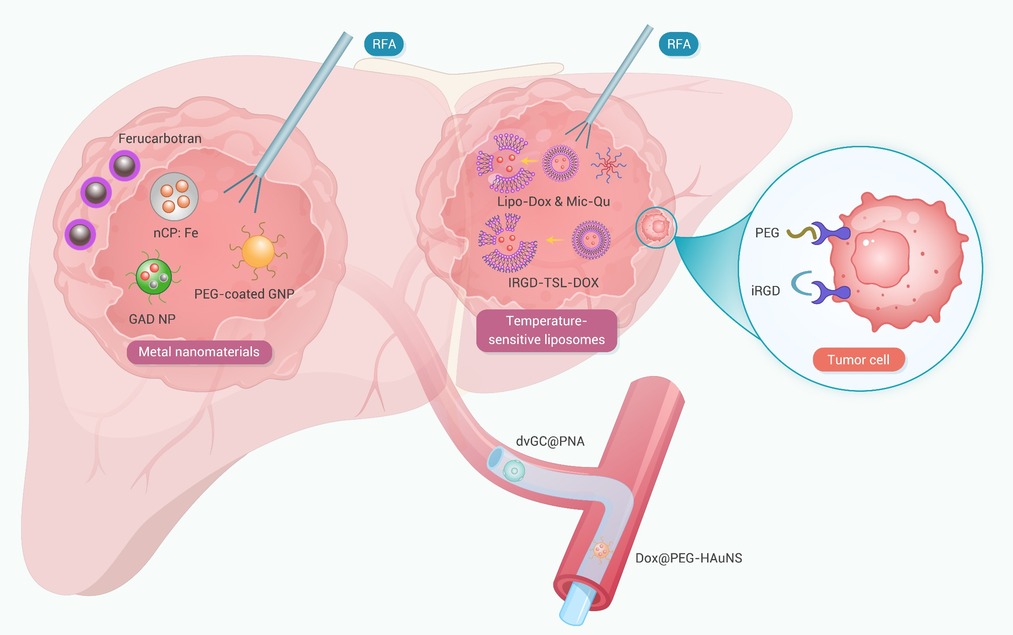
Export File
Citation
Li H., Lan J., Qiu Y., et al. (2026). Advances in nanomaterial-enhanced ablation therapy for hepatocellular carcinoma. The Innovation Oncology 1:100029. https://doi.org/10.59717/j.xinn-oncol.2026.100029

Format
Content
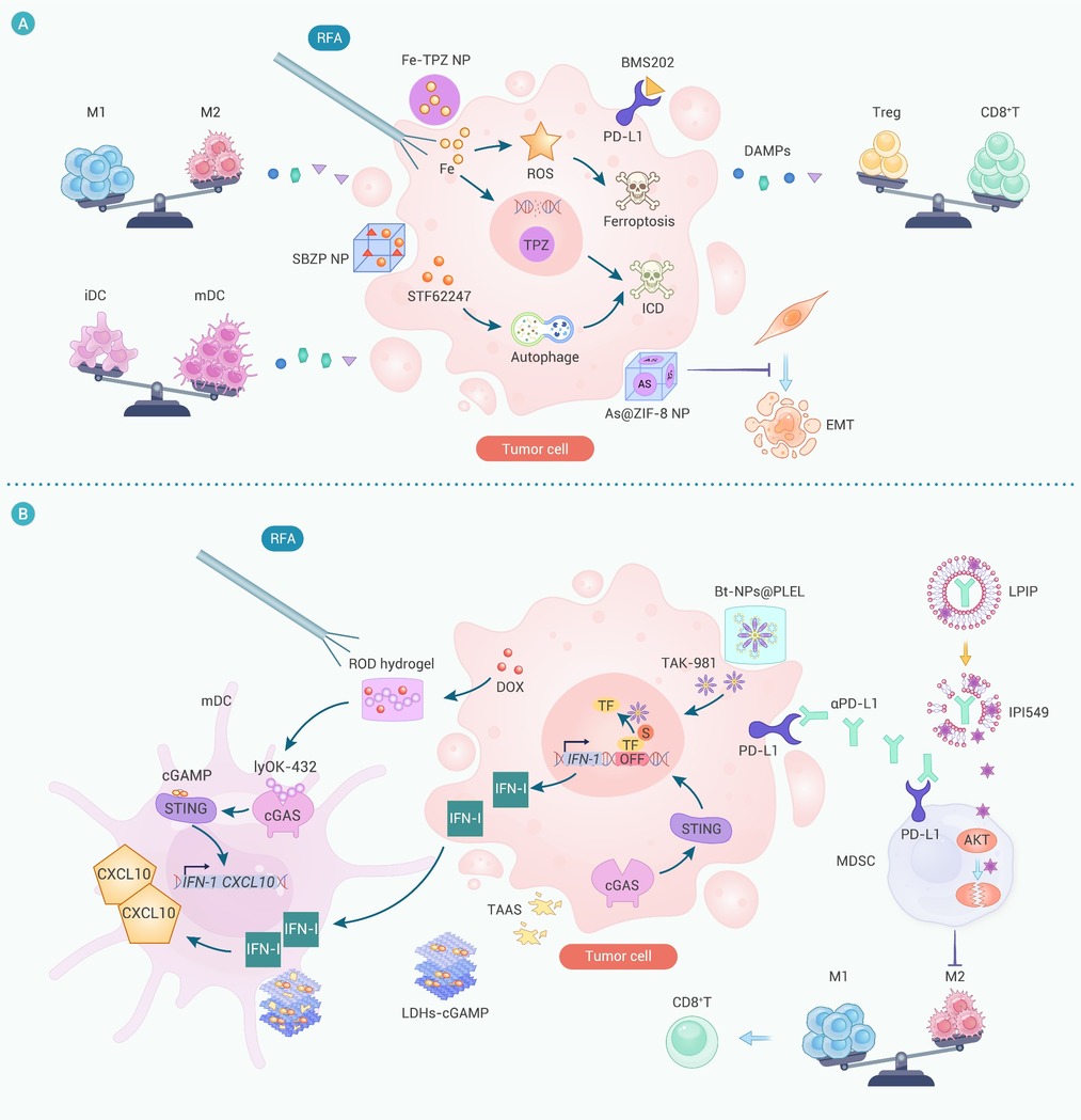
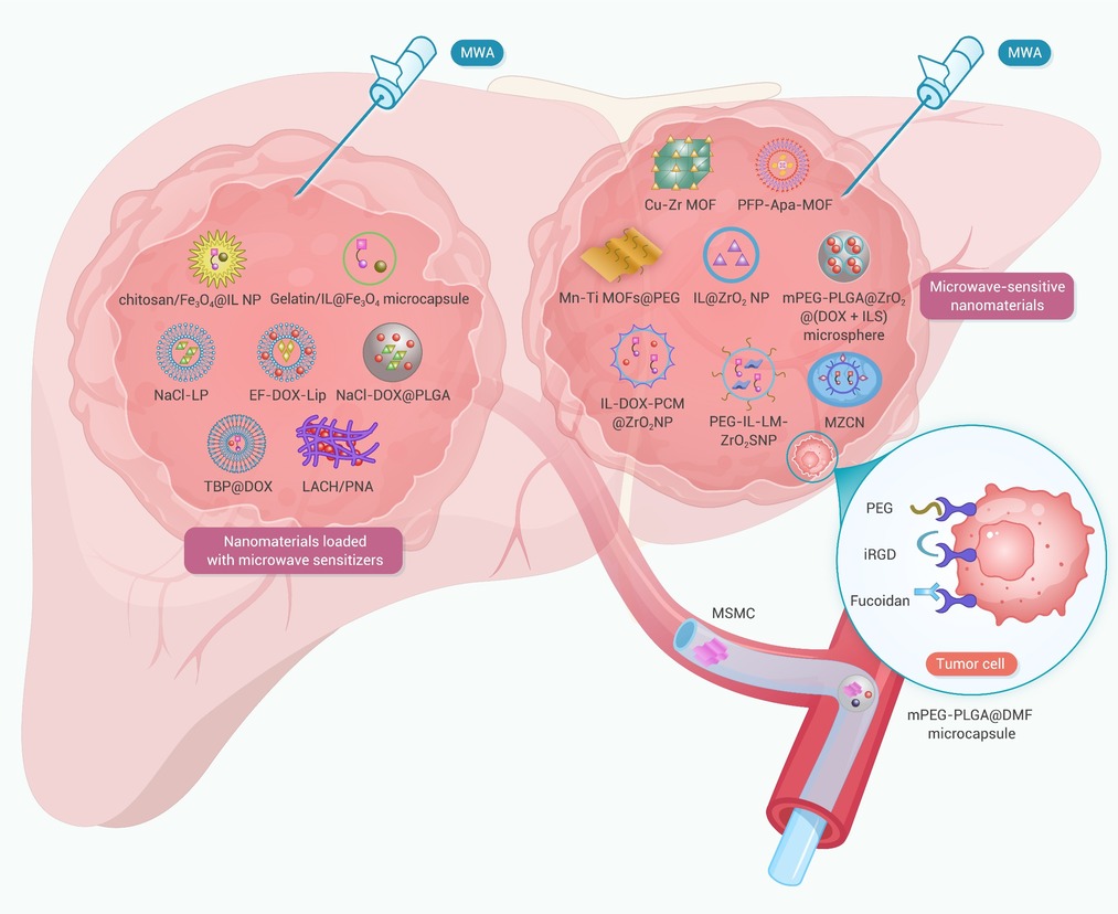
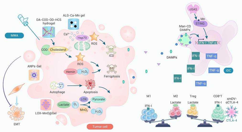
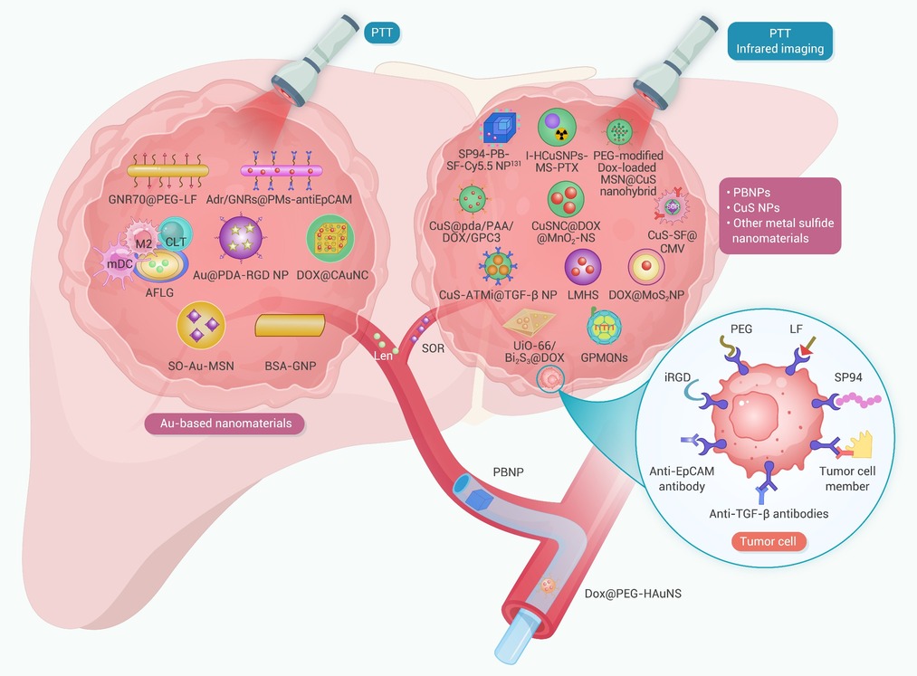
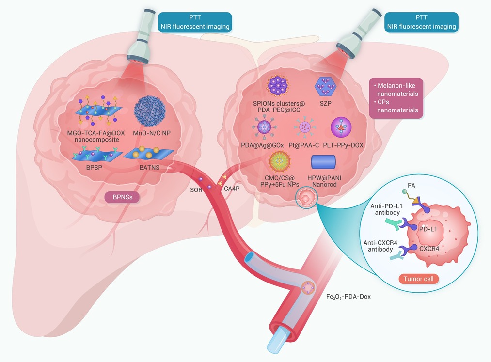
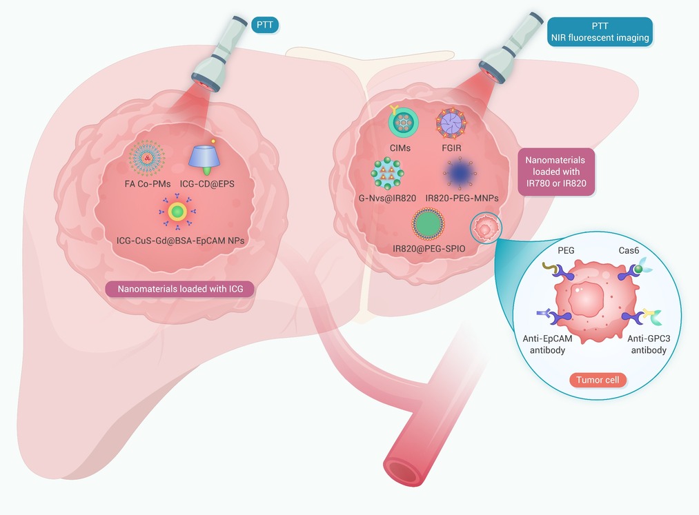
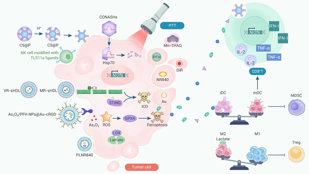
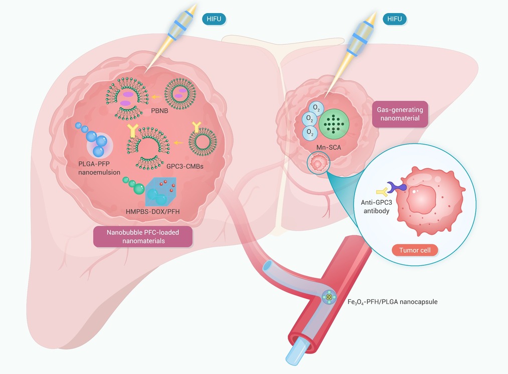
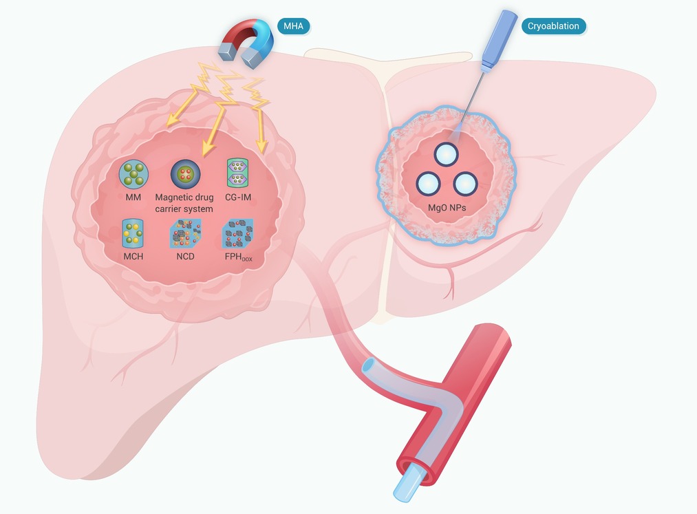
-
Figure 1.
Enhancement of RFA in HCC using different types of nanomaterials
-
Figure 2.
Nanomaterial-mediated modulation of TME enhances RFA in HCC
-
Figure 3.
Enhancement of MWA in HCC using different types of nanomaterials
-
Figure 4.
Nanomaterial-mediated modulation of TME enhances MWA for HCC
-
Figure 5.
Metal-based nanomaterials and partial inorganic nonmetallic nanomaterials enhance PTT for HCC
-
Figure 6.
Other inorganic nonmetallic nanomaterials enhance PTT for HCC
-
Figure 7.
Nanomaterials loaded with small molecular dyes enhance PTT for HCC
-
Figure 8.
Nanomaterial-mediated TME modulation enhances PTT in HCC
-
Figure 9.
Nanomaterials enhance HIFU for HCC
-
Figure 10.
Nanomaterials enhance MHA and cryoablation for HCC
