
Iron-based materials for sequential tumor microenvironment reprogramming: From mechanical decompression to immune remodeling
-
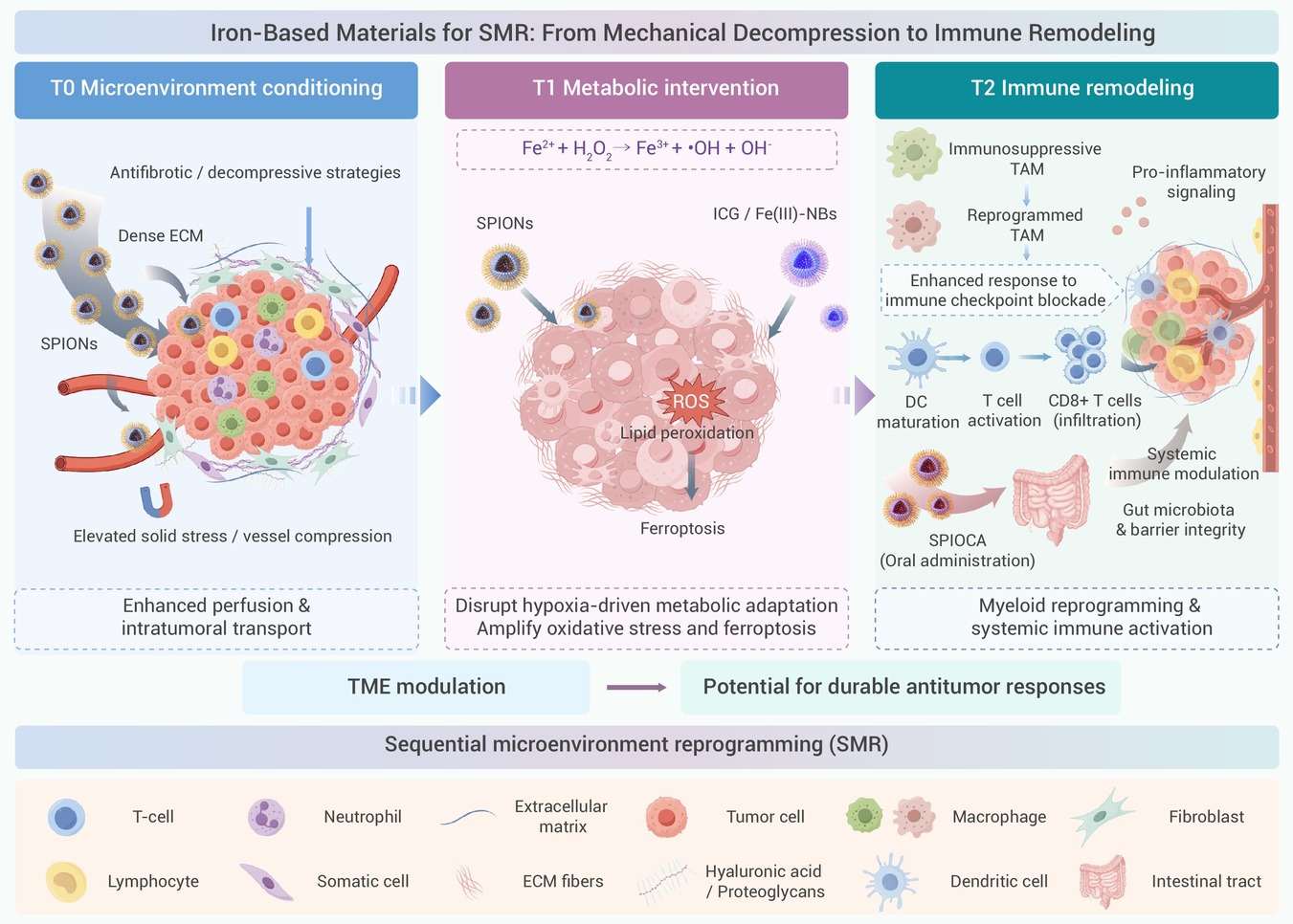
GRAPHICAL ABSTRACT
 DownLoad:
Full size image
-
PUBLIC SUMMARY
-
The tumor microenvironment creates physical, metabolic, and immune barriers that limit cancer therapy.
Iron-based materials offer magnetic, catalytic, and immune-regulating functions to address these barriers.
Sequential microenvironment reprogramming (SMR) links three stages, from transport to immune remodeling.
Rather than following a fixed sequence, SMR guides interventions based on biological responses.
Its clinical potential requires reliable biomarkers, patient stratification, and rigorous validation.
-
-
ABSTRACT
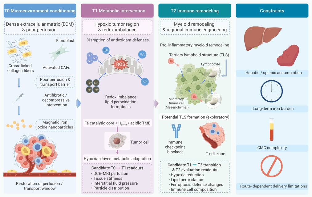
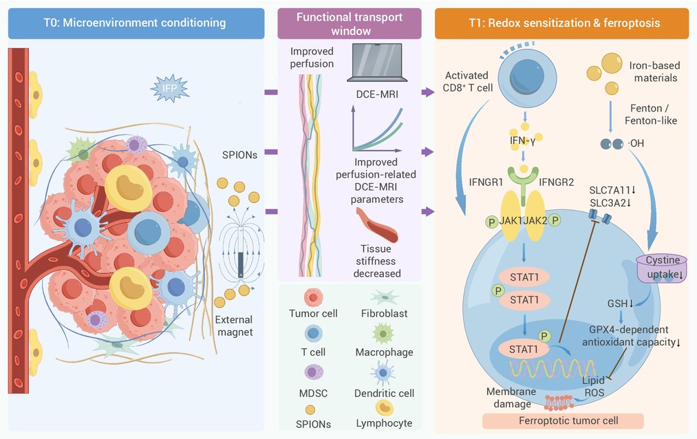
The tumor microenvironment (TME) is a dynamic ecosystem in which abnormal vasculature, stromal fibrosis, metabolic adaptation, and immune suppression collectively undermine the effectiveness of cancer therapy. Although iron-based materials have traditionally been developed as imaging agents or drug carriers, their magnetic responsiveness, catalytic activity, and immunomodulatory properties now provide opportunities to intervene in multiple components of the TME. We adapt and formalize sequential microenvironment reprogramming (SMR) as a framework that integrates the functions of iron-based materials across three interconnected intervention tiers (T0–T2). T0 represents baseline microenvironment conditioning, improving perfusion and intratumoral transport through magnetically assisted delivery together with antifibrotic or decompressive strategies. This creates a more permissive environment for T1, in which iron-catalyzed redox reactions and ferroptosis are exploited to disrupt tumor metabolic adaptation. T2 extends these local effects toward myeloid remodeling and regional immune activation to support more durable antitumor responses. SMR provides a framework for interpreting current evidence and identifying stage-specific therapeutic opportunities. Transitions between intervention tiers are guided by biological prerequisites rather than a fixed treatment sequence. We discuss the major challenges that remain, including biomarker-guided stage switching, patient stratification, translational feasibility, and the need for rigorous experimental validation of this staged therapeutic strategy. -

-
REFERENCES
[1] Mitchell M.J., Billingsley M.M., Haley R.M., et al. (2021). Engineering precision nanoparticles for drug delivery. Nat. Rev. Drug Discov. 20:101−124. DOI:10.1038/s41573-020-0090-8 [2] Shi J., Kantoff P.W., Wooster R., et al. (2017). Cancer nanomedicine: Progress, challenges and opportunities. Nat. Rev. Cancer 17:20−37. DOI:10.1038/nrc.2016.108 [3] Matsumura Y. and Maeda H. (1986). A new concept for macromolecular therapeutics in cancer chemotherapy: Mechanism of tumoritropic accumulation of proteins and the antitumor agent smancs. Cancer Res. 46:6387-6392. https://aacrjournals.org/cancerres/article/46/12_Part_1/6387/490212/A-New-Concept-for-Macromolecular-Therapeutics-in [4] Youn Y.S. and Bae Y.H. (2018). Perspectives on the past, present, and future of cancer nanomedicine. Adv. Drug Deliv. Rev. 130:3−11. DOI:10.1016/j.addr.2018.05.008 [5] Wilhelm S., Tavares A.J., Dai Q., et al. (2016). Analysis of nanoparticle delivery to tumours. Nat. Rev. Mater. 1:16014. DOI:10.1038/natrevmats.2016.14 [6] Fukumura D., Kloepper J., Amoozgar Z., et al. (2018). Enhancing cancer immunotherapy using antiangiogenics: Opportunities and challenges. Nat. Rev. Clin. Oncol. 15:325−340. DOI:10.1038/nrclinonc.2018.29 [7] De Palma M., Biziato D. and Petrova T.V. (2017). Microenvironmental regulation of tumour angiogenesis. Nat. Rev. Cancer 17:457−474. DOI:10.1038/nrc.2017.51 [8] Jain R.K. (2014). Antiangiogenesis strategies revisited: From starving tumors to alleviating hypoxia. Cancer Cell 26:605−622. DOI:10.1016/j.ccell.2014.10.006 [9] Sahai E., Astsaturov I., Cukierman E., et al. (2020). A framework for advancing our understanding of cancer-associated fibroblasts. Nat. Rev. Cancer 20:174−186. DOI:10.1038/s41568-019-0238-1 [10] Stylianopoulos T., Martin J.D., Chauhan V.P., et al. (2012). Causes, consequences, and remedies for growth-induced solid stress in murine and human tumors. Proc. Natl. Acad. Sci. USA 109:15101−15108. DOI:10.1073/pnas.1213353109 [11] Nia H.T., Munn L.L. and Jain R.K. (2020). Physical traits of cancer. Science 370. DOI:10.1126/science.aaz0868. [12] Semenza G.L. (2012). Hypoxia-inducible factors in physiology and medicine. Cell 148:399−408. DOI:10.1016/j.cell.2012.01.021 [13] Petrova V., Annicchiarico-Petruzzelli M., Melino G., et al. (2018). The hypoxic tumour microenvironment. Oncogenesis 7:10. DOI:10.1038/s41389-017-0011-9 [14] Pavlova Natalya N. and Thompson Craig B. (2016). The emerging hallmarks of cancer metabolism. Cell Metab. 23:27−47. DOI:10.1016/j.cmet.2015.12.006 [15] Zhang W., Wang P., Yuan G., et al. (2026). Fatty acid metabolic reprogramming in the tumor microenvironment: Unraveling mechanisms and therapeutic prospects. Genes Dis. 13:101772. DOI:10.1016/j.gendis.2025.101772 [16] Joyce J.A. and Fearon D.T. (2015). T cell exclusion, immune privilege, and the tumor microenvironment. Science 348:74−80. DOI:10.1126/science.aaa6204 [17] Hinshaw D.C. and Shevde L.A. (2019). The tumor microenvironment innately modulates cancer progression. Cancer Res. 79:4557−4566. DOI:10.1158/0008-5472.Can-18-3962 [18] Binnewies M., Roberts E.W., Kersten K., et al. (2018). Understanding the tumor immune microenvironment (TIME) for effective therapy. Nat. Med. 24:541−550. DOI:10.1038/s41591-018-0014-x [19] Anderson N.M. and Simon M.C. (2020). The tumor microenvironment. Curr. Biol. 30:R921−R925. DOI:10.1016/j.cub.2020.06.081 [20] Valkenburg K.C., de Groot A.E. and Pienta K.J. (2018). Targeting the tumour stroma to improve cancer therapy. Nat. Rev. Clin. Oncol. 15:366−381. DOI:10.1038/s41571-018-0007-1 [21] Martin J.D., Cabral H., Stylianopoulos T., et al. (2020). Improving cancer immunotherapy using nanomedicines: Progress, opportunities and challenges. Nat. Rev. Clin. Oncol. 17:251−266. DOI:10.1038/s41571-019-0308-z [22] Liu Y., Sun D., Li W., et al. (2026). Nanomedicine-mediated modulation of tumor metabolism for enhanced immunotherapy. J. Control. Release 390:114587. DOI:10.1016/j.jconrel.2025.114587 [23] Jain R.K. (2005). Normalization of tumor vasculature: An emerging concept in antiangiogenic therapy. Science 307:58−62. DOI:10.1126/science.1104819 [24] Galluzzi L., Buqué A., Kepp O., et al. (2017). Immunogenic cell death in cancer and infectious disease. Nat. Rev. Immunol. 17:97−111. DOI:10.1038/nri.2016.107 [25] Sengupta S., Eavarone D., Capila I., et al. (2005). Temporal targeting of tumour cells and neovasculature with a nanoscale delivery system. Nature 436:568−572. DOI:10.1038/nature03794 [26] Mpekris F., Panagi M., Voutouri C., et al. (2021). Normalizing the microenvironment overcomes vessel compression and resistance to nano-immunotherapy in breast cancer lung metastasis. Adv. Sci. (Weinh) 8:2001917. DOI:10.1002/advs.202001917 [27] Mpekris F., Voutouri C., Baish J.W., et al. (2020). Combining microenvironment normalization strategies to improve cancer immunotherapy. Proc. Natl. Acad. Sci. USA 117:3728−3737. DOI:10.1073/pnas.1919764117 [28] Yu Y., Song Z., Zhu A., et al. (2026). Sequential tumor microenvironment reprogramming by nanoplatform potentiates sonodynamic-chemodynamic therapy and immune checkpoint blockade in breast cancer. Adv. Sci. (Weinh) 13:e12135. DOI:10.1002/advs.202512135 [29] Yu E. Y., Bishop M., Zheng B., et al. (2017). Magnetic particle imaging: A novel in vivo imaging platform for cancer detection. Nano Lett. 17:1648−1654. DOI:10.1021/acs.nanolett.6b04865 [30] Bulte J.W. and Kraitchman D.L. (2004). Iron oxide MR contrast agents for molecular and cellular imaging. NMR Biomed. 17:484−499. DOI:10.1002/nbm.924 [31] Yang W. S., SriRamaratnam R., Welsch M.E., et al. (2014). Regulation of ferroptotic cancer cell death by GPX4. Cell 156:317−331. DOI:10.1016/j.cell.2013.12.010 [32] Huo M., Wang L., Chen Y., et al. (2017). Tumor-selective catalytic nanomedicine by nanocatalyst delivery. Nat. Commun. 8:357. DOI:10.1038/s41467-017-00424-8 [33] Tang Z., Liu Y., He M., et al. (2019). Chemodynamic therapy: Tumour microenvironment-mediated fenton and fenton-like reactions. Angew. Chem. Int. Ed. 58:946−956. DOI:10.1002/anie.201805664 [34] Dixon S.J., Lemberg K.M., Lamprecht M.R., et al. (2012). Ferroptosis: An iron-dependent form of nonapoptotic cell death. Cell 149:1060−1072. DOI:10.1016/j.cell.2012.03.042 [35] Jiang X., Stockwell B.R. and Conrad M. (2021). Ferroptosis: Mechanisms, biology and role in disease. Nat. Rev. Mol. Cell Biol. 22:266−282. DOI:10.1038/s41580-020-00324-8 [36] Wan X., Song L., Pan W., et al. (2020). Tumor-targeted cascade nanoreactor based on metal-organic frameworks for synergistic ferroptosis-starvation anticancer therapy. ACS Nano 14:11017−11028. DOI:10.1021/acsnano.9b07789 [37] Xue C.C., Li M.H., Zhao Y., et al. (2020). Tumor microenvironment-activatable Fe-doxorubicin preloaded amorphous CaCO(3) nanoformulation triggers ferroptosis in target tumor cells. Sci. Adv. 6:eaax1346. DOI:10.1126/sciadv.aax1346 [38] Chen X. and Song E. (2019). Turning foes to friends: Targeting cancer-associated fibroblasts. Nat. Rev. Drug Discov. 18:99−115. DOI:10.1038/s41573-018-0004-1 [39] Diop-Frimpong B., Chauhan V.P., Krane S., et al. (2011). Losartan inhibits collagen I synthesis and improves the distribution and efficacy of nanotherapeutics in tumors. Proc. Natl. Acad. Sci. USA 108:2909−2914. DOI:10.1073/pnas.1018892108 [40] Shapiro B., Kulkarni S., Nacev A., et al. (2015). Open challenges in magnetic drug targeting. Wiley Interdiscip. Rev. Nanomed. Nanobiotechnol. 7:446−457. DOI:10.1002/wnan.1311 [41] Liu J.F., Lan Z., Ferrari C., et al. (2020). Use of oppositely polarized external magnets to improve the accumulation and penetration of magnetic nanocarriers into solid tumors. ACS Nano 14:142−152. DOI:10.1021/acsnano.9b05660 [42] Kim D.H., Rozhkova E.A., Ulasov I.V., et al. (2010). Biofunctionalized magnetic-vortex microdiscs for targeted cancer-cell destruction. Nat. Mater. 9:165−171. DOI:10.1038/nmat2591 [43] Cheng Y., Muroski M.E., Petit D.C.M.C., et al. (2016). Rotating magnetic field induced oscillation of magnetic particles for in vivo mechanical destruction of malignant glioma. J. Control. Release 223:75−84. DOI:10.1016/j.jconrel.2015.12.028 [44] Shen Y., Wu C., Uyeda T.Q.P., et al. (2017). Elongated nanoparticle aggregates in cancer cells for mechanical destruction with low frequency rotating magnetic field. Theranostics 7:1735−1748. DOI:10.7150/thno.18352 [45] Chauhan V.P., Boucher Y., Ferrone C.R., et al. (2014). Compression of pancreatic tumor blood vessels by hyaluronan is caused by solid stress and not interstitial fluid pressure. Cancer Cell 26:14−15. DOI:10.1016/j.ccr.2014.06.003 [46] Gu Z., Liu T., Liu C., et al. (2021). Ferroptosis-strengthened metabolic and inflammatory regulation of tumor-associated macrophages provokes potent tumoricidal activities. Nano Lett. 21:6471−6479. DOI:10.1021/acs.nanolett.1c01401 [47] Zanganeh S., Hutter G., Spitler R., et al. (2016). Iron oxide nanoparticles inhibit tumour growth by inducing pro-inflammatory macrophage polarization in tumour tissues. Nat. Nanotechnol. 11:986−994. DOI:10.1038/nnano.2016.168 [48] Recalcati S., Locati M., Marini A., et al. (2010). Differential regulation of iron homeostasis during human macrophage polarized activation. Eur. J. Immunol. 40:824−835. DOI:10.1002/eji.200939889 [49] Costa da Silva M., Breckwoldt M.O., Vinchi F., et al. (2017). Iron induces anti-tumor activity in tumor-associated macrophages. Front. Immunol. 8:1479. DOI:10.3389/fimmu.2017.01479 [50] Gu Z., Liu T., Tang J., et al. (2019). Mechanism of iron oxide-induced macrophage activation: The impact of composition and the underlying signaling pathway. J. Am. Chem. Soc. 141:6122−6126. DOI:10.1021/jacs.8b10904 [51] Jansen C.S., Prokhnevska N., Master V.A., et al. (2019). An intra-tumoral niche maintains and differentiates stem-like CD8 T cells. Nature 576:465−470. DOI:10.1038/s41586-019-1836-5 [52] Sautès-Fridman C., Petitprez F., Calderaro J., et al. (2019). Tertiary lymphoid structures in the era of cancer immunotherapy. Nat. Rev. Cancer 19:307−325. DOI:10.1038/s41568-019-0144-6 [53] Schumacher T.N. and Thommen D.S. (2022). Tertiary lymphoid structures in cancer. Science 375:eabf9419. DOI:10.1126/science.abf9419 [54] Helmink B.A., Reddy S.M., Gao J., et al. (2020). B cells and tertiary lymphoid structures promote immunotherapy response. Nature 577:549−555. DOI:10.1038/s41586-019-1922-8 [55] Cabrita R., Lauss M., Sanna A., et al. (2020). Tertiary lymphoid structures improve immunotherapy and survival in melanoma. Nature 577:561−565. DOI:10.1038/s41586-019-1914-8 [56] Petitprez F., de Reyniès A., Keung E.Z., et al. (2020). B cells are associated with survival and immunotherapy response in sarcoma. Nature 577:556−560. DOI:10.1038/s41586-019-1906-8 [57] Stylianopoulos T., Martin J.D., Snuderl M., et al. (2013). Coevolution of solid stress and interstitial fluid pressure in tumors during progression: implications for vascular collapse. Cancer Res. 73:3833−3841. DOI:10.1158/0008-5472.Can-12-4521 [58] Brand A., Singer K., Koehl G.E., et al. (2016). LDHA-associated lactic acid production blunts tumor immunosurveillance by T and NK cells. Cell Metab. 24:657−671. DOI:10.1016/j.cmet.2016.08.011 [59] Recalcati S., Locati M., Gammella E., et al. (2012). Iron levels in polarized macrophages: Regulation of immunity and autoimmunity. Autoimmun. Rev. 11:883−889. DOI:10.1016/j.autrev.2012.03.003 [60] Manz D.H., Blanchette N.L., Paul B.T., et al. (2016). Iron and cancer: Recent insights. Ann. N. Y. Acad. Sci. 1368:149−161. DOI:10.1111/nyas.13008 [61] Yang Y., Liu Y., Song L., et al. (2023). Iron oxide nanoparticle-based nanocomposites in biomedical application. Trends Biotechnol. 41:1471−1487. DOI:10.1016/j.tibtech.2023.06.001 [62] Chen G., Roy I., Yang C., et al. (2016). Nanochemistry and nanomedicine for nanoparticle-based diagnostics and therapy. Chem. Rev. 116:2826−2885. DOI:10.1021/acs.chemrev.5b00148 [63] Lim E.K., Kim T., Paik S., et al. (2015). Nanomaterials for theranostics: Recent advances and future challenges. Chem. Rev. 115:327−394. DOI:10.1021/cr300213b [64] Gleich B. and Weizenecker J. (2005). Tomographic imaging using the nonlinear response of magnetic particles. Nature 435:1214−1217. DOI:10.1038/nature03808 [65] Makela A.V., Gaudet J.M., Schott M.A., et al. (2020). Magnetic particle imaging of macrophages associated with cancer: Filling the voids left by iron-based magnetic resonance imaging. Mol. Imaging Biol. 22:958−968. DOI:10.1007/s11307-020-01473-0 [66] Park S.J., Han S.R., Kang Y.H., et al. (2022). In vivo preclinical tumor-specific imaging of superparamagnetic iron oxide nanoparticles using magnetic particle imaging for cancer diagnosis. Int. J. Nanomedicine 17:3711−3722. DOI:10.2147/ijn.S372494 [67] Dadfar S.M., Camozzi D., Darguzyte M., et al. (2020). Size-isolation of superparamagnetic iron oxide nanoparticles improves MRI, MPI and hyperthermia performance. J. Nanobiotechnology 18:22. DOI:10.1186/s12951-020-0580-1 [68] Kumar P.P.P., Nafiujjaman M., Makela A.V., et al. (2025). Development of iron oxide nanochains as a sensitive magnetic particle imaging tracer for cancer detection. ACS Appl. Mater. Interfaces 17:20859−20871. DOI:10.1021/acsami.5c00332 [69] Ejima H., Richardson J.J., Liang K., et al. (2013). One-step assembly of coordination complexes for versatile film and particle engineering. Science 341:154−157. DOI:10.1126/science.1237265 [70] Zhang C., Bu W., Ni D., et al. (2016). Synthesis of iron nanometallic glasses and their application in cancer therapy by a localized Fenton reaction. Angew. Chem. Int. Ed. 55:2101−2106. DOI:10.1002/anie.201510031 [71] Yang G., Xu L., Chao Y., et al. (2017). Hollow MnO(2) as a tumor-microenvironment-responsive biodegradable nano-platform for combination therapy favoring antitumor immune responses. Nat. Commun. 8:902. DOI:10.1038/s41467-017-01050-0 [72] Wang C., Guan Y., Lv M., et al. (2018). Manganese increases the sensitivity of the cGAS-STING pathway for double-stranded DNA and is required for the host defense against DNA viruses. Immunity 48:675−687.e677. DOI:10.1016/j.immuni.2018.03.017 [73] Lin L.-S., Huang T., Song J., et al. (2019). Synthesis of copper peroxide nanodots for H2O2 self-supplying chemodynamic therapy. J. Am. Chem. Soc. 141:9937−9945. DOI:10.1021/jacs.9b03457 [74] Tsvetkov P., Coy S., Petrova B., et al. (2022). Copper induces cell death by targeting lipoylated TCA cycle proteins. Science 375:1254−1261. DOI:10.1126/science.abf0529 [75] Nam J., Son S., Ochyl L.J., et al. (2018). Chemo-photothermal therapy combination elicits anti-tumor immunity against advanced metastatic cancer. Nat. Commun. 9:1074. DOI:10.1038/s41467-018-03473-9 [76] Zheng P., Ding B., Shi R., et al. (2021). A Multichannel Ca(2+) Nanomodulator for multilevel mitochondrial destruction-mediated cancer therapy. Adv. Mater. 33:e2007426. DOI:10.1002/adma.202007426 [77] Rhim A.D., Oberstein P.E., Thomas D.H., et al. (2014). Stromal elements act to restrain, rather than support, pancreatic ductal adenocarcinoma. Cancer Cell 25:735−747. DOI:10.1016/j.ccr.2014.04.021 [78] Provenzano P.P., Cuevas C., Chang A.E., et al. (2012). Enzymatic targeting of the stroma ablates physical barriers to treatment of pancreatic ductal adenocarcinoma. Cancer Cell 21:418−429. DOI:10.1016/j.ccr.2012.01.007 [79] Özdemir B.C., Pentcheva-Hoang T., Carstens J.L., et al. (2014). Depletion of carcinoma-associated fibroblasts and fibrosis induces immunosuppression and accelerates pancreas cancer with reduced survival. Cancer Cell 25:719-734. DOI:10.1016/j.ccr.2014.04.005 [80] Zhang Z., Chen X., Zhang B., et al. (2026). Fibrosis-driven hepatocarcinogenesis, metastasis and immune evasion: Mechanisms and therapeutic targets. Biochim. Biophys. Acta Rev. Cancer 1881:189517. DOI:10.1016/j.bbcan.2025.189517 [81] Heldin C.H., Rubin K., Pietras K., et al. (2004). High interstitial fluid pressure - an obstacle in cancer therapy. Nat. Rev. Cancer 4:806−813. DOI:10.1038/nrc1456 [82] Chauhan V.P., Martin J.D., Liu H., et al. (2013). Angiotensin inhibition enhances drug delivery and potentiates chemotherapy by decompressing tumour blood vessels. Nat. Commun. 4:2516. DOI:10.1038/ncomms3516 [83] Tian L., Goldstein A., Wang H., et al. (2017). Mutual regulation of tumour vessel normalization and immunostimulatory reprogramming. Nature 544:250−254. DOI:10.1038/nature21724 [84] O'Connor J.P., Aboagye E.O., Adams J.E., et al. (2017). Imaging biomarker roadmap for cancer studies. Nat. Rev. Clin. Oncol. 14:169−186. DOI:10.1038/nrclinonc.2016.162 [85] Winkler J., Abisoye-Ogunniyan A., Metcalf K.J., et al. (2020). Concepts of extracellular matrix remodelling in tumour progression and metastasis. Nat. Commun. 11:5120. DOI:10.1038/s41467-020-18794-x [86] Levental K.R., Yu H., Kass L., et al. (2009). Matrix crosslinking forces tumor progression by enhancing integrin signaling. Cell 139:891−906. DOI:10.1016/j.cell.2009.10.027 [87] Kalluri R. (2016). The biology and function of fibroblasts in cancer. Nat. Rev. Cancer 16:582−598. DOI:10.1038/nrc.2016.73 [88] Huang Z., Chen J., Zhu T., et al. (2025). Cancer-associated fibroblasts in the tumor microenvironment: Heterogeneity, crosstalk mechanisms, and therapeutic implications. Mol. Cancer 25:19. DOI:10.1186/s12943-025-02533-1 [89] Hakim N., Patel R., Devoe C., et al. (2019). Why HALO 301 failed and implications for treatment of pancreatic cancer. Pancreas (Fairfax) 3:e1−e4. DOI:10.17140/POJ-3-e010 [90] Cai W., Chen Y., Dai S., et al. (2025). Hyaluronic acid and its role in pancreatic cancer: Mechanisms of tumor progression, drug resistance, and therapeutic targeting. Pharmacol Res. 221:107958. DOI:10.1016/j.phrs.2025.107958 [91] Li L., Wen J., Xu H., et al. (2026). ECM-responsive nanomedicine to enhance immunotherapy in pancreatic cancer. Colloids Surf. B Biointerfaces 260:115356. DOI:10.1016/j.colsurfb.2025.115356 [92] Song G., Chen Y., Liang C., et al. (2016). Catalase-loaded TaOx nanoshells as bio-nanoreactors combining high-Z element and enzyme delivery for enhancing radiotherapy. Adv. Mater. 28:7143−7148. DOI:10.1002/adma.201602111 [93] Chen Q., Feng L., Liu J., et al. (2016). Intelligent albumin-MnO2 nanoparticles as pH-/H2O2 -responsive dissociable nanocarriers to modulate tumor hypoxia for effective combination therapy. Adv. Mater. 28:7129−7136. DOI:10.1002/adma.201601902 [94] Cheng Y., Cheng H., Jiang C., et al. (2015). Perfluorocarbon nanoparticles enhance reactive oxygen levels and tumour growth inhibition in photodynamic therapy. Nat. Commun. 6:8785. DOI:10.1038/ncomms9785 [95] Gao S., Lin H., Zhang H., et al. (2019). Nanocatalytic tumor therapy by biomimetic dual inorganic nanozyme-catalyzed cascade reaction. Adv. Sci. (Weinh) 6:1801733. DOI:10.1002/advs.201801733 [96] Chen H., Tian J., He W., et al. (2015). H2O2-activatable and O2-evolving nanoparticles for highly efficient and selective photodynamic therapy against hypoxic tumor cells. J. Am. Chem. Soc. 137:1539−1547. DOI:10.1021/ja511420n [97] Yang L., Zhang W.H., Li Y., et al. (2025). Oxygen nanobubbles enhance ICG/Fe(III)-mediated dual-modal therapy to induce ferroptosis in tumor treatment. ACS Appl. Mater. Interfaces 17:11718−11730. DOI:10.1021/acsami.4c19604 [98] Prasad S.B., Shinde A., Srinivasrao D.A., et al. (2025). Metal-organic frameworks as therapeutic chameleons: Revolutionizing the cancer therapy employing novel nanoarchitectonics. Asian J. Pharm. Sci. 20:101054. DOI:10.1016/j.ajps.2025.101054 [99] Jiao L., Yan H., Wu Y., et al. (2020). When nanozymes meet single-atom catalysis. Angew. Chem. Int. Ed. 59:2565−2576. DOI:10.1002/anie.201905645 [100] Huang L., Chen J., Gan L., et al. (2019). Single-atom nanozymes. Sci. Adv. 5:eaav5490. DOI:10.1126/sciadv.aav5490 [101] Horcajada P., Chalati T., Serre C., et al. (2010). Porous metal-organic-framework nanoscale carriers as a potential platform for drug delivery and imaging. Nat. Mater. 9:172−178. DOI:10.1038/nmat2608 [102] Ghaemi B. and Javad Hajipour M. (2023). Tumor acidic environment directs nanoparticle impacts on cancer cells. J. Colloid Interface Sci. 634:684−692. DOI:10.1016/j.jcis.2022.12.019 [103] Liu Y., Cai X., Liu J., et al. (2025). The role of redox homeostasis in tumor progression: Implications for cancer therapy. Acta Biomater. 204:156−186. DOI:10.1016/j.actbio.2025.07.067 [104] Doll S., Proneth B., Tyurina Y.Y., et al. (2017). ACSL4 dictates ferroptosis sensitivity by shaping cellular lipid composition. Nat. Chem. Biol. 13:91−98. DOI:10.1038/nchembio.2239 [105] Kagan V.E., Mao G., Qu F., et al. (2017). Oxidized arachidonic and adrenic PEs navigate cells to ferroptosis. Nat. Chem. Biol. 13:81−90. DOI:10.1038/nchembio.2238 [106] Hu Y., Li Z., Li M., et al. (2025). Targeting PRDX6-dependent localization and function of GPX4 enhances ferroptosis-mediated tumor suppression. Mol. Cell 85:4602−4620.e4609. DOI:10.1016/j.molcel.2025.11.023 [107] Meng X., Deng J., Liu F., et al. (2019). Triggered all-active metal organic framework: Ferroptosis machinery contributes to the apoptotic photodynamic antitumor therapy. Nano Lett. 19:7866−7876. DOI:10.1021/acs.nanolett.9b02904 [108] Zhao Y., Yang S., Huang L., et al. (2025). MUC1 drives ferroptosis resistance in ICC via Src-mediated FSP1 deubiquitination and myristoylation. Clin. Transl. Med. 15:e70495. DOI:10.1002/ctm2.70495 [109] Hai Y., Lin R., Liao W., et al. (2025). Dihydroorotate dehydrogenase inhibition activates STING pathway and pyroptosis to enhance NK cell-dependent tumor immunotherapy. Mol. Biomed. 6:87. DOI:10.1186/s43556-025-00339-7 [110] Ma C., Han L., Yao H., et al. (2026). Modulating G6PD/PGD to overcome FSP1/DHODH-mediated ferroptosis defence: A novel oridonin derivative suppresses liver cancer. Br. J. Pharmacol. 183:249−267. DOI:10.1111/bph.70160 [111] Li W., Panacek D., Xie Q., et al. (2025). Precision fishing for enzyme-like materials: Identifying and engineering mesoporous catalytic sites for ferroptosis induction. Nano Lett. 25:11356−11364. DOI:10.1021/acs.nanolett.5c02476 [112] Stockwell B.R., Friedmann Angeli J.P., Bayir H., et al. (2017). Ferroptosis: A regulated cell death nexus linking metabolism, redox biology, and disease. Cell 171:273−285. DOI:10.1016/j.cell.2017.09.021 [113] Stockwell B.R. (2022). Ferroptosis turns 10: Emerging mechanisms, physiological functions, and therapeutic applications. Cell 185:2401−2421. DOI:10.1016/j.cell.2022.06.003 [114] Dixon S.J. and Stockwell B.R. (2019). The hallmarks of ferroptosis. Annu. Rev. Cancer Biol. 3:35−54. DOI:10.1146/annurev-cancerbio-030518-055844 [115] Lei G., Zhuang L. and Gan B. (2022). Targeting ferroptosis as a vulnerability in cancer. Nat. Rev. Cancer 22:381−396. DOI:10.1038/s41568-022-00459-0 [116] Hangauer M.J., Viswanathan V.S., Ryan M.J., et al. (2017). Drug-tolerant persister cancer cells are vulnerable to GPX4 inhibition. Nature 551:247−250. DOI:10.1038/nature24297 [117] Shen Z., Liu T., Li Y., et al. (2018). Fenton-reaction-acceleratable magnetic nanoparticles for ferroptosis therapy of orthotopic brain tumors. ACS Nano 12:11355−11365. DOI:10.1021/acsnano.8b06201 [118] Su L., Yang S., Yuan H., et al. (2026). Cell membrane biomimetic drug delivery systems based on ferroptosis mechanism for anticancer treatment. Adv. Healthc. Mater. 15:e04993. DOI:10.1002/adhm.202504993 [119] Gao Y., Xu J., Lai Y., et al. (2025). 2D nanomaterial-triggered ferroptosis in cancer therapy. J. Nanobiotechnology 24:81. DOI:10.1186/s12951-025-03975-x [120] Ye L.F., Chaudhary K.R., Zandkarimi F., et al. (2020). Radiation-induced lipid peroxidation triggers ferroptosis and synergizes with ferroptosis inducers. ACS Chem. Biol. 15:469−484. DOI:10.1021/acschembio.9b00939 [121] Lang X., Green M.D., Wang W., et al. (2019). Radiotherapy and immunotherapy promote tumoral lipid oxidation and ferroptosis via synergistic repression of SLC7A11. Cancer Discov. 9:1673−1685. DOI:10.1158/2159-8290.Cd-19-0338 [122] Lei G., Zhang Y., Koppula P., et al. (2020). The role of ferroptosis in ionizing radiation-induced cell death and tumor suppression. Cell Res. 30:146−162. DOI:10.1038/s41422-019-0263-3 [123] Zhu T., Shi L., Yu C., et al. (2019). Ferroptosis promotes photodynamic therapy: Supramolecular photosensitizer-inducer nanodrug for enhanced cancer treatment. Theranostics 9:3293−3307. DOI:10.7150/thno.32867 [124] Bae C., Hernández Millares R., Ryu S., et al. (2024). Synergistic effect of ferroptosis-inducing nanoparticles and X-ray irradiation combination therapy. Small 20:e2310873. DOI:10.1002/smll.202310873 [125] Duo K., Feng X., Tian X., et al. (2025). Ferroptosis inhibitors: Mechanisms of action and therapeutic potential. Cell. Mol. Life Sci. 82:441. DOI:10.1007/s00018-025-05958-5 [126] Tang D., Chen X., Kang R., et al. (2021). Ferroptosis: Molecular mechanisms and health implications. Cell Res. 31:107−125. DOI:10.1038/s41422-020-00441-1 [127] Hirayama T., Niwa M., Hirosawa S., et al. (2020). High-throughput screening for the discovery of iron homeostasis modulators using an extremely sensitive fluorescent probe. ACS Sens. 5:2950−2958. DOI:10.1021/acssensors.0c01445 [128] Wiernicki B., Maschalidi S., Pinney J., et al. (2022). Cancer cells dying from ferroptosis impede dendritic cell-mediated anti-tumor immunity. Nat. Commun. 13:3676. DOI:10.1038/s41467-022-31218-2 [129] Ma X., Xiao L., Liu L., et al. (2021). CD36-mediated ferroptosis dampens intratumoral CD8(+) T cell effector function and impairs their antitumor ability. Cell Metab. 33:1001−1012.e1005. DOI:10.1016/j.cmet.2021.02.015 [130] Xu S., Chaudhary O., Rodríguez-Morales P., et al. (2021). Uptake of oxidized lipids by the scavenger receptor CD36 promotes lipid peroxidation and dysfunction in CD8(+) T cells in tumors. Immunity 54:1561−1577.e1567. DOI:10.1016/j.immuni.2021.05.003 [131] Kim R., Hashimoto A., Markosyan N., et al. (2022). Ferroptosis of tumour neutrophils causes immune suppression in cancer. Nature 612:338−346. DOI:10.1038/s41586-022-05443-0 [132] Liu D., Ling Y., Liu W., et al. (2025). Ultrasound-responsive micro/nanobubbles based intelligent theranostic systems for precision tumor therapy. J. Nanobiotechnology 24:83. DOI:10.1186/s12951-025-03959-x [133] Liu J., Dong R., Yuan B., et al. (2025). Immune cells dying from ferroptosis: Mechanisms and therapeutic opportunities. Cell Death Dis. 16:878. DOI:10.1038/s41419-025-08204-9 [134] Galluzzi L., Kepp O., Zitvogel L., et al. (2026). Cancer cell death: Cell-autonomous and immunogenic dimensions. Cancer Cell 44:281−305. DOI:10.1016/j.ccell.2025.12.005 [135] Obeid M., Tesniere A., Ghiringhelli F., et al. (2007). Calreticulin exposure dictates the immunogenicity of cancer cell death. Nat. Med. 13:54−61. DOI:10.1038/nm1523 [136] Tang D., Kepp O. and Kroemer G. (2020). Ferroptosis becomes immunogenic: Implications for anticancer treatments. Oncoimmunology 10:1862949. DOI:10.1080/2162402x.2020.1862949 [137] Krysko D.V., Garg A.D., Kaczmarek A., et al. (2012). Immunogenic cell death and DAMPs in cancer therapy. Nat. Rev. Cancer 12:860−875. DOI:10.1038/nrc3380 [138] Efimova I., Catanzaro E., Van der Meeren L., et al. (2020). Vaccination with early ferroptotic cancer cells induces efficient antitumor immunity. J. Immunother. Cancer 8. DOI:10.1136/jitc-2020-001369. [139] Wang W., Green M., Choi J.E., et al. (2019). CD8(+) T cells regulate tumour ferroptosis during cancer immunotherapy. Nature 569:270−274. DOI:10.1038/s41586-019-1170-y [140] Liao P., Wang W., Wang W., et al. (2022). CD8(+) T cells and fatty acids orchestrate tumor ferroptosis and immunity via ACSL4. Cancer Cell 40:365−378.e366. DOI:10.1016/j.ccell.2022.02.003 [141] Huang Z., Zhou H., Huang S., et al. (2026). Amifostine-iron nanoparacrystalline for tumor-selective immunogenic ferroptosis. Nano Lett. 26:278−287. DOI:10.1021/acs.nanolett.5c05065 [142] Son S.W., Lee H.J., Kang N., et al. (2025). The E2F1-HMGCR axis promotes ferroptosis resistance in immune refractory tumor cells. Nat. Commun. 16:10849. DOI:10.1038/s41467-025-66282-x [143] Hu K., Li J., Chen K., et al. (2025). Hypoxia-induced USP13 expression drives ferroptosis resistance and tumor immune evasion in hepatocellular carcinoma through the stabilization of ACLY. Cell Death Discov. 12:28. DOI:10.1038/s41420-025-02869-z [144] Zhang J., Jin X., Abulaihaiti M., et al. (2025). Hypoxic tumor exosomes suppress macrophage inflammation and ferroptosis via NDUFV2 to enhance bystander tumor radioresistance. Cell Death Dis. 17:109. DOI:10.1038/s41419-025-08357-7 [145] Li W., Liang L., Liu S., et al. (2025). Inhibin beta A drives colorectal cancer progression through macrophage M2 polarization and mitochondria-dependent ferroptosis suppression. Signal Transduct. Target. Ther. 10:420. DOI:10.1038/s41392-025-02518-y [146] He Q. and Zhang L. (2025). Nanozyme-driven remodeling of the immune microenvironment: From catalytic therapy to precise immune regulation. Nanoscale 17:25947−25963. DOI:10.1039/d5nr03420e [147] Ren Y., Cui Y., Wang Z., et al. (2025). ALDH3A2 negatively orchestrates gastric cancer progression through a synergistic induction of ferroptosis and ferroptosis-driven macrophage reprogramming. Cell Death Dis. 17:97. DOI:10.1038/s41419-025-08364-8 [148] Hefetz R., Lianski S., Ghantous L., et al. (2025). Macrophages rescue cells from ferroptotic death. Cell Death Dis. 17:66. DOI:10.1038/s41419-025-08277-6 [149] Wang Z., Xu W., Zhao L., et al. (2026). IL4I1+ Macrophages and TDO2+ myofibroblasts drive AhR-mediated immunosuppression and ferroptosis resistance in solid predominant lung adenocarcinoma. Adv. Sci. (Weinh) 13:e13606. DOI:10.1002/advs.202513606 [150] Vitale I., Manic G., Coussens L.M., et al. (2019). Macrophages and metabolism in the tumor microenvironment. Cell Metab. 30:36−50. DOI:10.1016/j.cmet.2019.06.001 [151] DeNardo D.G. and Ruffell B. (2019). Macrophages as regulators of tumour immunity and immunotherapy. Nat. Rev. Immunol. 19:369−382. DOI:10.1038/s41577-019-0127-6 [152] Liu Y., Dai Y. and Wang L. (2026). Spatial omics at the forefront: Emerging technologies, analytical innovations, and clinical applications. Cancer Cell 44:24−49. DOI:10.1016/j.ccell.2025.12.009 [153] He Y., Zhang W., Guo Z., et al. (2026). Superparamagnetic iron oxide nanoparticle restores gut microbiota homeostasis to enhance lung cancer immunotherapy. Natl. Sci. Rev. 13:nwaf565. DOI:10.1093/nsr/nwaf565 [154] Liu X., Fang S., Ren X., et al. (2025). GBP2 condensates promote ferroptosis to sensitize anti-PD-L1 immunotherapy in melanoma. Nat. Commun. 17:964. DOI:10.1038/s41467-025-67690-9 [155] Lyu Q., Liu C., Li S., et al. (2025). Pre-activation of T cell immunity potentiates ferroptotic cell death through arachidonic acid hybridized nanovesicles. J. Nanobiotechnology 23:718. DOI:10.1186/s12951-025-03797-x [156] Tang C., He C., Wang D., et al. (2025). Co-delivery of sorafenib and an FSP1 inhibitor triggers dual ferroptosis in tumor cells and immunosuppressive macrophages for enhanced immunotherapy in mouse models of hepatocellular carcinoma. Nat. Commun. 16:10096. DOI:10.1038/s41467-025-65056-9 [157] Su G.L., Zhang M.J., Li H., et al. (2025). Dissecting tertiary lymphoid structures in cancer: Maturation, localization and density. Theranostics 15:9459−9485. DOI:10.7150/thno.113940 [158] Wu X., Huang Q., Chen X., et al. (2025). B cells and tertiary lymphoid structures in tumors: Immunity cycle, clinical impact, and therapeutic applications. Theranostics 15:605−631. DOI:10.7150/thno.105423 [159] Zhu G., Nemoto S., Mailloux A.W., et al. (2018). Induction of tertiary lymphoid structures with antitumor function by a lymph node-derived stromal cell line. Front. Immunol. 9:1609. DOI:10.3389/fimmu.2018.01609 [160] Argyris D.G., Johnson L., Hägglöf T., et al. (2026). Emerging involvement of CXCL13 in cancer development and progression. Cytokine Growth Factor Rev. 87:73−88. DOI:10.1016/j.cytogfr.2025.12.005 [161] Rodriguez A.B., Peske J.D., Woods A.N., et al. (2021). Immune mechanisms orchestrate tertiary lymphoid structures in tumors via cancer-associated fibroblasts. Cell Rep. 36:109422. DOI:10.1016/j.celrep.2021.109422 [162] Lei L., Long J., Chen C., et al. (2026). Tumor vasculature-mediated formation of tertiary lymphoid structures: Unraveling spatiotemporal coupling to optimize vasculo-immunotherapy. Biochem. Pharmacol. 244:117574. DOI:10.1016/j.bcp.2025.117574 [163] Zhang M.J., Wen Y. and Sun Z.J. (2025). The impact of metabolic reprogramming on tertiary lymphoid structure formation: Enhancing cancer immunotherapy. BMC Med. 23:217. DOI:10.1186/s12916-025-04037-7 [164] Xu C., Sun S., Johnson T., et al. (2021). The glutathione peroxidase Gpx4 prevents lipid peroxidation and ferroptosis to sustain Treg cell activation and suppression of antitumor immunity. Cell Rep. 35:109235. DOI:10.1016/j.celrep.2021.109235 [165] Pacella I., Pinzon Grimaldos A., Rossi A., et al. (2024). Iron capture through CD71 drives perinatal and tumor-associated Treg expansion. JCI Insight 9. DOI:10.1172/jci.insight.167967 [166] Marine J.C., Dawson S.J. and Dawson M.A. (2020). Non-genetic mechanisms of therapeutic resistance in cancer. Nat. Rev. Cancer 20:743−756. DOI:10.1038/s41568-020-00302-4 [167] Sharma P., Hu-Lieskovan S., Wargo J.A., et al. (2017). Primary, adaptive, and acquired resistance to cancer immunotherapy. Cell 168:707−723. DOI:10.1016/j.cell.2017.01.017 [168] Boumahdi S. and de Sauvage F.J. (2020). The great escape: Tumour cell plasticity in resistance to targeted therapy. Nat. Rev. Drug Discov. 19:39−56. DOI:10.1038/s41573-019-0044-1 [169] Zhu J.F., Guo D.P., Lv H.N., et al. (2025). Histone lactylation-mediated up-regulation of IGF2BP2 enhances ferroptosis resistance via Nrf2 in colorectal cancer. Clin. Transl. Med. 15:e70551. DOI:10.1002/ctm2.70551 [170] Zaretsky J.M., Garcia-Diaz A., Shin D.S., et al. (2016). Mutations associated with acquired resistance to PD-1 blockade in melanoma. N. Engl. J. Med. 375:819−829. DOI:10.1056/NEJMoa1604958 [171] Clogston J.D., Foss W., Harris D., et al. (2024). Current state of nanomedicine drug products: An industry perspective. J. Pharm. Sci. 113:3395−3405. DOI:10.1016/j.xphs.2024.09.005 [172] Dadfar S.M., Roemhild K., Drude N.I., et al. (2019). Iron oxide nanoparticles: Diagnostic, therapeutic and theranostic applications. Adv. Drug Deliv. Rev. 138:302−325. DOI:10.1016/j.addr.2019.01.005 [173] Tian J., Song X., Wang Y., et al. (2022). Regulatory perspectives of combination products. Bioact. Mater. 10:492−503. DOI:10.1016/j.bioactmat.2021.09.002 [174] Singh N., Jenkins G.J., Asadi R., et al. (2010). Potential toxicity of superparamagnetic iron oxide nanoparticles (SPION). Nano Rev. 1. DOI:10.3402/nano.v1i0.5358 [175] Arami H., Khandhar A., Liggitt D., et al. (2015). In vivo delivery, pharmacokinetics, biodistribution and toxicity of iron oxide nanoparticles. Chem. Soc. Rev. 44:8576−8607. DOI:10.1039/c5cs00541h [176] Mazuel F., Espinosa A., Luciani N., et al. (2016). Massive intracellular biodegradation of iron oxide nanoparticles evidenced magnetically at single-endosome and tissue levels. ACS Nano 10:7627−7638. DOI:10.1021/acsnano.6b02876 [177] Lartigue L., Alloyeau D., Kolosnjaj-Tabi J., et al. (2013). Biodegradation of iron oxide nanocubes: High-resolution in situ monitoring. ACS Nano 7:3939−3952. DOI:10.1021/nn305719y [178] Xie W., Guo Z., Gao F., et al. (2018). Shape-, size- and structure-controlled synthesis and biocompatibility of iron oxide nanoparticles for magnetic theranostics. Theranostics 8:3284−3307. DOI:10.7150/thno.25220 [179] Auerbach M., Chertow G.M. and Rosner M. (2018). Ferumoxytol for the treatment of iron deficiency anemia. Expert Rev. Hematol. 11:829−834. DOI:10.1080/17474086.2018.1518712 [180] Yang C., Li S. and Wang L. (2025). Engineered iron oxide nanoplatforms: Reprogramming immunosuppressive niches for precision cancer theranostics. Mol. Cancer 24:225. DOI:10.1186/s12943-025-02443-2 [181] Moloney C., Roy Chaudhuri T., Spernyak J.A., et al. (2023). Long-circulating magnetoliposomes as surrogates for assessing pancreatic tumour permeability and nanoparticle deposition. Acta Biomater. 158:611−624. DOI:10.1016/j.actbio.2022.12.057 -
ABOUT THIS ARTICLE
Cite this article:
Chen S., Wang H., Gan Y., et al. (2026). Iron-based materials for sequential tumor microenvironment reprogramming: From mechanical decompression to immune remodeling. The Innovation Oncology 1:100044. https://doi.org/10.59717/j.xinn-oncol.2026.100044
Welcome!
To request copyright permission to republish or share portions of our works, please visit Copyright Clearance Center's (CCC) Marketplace website at marketplace.copyright.com.
Export File
Citation
Chen S., Wang H., Gan Y., et al. (2026). Iron-based materials for sequential tumor microenvironment reprogramming: From mechanical decompression to immune remodeling. The Innovation Oncology 1:100044. https://doi.org/10.59717/j.xinn-oncol.2026.100044

Format
Content
-
Figure 1.
Conceptual framework of SMR (T0-T2) by iron-based nanomaterials
-
Figure 2.
T0-T1 SMR mediated by iron-based nanomaterials
-
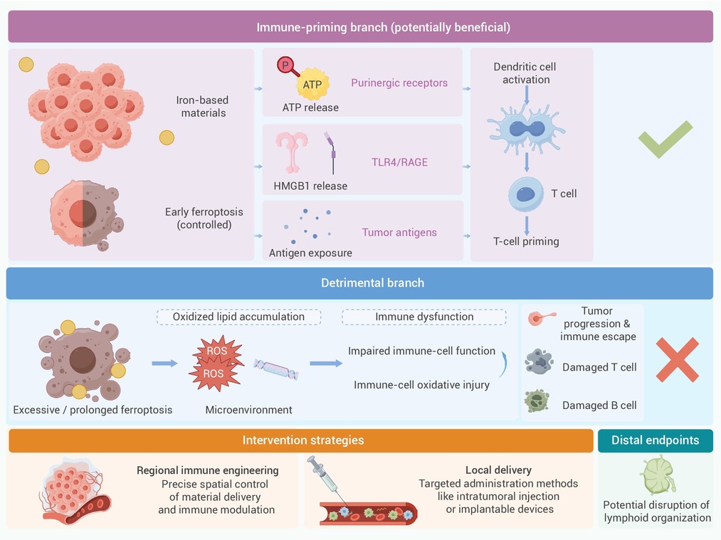
Figure 3.
Beneficial and detrimental outcomes of ferroptosis during SMR
