
Detecting shale oil hidden in nanopores using a novel electron microscopy method
-
GRAPHICAL ABSTRACT

 DownLoad:
Full size image
DownLoad:
Full size image
-
PUBLIC SUMMARY
-
Oil in shale matrix can cause local charging effect during high resolution electron microscopy imaging.
Shale oil reserved in nano pores with small amount of below pmol can be detected by controlled charging imaging.
High resolution large-area 2D controlled charging imaging provides a powerful tool for highly anisotropic samples.
Microscopic 3D imaging reveals the spatial distribution of shale oil and its relationship with pore system.
-
-
ABSTRACT
Shale oil has revolutionized the global energy landscape, mostly existing in nanopores. However, there is no method for directly characterizing such a small oil cluster. This paper proposes a novel method for the direct characterization of shale oil at the nanoscale via controllable local charging imaging using electron microscopy. With this method, the potentially movable hydrocarbons of below one pmol (10−12 mol) reserved in nanopores can be detected. The distribution of the shale oil clusters can be obtained through large-area two dimensional (2D) and three dimensional (3D) visualizations. Additionally, the amount of these resources can be calculated. Comparative analyses were conducted on reservoir samples from typical shale oil blocks in China and the US, and the results indicate the universality of the novel method, paving the way for accurate evaluation of shale oil resources and reasonable choice of exploitation technologies such as hydrofracturing and underground in-situ conversion processing. -

-
REFERENCES
[1] EIA. (2015). Energy Information Administration (EIA) Reports, U.S. [2] Jarvie, D.M. (2012). Shale Resource systems for oil and gas Part 2—Shale-oil resource systems. Breyer, J.A. ed., Shale Reservoirs—Giant resources for the 21st Century. DOI: 10.1306/13321447m973489. [3] Zou, C., Yang, Z., Tao, S., et al. (2012). Nano-hydrocarbon and the accumulation in coexisting source and reservoir. Petrol. Explor. Develop. 39: 15−32. DOI: 10.1016/s1876-3804(12)60011-1. [4] Jia, C., Zheng, M., and Zhang, Y. (2016). Some key issues on the unconventional petroleum systems. Petrol. Res. 1: 113−122. DOI: 10.1016/s2096-2495(17)30036-4. [5] Zhang, X. (2023). Uncover the black box of black shales. The Innovation Geoscience 1: 10050. DOI: 10.59717/j.xinn-geo.2023.100005. [6] Yang, H., Li, S., and Liu, X. (2016). Characteristics and resource prospects of tight oil in Ordos Basin, China. Petrol. Res. 1: 27−38. DOI: 10.1016/s2096-2495(17)30028-5. [7] Yang, H., Niu, X., Xu, L., et al. (2016). Exploration potential of shale oil in Chang7 Member, Upper Triassic Yanchang Formation, Ordos Basin, NW China. Petrol. Explor. Develop. 43: 560−569. DOI: 10.1016/s1876-3804(16)30066-0. [8] Zou, C., Yang, Z., Cui, J., et al. (2013). Formation mechanism, geological characteristics and development strategy of nonmarine shale oil in China. Petrol. Explor. Develop. 40: 15−27. DOI: 10.1016/s1876-3804(13)60002-6. [9] Liao, L., Wang, Y., and Lu, J. (2016). Experimental Study on Fractional Compositions of Residual Oil from Shale and Coal of China Using Grain-Based MSSV Pyrolysis. Energ. Fuel. 30: 256−263. DOI: 10.1021/acs.energyfuels.5b02486. [10] Li, M., Chen, Z., Cao, T., et al. (2018). Expelled oils and their impacts on Rock-Eval data interpretation, Eocene Qianjiang Formation in Jianghan Basin, China. Int. J. Coal. Geol. 191: 37−48. DOI: 10.1016/j.coal.2018.03.001. [11] Ma, Y., Cao, T., Snowdon, L., et al. (2017). Impact of different experimental heating rates on calculated hydrocarbon generation kinetics. Energ. Fuel. 31: 10378−10392. DOI: 10.1021/acs.energyfuels.7b01035. [12] Hu, Q., Zhang, Y., Meng, X., et al. (2017). Characterization of micro-nano pore networks in shale oil reservoirs of Paleogene Shahejie Formation in Dongying Sag of Bohai Bay Basin, East China. Petrol. Explor. Develop. 44: 720−730. DOI: 10.1016/s1876-3804(17)30083-6. [13] Jin, X., Wang, X., Yan, W., et al. (2019). Exploration and casting of large scale microscopic pathways for shale using electrodeposition. Appl. Energ. 247: 32−39. DOI: 10.1016/j.apenergy.2019.03.197. [14] Saif, T., Lin, Q., Butcher, A.R., et al. (2017). Multi-scale multi-dimensional microstructure imaging of oil shale pyrolysis using X-ray micro-tomography, automated ultra-high resolution SEM, MAPS Mineralogy and FIB-SEM. Appl. Energ. 202: 628−647. DOI: 10.1016/j.apenergy.2017.05.039. [15] Saif, T., Lin, Q., Gao, Y., et al. (2019). 4D in situ synchrotron X-ray tomographic microscopy and laser-based heating study of oil shale pyrolysis. Appl. Energ. 235: 1468−1475. DOI: 10.1016/j.apenergy.2018.11.044. [16] Loucks, R.G., Reed, R.M., Ruppel, S.C., et al. (2009). Morphology, genesis, and distribution of nanometer-scale pores in siliceous mudstones of the Mississippian Barnett Shale. J. Sediment. Res. 79: 848−861. DOI: 10.2110/jsr.2009.092. [17] Loucks, R.G., Reed, R.M., Ruppel, S.C., et al. (2012). Spectrum of pore types and networks in mudrocks and a descriptive classification for matrix-related mudrock pores. AAPG. Bull. 96: 1071−1098. DOI: 10.1306/08171111061. [18] Liu, K., Ostadhassan, M., Zhou, J., et al. (2017). Nanoscale pore structure characterization of the Bakken shale in the USA. Fuel 209: 567−578. DOI: 10.1016/j.fuel.2017.08.034. [19] Bera, B., Mitra, S.K., and Vick, D. (2011). Understanding the micro structure of Berea Sandstone by the simultaneous use of micro-computed tomography (micro-CT) and focused ion beam-scanning electron microscopy (FIB-SEM). Micron. 42: 412−418. DOI: 10.1016/j.micron.2010.12.002. [20] Bera, B., Gunda, N.S., Mitra, S.K., et al. (2012). Characterization of nanometer-scale porosity in reservoir carbonate rock by focused ion beam-scanning electron microscopy. Microsc. Microanal. 18: 171−178. DOI: 10.1017/S1431927611012505. [21] Zhu, J., Hu, Z., Guo, S., et al. (2023). Non-epitaxial growth of highly oriented transition metal dichalcogenides with density-controlled twin boundaries. The Innovation 4: 100502. DOI: 10.1016/j.xinn.2023.100502. [22] Chalmers, G.R., Bustin, R.M., and Power, I.M. (2012). Characterization of gas shale pore systems by porosimetry, pycnometry, surface area, and field emission scanning electron microscopy/transmission electron microscopy image analyses: Examples from the Barnett, Woodford, Haynesville, Marcellus, and Doig units. AAPG. Bull. 96: 1099−1119. DOI: 10.1306/10171111052. [23] Passey, Q.R., Bohacs, K.M., Esch, W.L., et al. (2010). From oil-prone source rock to gas-producing shale reservoir-geologic and petrophysical characterization of unconventional shale-gas reservoirs. SPE 131350. DOI: 10.2118/131350-MS. [24] Zhang, G., Long, H., and Yang, F. (2023). Understanding the formation time of black soils. The Innovation Geoscience 1: 100010. DOI: 10.59717/j.xinn-geo.2023.100010. [25] Yang, W., Wang, Y., Gao, L., et al. (2022). Sci-tech arts on Chang'e-5 lunar soil. The Innovation 3: 100300. DOI: 10.1016/j.xinn.2022.100300. [26] Wang, W., Tschauner, O., Huang, S., et al. (2021). Coupled deep-mantle carbon-water cycle: Evidence from lower-mantle diamonds. The Innovation 2: 100117. DOI: 10.1016/j.xinn.2021.100117. [27] Zamiri, M.S., Marica, F., Romero-Zerón, L., et al. (2022). Monitoring shale water uptake using 2D magnetic resonance relaxation correlation and SPRITE MRI. Chem. Eng. J. 428: 131042. DOI: 10.1016/j.cej.2021.131042. [28] Khatibi, S., Ostadhassan, M., Xie, Z.H., et al. (2019). NMR relaxometry a new approach to detect geochemical properties of organic matter in tight shales. Fuel 235: 167−177. DOI: 10.1016/j.fuel.2018.07.100. [29] Romero-Sarmiento, M.-F., Ramiro-Ramirez, S., Berthe, G., et al. (2017). Geochemical and petrophysical source rock characterization of the Vaca Muerta Formation, Argentina: Implications for unconventional petroleum resource estimations. Int. J. Coal. Geol. 184: 27−41. DOI: 10.1016/j.coal.2017.11.004. [30] Wang, X., Sun, L., Zhu, R., et al. (2015). Application of charging effects in evaluating storage space of tight reservoirs: A case study from Permian Lucaogou Formation in Jimusar sag, Junggar Basin, NW China. Petrol. Explor. Develop. 42: 516−524. DOI: 10.1016/s1876-3804(15)30044-6. [31] Pawley, J. (1984). Low voltage scanning electron microscopy. J. Microsc-Oxford. 136: 45−68. DOI: 10.1017/S1431927600029883. [32] Le Gressus, C., Valin, F., Gautier, M., et al. (1990). Charging phenomena on insulating materials: Mechanisms and applications. Scanning 12: 203−210. DOI: 10.1002/sca.4950120406. [33] Jbara, O., Fakhfakh, S., Belhaj, M., et al. (2008). Charging effects of PET under electron beam irradiation in a SEM. J. Phys. D. Appl. Phys. 41. DOI: 10.1088/0022-3727/41/24/245504. [34] Brunner, E. (1938). The Theory of the Continuous X-Ray Spectrum Near the Duane-Hunt Limit. Phys. Rev. 53: 457−459. DOI: 10.1103/PhysRev.53.457. [35] Wang, Y., Kong, M., Khan, M., et al. (2018). Determining E1 and E2 values for yttrium aluminum garnet ceramics using the Duane-Hunt limit. Microsc. Res. Tech. 81: 1203−1207. DOI: 10.1002/jemt.23118. [36] Brochu, M., Demers, H., Gauvin, R., et al. (2005). Determination of E2 for nitride ceramics using FE-SEM and the Duane-Hunt limit procedure. Microsc. Microanal. 11: 56−65. DOI: 10.1017/S143192760505004X. [37] Jbara, O., Fakhfakh, S., Belhaj, M., et al. (2004). Charge implantation measurement on electron-irradiated insulating materials by means of an SEM technique. Microsc. Microanal. 10: 697−710. DOI: 10.1017/s1431927604040644. [38] Liu, B., Wang, H., Fu, X., et al. (2019). Lithofacies and depositional setting of a highly prospective lacustrine shale oil succession from the Upper Cretaceous Qingshankou Formation in the Gulong sag, northern Songliao Basin, northeast China. AAPG. Bull. 103: 405−432. DOI: 10.1306/08031817416. [39] Tong, J., Jiang, X., Han, X., et al. (2016). Evaluation of the macromolecular structure of Huadian oil shale kerogen using molecular modeling. Fuel 181: 330−339. DOI: 10.1016/j.fuel.2016.04.139. [40] Li, Z., Zou, Y.-R., Xu, X.-Y., et al. (2016). Adsorption of mudstone source rock for shale oil – Experiments, model and a case study. Org. Geochem. 92: 55−62. DOI: 10.1016/j.orggeochem.2015.12.009. [41] Falk, K., Coasne, B., Pellenq, R., et al. (2015). Subcontinuum mass transport of condensed hydrocarbons in nanoporous media. Nat. Commun. 6: 6949. DOI: 10.1038/ncomms7949. [42] Lee, T., Bocquet, L., and Coasne, B. (2016). Activated desorption at heterogeneous interfaces and long-time kinetics of hydrocarbon recovery from nanoporous media. Nat. Commun. 7: 11890. DOI: 10.1038/ncomms11890. [43] Bousige, C., Ghimbeu, C.M., Vix-Guterl, C., et al. (2016). Realistic molecular model of kerogen's nanostructure. Nat. Mater. 15: 576−582. DOI: 10.1038/nmat4541. [44] Dijkmans, T., Djokic, M.R., Van Geem, K.M., et al. (2015). Comprehensive compositional analysis of sulfur and nitrogen containing compounds in shale oil using GC×GC – FID/SCD/NCD/TOF-MS. Fuel 140: 398−406. DOI: 10.1016/j.fuel.2014.09.055. [45] Gamadi, T.D., Sheng, J.J., Soliman, M.Y., et al. (2014). An experimental study of cyclic CO2 injection to improve shale oil recovery. SPE-169142-MS. DOI: 10.2118/169142-MS. [46] Sheng, J.J. (2015). Enhanced oil recovery in shale reservoirs by gas injection. J. Gas. Sci. Eng. 22: 252−259. DOI: 10.1016/j.jngse.2014.12.002. [47] Wang, Y.P., Wang, Y.W., Meng, X.L., et al. (2013). Enlightenment of American’s oil shale in-situ retorting technology. Oil Dril. Product. Tech. 35(6): 55-59. (in Chinese). DOI:10.13639/j.odpt.2013.06.032. [48] Zhao, W., Hu, S., and Hou, L. (2018). Connotation and strategic role of in-situ conversion processing of shale oil underground in the onshore China. Petrol. Explor. Develop. 45: 563−572. DOI: 10.1016/s1876-3804(18)30063-6. [49] Needham, R.B., Judzis, A., and Cornelius, A.J. (1976). Oil yield and quality from simulated in-situ retorting of green river oil shale. SPE Annual Fall Technical Conference and Exhibition, New Orleans, Louisiana, SPE 6069. DOI: 10.2118/6069-MS. -
ABOUT THIS ARTICLE
Cite this article:
Wang X., Meng S., Ostadhassan M., et al., (2024). Detecting shale oil hidden in nanopores using a novel electron microscopy method. The Innovation Energy 1(1): 100011. https://doi.org/10.59717/j.xinn-energy.2024.100011
Welcome!
To request copyright permission to republish or share portions of our works, please visit Copyright Clearance Center's (CCC) Marketplace website at marketplace.copyright.com.

Article Metrics
Relative Articles
Cited by
Export File
Citation
Wang X., Meng S., Ostadhassan M., et al., (2024). Detecting shale oil hidden in nanopores using a novel electron microscopy method. The Innovation Energy 1(1): 100011. https://doi.org/10.59717/j.xinn-energy.2024.100011

Format
Content
-
Figure 1.
Pathway of a movable oil cluster composed of hydraulic fractures and natural microcracks
-
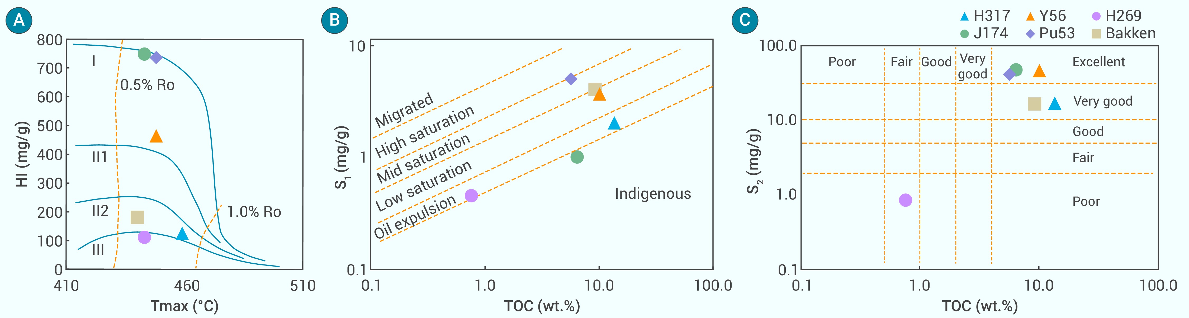
Figure 2.
Organic geochemical characteristics of samples
-
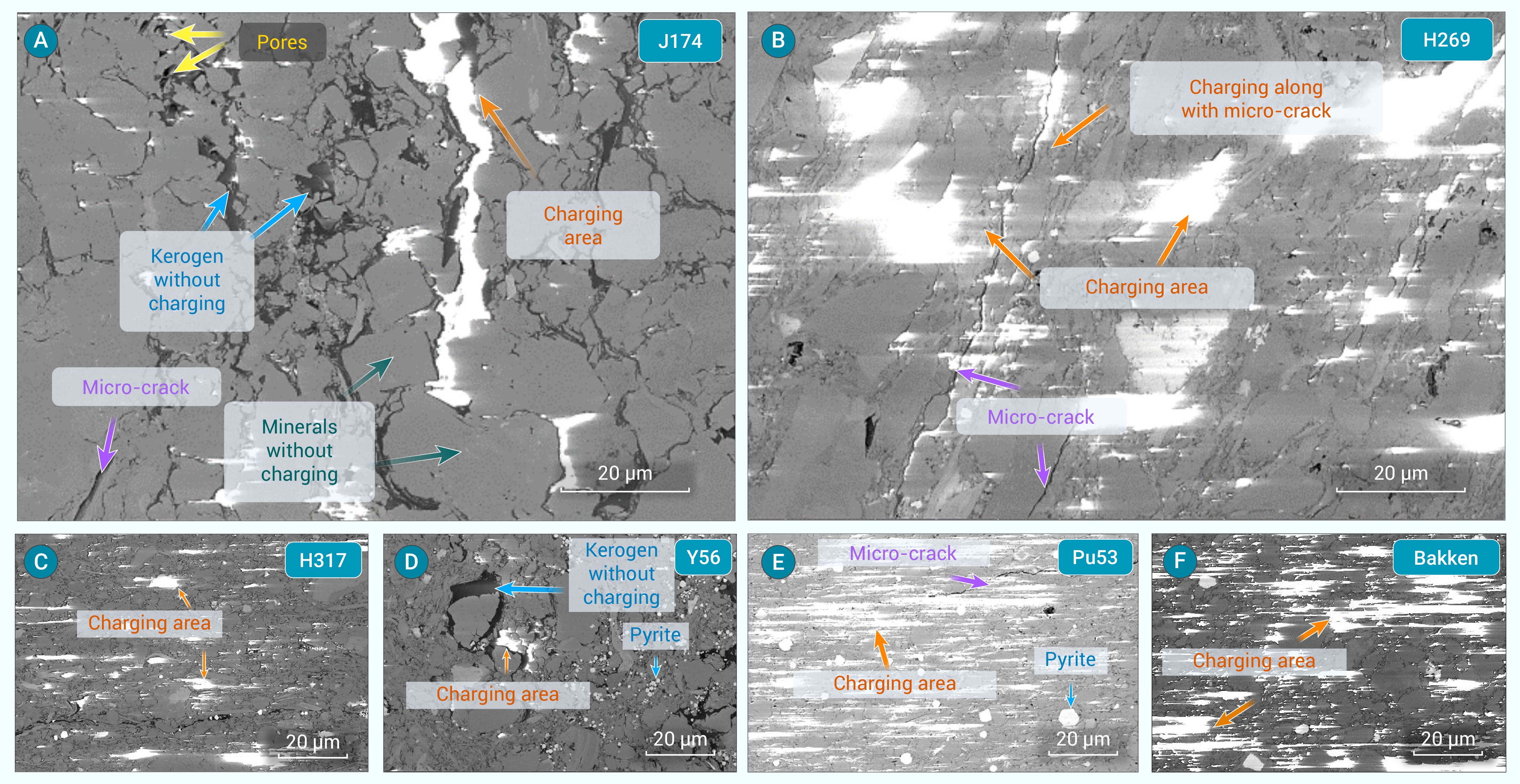
Figure 3.
Local charging effects observed in the samples during electron microscopy
-
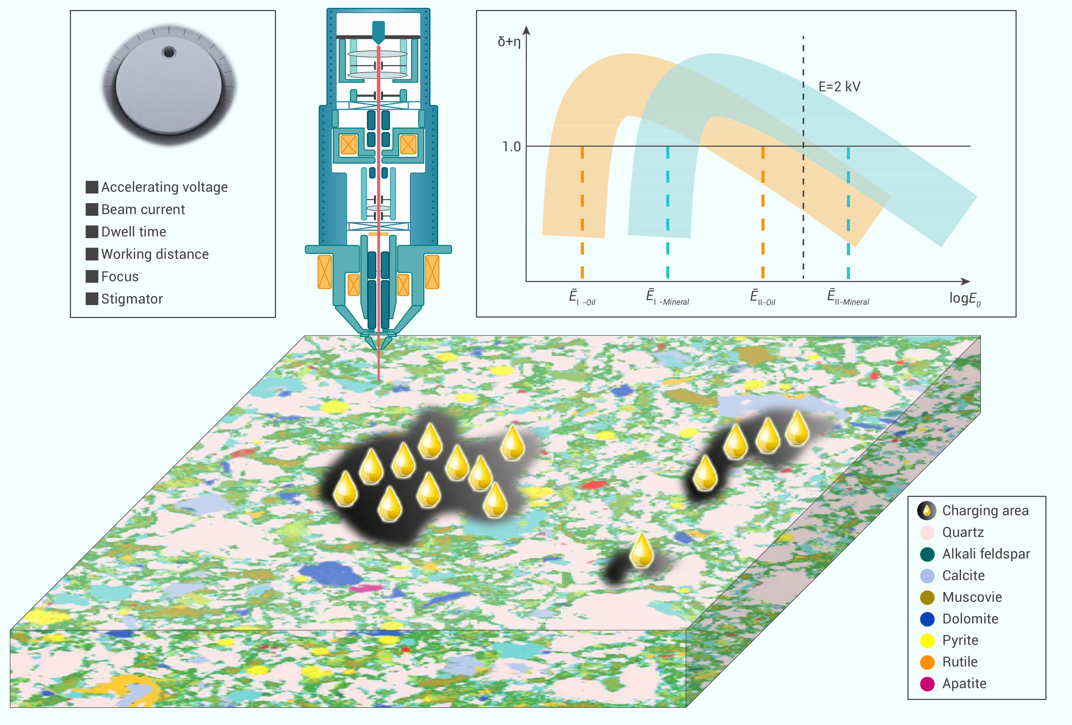
Figure 4.
Schematic of charging effect and key parameters that affect the degree of charging
-
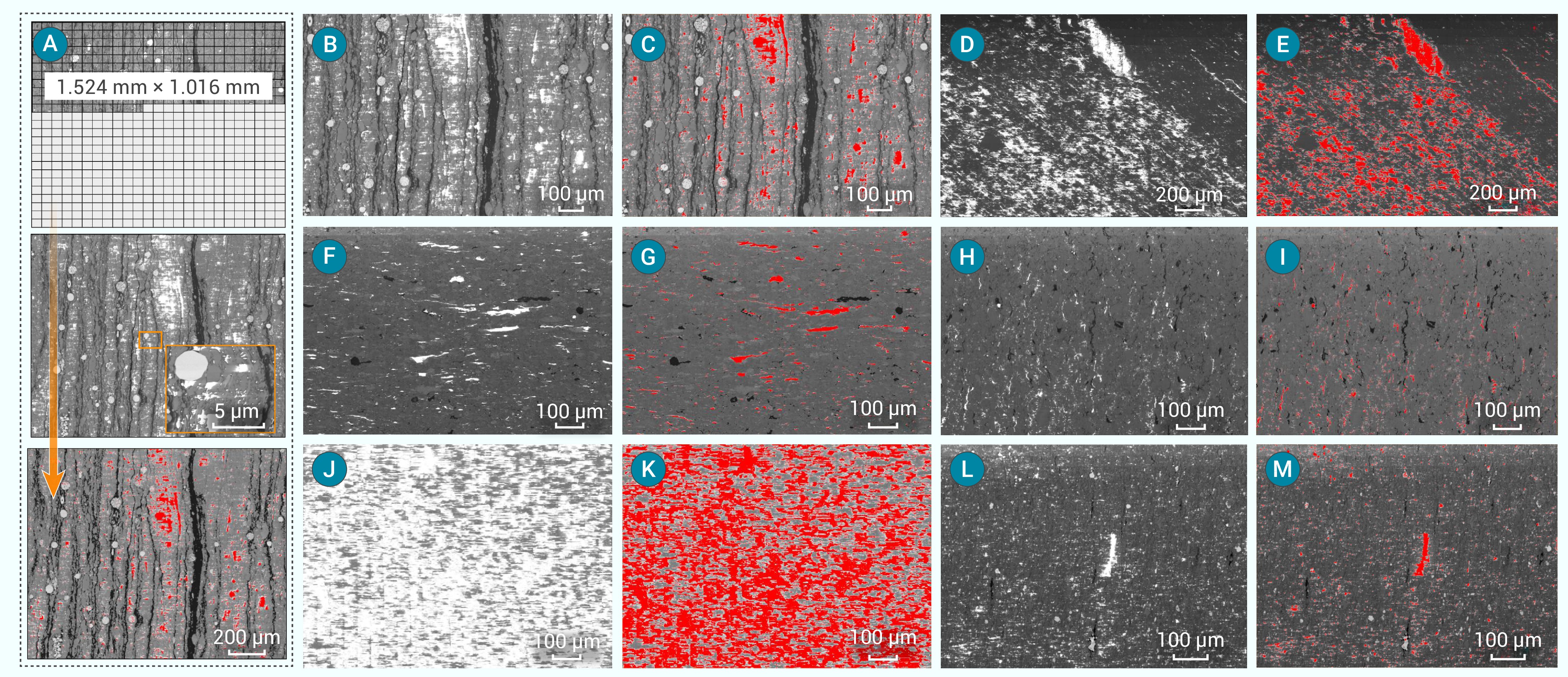
Figure 5.
Charging distribution of samples obtained from a large-area image mosaic at the millimeter scale
-
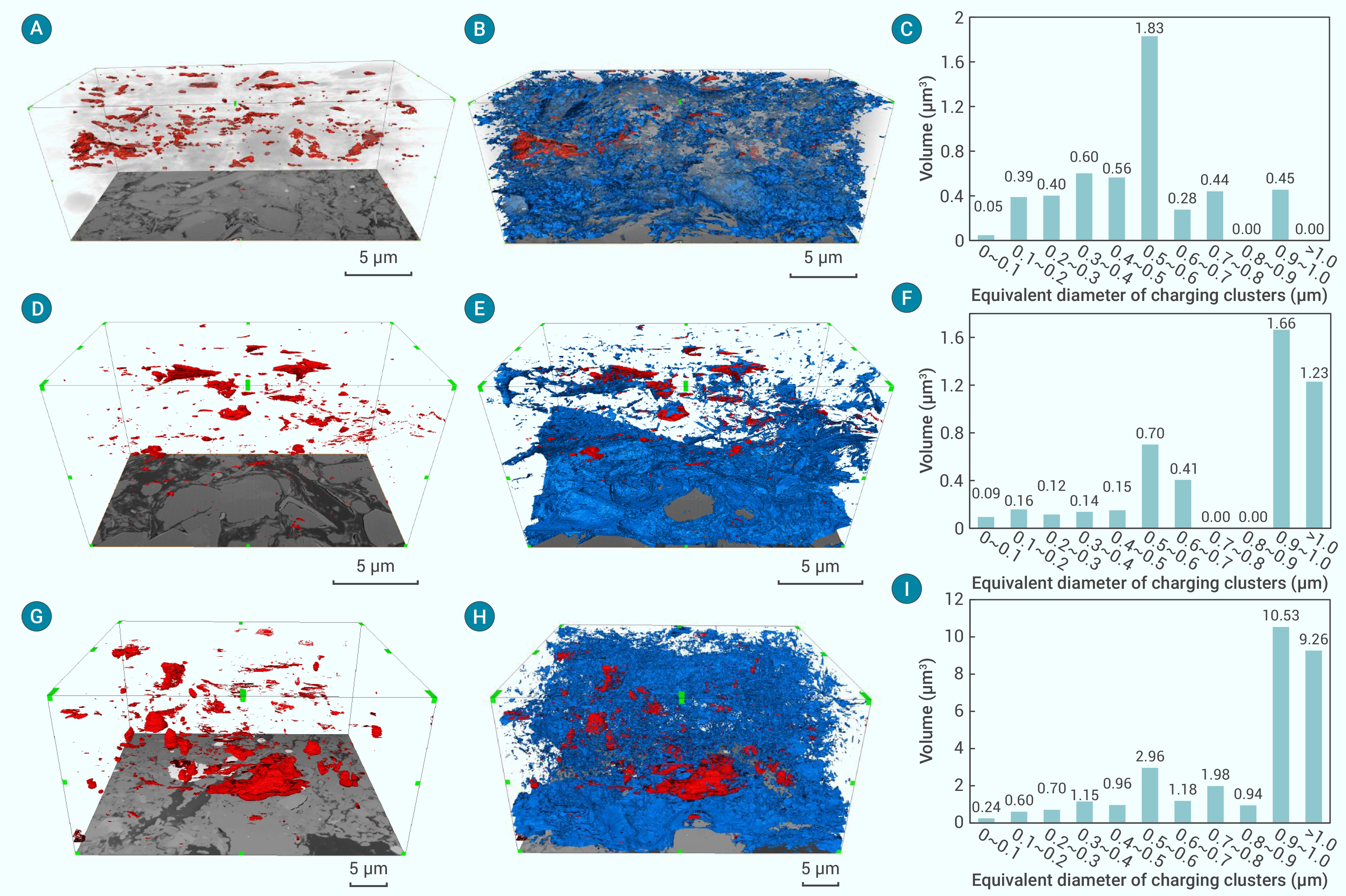
Figure 6.
3D charging distribution of residual oil in samples
-
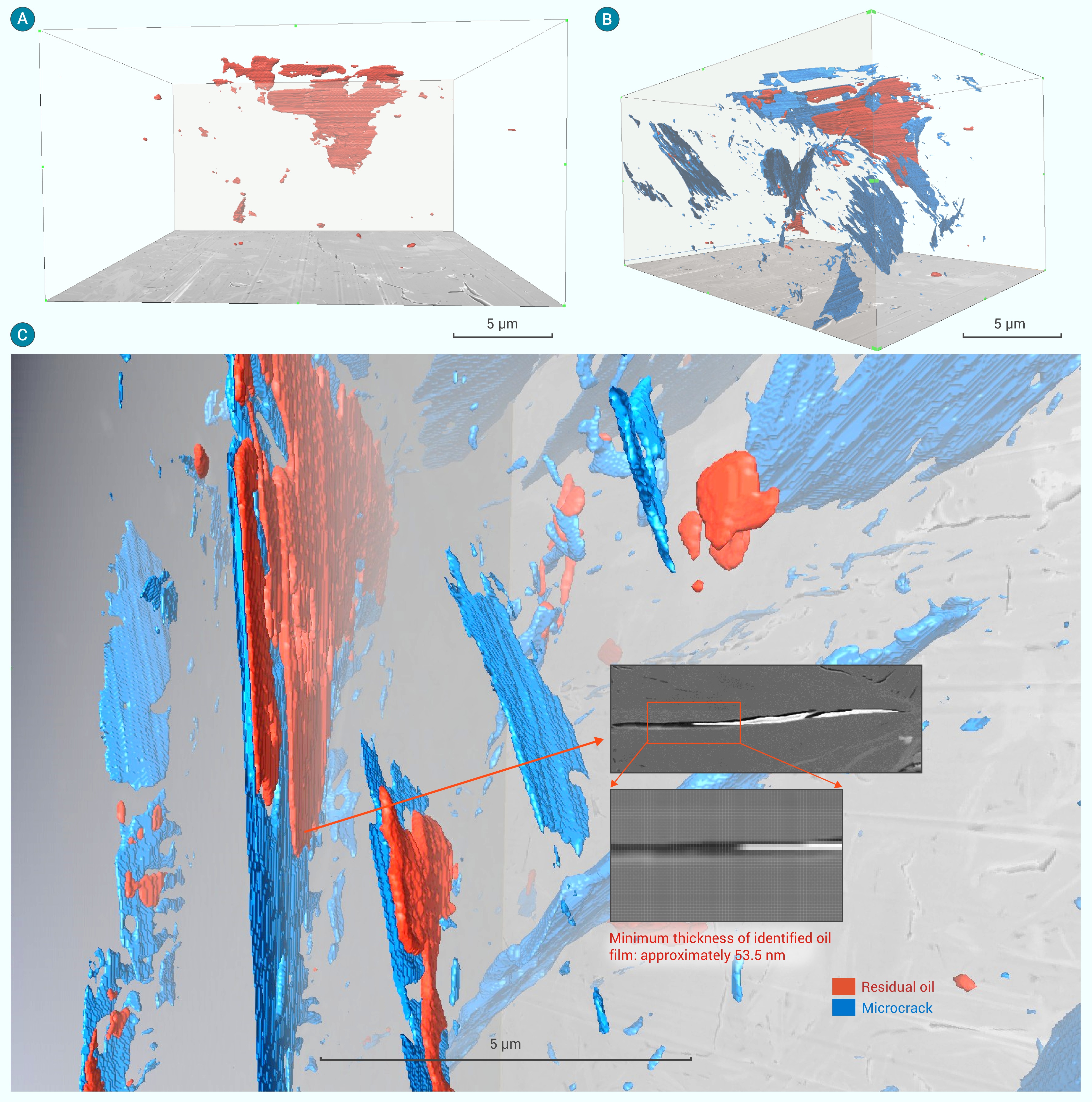
Figure 7.
3D charging distribution of residual oil in the H269 sample
