
3D structural analysis of bacteriocytes in a deep-sea mussel Gigantidas platifrons with methanotrophic symbionts
-
GRAPHICAL ABSTRACT
 DownLoad:
Full size image
DownLoad:
Full size image
-
PUBLIC SUMMARY
-
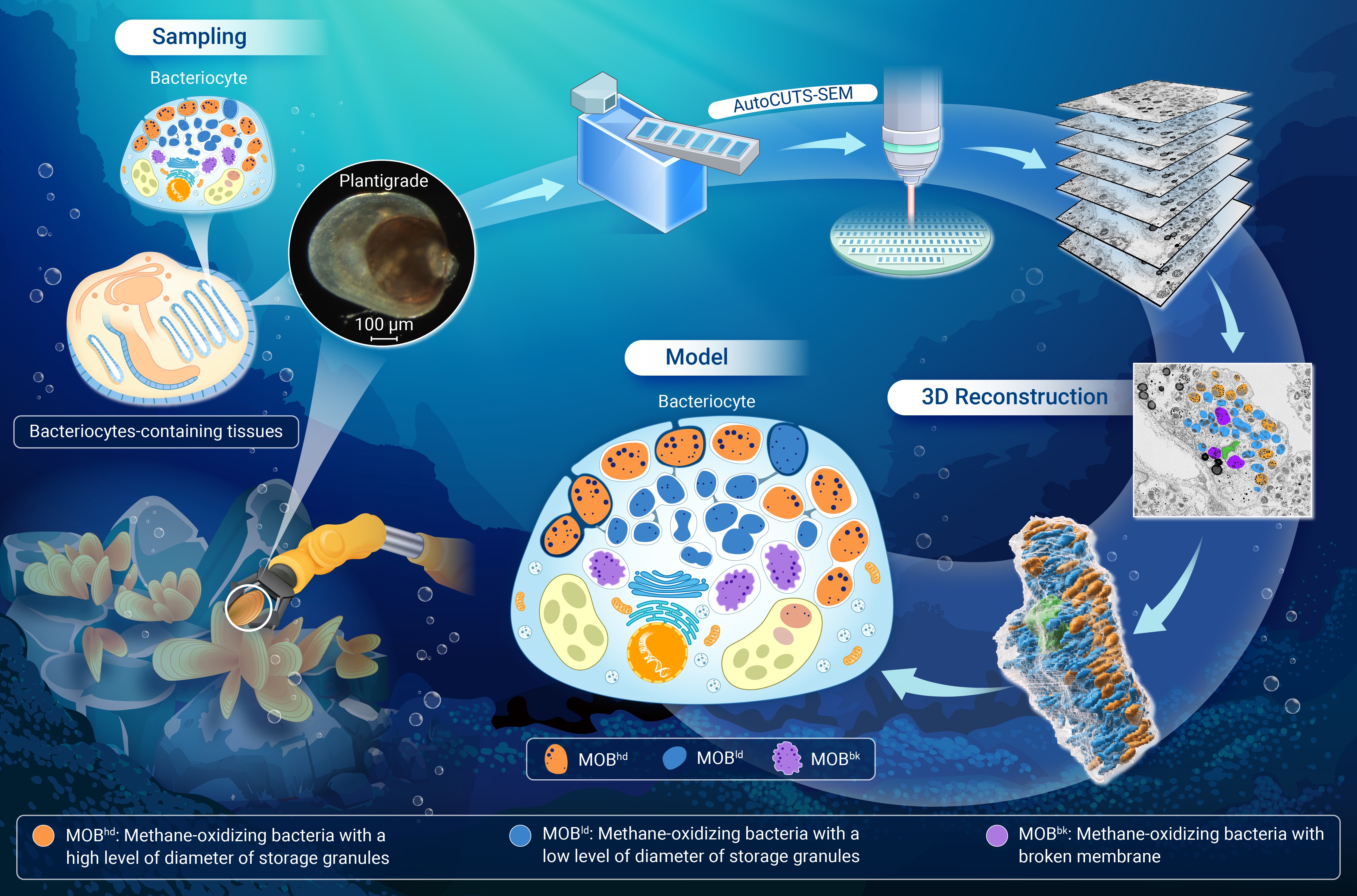
3D imaging shows bacteriocyte structure with methanotrophic bacteria (MOB) in Gigantidas platifrons.
High- and low-density granule content MOB (MOBhd) at apex and MOB (MOBld) in the interior were identified.
MOBhd lyses for nutrients, MOBld divides to maintain symbiont population, all MOB in a connected channel system.
Gill bacteriocytes exhibit more organized MOB distribution and controlled symbiont management strategies.
Unprecedented insights into the structural basis of stable symbiosis in deep-sea mussels.
-
-
ABSTRACT
Deep-sea mussels inhabit extreme environments through symbiosis with chemosynthetic bacteria, yet the three-dimensional (3D) ultrastructure of their bacteriocytes—the fundamental symbiotic units—remains elusive. This study employed advanced volume electron microscopy (vEM) to generate high-resolution, 3D models of bacteriocytes, revealing insights into the structural basis of stable symbiosis. We identified two distinct methanotrophic bacterial (MOB) morphotypes within the bacteriocytes: MOBhd (high-density granule content) and MOBld (low-density granule content). MOBhd, located in the apical region, undergoes lysis for nutrient release, while MOBld, situated in the interior, proliferates to maintain the symbiont population. Comparative analyses between gill and non-gill bacteriocytes revealed differential patterns of symbiont management, with gill bacteriocytes exhibiting more organized MOB distribution and orderly lysis. The 3D model unveiled a novel channel membrane system, potentially enhancing intracellular symbiont connectivity and facilitating distinct micro-niche occupation within the host cell. This organized structure maintains stable symbiosis, particularly in gill tissues. These findings advance our understanding of cellular mechanisms in symbiosis and adaptive strategies in extreme deep-sea environments. -

-
REFERENCES
[1] Fronk D.C. and Sachs J.L. (2022). Symbiotic organs: The nexus of host-microbe evolution. Trends Ecol. Evol. 37:599−610. DOI:10.1016/j.tree.2022.02.014 [2] Dubilier N., Bergin C. and Lott C. (2008). Symbiotic diversity in marine animals: The art of harnessing chemosynthesis. Nat. Rev. Microbiol. 6:725−740. DOI:10.1038/nrmicro1992 [3] McFall-Ngai, M., Hadfield, M.G., Bosch, T.C., et al. (2013). Animals in a bacterial world, a new imperative for the life sciences. Proc. Natl. Acad. Sci. USA 110:3229−3236. DOI:10.1073/pnas.1218525110 [4] Moya A., Peretó J., Gil R. and Latorre A. (2008). Learning how to live together: genomic insights into prokaryote–animal symbioses. Nat. Rev. Genet. 9:218−229. DOI:10.1038/nrg2319 [5] McFall-Ngai M. (2024). Symbiosis takes a front and center role in biology. PLoS Biol. 22:e3002571. DOI:10.1371/journal.pbio.3002571 [6] Sogin E.M., Kleiner M., Borowski C., et al. (2021). Life in the dark: Phylogenetic and physiological diversity of chemosynthetic symbioses. Annu. Rev. Microbiol. 75:695−718. DOI:10.1146/annurev-micro-051021-123130 [7] Sogin E.M., Leisch N. and Dubilier N. (2020). Chemosynthetic symbioses. Curr. Biol. 30:R1137−R1142. DOI:10.1016/j.cub.2020.07.050 [8] He X., Xu T., Chen C., et al. (2023). Same (sea) bed different dreams: Biological community structure of the Haima seep reveals distinct biogeographic affinities. The Innovation Geoscience 1:100019. DOI:10.59717/j.xinn-geo.2023.100019 [9] Cavanaugh C.M., Gardiner S.L., Jones M.L., et al. (1981). Prokaryotic cells in the hydrothermal vent tube worm riftia pachyptila Jones: Possible chemoautotrophic symbionts. Science 213:340−342. DOI:10.1126/science.213.4505.340 [10] Cavanaugh C.M., Levering P.R., Maki J.S., et al. (1987). Symbiosis of methylotrophic bacteria and deep-sea mussels. Nature 325:346−348. DOI:10.1038/325346a0 [11] Breusing C., Mitchell J., Delaney J., et al. (2020). Physiological dynamics of chemosynthetic symbionts in hydrothermal vent snails. ISME J. 14:2568−2579. DOI:10.1038/s41396-020-0707-2 [12] Roeselers G. and Newton I.L. (2012). On the evolutionary ecology of symbioses between chemosynthetic bacteria and bivalves. Appl. Microbiol. Biot. 94:1−10. DOI:10.1007/s00253-011-3819-9 [13] Zheng P., Wang M., Li C., et al. (2017). Insights into deep-sea adaptations and host-symbiont interactions: A comparative transcriptome study on Bathymodiolus mussels and their coastal relatives. Mol. Ecol. 26:5133−5148. DOI:10.1111/mec.14160 [14] Sun Y., Wang M., Zhong Z., et al. (2022). Adaption to hydrogen sulfide-rich environments: Strategies for active detoxification in deep-sea symbiotic mussels, Gigantidas platifrons. Sci. Total Environ. 804:150054. DOI:10.1016/j.scitotenv.2021.150054 [15] Gibson C.M. and Hunter M.S. (2010). Extraordinarily widespread and fantastically complex: Comparative biology of endosymbiotic bacterial and fungal mutualists of insects. Ecol. Letters 13:223−234. DOI:10.1111/j.1461-0248.2009.01416.x [16] Braendle C., Miura T., Bickel R., et al. (2003). Developmental origin and evolution of bacteriocytes in the aphid–buchnera symbiosis. PLOS Biol. 1:e21. DOI:10.1371/journal.pbio.0000021 [17] Luan J.-B. (2024). Insect bacteriocytes: Adaptation, development, and evolution. Annu. Rev. Entomol. 69:81−98. DOI:10.1146/annurev-ento-010323-124159 [18] Douglas A.E. (2020). Housing microbial symbionts: Evolutionary origins and diversification of symbiotic organs in animals. Philosophical Transactions of the Royal Society B 375:20190603. DOI:10.1098/rstb.2019.0603 [19] Simonet P., Gaget K., Balmand S., et al. (2018). Bacteriocyte cell death in the pea aphid/Buchnera symbiotic system. Proc. Natl. Acad. Sci. USA 115:E1819−E1828. DOI:10.1073/pnas.1720237115 [20] Stoll S., Feldhaar H., Fraunholz M.J., et al. (2010). Bacteriocyte dynamics during development of a holometabolous insect, the carpenter ant Camponotus floridanus. BMC Microbiol. 10:308. DOI:10.1186/1471-2180-10-308 [21] Maire J., Parisot N., Galvao Ferrarini M., et al. (2020). Spatial and morphological reorganization of endosymbiosis during metamorphosis accommodates adult metabolic requirements in a weevil. Proc. Natl. Acad. Sci. USA DOI:10.1073/pnas.2007151117 [22] Nussbaumer A.D., Fisher C.R. and Bright M. (2006). Horizontal endosymbiont transmission in hydrothermal vent tubeworms. Nature 441:345−348. DOI:10.1038/nature04793 [23] Wentrup C., Wendeberg A., Huang J.Y., et al. (2013). Shift from widespread symbiont infection of host tissues to specific colonization of gills in juvenile deep-sea mussels. ISME J. 7:1244−1247. DOI:10.1038/ismej.2013.5 [24] Laming S.R., Duperron S., Cunha M.R., et al. (2014). Settled, symbiotic, then sexually mature: adaptive developmental anatomy in the deep-sea, chemosymbiotic mussel Idas modiolaeformis. Marine Biology 161:1319−1333. DOI:10.1007/s00227-014-2421-y [25] Ikuta T., Takaki Y., Nagai Y., et al. (2016). Heterogeneous composition of key metabolic gene clusters in a vent mussel symbiont population. ISME J. 10:990−1001. DOI:10.1038/ismej.2015.176 [26] Duperron S., Lorion J., Samadi S., et al. (2009). Symbioses between deep-sea mussels (Mytilidae: Bathymodiolinae) and chemosynthetic bacteria: Diversity, function and evolution. CR. Biol. 332:298−310. DOI:10.1016/j.crvi.2008.08.003 [27] Kádár E. and Bettencourt R. (2008). Ultrastructural and molecular evidence for potentially symbiotic bacteria within the byssal plaques of the deep-sea hydrothermal vent mussel Bathymodiolus azoricus. BioMetals 21:395−404. DOI:10.1007/s10534-007-9128-1 [28] Franke M., Geier B., Hammel J.U., et al. (2021). Coming together-symbiont acquisition and early development in deep-sea bathymodioline mussels. P. Roy. Soc. B-Biol. Sci. 288:20211044. DOI:10.1098/rspb.2021.1044 [29] Halary S., Riou V., Gaill F., et al. (2008). 3D FISH for the quantification of methane- and sulphur-oxidizing endosymbionts in bacteriocytes of the hydrothermal vent mussel Bathymodiolus azoricus. ISME J. 2:284−292. DOI:10.1038/ismej.2008.3 [30] Li X., Ji G., Chen X., et al. (2017). Large scale three-dimensional reconstruction of an entire Caenorhabditis elegans larva using AutoCUTS-SEM. J. Struct. Biol. 200:87−96. DOI:10.1016/j.jsb.2017.09.010 [31] Xu T., Sun J., Watanabe H.K., et al. (2018). Population genetic structure of the deep-sea mussel Bathymodiolus platifrons (Bivalvia: Mytilidae) in the Northwest Pacific. Evol. Appl. 11:1915−1930. DOI:10.1111/eva.12696 [32] Barry, J.P., Buck, K.R., Kochevar, R.K., et al. (2002). Methane‐based symbiosis in a mussel, Bathymodiolus platifrons, from cold seeps in Sagami Bay, Japan. Invertebr. Biol. 121:47−54. DOI:10.1111/j.1744-7410.2002.tb00128.x [33] Wang H., Zhang H., Wang M., et al. (2019). Comparative transcriptomic analysis illuminates the host-symbiont interactions in the deep-sea mussel Bathymodiolus platifrons. Deep Sea Research Part I: Oceanographic Research Papers 151:103082. DOI:10.1016/j.dsr.2019.103082 [34] Sun Y., Wang M., Cao L., et al. (2023). Mosaic environment-driven evolution of the deep-sea mussel Gigantidas platifrons bacterial endosymbiont. Microbiome 11 :253. DOI:10.1186/s40168-023-01695-8 [35] Gottlieb Y., Ghanim M., Gueguen G., et al. (2008). Inherited intracellular ecosystem: Symbiotic bacteria share bacteriocytes in whiteflies. FASEB J. 22:2591−2599. DOI:10.1096/fj.07-101162 [36] Duperron S., Nadalig T., Caprais J.C., et al. (2005). Dual symbiosis in a Bathymodiolus sp. mussel from a methane seep on the Gabon continental margin (Southeast Atlantic): 16S rRNA phylogeny and distribution of the symbionts in gills. Appl. Environ. Microb. 71 :1694-1700. DOI:10.1128/AEM.71.4.1694-1700.2005 [37] Wang H., He K., Zhang H., et al. (2024). Deciphering deep-sea chemosynthetic symbiosis by single-nucleus RNA-sequencing. eLife 12:RP88294. DOI:10.7554/eLife.88294 [38] Titze B. and Genoud C. (2016). Volume scanning electron microscopy for imaging biological ultrastructure. Biol. Cell 108:307−323. DOI:10.1111/boc.201600024 [39] Preibisch S., Saalfeld S. and Tomancak P. (2009). Globally optimal stitching of tiled 3D microscopic image acquisitions. Bioinformatics 25:1463−1465. DOI:10.1093/bioinformatics/btp184 [40] Saalfeld S., Cardona A., Hartenstein V., et al. (2010). As-rigid-as-possible mosaicking and serial section registration of large ssTEM datasets. Bioinformatics 26:i57−i63. DOI:10.1093/bioinformatics/btq219 [41] Belevich I., Joensuu M., Kumar D., et al. (2016). Microscopy image browser: a platform for segmentation and analysis of multidimensional datasets. PLoS biology 14:e1002340. DOI:10.1371/journal.pbio.1002340 [42] Chen S., Zhou Y., Chen Y., et al. (2018). fastp: An ultra-fast all-in-one FASTQ preprocessor. 34 :i884-i890. DOI:10.1093/bioinformatics/bty560 [43] Nurk S., Meleshko D., Korobeynikov A., et al. (2017). metaSPAdes: A new versatile metagenomic assembler. Genome Res. 27:824−834. DOI:10.1101/gr.213959.116 [44] Uritskiy G.V., DiRuggiero J. and Taylor J. (2018). MetaWRAP—a flexible pipeline for genome-resolved metagenomic data analysis. Microbiome 6:1−13. DOI:doi.org/10.1186/s40168-018-0541-1 [45] Parks D.H., Imelfort M., Skennerton C.T., et al. (2015). CheckM: assessing the quality of microbial genomes recovered from isolates single cells and metagenomes. Genome Res. 25:1043−1055. DOI:10.1101/gr.186072.114 [46] Fu L., Niu B., Zhu Z., et al. (2012). CD-HIT: Accelerated for clustering the next-generation sequencing data. Bioinformatics 28:3150−3152. DOI:10.1093/bioinformatics/bts565 [47] Li H. (2018). Minimap2: Pairwise alignment for nucleotide sequences. Bioinformatics 34:3094−3100. DOI:10.1093/bioinformatics/bty191 [48] McKenna A., Hanna M., Banks E., et al. (2010). The genome analysis toolkit: A MapReduce framework for analyzing next-generation DNA sequencing data. Genome Res. 20:1297−1303. DOI:10.1101/gr.107524.110 [49] Quince C., Delmont T.O., Raguideau S., et al. (2017). DESMAN: A new tool for de novo extraction of strains from metagenomes. Genome Biol. 18:181. DOI:10.1186/s13059-017-1309-9 [50] Kolde R. and Kolde M.R. (2015). Package ‘pheatmap’. R. package 1:790. [51] Vincent A.E., White K., Davey T., et al. (2019). Quantitative 3D mapping of the human skeletal muscle mitochondrial network. Cell reports 26 :996-1009. e1004. DOI:10.1016/j.celrep.2019.01.010 [52] Tavormina P.L., Kellermann M.Y., Antony C.P., et al. (2017). Starvation and recovery in the deep-sea methanotroph Methyloprofundus sedimenti. Mol. Microbiol. 103:242−252. DOI:10.1111/mmi.13553 [53] Tavormina P.L., Hatzenpichler R., McGlynn S., et al. (2015). Methyloprofundus sedimenti gen. nov., sp. nov., an obligate methanotroph from ocean sediment belonging to the ‘deep sea-1’ clade of marine methanotrophs. Int. J. Syst. Evol. Micr. 65 :251-259. DOI:10.1099/ijs.0.062927-0 [54] Martin W., Baross J., Kelley D., et al. (2008). Hydrothermal vents and the origin of life. Nat. Rev. Microbiol. 6:805−814. DOI:10.1038/nrmicro1991 [55] Van Dover C.L., German C.R., Speer K.G., et al. (2002). Evolution and biogeography of deep-sea vent and seep invertebrates. Science 295:1253−1257. DOI:10.1126/science.1067361 [56] Osman E.O. and Weinnig A.M. (2022). Microbiomes and obligate symbiosis of deep-sea animals. Annu. Rev. Anim. Biosci. 10 :151-176. DOI:10.1146/annurev-animal-081621-112021 [57] Carrier T.J. and Reitzel A.M. (2017). The hologenome across environments and the implications of a host-associated microbial repertoire. F. Microbiol. 8:802. DOI:10.3389/fmicb.2017.00802 [58] Ansorge R., Romano S., Sayavedra L., et al. (2019). Functional diversity enables multiple symbiont strains to coexist in deep-sea mussels. Nat. Microbiol. 4 :2487-2497. DOI:10.1038/s41564-019-0572-9 [59] Uzum Z., Ershov D., Pavia M.J., et al. (2023). Three-dimensional images reveal the impact of the endosymbiont Midichloria mitochondrii on the host mitochondria. Nat. Commun. 14:4133. DOI:10.1038/s41467-023-39758-x [60] Hirayama H., Takaki Y., Abe M., et al. (2022). Multispecies populations of methanotrophic Methyloprofundus and cultivation of a likely dominant species from the Iheya North deep-sea hydrothermal field. Appl. Environ. Microbiol. 88:e00758−00721. DOI:10.1128/AEM.00758-21 [61] Liberton M., Austin J.R., Berg R.H., et al. (2011). Unique thylakoid membrane architecture of a unicellular N2-fixing cyanobacterium revealed by electron tomography. Plant Physiol. 155:1656−1666. DOI:10.1104/pp.110.165332 [62] Young K.D. (2006). The selective value of bacterial shape. Microbiol. Mol. Biol. R. 70:660−703. DOI:10.1128/mmbr.00001-06 [63] Hinzke T., Kleiner M., Meister M., et al. (2021). Bacterial symbiont subpopulations have different roles in a deep-sea symbiosis. elife 10: e58371. DOI:10.7554/eLife.58371 [64] Wang H., Xiao H., Feng B., et al. (2024). Single-cell RNA-seq reveals distinct metabolic “microniches” and close host-symbiont interactions in deep-sea chemosynthetic tubeworm. Sci. Adv. 10:eadn3053. DOI:10.1126/sciadv.adn3053 [65] Wang H., Zhang H., Zhong Z., et al. (2021). Molecular analyses of the gill symbiosis of the bathymodiolin mussel Gigantidas platifrons. iScience 24:101894. DOI:10.1016/j.isci.2020.101894 [66] Sun Y., Wang M., Chen H., et al. (2022). Insights into symbiotic interactions from metatranscriptome analysis of deep-sea mussel Gigantidas platifrons under long-term laboratory maintenance. Mol. Ecol. 32 :444-459 DOI:10.1111/mec.16765 [67] Matthews J.L., Crowder C.M., Oakley C.A., et al. (2017). Optimal nutrient exchange and immune responses operate in partner specificity in the cnidarian-dinoflagellate symbiosis. Proc. Natl. Acad. Sci. USA 114:13194−13199. DOI:10.1073/pnas.1710733114 [68] Wein, T., Picazo, D.R., Blow, F., et al. (2019). Currency, exchange, and inheritance in the evolution of symbiosis. Trends Microbiol. 27:836−849. DOI:10.1016/j.tim.2019.05.010 [69] Ponnudurai R., Kleiner M., Sayavedra L., et al. (2017). Metabolic and physiological interdependencies in the Bathymodiolus azoricus symbiosis. ISME J. 11:463−477. DOI:10.1038/ismej.2016.124 [70] Takishita K., Takaki Y., Chikaraishi Y., et al. (2017). Genomic evidence that methanotrophic endosymbionts likely provide deep-sea bathymodiolus mussels with a sterol intermediate in cholesterol biosynthesis. Genome Biol. Evol. 9:1148−1160. DOI:10.1093/gbe/evx082 [71] Streams M.E., Fisher C.R. and Fiala-Médioni A. (1997). Methanotrophic symbiont location and fate of carbon incorporated from methane in a hydrocarbon seep mussel. Marine Biology 129:465−476. DOI:10.1007/s002270050187 [72] Tame A., Maruyama T., Ikuta T., et al. (2023). mTORC1 regulates phagosome digestion of symbiotic bacteria for intracellular nutritional symbiosis in a deep-sea mussel. Sci. Adv. 9:eadg8364. DOI:10.1126/sciadv.adg8364 [73] Li M., Chen H., Wang M., et al. (2022). Robust phenotypic plasticity of symbiotic organs facilitates the adaptation and evolution of deep-sea mussels. bioRxiv. DOI:10.1101/2022.08.11.503589 [74] Wang D., Griffitts J., Starker C., et al. (2010). A nodule-specific protein secretory pathway required for nitrogen-fixing symbiosis. Science 327:1126−1129. DOI:10.1126/science.1184096 [75] Kádár E., Davis S.A. and Lobo-da-Cunha A. (2008). Cytoenzymatic investigation of intracellular digestion in the symbiont-bearing hydrothermal bivalve Bathymodiolus azoricus. Marine biology 153:995−1004. DOI:10.1007/s00227-007-0872-0 [76] Tame A., Maruyama T., and Yoshida T. (2022). Phagocytosis of exogenous bacteria by gill epithelial cells in the deep-sea symbiotic mussel Bathymodiolus japonicus. Roy. Soc. Open Sci. 9:211384. DOI:10.1098/rsos.211384 [77] Wentrup C., Wendeberg A., Schimak M.P., et al. (2014). Forever competent: Deep-sea bivalves are colonized by their chemosynthetic symbionts throughout their lifetime. Environ. Microbiol. 16:3699−3713. DOI:10.1111/1462-2920.12597 [78] Piquet B., Le Panse S., Lallier F.H., et al. (2022). “There and back again” - Ultrastructural changes in the gills of Bathymodiolus vent-mussels during symbiont loss: Back to a regular filter-feeding epidermis. Front. Mar. Sci. 9 :968331. DOI:10.3389/fmars.2022.968331 [79] Fujiwara A., Meng X.-Y., Kamagata Y., et al. (2022). Subcellular niche segregation of co-obligate symbionts in whiteflies. Microbiol. Spectr. 11:e04684−04622. DOI:10.1128/spectrum.04684-22 [80] Strunov A., Schmidt K., Kapun M., et al. (2022). Restriction of Wolbachia bacteria in early embryogenesis of neotropical Drosophila species via endoplasmic reticulum-mediated autophagy. MBio 13:e03863−03821. DOI:10.1128/mbio.03863-21 [81] Ikuta T., Amari Y., Tame A., et al. (2021). Inside or out? Clonal thiotrophic symbiont populations occupy deep-sea mussel bacteriocytes with pathways connecting to the external environment. ISME Commun. 1:38. DOI:10.1038/s43705-021-00043-x -
ABOUT THIS ARTICLE
Cite this article:
Zhong Z., Sun W., Zhang Y., et al. (2025). 3D structural analysis of bacteriocytes in a deep-sea mussel Gigantidas platifrons with methanotrophic symbionts. The Innovation Geoscience 3:100110. https://doi.org/10.59717/j.xinn-geo.2024.100110
Welcome!
To request copyright permission to republish or share portions of our works, please visit Copyright Clearance Center's (CCC) Marketplace website at marketplace.copyright.com.
Article Metrics
Relative Articles
Cited by
Export File
Citation
Zhong Z., Sun W., Zhang Y., et al. (2025). 3D structural analysis of bacteriocytes in a deep-sea mussel Gigantidas platifrons with methanotrophic symbionts. The Innovation Geoscience 3:100110. https://doi.org/10.59717/j.xinn-geo.2024.100110

Format
Content

-
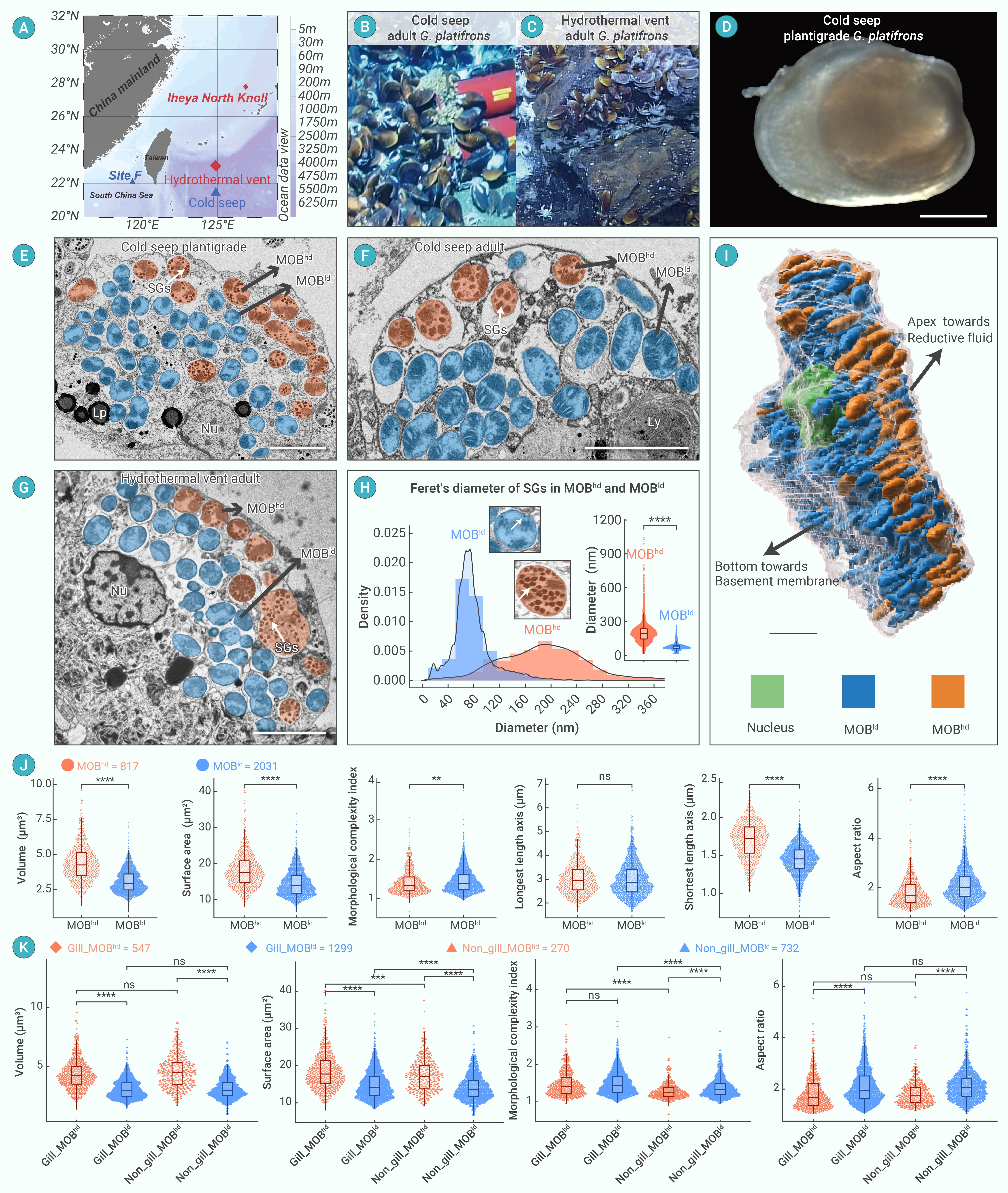
Figure 1.
Two MOB morphotypes identified in deep-sea mussel G. platifrons bacteriocytes
-
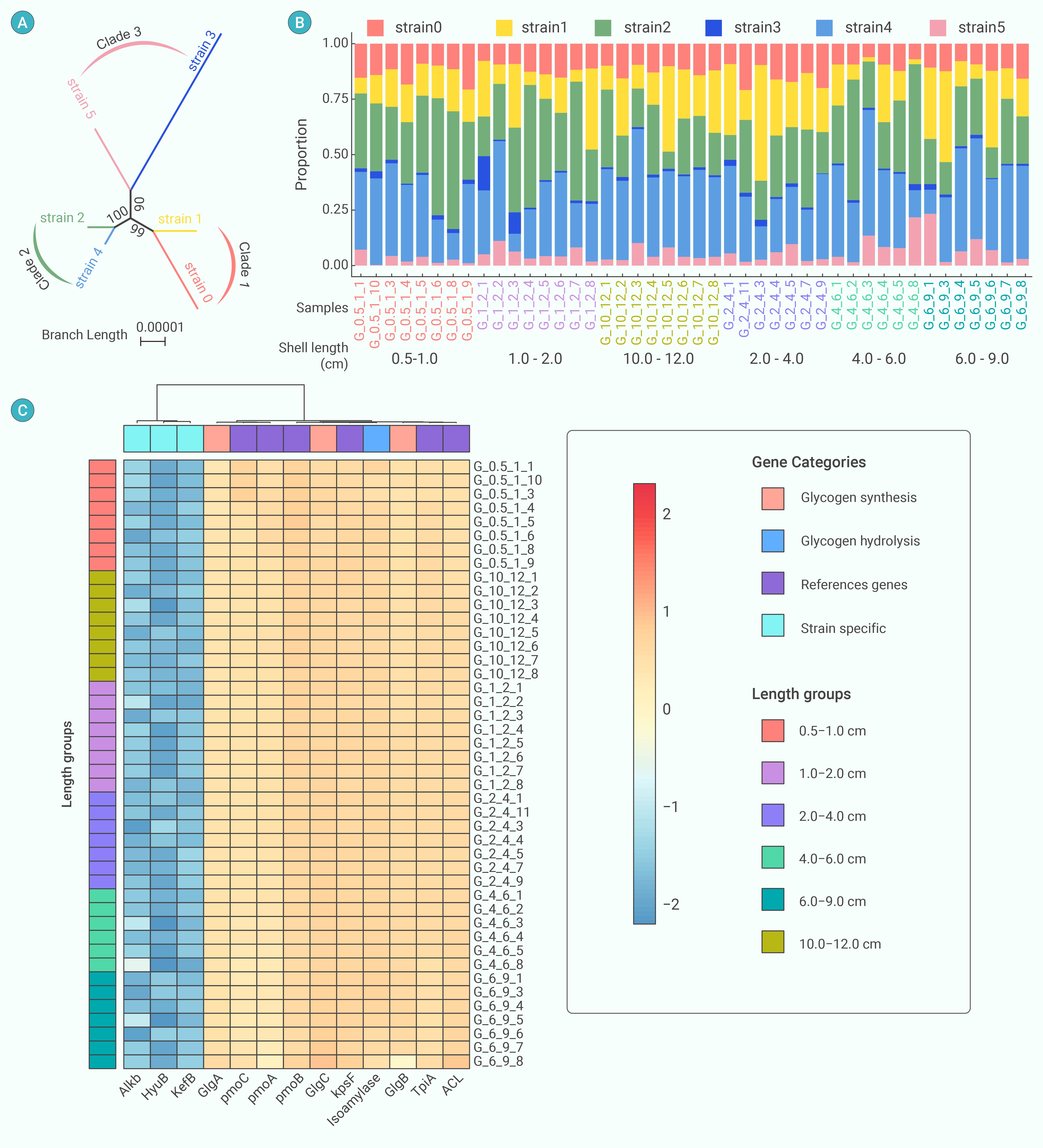
Figure 2.
Deconvolution of G. platifrons individuals of various lengths
-
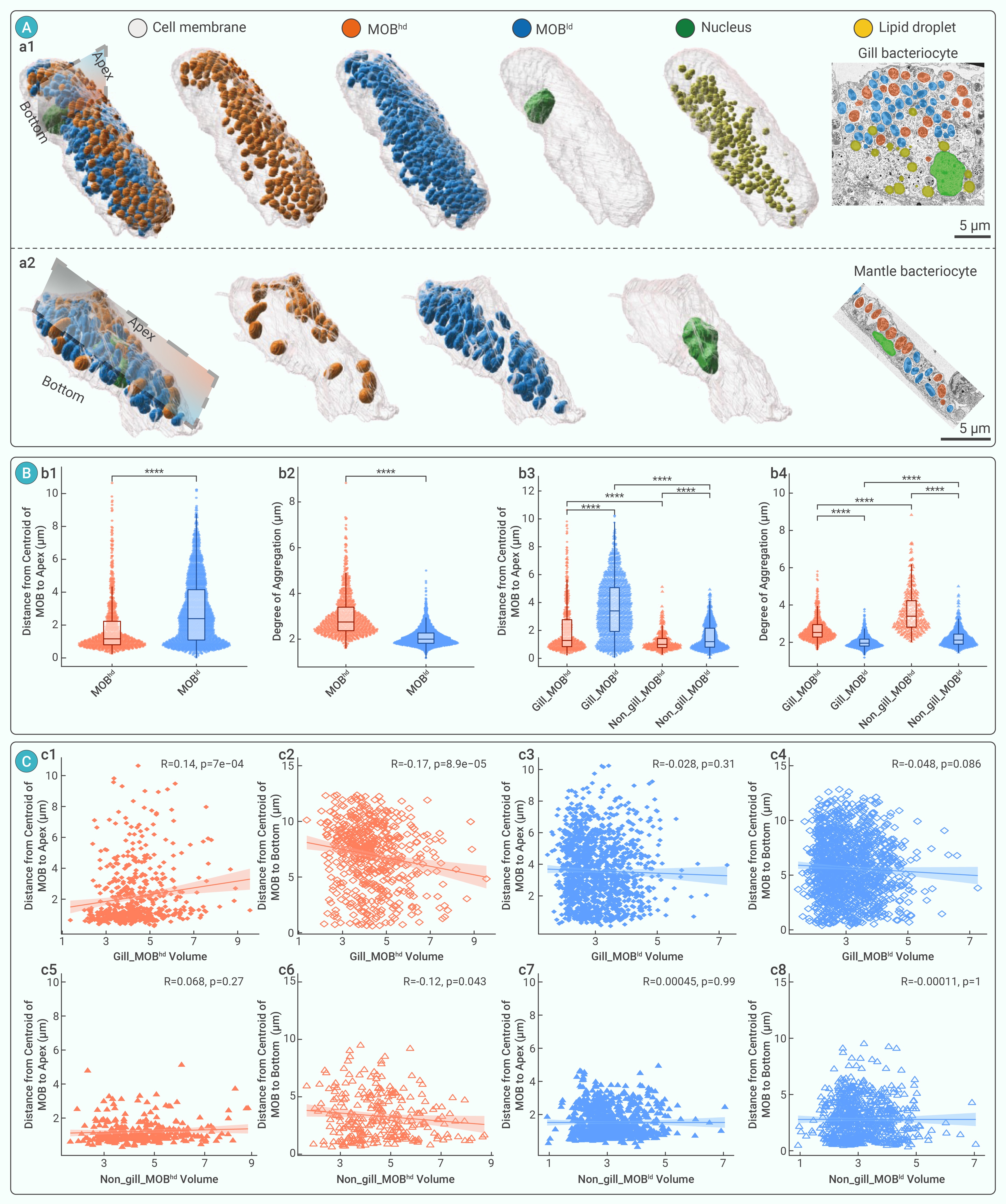
Figure 3.
Spatial distribution of two MOB morphotypes within bacteriocyte
-
Figure 4.
MOB lysis without surrounding lysosomes
-
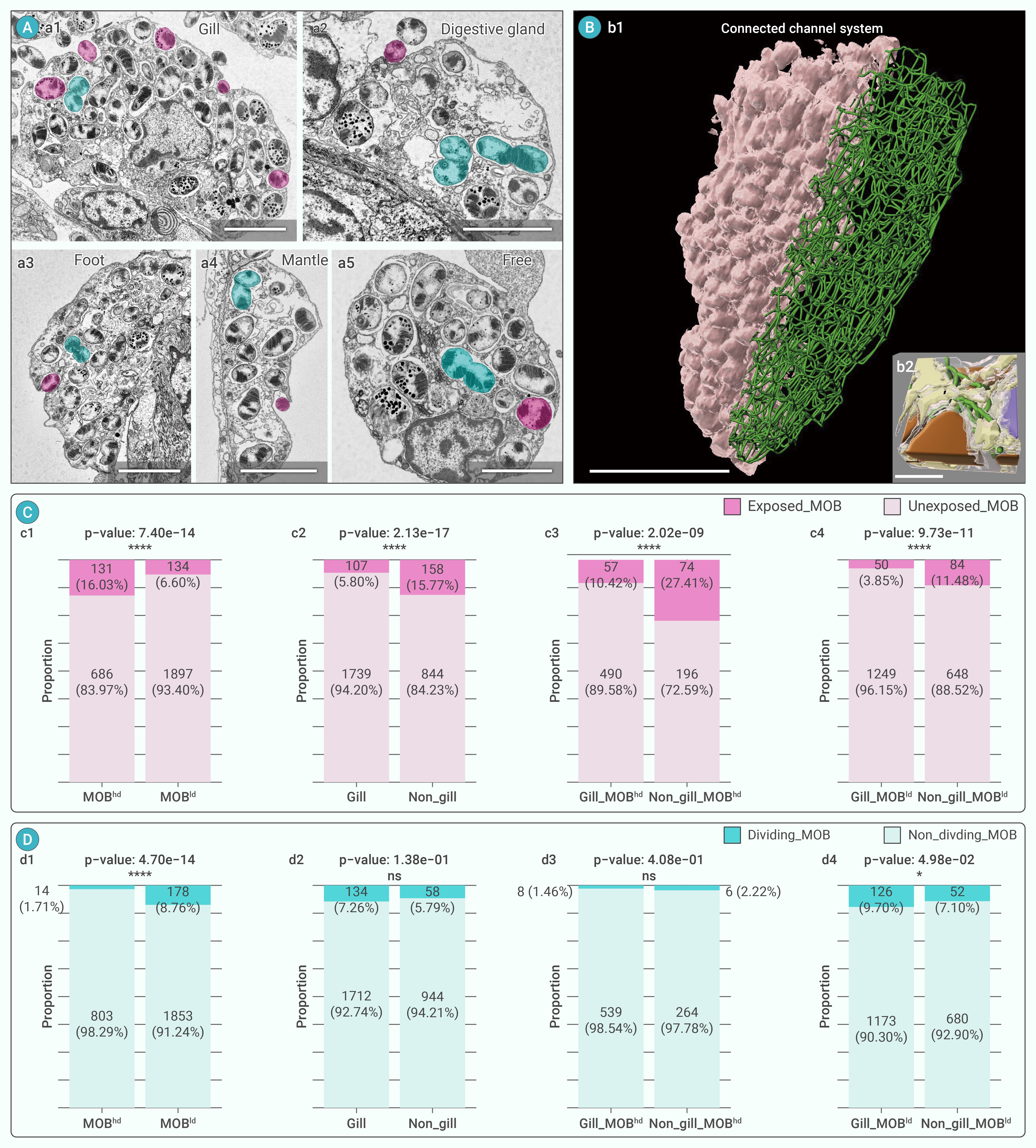
Figure 5.
MOB exposed to external environment and MOB division within bacteriocytes
-
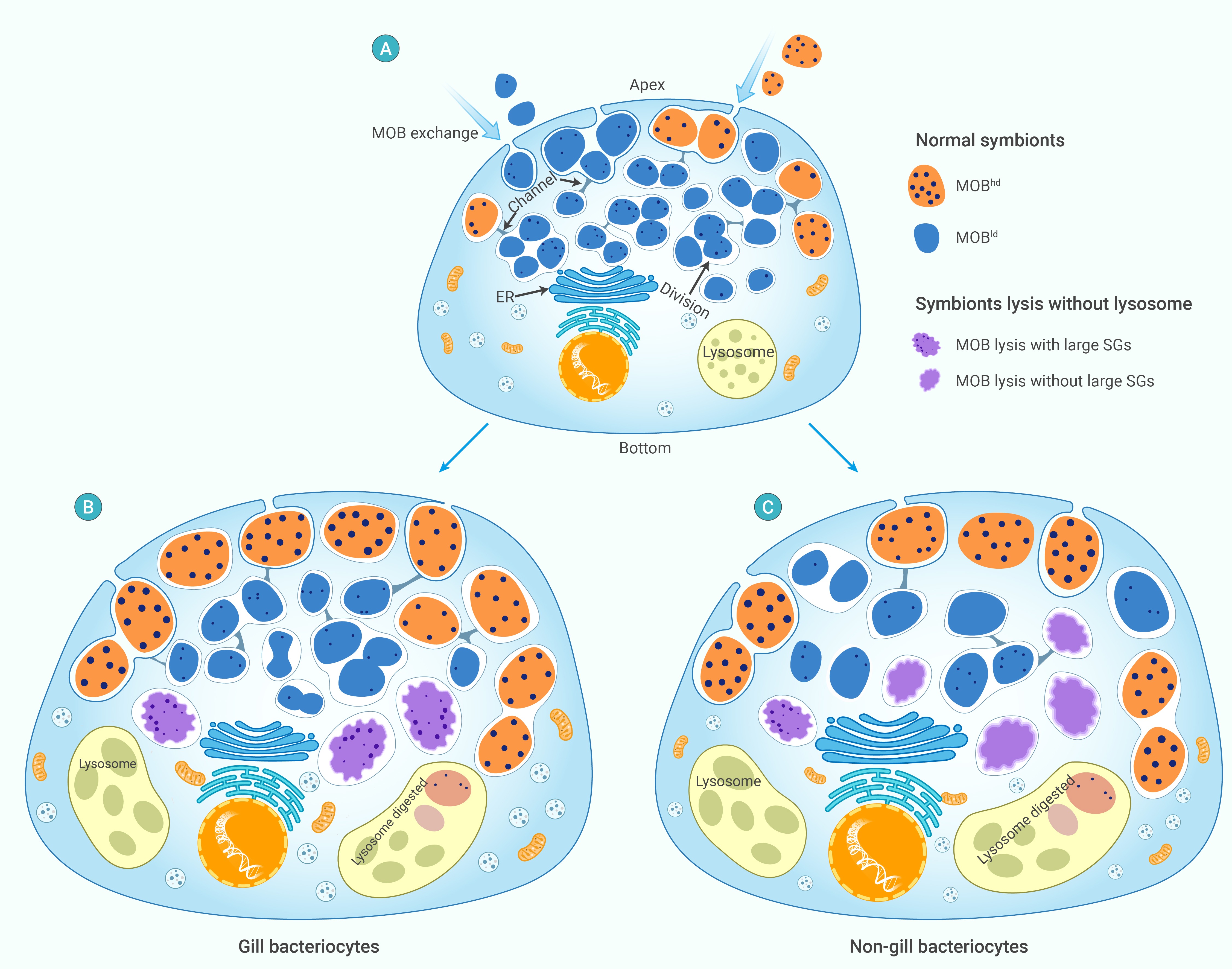
Figure 6.
Theoretical model of MOB population dynamics and long-term gill bacteriocyte maintenance
