
Mechanisms utilized by Methanobacterium sp. YSL for growth on zero-valent iron
-
GRAPHICAL ABSTRACT
 DownLoad:
Full size image
DownLoad:
Full size image
-
PUBLIC SUMMARY
-
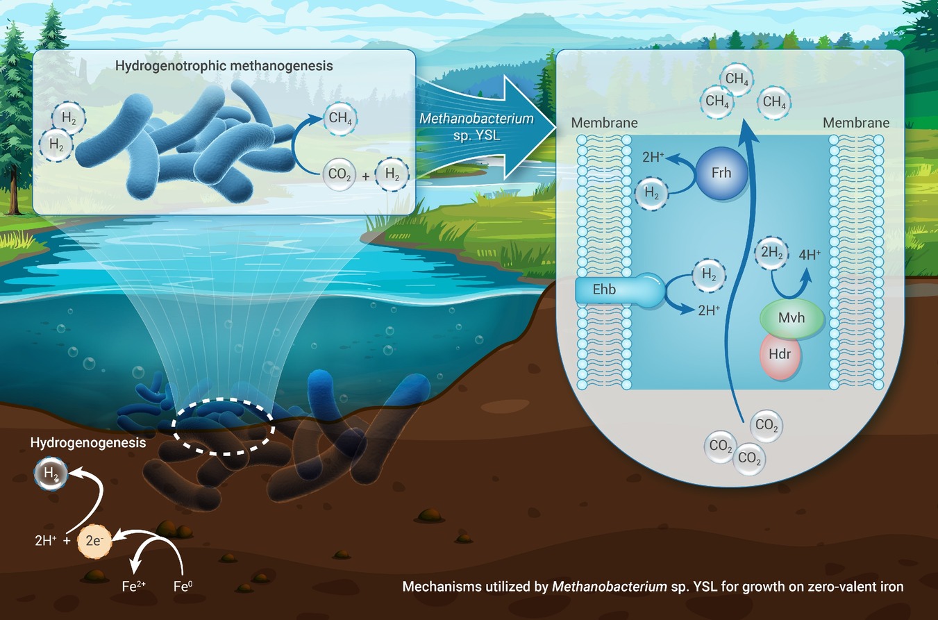
Strain YSL employs H2 as an intermediary electron carrier to extract electrons from zero-valent iron.
Mutations in key subunits of the Eha complex likely render it nonfunctional in strain YSL.
Cytoplasmic hydrogenases compensate for Eha’s inactivity and enable slow H2-dependent growth of strain YSL.
The results emphasize the environmental importance of microbial reactions that seem slow in lab culture.
-
-
ABSTRACT
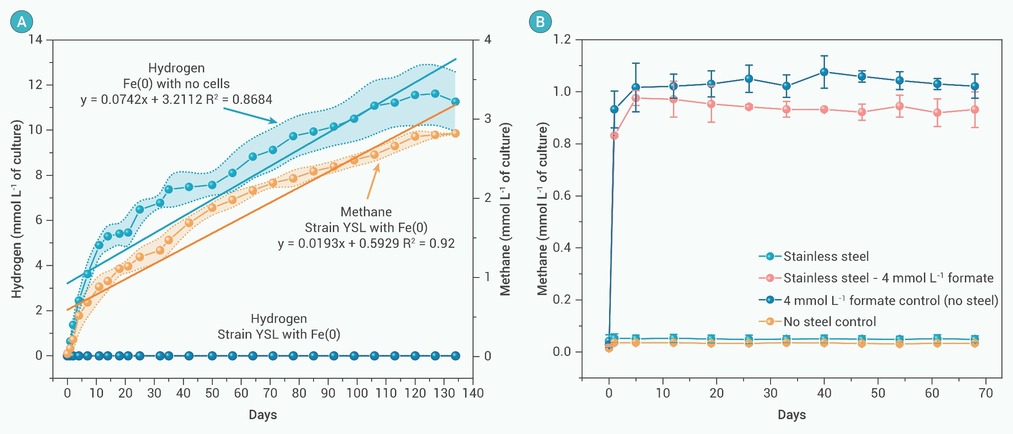
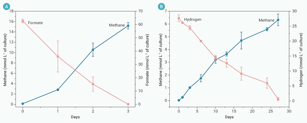
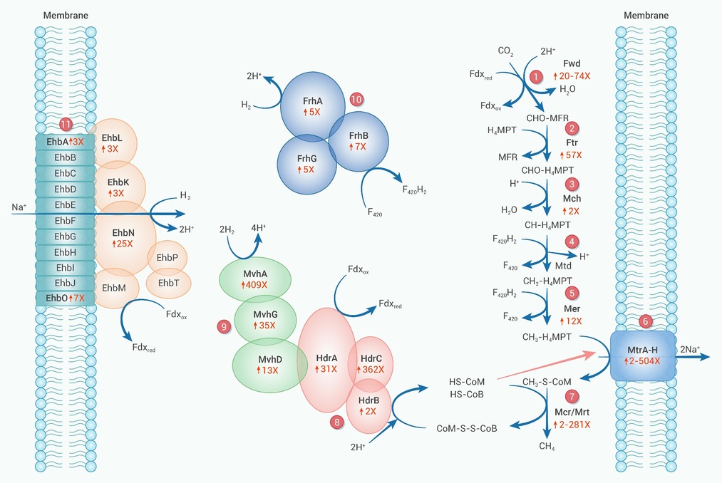
Electrobiocorrosion (i.e. direct metal-to-microbe electron transfer) is a recently recognized, highly aggressive form of microbial metal corrosion. Methanobacterium subterraneum strain YSL grows as an electron accepting partner in direct interspecies electron transfer, suggesting that it might have the capacity for electrobiocorrosion. To evaluate this possibility, strain YSL was grown with either pure Fe0, which abiotically generates H2, or 316L stainless steel, which does not generate H2, as the sole potential electron donor. There was a steady accumulation of H2 in uninoculated controls with Fe0. No H2 was detected in strain YSL cultures with Fe0. Rather, strain YSL produced methane at a rate consistent with conversion of the H2 abiotically generated from Fe0 to methane. Strain YSL did not produce methane in incubations with stainless steel. These results indicated that strain YSL relies on H2 as an intermediary electron carrier between Fe0 and cells, a result in conflict with the previous report that strain YSL was incapable of H2 utilization. Further investigation revealed that strain YSL can grow on H2, but more than 50-fold slower than is typical for other Methanobacterium strains. Genomic analysis revealed that mutations in genes for seven subunits of Eha, the primary energy-converting hydrogenase of Methanobacterium species, likely disabled its function in strain YSL. Proteomic analysis suggested that a compensatory high expression of cytoplasmic hydrogenases enabled slow growth on H2. These studies highlight the importance of analyzing slow metabolic capabilities, which are difficult to detect with standard methods yet can have significant environmental consequences. -

-
REFERENCES
[1] Knisz J., Eckert R., Gieg L.M., et al. (2023). Microbiologically influenced corrosion-more than just microorganisms. FEMS Microbiol. Rev. 47:fuad041. DOI:10.1093/femsre/fuad041 [2] Little B.J., Hinks J. and Blackwood D.J. (2020). Microbially influenced corrosion: Towards an interdisciplinary perspective on mechanisms. Int. Biodeterior. Biodegr. 154:105062. DOI:10.1016/j.ibiod.2020.105062 [3] Xu D.K., Gu T.Y. and Lovley D.R. (2023). Microbially mediated metal corrosion. Nat. Rev. Microbiol. 21:705−718. DOI:10.1038/s41579-023-00920-3 [4] Daniels L., Belay N., Rajagopal B.S., et al. (1987). Bacterial methanogenesis and growth from CO2 with elemental iron as the sole source of electrons. Science 237:509−511. DOI:10.1126/science.237.4814.509 [5] Lahme S., Mand J., Longwell J., et al. (2021). Severe corrosion of carbon steel in oil field produced water can be linked to methanogenic archaea containing a special type of [NiFe] hydrogenase. Appl. Environ. Microbiol. 87:e01819−01820. DOI:10.1128/aem.01819-20 [6] Tamisier M., Schmidt M., Vogt C., et al. (2022). Iron corrosion by methanogenic archaea characterized by stable isotope effects and crust mineralogy. Environ. Microbiol. 24:583−595. DOI:10.1111/1462-2920.15658 [7] Usher K.M., Kaksonen A.H. and MacLeod I.D. (2014). Marine rust tubercles harbour iron corroding archaea and sulphate reducing bacteria. Corros. Sci. 83:189−197. DOI:10.1016/j.corsci.2014.02.014 [8] Deutzmann J.S., Sahin M. and Spormann A.M. (2015). Extracellular enzymes facilitate electron uptake in biocorrosion and bioelectrosynthesis. mBio 6:e00496−00415. DOI:10.1128/mBio.00496-15 [9] Holmes D.E., Woodard T.L., Smith J.A., et al. (2024). Electrobiocorrosion by microbes without outer-surface cytochromes. mLife 3:110−118. DOI:10.1002/mlf2.12111 [10] Kawaichi S., Kotoky R., Fiutowski J., et al. (2024). Adaptation of a methanogen to Fe0 corrosion via direct contact. Npj Biofilms and Microbi. 10:100. DOI:10.1038/s41522-024-00574-w [11] Tsurumaru H., Ito N., Mori K., et al. (2018). An extracellular [NiFe] hydrogenase mediating iron corrosion is encoded in a genetically unstable genomic island in Methanococcus maripaludis. Sci. Rep. 8:15149. DOI:10.1038/s41598-018-33541-5 [12] Lienemann M., Deutzmann J.S., Milton R.D., et al. (2018). Mediator-free enzymatic electrosynthesis of formate by the Methanococcus maripaludis heterodisulfide reductase supercomplex. Bioresource Technol. 254:278−283. DOI:10.1016/j.biortech.2018.01.036 [13] Holmes D.E., Tang H., Woodard T.L., et al. (2022). Cytochrome‐mediated direct electron uptake from metallic iron by Methanosarcina acetivorans. mLife 1:443−447. DOI:10.1002/mlf2.12044 [14] Liang D.D., Liu X.Y., Woodard T.L., et al. (2021). Extracellular electron exchange capabilities of Desulfovibrio ferrophilus and Desulfopila corrodens. Environ. Sci. Technol. 55:16195−16203. DOI:10.1021/acs.est.1c04071 [15] Tang H.-Y., Yang C., Ueki T., et al. (2021). Stainless steel corrosion via direct iron-to-microbe electron transfer by Geobacter species. The ISME J. 15:3084−3093. DOI:10.1038/s41396-021-00990-2 [16] Tang H.-Y., Holmes D.E., Ueki T., et al. (2019). Iron corrosion via direct metal-microbe electron transfer. mBio 10:e00303−00319. DOI:10.1128/mBio.00303-19 [17] Hernandez-Santana A., Suflita J.M. and Nanny M.A. (2022). Shewanella oneidensis MR-1 accelerates the corrosion of carbon steel using multiple electron transfer mechanisms. Int. Biodeterior. Biodegr. 173:105439. DOI:10.1016/j.ibiod.2022.105439 [18] Zhou E.Z., Li F., Zhang D.W., et al. (2022). Direct microbial electron uptake as a mechanism for stainless steel corrosion in aerobic environments. Water Res. 219:118553. DOI:10.1016/j.watres.2022.118553 [19] Holmes D.E., Zhou J.J., Ueki T., et al. (2021). Mechanisms for electron uptake by Methanosarcina acetivorans during direct interspecies electron transfer. mBio 12:e0234421. DOI:10.1128/mBio.02344-21 [20] Yee M.O. and Rotaru A.-E. (2020). Extracellular electron uptake in Methanosarcinales is independent of multiheme c-type cytochromes. Sci. Rep. 10:747485. DOI:10.1101/747485 [21] Zhou J.J., Holmes D.E., Tang, H.-Y., et al. (2021). Correlation of key physiological properties of Methanosarcina isolates with environment of origin. Appl. Environ. Microbiol. 87:e00731−00721. DOI:10.1128/aem.00731-21 [22] Zheng S.L., Liu F.H., Wang B.C., et al. (2020). Methanobacterium capable of direct interspecies electron transfer. Environ. Sci. Technol. 54:15347−15354. DOI:10.1021/acs.est.0c05525 [23] Zheng S.L. and Liu F.H. (2021). Complete genome sequence of Methanobacterium electrotrophus strain YSL, isolated from coastal riverine sediments. Microbiol. Resour. Ann 10:e0075221. DOI:10.1128/mra.00752-21 [24] Lovley D.R. and Phillips E.J.P. (1987). Rapid assay for microbially reducible ferric iron in aquatic sediments. Appl. Environ. Microbiol. 53:1536−1540. DOI:10.1128/aem.53.7.1536-1540.1987 [25] Kosem N. (2023). H2 production from methyl viologen-dependent hydrogenase activity monitored by gas chromatography. Bio-protocol 13:e4895. DOI:10.21769/BioProtoc.4895 [26] Lazar C., Gatto L., Ferro M., et al. (2016). Accounting for the multiple natures of missing values in label-free quantitative proteomics data sets to compare imputation strategies. J. Proteome Res. 15:1116−1125. DOI:10.1021/acs.jproteome.5b00981 [27] Chen T., Ma J., Liu Y., et al. (2022). iProX in 2021: Connecting proteomics data sharing with big data. Nucleic Acids Res. 50:D1522−D1527. DOI:10.1093/nar/gkab1081 [28] Ma J., Chen T., Wu S.F., et al. (2019). iProX: An integrated proteome resource. Nucleic Acids Res. 47:D1211−D1217. DOI:10.1093/nar/gky869 [29] Kotelnikova S., Macario A.J. and Pedersen K. (1998). Methanobacterium subterraneum sp. nov., a new alkaliphilic, eurythermic and halotolerant methanogen isolated from deep granitic groundwater. Int. J. Syst. Evol. Microbiol. 48:357−367. DOI:10.1099/00207713-48-2-357 [30] Wu W.M., Hickey R.F., Jain M.K., et al. (1993). Energetics and regulations of formate and hydrogen metabolism by Methanobacterium formicicum. Arch. Microbiol. 159:57−65. DOI:10.1007/bf00244265 [31] Lie T.J., Costa K.C., Lupa B., et al. (2012). Essential anaplerotic role for the energy-converting hydrogenase Eha in hydrogenotrophic methanogenesis. Proc. Natl. Acad. Sci. USA 109:15473−15478. DOI:10.1073/pnas.1208779109 [32] Porat I., Kim W., Hendrickson E.L., et al. (2006). Disruption of the operon encoding Ehb hydrogenase limits anabolic CO2 assimilation in the archaeon Methanococcus maripaludis. J. Bacteriol. 188:1373−1380. DOI:10.1128/jb.188.4.1373-1380.2006 [33] Tersteegen A. and Hedderich R. (1999). Methanobacterium thermoautotrophicum encodes two multisubunit membrane-bound [NiFe] hydrogenases -: Transcription of the operons and sequence analysis of the deduced proteins. Eur. J. Biochem. 264:930−943. DOI:10.1046/j.1432-1327.1999.00692.x [34] Thauer R.K., Kaster A.K., Goenrich M., et al. (2010). Hydrogenases from methanogenic archaea, nickel, a novel cofactor, and H2 storage. Kornberg R.D., Raetz C.R.H., Rothman J.E., et al. (eds). Annual Review of Biochemistry (Annual Reviews), pp:507-536. DOI:10.1146/annurev.biochem.030508.152103 [35] Major T.A., Liu Y.C. and Whitman W.B. (2010). Characterization of energy-conserving hydrogenase B in Methanococcus maripaludis. J. Bacteriol. 192:4022−4030. DOI:10.1128/jb.01446-09 [36] Appel L., Willistein M., Dahl C., et al. (2021). Functional diversity of prokaryotic HdrA(BC) modules: Role in flavin-based electron bifurcation processes and beyond. BBA-Bioenergetics 1862:148379. DOI:10.1016/j.bbabio.2021.148379 [37] Wagner T., Koch J., Ermler U., et al. (2017). Methanogenic heterodisulfide reductase (HdrABC-MvhAGD) uses two noncubane [4Fe-4S] clusters for reduction. Science 357:699−702. DOI:10.1126/science.aan0425 [38] Watanabe T., Wagner T., Huang G.F., et al. (2019). The bacterial [Fe]-hydrogenase paralog HmdII uses tetrahydrofolate derivatives as substrates. Angew. Chem. Int. Edit. 58:3506−3510. DOI:10.1002/anie.201813465 [39] Guss A.M., Kulkarni G. and Metcalf W.W. (2009). Differences in hydrogenase gene expression between Methanosarcina acetivorans and Methanosarcina barkeri. J. Bacteriol. 191:2826−2833. DOI:10.1128/jb.00563-08 [40] Blokesch M., Paschos A., Theodoratou E., et al. (2002). Metal insertion into NiFe-hydrogenases. Biochem. Soc. Trans. 30:674−680. DOI:10.1042/bst0300674 [41] Buhrke T., Bleijlevens B., Albracht S.P.J., et al. (2001). Involvement of hyp gene products in maturation of the H2-sensing [NiFe] hydrogenase of Ralstonia eutropha. J. Bacteriol. 183:7087−7093. DOI:10.1128/jb.183.24.7087-7093.2001 [42] Lacasse M.J. and Zamble D.B. (2016). [NiFe]-hydrogenase maturation. Biochemistry-US 55:1689−1701. DOI:10.1021/acs.biochem.5b01328 [43] Caserta G., Hartmann S., Van Stappen C., et al. (2023). Stepwise assembly of the active site of [NiFe]-hydrogenase. Nat. Chem. Biol. 19:498. DOI:10.1038/s41589-022-01226-w [44] Miki K., Atomi H. and Watanabe S. (2020). Structural insight into [NiFe] hydrogenase maturation by transient complexes between Hyp proteins. Acc. Chem. Res. 53:875−886. DOI:10.1021/acs.accounts.0c00022 [45] Sawers R.G., Hardelt M., Haase A., et al. (2025). Biosynthesis and assembly of hydrogenase [NiFe]-cofactor: recent advances and perspectives. Metallomics 17:mfaf015. DOI:10.1093/mtomcs/mfaf015 [46] Abdul Halim M.F., Fonseca D.R., Niehaus T.D., et al. (2024). Functionally redundant formate dehydrogenases enable formate-dependent growth in Methanococcus maripaludis. J. Biol. chem. 300:105550. DOI:10.1016/j.jbc.2023.105550 [47] Costa K.C., Wong P.M., Wang T.S., et al. (2010). Protein complexing in a methanogen suggests electron bifurcation and electron delivery from formate to heterodisulfide reductase. Proc. Natl. Acad. Sci. USA 107:11050−11055. DOI:10.1073/pnas.1003653107 [48] Rotaru A.-E., Shrestha P.M., Liu F.H., et al. (2014). A new model for electron flow during anaerobic digestion: Direct interspecies electron transfer to Methanosaeta for the reduction of carbon dioxide to methane. Energy Environ. Sci. 7:408−415. DOI:10.1039/c3ee42189a [49] Rotaru A.E., Shrestha P.M., Liu F.H., et al. (2012). Interspecies electron transfer via hydrogen and formate rather than direct electrical connections in cocultures of Pelobacter carbinolicus and Geobacter sulfurreducens Appl. Environ. Microbiol. 78:7645−7651. DOI:10.1128/aem.01946-12 [50] Shrestha P.M., Rotaru A.-E., Summers Z.M., et al. (2013). Transcriptomic and genetic analysis of direct interspecies electron transfer. Appl. Environ. Microbiol. 79:2397−2404. DOI:10.1128/aem.03837-12 [51] Summers Z.M., Fogarty H.E., Leang C., et al. (2010). Direct exchange of electrons within aggregates of an evolved syntrophic coculture of anaerobic bacteria. Science 330:1413−1415. DOI:10.1126/science.1196526 [52] Wang D., Zhou E.Z., Xu D.K., et al. (2023). Burning question: Are there sustainable strategies to prevent microbial metal corrosion. Microb. Biotechnol. 16:2026−2035. DOI:10.1111/1751-7915.14347 [53] Holmes D.E., Zhou J.J., Smith J.A., et al. (2022). Different outer membrane c-type cytochromes are involved in direct interspecies electron transfer to Geobacter or Methanosarcina species. mLife 1:272−286. DOI:10.1002/mlf2.12037 [54] Holmes D.E., Rotaru A.-E., Ueki T., et al. (2018). Electron and proton flux for carbon dioxide reduction in Methanosarcina barkeri during direct interspecies electron transfer. Front. Microbiol. 9:3109. DOI:10.3389/fmicb.2018.03109 [55] Rotaru A.-E., Shrestha P.M., Liu F.H., et al. (2014). Direct interspecies electron transfer between Geobacter metallireducens and Methanosarcina barkeri. Appl. Environ. Microbiol. 80:4599−4605. DOI:10.1128/aem.00895-14 [56] Zhou J.J., Smith J.A., Li M., et al. (2023). Methane production by Methanothrix thermoacetophila via direct interspecies electron transfer with Geobacter metallireducens. mBio 14:e00360−00323. DOI:10.1128/mbio.00360-23 -
ABOUT THIS ARTICLE
Cite this article:
Zheng S., Li X., Liu F., et al. (2025). Mechanisms utilized by Methanobacterium sp. YSL for growth on zero-valent iron. The Innovation Geoscience 3:100166. https://doi.org/10.59717/j.xinn-geo.2025.100166
Welcome!
To request copyright permission to republish or share portions of our works, please visit Copyright Clearance Center's (CCC) Marketplace website at marketplace.copyright.com.
Supplementary Information
-
download_supplementary
Table(1-2)
Article Metrics
Relative Articles
Cited by
Export File
Citation
Zheng S., Li X., Liu F., et al. (2025). Mechanisms utilized by Methanobacterium sp. YSL for growth on zero-valent iron. The Innovation Geoscience 3:100166. https://doi.org/10.59717/j.xinn-geo.2025.100166

Format
Content
-
Figure 1.
Growth of YSL with Fe(0) granules or stainless steel provided as an electron donor for methanogenesis
-
Figure 2.
Growth of strain YSL with formate or H2/CO2 provided as substrates for methanogenesis
-
Figure 3.
Proposed pathway for H2 oxidation during growth on Fe(0) by strain YSL
