
The revolution of 3D pathology for clinical practice in China
-

-
REFERENCES
[1] Liu J.T.C., Glaser A.K., Bera K., et al. (2021). Harnessing non-destructive 3D pathology. Nat. Biomed. Eng. 5:203−218. DOI:10.1038/s41551-020-00681-x [2] Glaser A.K., Reder N.P., Chen Y., et al. (2017). Light-sheet microscopy for slide-free non-destructive pathology of large clinical specimens. Nat. Biomed. Eng. 1:0084. DOI:10.1038/s41551-017-0084 [3] Intoto-Biotech (2024). Patho One & Pano One. https://intotobiotech.com/cpzx [4] Song A.H., Williams M., Williamson D.F.K., et al. (2024). Analysis of 3D pathology samples using weakly supervised AI. Cell 187:2502-2520. DOI:https://doi.org/10.1016/j.cell.2024.03.035 [5] Liu Y., Dai Y. and Wang L. (2026). Spatial omics at the forefront: Emerging technologies, analytical innovations, and clinical applications. Cancer Cell 44:24−49. DOI:10.1016/j.ccell.2025.12.009 -
ABOUT THIS ARTICLE
Cite this article:
Xu T., Li J., Qin W., et al. (2026). The revolution of 3D pathology for clinical practice in China. The Innovation Informatics 2:100034. https://doi.org/10.59717/j.xinn-inform.2026.100034
Welcome!
To request copyright permission to republish or share portions of our works, please visit Copyright Clearance Center's (CCC) Marketplace website at marketplace.copyright.com.
Export File
Citation
Xu T., Li J., Qin W., et al. (2026). The revolution of 3D pathology for clinical practice in China. The Innovation Informatics 2:100034. https://doi.org/10.59717/j.xinn-inform.2026.100034

Format
Content
 DownLoad:
-
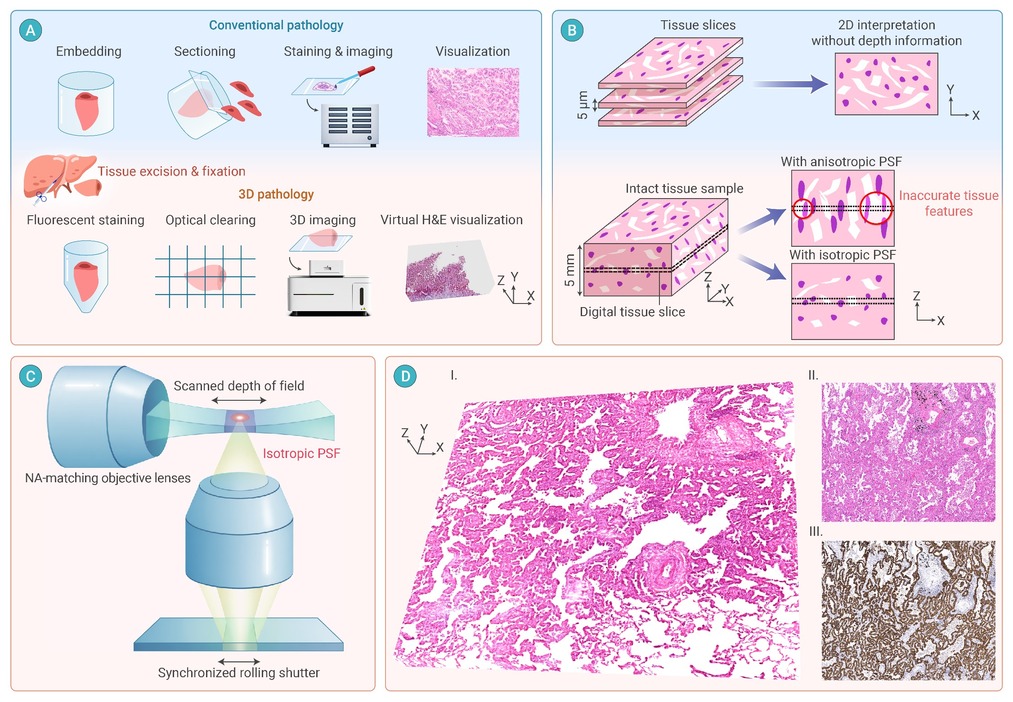
Figure 1.
Concepts and applications of Patho One for 3D pathology
