
Microsnoop: A generalist tool for microscopy image representation
-
GRAPHICAL ABSTRACT
 DownLoad:
Full size image
DownLoad:
Full size image
-
PUBLIC SUMMARY
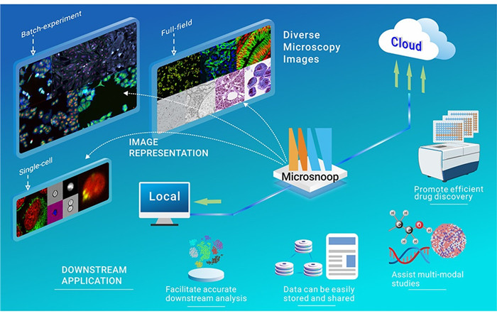
- ■ Microsnoop is a deep learning tool for profiling heterogeneous microscopy images.
- ■ Microsnoop provides generalist pipelines for processing various types of images.
- ■ Microsnoop achieves cutting-edge microscopy image representation ability with great potential for expansion.
- ■ Microsnoop is highly scalable for studies from small scale to high throughput.
-
ABSTRACT
Accurate profiling of microscopy images from small scale to high throughput is an essential procedure in basic and applied biological research. Here, we present Microsnoop, a novel deep learning–based representation tool trained on large-scale microscopy images using masked self-supervised learning. Microsnoop can process various complex and heterogeneous images, and we classified images into three categories: single-cell, full-field, and batch-experiment images. Our benchmark study on 10 high-quality evaluation datasets, containing over 2,230,000 images, demonstrated Microsnoop’s robust and state-of-the-art microscopy image representation ability, surpassing existing generalist and even several custom algorithms. Microsnoop can be integrated with other pipelines to perform tasks such as superresolution histopathology image and multimodal analysis. Furthermore, Microsnoop can be adapted to various hardware and can be easily deployed on local or cloud computing platforms. We will regularly retrain and reevaluate the model using community-contributed data to consistently improve Microsnoop. -

-
REFERENCES
[1] Caicedo, J.C., Singh, S., and Carpenter, A.E. (2016). Applications in image-based profiling of perturbations. Curr. Opin. Biotechnol. 39, 134-142. [2] Pratapa, A., Doron, M., and Caicedo, J.C. (2021). Image-based cell phenotyping with deep learning. Curr. Opin. Chem. Biol. 65, 9-17. [3] Lu, A.X., Chong, Y.T., Hsu, I.S., et al. (2018). Integrating images from multiple microscopy screens reveals diverse patterns of change in the subcellular localization of proteins. Elife 7, e31872. [4] Eulenberg, P., Köhler, N., Blasi, T., et al. (2017). Reconstructing cell cycle and disease progression using deep learning. Nat. Commun. 8, 463. [5] Pawlowski, N., Caicedo, J.C., Singh, S., et al. (2016). Automating morphological profiling with generic deep convolutional networks. Preprint at. bioRxiv. http://biorxiv.org/lookup/doi/10.1101/085118. [6] Cuccarese, M.F., Earnshaw, B.A., Heiser, K., et al. (2020). Functional immune mapping with deep-learning enabled phenomics applied to immunomodulatory and COVID-19 drug discovery. Preprint at. bioRxiv. http://biorxiv.org/lookup/doi/10.1101/2020.08.02.233064. [7] Schürch, C.M., Bhate, S.S., Barlow, G.L., et al. (2020). Coordinated cellular neighborhoods orchestrate antitumoral immunity at the colorectal cancer invasive front. Cell 182, 1341-1359.e19. [8] Eng, C.-H.L., Lawson, M., Zhu, Q., et al. (2019). Transcriptome-scale super-resolved imaging in tissues by RNA seqFISH+. Nature 568, 235-239. [9] Carpenter, A.E., Jones, T.R., Lamprecht, M.R., et al. (2006). CellProfiler: image analysis software for identifying and quantifying cell phenotypes. Genome Biol. 7, R100. [10] Pau, G., Fuchs, F., Sklyar, O., et al. (2010). EBImage–an R package for image processing with applications to cellular phenotypes. Bioinformatics 26, 979-981. [11] Singh, S., Bray, M.-A., Jones, T.R., et al. (2014). Pipeline for illumination correction of images for high-throughput microscopy. J. Microsc. 256, 231-236. [12] Caicedo, J.C., McQuin, C., Goodman, A., et al. (2018). Weakly supervised learning of singlecell feature embeddings. In Proc. IEEE Conference on Computer Vision and Pattern Recognition, 2018, pp. 9309–9318. [13] Lu, A.X., Kraus, O.Z., Cooper, S., et al. (2019). Learning unsupervised feature representations for single cell microscopy images with paired cell inpainting. PLoS Comput. Biol. 15, e1007348. [14] Adnan, M., Kalra, S., and Tizhoosh, H.R. (2020). Representation learning of histopathology images using graph neural networks. In Proc. IEEE Conference on Computer Vision and Pattern Recognition, pp. 988–989. [15] Perakis, A., Gorji, A., Jain, S., et al. (2021). Contrastive learning of single-cell phenotypic representations for treatment classification. In Machine Learning in Medical Imaging, 12966, C. Lian, X. Cao, I. Rekik, X. Xu, and P. Yan, eds., pp. 565–575. [16] Russakovsky, O., Deng, J., Su, H., et al. (2015). ImageNet large scale visual recognition challenge. Int. J. Comput. Vis. 115, 211-252. [17] Bao, F., Deng, Y., Wan, S., et al. (2022). Integrative spatial analysis of cell morphologies and transcriptional states with MUSE. Nat. Biotechnol. 40, 1200-1209. [18] Hua, S.B.Z., Lu, A.X., and Moses, A.M. (2021). CytoImageNet: a large-scale pretraining dataset for bioimage transfer learning. In Proc. Advances in Neural Information Processing Systems. [19] Kobayashi, H., Cheveralls, K.C., Leonetti, M.D., et al. (2022). Self-supervised deep learning encodes high-resolution features of protein subcellular localization. Nat. Methods 19, 995-1003. [20] Wong, D.R., Conrad, J., Johnson, N., et al. (2022). Trans-channel fluorescence learning improves high-content screening for Alzheimer’s disease therapeutics. Nat. Mach. Intell. 4, 583-595. [21] He, K., Chen, X., Xie, S., et al. (2022). Masked autoencoders are scalable vision learners. In Proc. IEEE Conference on Computer Vision and Pattern Recognition, pp. 16000–16009. [22] Liu, X., Zhou, J., Kong, T., et al. (2022). Exploring target representations for masked autoencoders. Preprint at. arXiv. https://arxiv.org/abs/2209.03917. [23] Li, Z., Chen, Z., Yang, F., et al. (2021). MST: masked self-supervised transformer for visual representation. In SAVE Proc. Advances in Neural Information Processing Systems, p. 35. [24] Wei, C., Fan, H., Xie, S., et al. (2022). Masked feature prediction for self-supervised visual pretraining. In Proc. IEEE Conference on Computer Vision and Pattern Recognition, pp. 14668–14678. [25] Pandey, V., Brune, C., and Strisciuglio, N. (2022). Self-supervised learning through colorization for microscopy images. In Image Analysis and Processing - ICIAP 2022, S. Sclaroff, C. Distante, and M. Leo, et al., eds., pp. 621–632. [26] Mascolini, A., Cardamone, D., Ponzio, F., et al. (2022). Exploiting generative self-supervised learning for the assessment of biological images with lack of annotations. BMC Bioinf. 23, 295. [27] Stringer, C., Wang, T., Michaelos, M., et al. (2021). Cellpose: a generalist algorithm for cellular segmentation. Nat. Methods 18, 100-106. [28] Edlund, C., Jackson, T.R., Khalid, N., et al. (2021). LIVECell-a large-scale dataset for label-free live cell segmentation. Nat. Methods 18, 1038-1045. [29] Greenwald, N.F., Miller, G., Moen, E., et al. (2022). Whole-cell segmentation of tissue images with human-level performance using large-scale data annotation and deep learning. Nat. Biotechnol. 40, 555-565. [30] Bray, M.-A., Singh, S., Han, H., et al. (2016). Cell Painting, a high-content image-based assay for morphological profiling using multiplexed fluorescent dyes. Nat. Protoc. 11, 1757-1774. [31] Leek, J.T., Scharpf, R.B., Bravo, H.C., et al. (2010). Tackling the widespread and critical impact of batch effects in high-throughput data. Nat. Rev. Genet. 11, 733-739. [32] Lin, A., and Lu, A.X. (2022). Incorporating knowledge of plates in batch normalization improves generalization of deep learning for microscopy images. In Proc. International Conference on Machine Learning, pp. 74–93. [33] Kumar, N., Verma, R., Anand, D., et al. (2020). A multi-organ nucleus segmentation challenge. IEEE Trans. Med. Imag. 39, 1380-1391. [34] Verma, R., Kumar, N., Patil, A., et al. (2021). MoNuSAC2020: a multi-organ nuclei segmentation and classification challenge. IEEE Trans. Med. Imag. 40, 3413-3423. [35] Amgad, M., Atteya, L.A., Hussein, H., et al. (2022). NuCLS: a scalable crowdsourcing, deep learning approach and dataset for nucleus classification, localization and segmentation. GigaScience 11, giac037. [36] Ronneberger, O., Fischer, P., and Brox, T. (2015). U-Net: convolutional networks for biomedical image segmentation. In Proc. International Conference on Medical Image Computing and Computer-Assisted Intervention, pp. 234–241. [37] Vaswani, A., Shazeer, N., Parmar, N., et al. (2017). Attention is all you need. In Proc. Advances in Neural Information Processing Systems, 30. [38] Dosovitskiy, A., Beyer, L., Kolesnikov, A., et al. (2021). An image is worth 16x16 words: transformers for image recognition at scale. In International Conference on Learning Representations. [39] Ando, D.M., McLean, C.Y., and Berndl, M. (2017). Improving phenotypic measurements in high-content imaging screens. Preprint at. bioRxiv. http://biorxiv.org/lookup/doi/10.1101/161422. [40] Bray, M.-A., Weck, A.D., Durand, E., et al. (2022). High-content cellular screen image analysis benchmark study. Preprint at. bioRxiv. https://www.biorxiv.org/content/10.1101/2022.05.15.491989v1.abstract. [41] Lu, A., Lu, A., Schormann, W., et al. (2019). The Cells Out of Sample (COOS) dataset and benchmarks for measuring out-of-sample generalization of image classifiers. In Proc. Advances in Neural Information Processing Systems, p. 32. [42] Maaten, L., and Hinton, G. (2008). Visualizing data using t-SNE. J. Mach. Learn. Res. 9, 2579-2605. [43] Caie, P.D., Walls, R.E., Ingleston-Orme, A., et al. (2010). High-content phenotypic profiling of drug response signatures across distinct cancer cells. Mol. Cancer Therapeut. 9, 1913-1926. [44] Graham, S., Vu, Q.D., Raza, S.E.A., et al. (2019). Hover-Net: simultaneous segmentation and classification of nuclei in multi-tissue histology images. Med. Image Anal. 58, 101563. [45] Schraivogel, D., Kuhn, T.M., Rauscher, B., et al. (2022). High-speed fluorescence image-enabled cell sorting. Science 375, 315-320. [46] Tan, M., and Le, Q.V. (2019). EfficientNet: rethinking model scaling for convolutional neural networks. In Proc. International Conference on Machine Learning, pp. 6105–6114. [47] Szegedy, C., Vanhoucke, V., Ioffe, S., et al. (2016). Rethinking the Inception architecture for computer vision. In Proc. IEEE Conference on Computer Vision and Pattern Recognition, pp. 2818–2826. [48] Xun, D., Chen, D., Zhou, Y., et al. (2022). Scellseg: a style-aware deep learning tool for adaptive cell instance segmentation by contrastive fine-tuning. iScience 25, 105506. [49] Pachitariu, M., and Stringer, C. (2022). Cellpose 2.0: how to train your own model. Nat. Methods 19, 1634-1641. [50] Lazard, T., Bataillon, G., Naylor, P., et al. (2022). Deep learning identifies morphological patterns of homologous recombination deficiency in luminal breast cancers from whole slide images. Cell Rep. Med. 3, 100872. [51] Moshkov, N., Bornholdt, M., Benoit, S., et al. (2022). Learning representations for imagebased profiling of perturbations. Preprint at. bioRxiv. http://biorxiv.org/lookup/doi/10.1101/2022.08.12.503783. [52] Taylor, J., Earnshaw, B., Mabey, B., et al. (2019). RxRx1: an image set for cellular morphological variation across many experimental batches. In 7th International Conference on Learning Representations. [53] Caicedo, J.C., Cooper, S., Heigwer, F., et al. (2017). Data-analysis strategies for image-based cell profiling. Nat. Methods 14, 849-863. [54] Chandrasekaran, S.N., Ceulemans, H., Boyd, J.D., et al. (2021). Image-based profiling for drug discovery: due for a machine-learning upgrade? Nat. Rev. Drug Discov. 20, 145-159. [55] Devlin, J., Chang, M.-W., Lee, K., et al. (2018). BERT: pre-training of deep bidirectional transformers for language understanding. Preprint at. arXiv. https://arxiv.org/abs/1810.04805. [56] Brown, T.B., Mann, B., Ryder, N., et al. (2020). Language models are few-shot learners. In Proc. Advances in Neural Information Processing Systems, p. 33. [57] Min, B., Ross, H., Sulem, E., et al. (2021). Recent advances in natural language processing via large pre-trained language models: a survey. Preprint at. arXiv. http://arxiv.org/abs/2111.01243. [58] Davari, M., Asadi, N., Mudur, S., et al. (2022). Probing representation forgetting in supervised and unsupervised continual learning. In Proc. IEEE Conference on Computer Vision and Pattern Recognition, pp. 16691–16700. [59] Mundt, M., Hong, Y., Pliushch, I., et al. (2023). A wholistic view of continual learning with deep neural networks: forgotten lessons and the bridge to active and open world learning. Neural Network. 160, 306-336. [60] Abdalla, M., Abdalla, M., Hirst, G., et al. (2020). Exploring the privacy-preserving properties of word embeddings: algorithmic validation study. J. Med. Internet Res. 22, e18055. [61] Wang, B., Li, Y., Zhou, M., et al. (2023). Smartphone-based platforms implementing microfluidic detection with image-based artificial intelligence. Nat. Commun. 14, 1341. [62] Sanchez-Fernandez, A., Rumetshofer, E., and Hochreiter, S. (2022). Contrastive learning of image- and structure- based representations in drug discovery. In International Conference on Learning Representations. [63] Tian, G., Harrison, P.J., Sreenivasan, A.P., et al. (2022). Combining molecular and cell painting image data for mechanism of action prediction. Preprint at. bioRxiv. http://biorxiv.org/lookup/doi/10.1101/2022.10.04.510834. [64] Haghighi, M., Caicedo, J.C., Cimini, B.A., et al. (2022). High-dimensional gene expression and morphology profiles of cells across 28,000 genetic and chemical perturbations. Nat. Methods 19, 1550-1557. [65] Liu, L., Bi, M., Wang, Y., et al. (2021). Artificial intelligence-powered microfluidics for nanomedicine and materials synthesis. Nanoscale 13, 19352-19366. [66] Wang, X., Xie, P., Chen, B., et al. (2022). Chip-based high-dimensional optical neural network. Nano-Micro Lett. 14, 221. -
ABOUT THIS ARTICLE
Cite this article:
Dejin Xun, Rui Wang, Xingcai Zhang, Yi Wang. Microsnoop: A generalist tool for microscopy image representation[J]. The Innovation, 2024, 5(1). https://doi.org/10.1016/j.xinn.2023.100541
Welcome!
To request copyright permission to republish or share portions of our works, please visit Copyright Clearance Center's (CCC) Marketplace website at marketplace.copyright.com.

Export File
Citation
Dejin Xun, Rui Wang, Xingcai Zhang, Yi Wang. Microsnoop: A generalist tool for microscopy image representation[J]. The Innovation, 2024, 5(1). https://doi.org/10.1016/j.xinn.2023.100541

Format
Content
-
Figure 1.
Design of Microsnoop for microscopy image representation
-
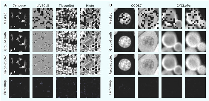
Figure 2.
Reconstruction results with Microsnoop
-
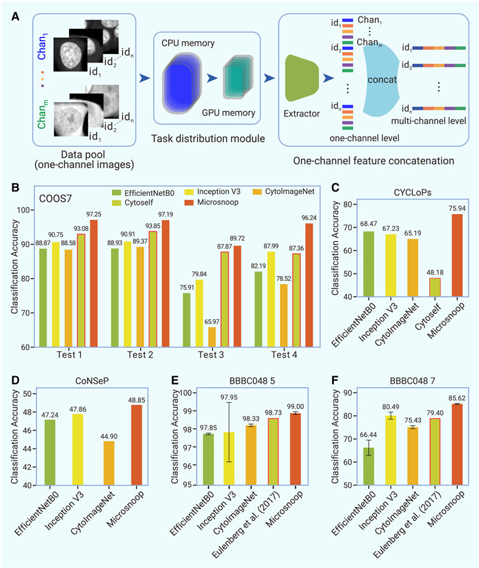
Figure 3.
Profiling with Microsnoop on single-cell images
-
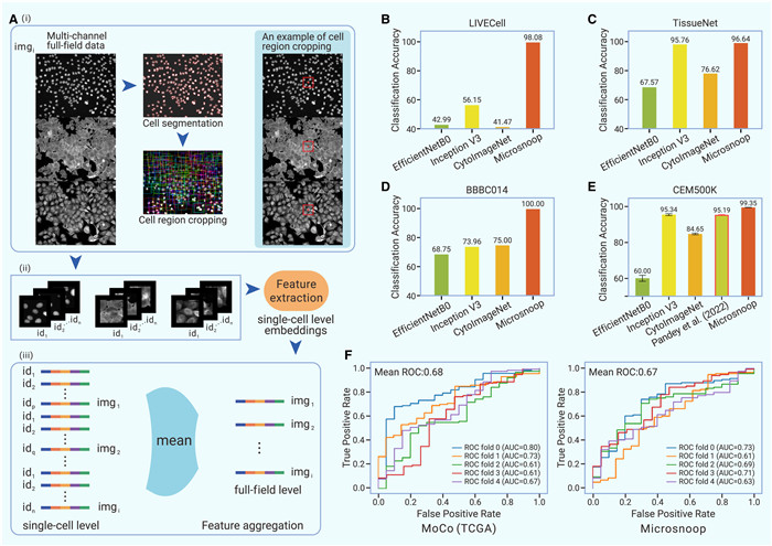
Figure 4.
Profiling with Microsnoop on full-field images
-
Figure 5.
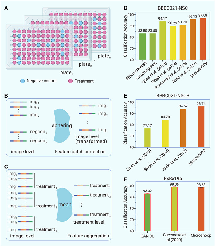
Profiling with Microsnoop on batch-experiment images
-
Figure 6.
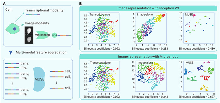
Joint use of Microsnoop and MUSE
