
Dynamic immune recovery process after liver transplantation revealed by single-cell multi-omics analysis
-
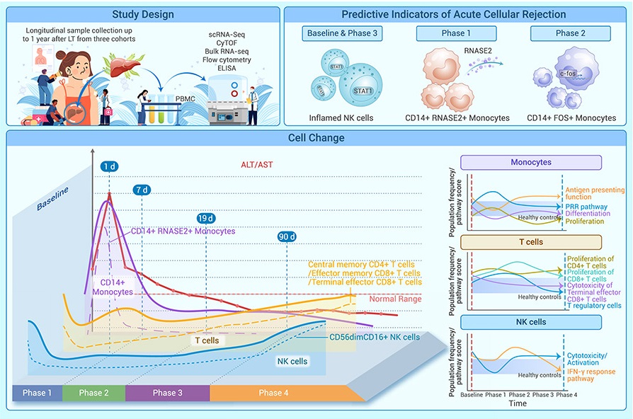
GRAPHICAL ABSTRACT
 DownLoad:
Full size image
DownLoad:
Full size image
-
PUBLIC SUMMARY
- ■ A dynamic immune landscape after liver transplantation unveils a four-phase recovery process, facilitating the precise management of liver transplantation patients.
- ■ T cell activity deficiency does not completely suppress the allogeneic cell rejection.
- ■ The elevated proportion of inflamed NK cell subset is a potential driver for graft rejection in liver transplantation patients.
- ■ Monocytes serve as the major signal source to initialize immune responses after liver transplantation.
- ■ CD14+RNASE2+ monocytes and FOS-expressing monocytes are potential early predictive indicators for graft rejection.
-
ABSTRACT
Elucidating the temporal process of immune remodeling under immunosuppressive treatment after liver transplantation (LT) is critical for precise clinical management strategies. Here, we performed a single-cell multi-omics analysis of peripheral blood mononuclear cells (PBMCs) collected from LT patients (with and without acute cellular rejection [ACR]) at 13 time points. Validation was performed in two independent cohorts with additional LT patients and healthy controls. Our study revealed a four-phase recovery process after LT and delineated changes in immune cell composition, expression programs, and interactions along this process. The intensity of the immune response differs between the ACR and non-ACR patients. Notably, the newly identified inflamed NK cells, CD14+RNASE2+ monocytes, and FOSexpressing monocytes emerged as predictive indicators of ACR. This study illuminates the longitudinal evolution of the immune cell landscape under tacrolimus-based immunosuppressive treatment during LT recovery, providing a four-phase framework that aids the clinical management of LT patients. -

-
REFERENCES
[1] Post, D.J., Douglas, D.D., and Mulligan, D.C. (2005). Immunosuppression in liver transplantation. Liver Transpl. 11(11): 1307–1314. https://doi.org/10.1002/lt.20614. [2] Wei, X., Xie, W., Yin, W., et al. (2022). Prediction of tumor recurrence by distinct immunoprofiles in liver transplant patients based on mass cytometry. Am. J. Cancer Res. 12(9): 4160–4176. [3] Huang, H., Chen, R., Lin, Y., et al. (2023). Decoding Single-cell Landscape and Intercellular Crosstalk in the Transplanted Liver. Transplantation 107(4): 890–902. https://doi.org/10.1097/TP.0000000000004365. [4] Li, X., Li, S., Wu, B., et al. (2022). Landscape of Immune Cells Heterogeneity in Liver Transplantation by Single-Cell RNA Sequencing Analysis. Front. Immunol. 13: 890019. https://doi.org/10.3389/fimmu.2022.890019. [5] Huang, H., Zhang, X., Zhang, C., et al. (2020). The time-dependent shift in the hepatic graft and recipient macrophage pool following liver transplantation. Cell. Mol. Immunol. 17(4): 412–414. https://doi.org/10.1038/s41423-019-0253-x. [6] Wang, L., Li, J., He, S., et al. (2021). Resolving the graft ischemia-reperfusion injury during liver transplantation at the single cell resolution. Cell Death Dis. 12(6): 589. https://doi.org/10.1038/s41419-021-03878-3. [7] Shan, Y., Qi, D., Zhang, L., et al. (2023). Single-cell RNA-seq revealing the immune features of donor liver during liver transplantation. Front. Immunol. 14: 1096733. https://doi.org/10.3389/fimmu.2023.1096733. [8] Mederacke, Y.-S., Nienen, M., Jarek, M., et al. (2021). T cell receptor repertoires within liver allografts are different from those in the peripheral blood. J. Hepatol. 74(5): 1167–1175. https://doi.org/10.1016/j.jhep.2020.12.014. [9] Wu, T., Hu, E., Xu, S., et al. (2021). clusterProfiler 4.0: A universal enrichment tool for interpreting omics data. Innovation 2(3): 100141. https://doi.org/10.1016/j.xinn.2021.100141. [10] Kumar, L., and E Futschik, M. (2007). Mfuzz: a software package for soft clustering of microarray data. Bioinformation 2(1): 5–7. https://doi.org/10.6026/97320630002005. [11] Ivashkiv, L.B., and Donlin, L.T. (2014). Regulation of type Ⅰ interferon responses. Nat. Rev. Immunol. 14(1): 36–49. https://doi.org/10.1038/nri3581. [12] Obara, H., Nagasaki, K., Hsieh, C.L., et al. (2005). IFN-gamma, produced by NK cells that infiltrate liver allografts early after transplantation, links the innate and adaptive immune responses. Am. J. Transplant. 5(9): 2094–2103. https://doi.org/10.1111/j.1600-6143.2005.00995.x. [13] Wang, E., Worschech, A., and Marincola, F.M. (2008). The immunologic constant of rejection. Trends Immunol. 29(6): 256–262. https://doi.org/10.1016/j.it.2008.03.002. [14] Gwon, Y., Maxwell, B.A., Kolaitis, R.-M., et al. (2021). Ubiquitination of G3BP1 mediates stress granule disassembly in a context-specific manner. Science 372(6549): eabf6548. https://doi.org/10.1126/science.abf6548. [15] Fang, N.N., Chan, G.T., Zhu, M., et al. (2014). Rsp5/Nedd4 is the main ubiquitin ligase that targets cytosolic misfolded proteins following heat stress. Nat. Cell Biol. 16(12): 1227–1237. https://doi.org/10.1038/ncb3054. [16] Turakhiya, A., Meyer, S.R., Marincola, G., et al. (2018). ZFAND1 Recruits p97 and the 26S Proteasome to Promote the Clearance of Arsenite-Induced Stress Granules. Mol. Cell 70(5): 906–919.e7. https://doi.org/10.1016/j.molcel.2018.04.021. [17] Balagopal, V., and Parker, R. (2009). Polysomes, P bodies and stress granules: states and fates of eukaryotic mRNAs. Curr. Opin. Cell Biol. 21(3): 403–408. https://doi.org/10.1016/j.ceb.2009.03.005. [18] Shalgi, R., Hurt, J.A., Lindquist, S., et al. (2014). Widespread inhibition of posttranscriptional splicing shapes the cellular transcriptome following heat shock. Cell Rep. 7(5): 1362–1370. https://doi.org/10.1016/j.celrep.2014.04.044. [19] Thomson, A.W., Vionnet, J., and Sanchez-Fueyo, A. (2020). Understanding, predicting and achieving liver transplant tolerance: from bench to bedside. Nat. Rev. Gastroenterol. Hepatol. 17(12): 719–739. https://doi.org/10.1038/s41575-020-0334-4. [20] Ogawa, K., Hirai, M., Katsube, T., et al. (2000). Suppression of cellular immunity by surgical stress. Surgery 127(3): 329–336. https://doi.org/10.1067/msy.2000.103498. [21] Goldrath, A.W., Bogatzki, L.Y., and Bevan, M.J. (2000). Naive T cells transiently acquire a memory-like phenotype during homeostasis-driven proliferation. J. Exp. Med. 192(4): 557–564. https://doi.org/10.1084/jem.192.4.557. [22] Lanzavecchia, A., and Sallusto, F. (2005). Understanding the generation and function of memory T cell subsets. Curr. Opin. Immunol. 17(3): 326–332. https://doi.org/10.1016/j.coi.2005.04.010. [23] Li, Y., An, H., Shen, C., et al. (2021). Deep phenotyping of T cell populations under long-term treatment of tacrolimus and rapamycin in patients receiving renal transplantations by mass cytometry. Clin. Transl. Med. 11(11): e629. https://doi.org/10.1002/ctm2.629. [24] Presser, D., Sester, U., Mohrbach, J., et al. (2009). Differential kinetics of effector and regulatory T cells in patients on calcineurin inhibitor-based drug regimens. Kidney Int. 76(5): 557–566. https://doi.org/10.1038/ki.2009.198. [25] Schwartz, R.H. (2003). T cell anergy. Annu. Rev. Immunol. 21: 305–334. https://doi.org/10.1146/annurev.immunol.21.120601.141110. [26] ElTanbouly, M.A., and Noelle, R.J. (2021). Rethinking peripheral T cell tolerance: checkpoints across a T cell’s journey. Nat. Rev. Immunol. 21(4): 257–267. https://doi.org/10.1038/s41577-020-00454-2. [27] Strom, T.B., Tilney, N.L., Carpenter, C.B., et al. (1975). Identity and cytotoxic capacity of cells infiltrating renal allografts. N. Engl. J. Med. 292(24): 1257–1263. https://doi.org/10.1056/NEJM197506122922402. [28] Kubota, N., Sugitani, M., Takano, S., et al. (2006). Correlation between acute rejection severity and CD8-positive T cells in living related liver transplantation. Transpl. Immunol. 16(1): 60–64. https://doi.org/10.1016/j.trim.2006.03.002. [29] Orr, M.T., and Lanier, L.L. (2010). Natural killer cell education and tolerance. Cell 142(6): 847–856. https://doi.org/10.1016/j.cell.2010.08.031. [30] Tai, L.-H., de Souza, C.T., Bélanger, S., et al. (2013). Preventing postoperative metastatic disease by inhibiting surgery-induced dysfunction in natural killer cells. Cancer Res. 73(1): 97–107. https://doi.org/10.1158/0008-5472.CAN-12-1993. [31] Angka, L., Tanese de Souza, C., Baxter, K.E., et al. (2022). Perioperative arginine prevents metastases by accelerating natural killer cell recovery after surgery. Mol. Ther. 30(10): 3270–3283. https://doi.org/10.1016/j.ymthe.2022.05.024. [32] Wang, Y.-Y., Chang, E.-Q., Zhu, R.-L., et al. (2022). An atlas of dynamic peripheral blood mononuclear cell landscapes in human perioperative anaesthesia/surgery. Clin. Transl. Med. 12(1): e663. https://doi.org/10.1002/ctm2.663. [33] Kim, T.-J., Kim, N., Kang, H.J., et al. (2010). FK506 causes cellular and functional defects in human natural killer cells. J. Leukoc. Biol. 88(6): 1089–1097. https://doi.org/10.1189/jlb.0310148. [34] Wang, H., Grzywacz, B., Sukovich, D., et al. (2007). The unexpected effect of cyclosporin A on CD56+CD16- and CD56+CD16+ natural killer cell subpopulations. Blood 110(5): 1530–1539. https://doi.org/10.1182/blood-2006-10-048173. [35] Kitchens, W.H., Uehara, S., Chase, C.M., et al. (2006). The changing role of natural killer cells in solid organ rejection and tolerance. Transplantation 81(6): 811–817. https://doi.org/10.1097/01.tp.0000202844.33794.0e. [36] Knorr, D.A., Bachanova, V., Verneris, M.R., et al. (2014). Clinical utility of natural killer cells in cancer therapy and transplantation. Semin. Immunol. 26(2): 161–172. https://doi.org/10.1016/j.smim.2014.02.002. [37] Cooper, M.A., Fehniger, T.A., and Caligiuri, M.A. (2001). The biology of human natural killercell subsets. Trends Immunol. 22(11): 633–640. https://doi.org/10.1016/s1471-4906(01)02060-9. [38] Zhang, Q., Bi, J., Zheng, X., et al. (2018). Blockade of the checkpoint receptor TIGIT prevents NK cell exhaustion and elicits potent anti-tumor immunity. Nat. Immunol. 19(7): 723–732. https://doi.org/10.1038/s41590-018-0132-0. [39] Taveirne, S., Wahlen, S., Van Loocke, W., et al. (2020). The transcription factor ETS1 is an important regulator of human NK cell development and terminal differentiation. Blood 136(3): 288–298. https://doi.org/10.1182/blood.2020005204. [40] Yang, C., Siebert, J.R., Burns, R., et al. (2019). Heterogeneity of human bone marrow and blood natural killer cells defined by single-cell transcriptome. Nat. Commun. 10(1): 3931. https://doi.org/10.1038/s41467-019-11947-7. [41] Smith, S.L., Kennedy, P.R., Stacey, K.B., et al. (2020). Diversity of peripheral blood human NK cells identified by single-cell RNA sequencing. Blood Adv. 4(7): 1388–1406. https://doi.org/10.1182/bloodadvances.2019000699. [42] Marçais, A., Cherfils-Vicini, J., Viant, C., et al. (2014). The metabolic checkpoint kinase mTOR is essential for IL-15 signaling during the development and activation of NK cells. Nat. Immunol. 15(8): 749–757. https://doi.org/10.1038/ni.2936. [43] Chiossone, L., Vitale, C., Cottalasso, F., et al. (2007). Molecular analysis of the methylprednisolone-mediated inhibition of NK-cell function: evidence for different susceptibility of IL-2- versus IL-15-activated NK cells. Blood 109(9): 3767–3775. https://doi.org/10.1182/blood-2006-07-037846. [44] Takeuchi, O., and Akira, S. (2010). Pattern recognition receptors and inflammation. Cell 140(6): 805–820. https://doi.org/10.1016/j.cell.2010.01.022. [45] van den Bosch, T.P.P., Kannegieter, N.M., Hesselink, D.A., et al. (2017). Targeting the Monocyte-Macrophage Lineage in Solid Organ Transplantation. Front. Immunol. 8: 153. https://doi.org/10.3389/fimmu.2017.00153. [46] Ginhoux, F., and Jung, S. (2014). Monocytes and macrophages: developmental pathways and tissue homeostasis. Nat. Rev. Immunol. 14(6): 392–404. https://doi.org/10.1038/nri3671. [47] Patel, A.A., Zhang, Y., Fullerton, J.N., et al. (2017). The fate and lifespan of human monocyte subsets in steady state and systemic inflammation. J. Exp. Med. 214(7): 1913–1923. https://doi.org/10.1084/jem.20170355. [48] Hop, H.T., Arayan, L.T., Huy, T.X.N., et al. (2018). The Key Role of c-Fos for Immune Regulation and Bacterial Dissemination in Brucella Infected Macrophage. Front. Cell. Infect. Microbiol. 8: 287. https://doi.org/10.3389/fcimb.2018.00287. [49] Larsen, S.B., Cowley, C.J., Sajjath, S.M., et al. (2021). Establishment, maintenance, and recall of inflammatory memory. Cell Stem Cell 28(10): 1758–1774.e8. https://doi.org/10.1016/j.stem.2021.07.001. [50] Rosenberg, H.F., Tenen, D.G., and Ackerman, S.J. (1989). Molecular cloning of the human eosinophil-derived neurotoxin: a member of the ribonuclease gene family. Proc. Natl. Acad. Sci. USA 86(12): 4460–4464. https://doi.org/10.1073/pnas.86.12.4460. [51] Okoye, I., Xu, L., Motamedi, M., et al. (2020). Galectin-9 expression defines exhausted T cells and impaired cytotoxic NK cells in patients with virus-associated solid tumors. J. Immunother. Cancer 8(2): e001849. https://doi.org/10.1136/jitc-2020-001849. [52] Motamedi, M., Shahbaz, S., Fu, L., et al. (2019). Galectin-9 Expression Defines a Subpopulation of NK Cells with Impaired Cytotoxic Effector Molecules but Enhanced IFNg Production, Dichotomous to TIGIT, in HIV-1 Infection. Immunohorizons 3(11): 531–546. https://doi.org/10.4049/immunohorizons.1900087. [53] Golden-Mason, L., McMahan, R.H., Strong, M., et al. (2013). Galectin-9 functionally impairs natural killer cells in humans and mice. J. Virol. 87(9): 4835–4845. https://doi.org/10.1128/JVI.01085-12. [54] Sánchez-Fueyo, A., and Strom, T.B. (2011). Immunologic basis of graft rejection and tolerance following transplantation of liver or other solid organs. Gastroenterology 140(1): 51–64. https://doi.org/10.1053/j.gastro.2010.10.059. [55] Wiesner, R.H., and Fung, J.J. (2011). Present state of immunosuppressive therapy in liver transplant recipients. Liver Transpl. 17(Suppl 3): S1–S9. https://doi.org/10.1002/lt.22410. [56] Wang, Z.-X., Song, S.-H., Teng, F., et al. (2010). A single-center retrospective analysis of liver transplantation on 255 patients with hepatocellular carcinoma. Clin. Transplant. 24(6): 752–757. https://doi.org/10.1111/j.1399-0012.2009.01172.x. [57] Li, C.-J., and Li, L. (2015). Tacrolimus in preventing transplant rejection in Chinese patients–optimizing use. Drug Des. Devel. Ther. 9: 473–485. https://doi.org/10.2147/DDDT.S41349. [58] Liu, Y., Zhang, C., Li, L., et al. (2019). Genome-Wide Association Study of Tacrolimus Pharmacokinetics Identifies Novel Single Nucleotide Polymorphisms in the Convalescence and Stabilization Periods of Post-transplant Liver Function. Front. Genet. 10: 528. https://doi.org/10.3389/fgene.2019.00528. [59] Yu, S., Su, C., and Luo, X. (2019). Impact of infection on transplantation tolerance. Immunol. Rev. 292(1): 243–263. https://doi.org/10.1111/imr.12803. [60] Wood, K.J., and Goto, R. (2012). Mechanisms of rejection: current perspectives. Transplantation 93(1): 1–10. https://doi.org/10.1097/TP.0b013e31823cab44. [61] Li, H., Yu, X., Shi, B., et al. (2021). Reduced pannexin 1-IL-33 axis function in donor livers increases risk of MRSA infection in liver transplant recipients. Sci. Transl. Med. 13(606): eaaz6169. https://doi.org/10.1126/scitranslmed.aaz6169. [62] Rodríguez-Perálvarez, M., Tsochatzis, E., Naveas, M.C., et al. (2013). Reduced exposure to calcineurin inhibitors early after liver transplantation prevents recurrence of hepatocellular carcinoma. J. Hepatol. 59(6): 1193–1199. https://doi.org/10.1016/j.jhep.2013.07.012. [63] Marik, P.E., and Flemmer, M. (2012). The immune response to surgery and trauma: Implications for treatment. J. Trauma Acute Care Surg. 73(4): 801–808. https://doi.org/10.1097/TA.0b013e318265cf87. [64] Gao, J., Duan, Z., Zhang, L., et al. (2016). Failure recovery of circulating NKG2D+CD56dimNK cells in HBV-associated hepatocellular carcinoma after hepatectomy predicts early recurrence. OncoImmunology 5(1): e1048061. https://doi.org/10.1080/2162402X.2015.1048061. [65] Rodríguez-Perálvarez, M., Germani, G., Papastergiou, V., et al. (2013). Early tacrolimus exposure after liver transplantation: relationship with moderate/severe acute rejection and long-term outcome. J. Hepatol. 58(2): 262–270. https://doi.org/10.1016/j.jhep.2012.09.019. [66] Montano-Loza, A.J., Rodríguez-Perálvarez, M.L., Pageaux, G.-P., et al. (2023). Liver transplantation immunology: Immunosuppression, rejection, and immunomodulation. J. Hepatol. 78(6): 1199–1215. https://doi.org/10.1016/j.jhep.2023.01.030. [67] Charlton, M., Levitsky, J., Aqel, B., et al. (2018). International Liver Transplantation Society Consensus Statement on Immunosuppression in Liver Transplant Recipients. Transplantation 102(5): 727–743. https://doi.org/10.1097/TP.0000000000002147. [68] (2016). EASL Clinical Practice Guidelines: Liver transplantation. J. Hepatol. 64(2): 433–485. https://doi.org/10.1016/j.jhep.2015.10.006. [69] Taubert, R., Hardtke-Wolenski, M., Noyan, F., et al. (2014). Intrahepatic regulatory T cells in autoimmune hepatitis are associated with treatment response and depleted with current therapies. J. Hepatol. 61(5): 1106–1114. https://doi.org/10.1016/j.jhep.2014.05.034. [70] Adams, D.H., Sanchez-Fueyo, A., and Samuel, D. (2015). From immunosuppression to tolerance. J. Hepatol. 62(1 Suppl): S170–S185. https://doi.org/10.1016/j.jhep.2015.02.042. [71] Knechtle, S.J., and Kwun, J. (2009). Unique aspects of rejection and tolerance in liver transplantation. Semin. Liver Dis. 29(1): 91–101. https://doi.org/10.1055/s-0029-1192058. [72] Ogura, Y., Martinez, O.M., Villanueva, J.C., et al. (2001). Apoptosis and allograft rejection in the absence of CD8+ T cells. Transplantation 71(12): 1827–1834. https://doi.org/10.1097/00007890-200106270-00020. [73] Navarro, F., Portalès, P., Candon, S., et al. (2000). Natural killer cell and alphabeta and gammadelta lymphocyte traffic into the liver graft immediately after liver transplantation. Transplantation 69(4): 633–639. https://doi.org/10.1097/00007890-200002270-00027. [74] Moroso, V., Metselaar, H.J., Mancham, S., et al. (2010). Liver grafts contain a unique subset of natural killer cells that are transferred into the recipient after liver transplantation. Liver Transpl. 16(7): 895–908. https://doi.org/10.1002/lt.22080. [75] Halloran, P.F., Afrouzian, M., Ramassar, V., et al. (2001). Interferon-gamma acts directly on rejecting renal allografts to prevent graft necrosis. Am. J. Pathol. 158(1): 215–226. https://doi.org/10.1016/s0002-9440(10)63960-0. [76] Millán, O., Rafael-Valdivia, L., San Segundo, D., et al. (2014). Should IFN-g, IL-17 and IL-2 be considered predictive biomarkers of acute rejection in liver and kidney transplant? Results of a multicentric study. Clin. Immunol. 154(2): 141–154. https://doi.org/10.1016/j.clim.2014.07.007. [77] Cui, A., Li, B., Wallace, M.S., et al. (2024). Single-cell atlas of the liver myeloid compartment before and after cure of chronic viral hepatitis. J. Hepatol. 80: 251–267. https://doi.org/10.1016/j.jhep.2023.02.040. -
ABOUT THIS ARTICLE
Cite this article:
Wang R., Peng X., Yuan Y., et al., (2024). Dynamic immune recovery process after liver transplantation revealed by single-cell multi-omics analysis. The Innovation 5(3), 100599. https://doi.org/10.1016/j.xinn.2024.100599
Welcome!
To request copyright permission to republish or share portions of our works, please visit Copyright Clearance Center's (CCC) Marketplace website at marketplace.copyright.com.
Export File
Citation
Wang R., Peng X., Yuan Y., et al., (2024). Dynamic immune recovery process after liver transplantation revealed by single-cell multi-omics analysis. The Innovation 5(3), 100599. https://doi.org/10.1016/j.xinn.2024.100599

Format
Content

-
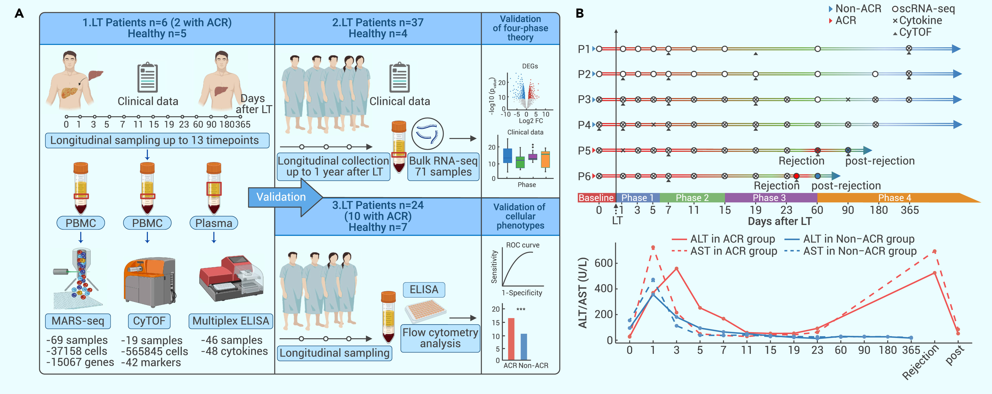
Figure 1.
Study design
-
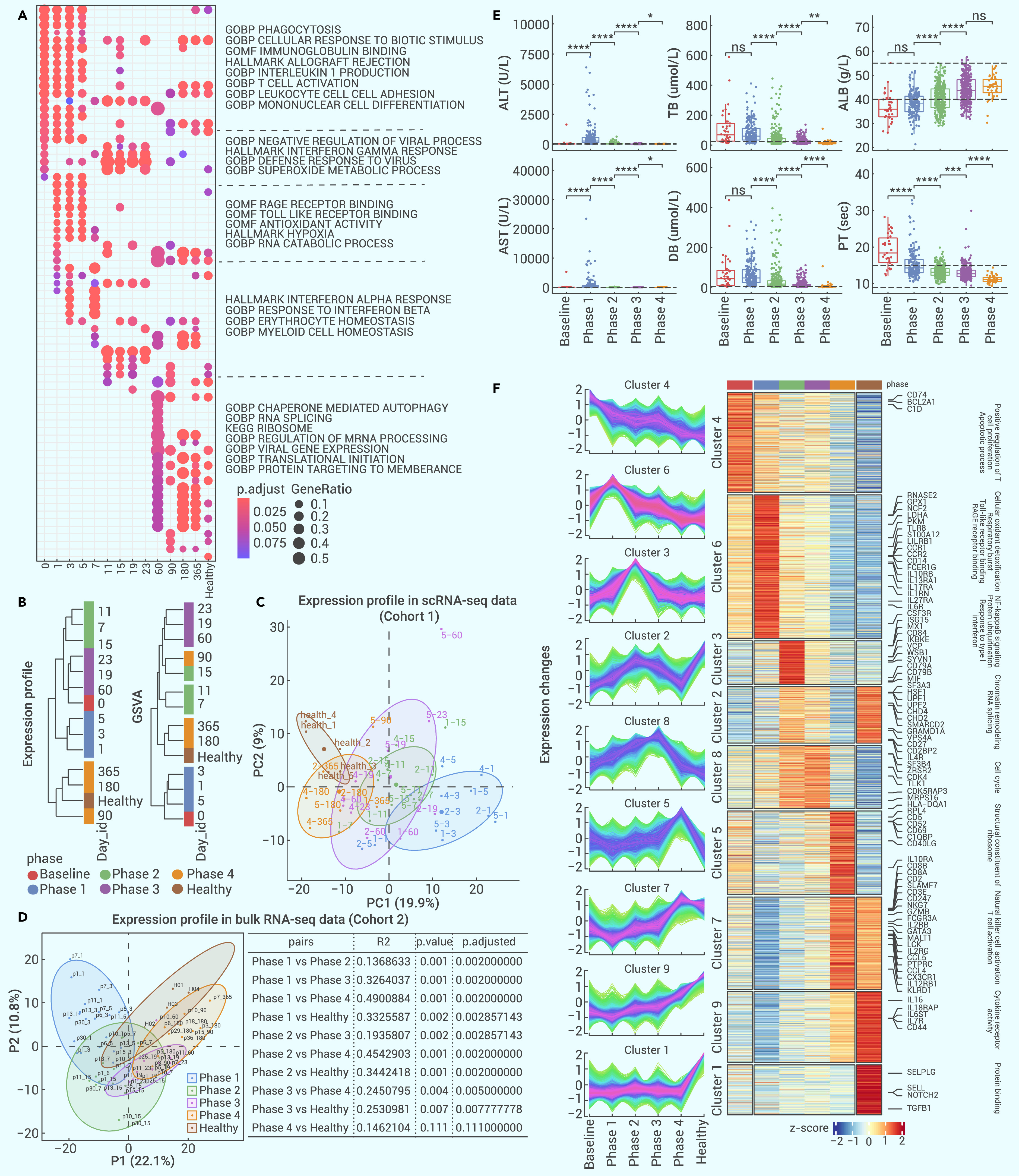
Figure 2.
Definition of a four-phase recovery process in LT
-
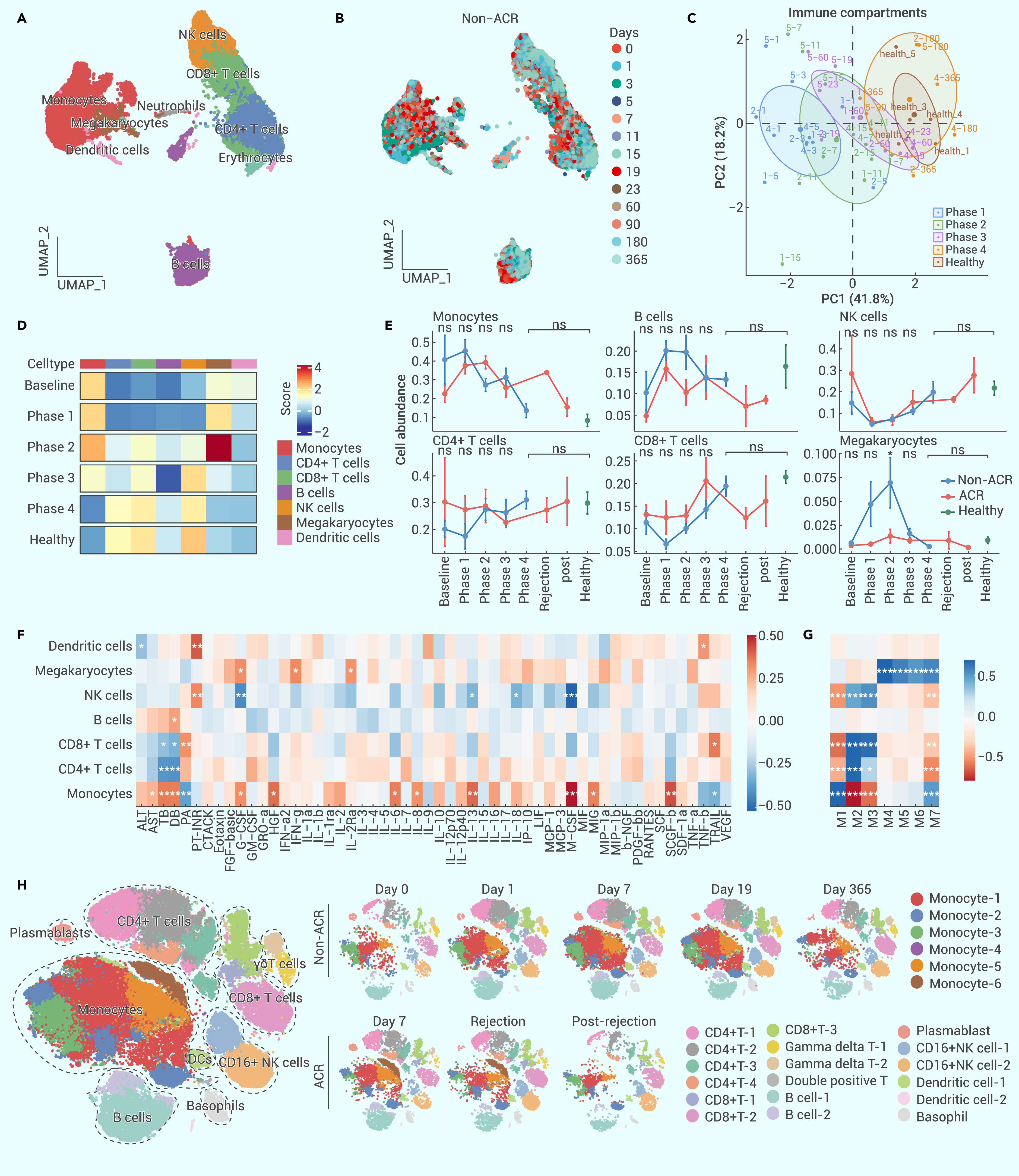
Figure 3.
Global changes in immune cells across the four-phase recovery process
-
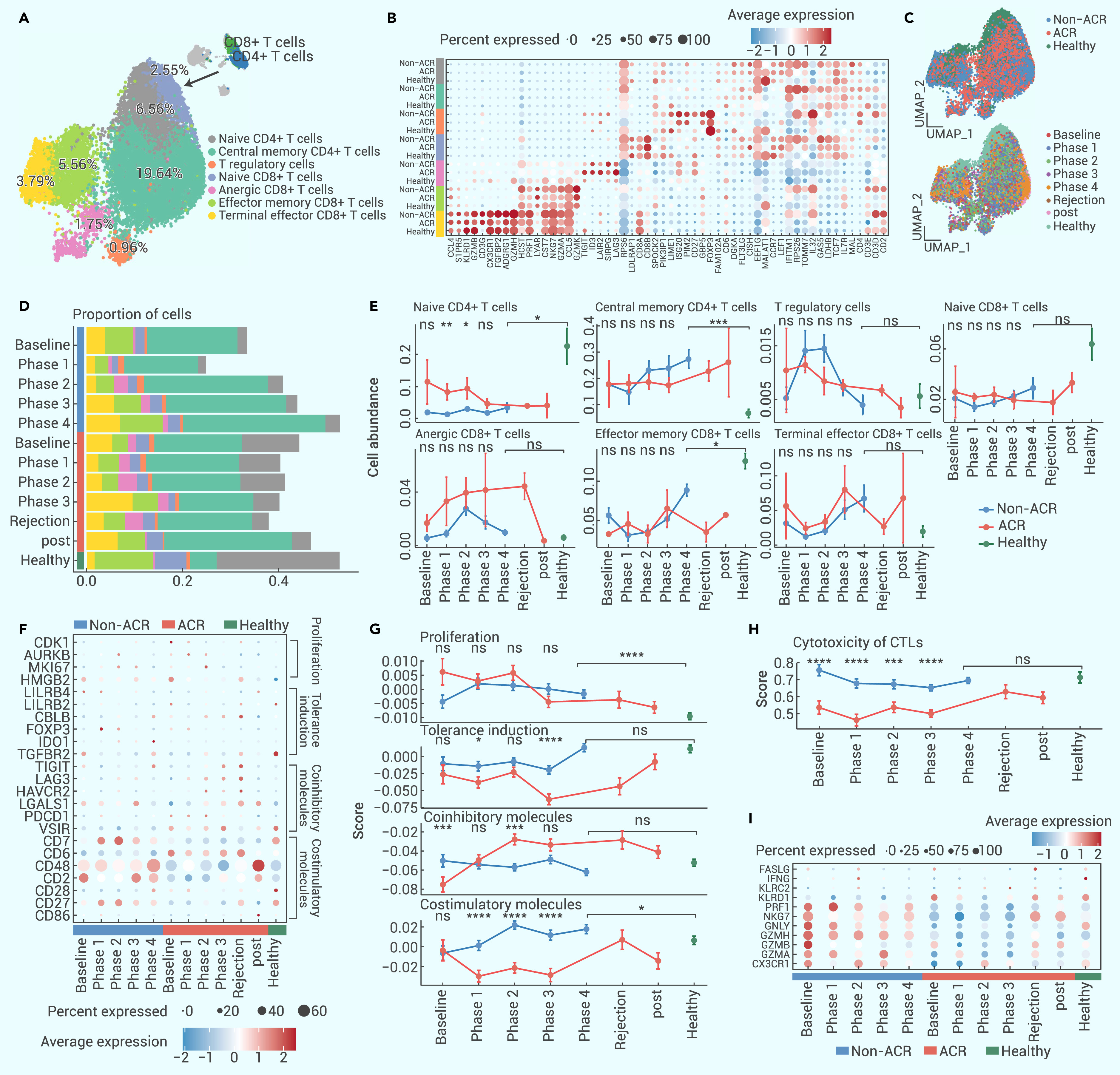
Figure 4.
Dynamic changes in subpopulations of T cells after LT
-
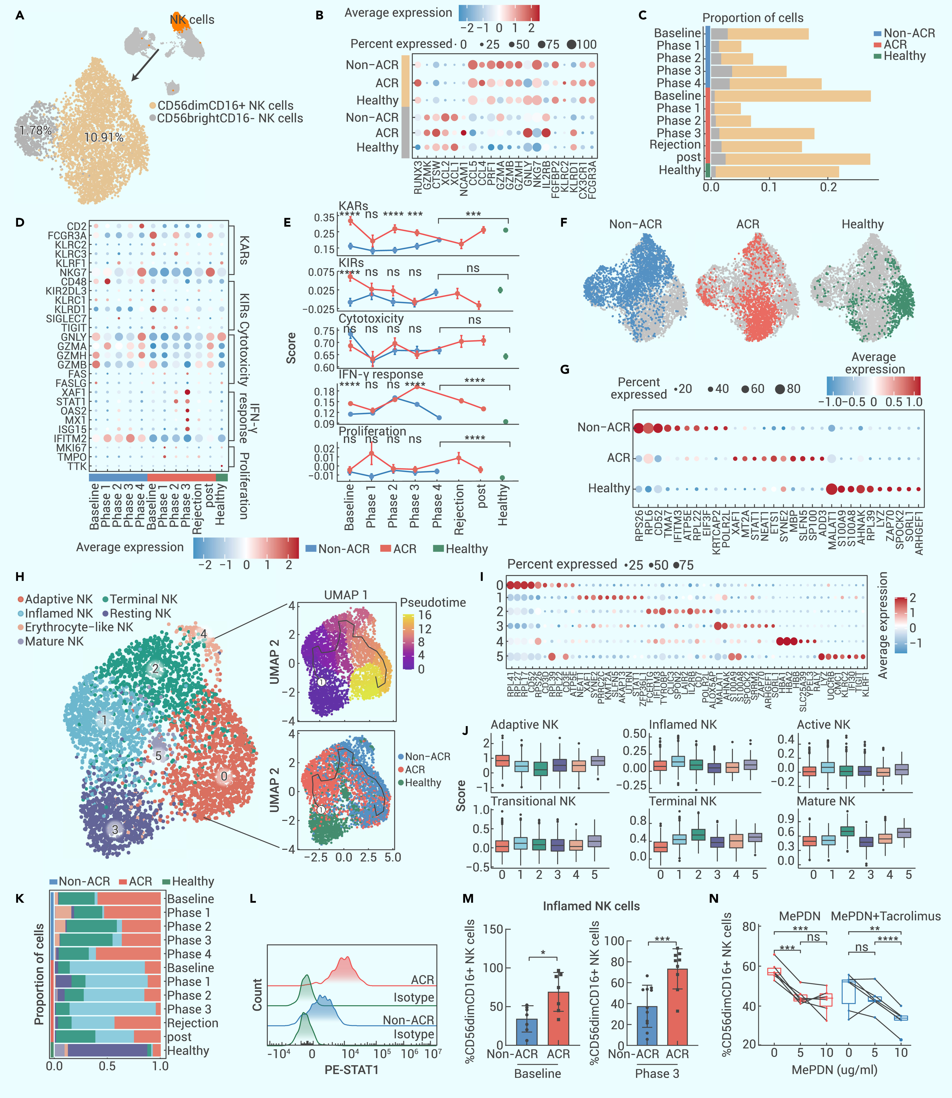
Figure 5.
Heterogeneity of NK cells across the LT recovery process
-
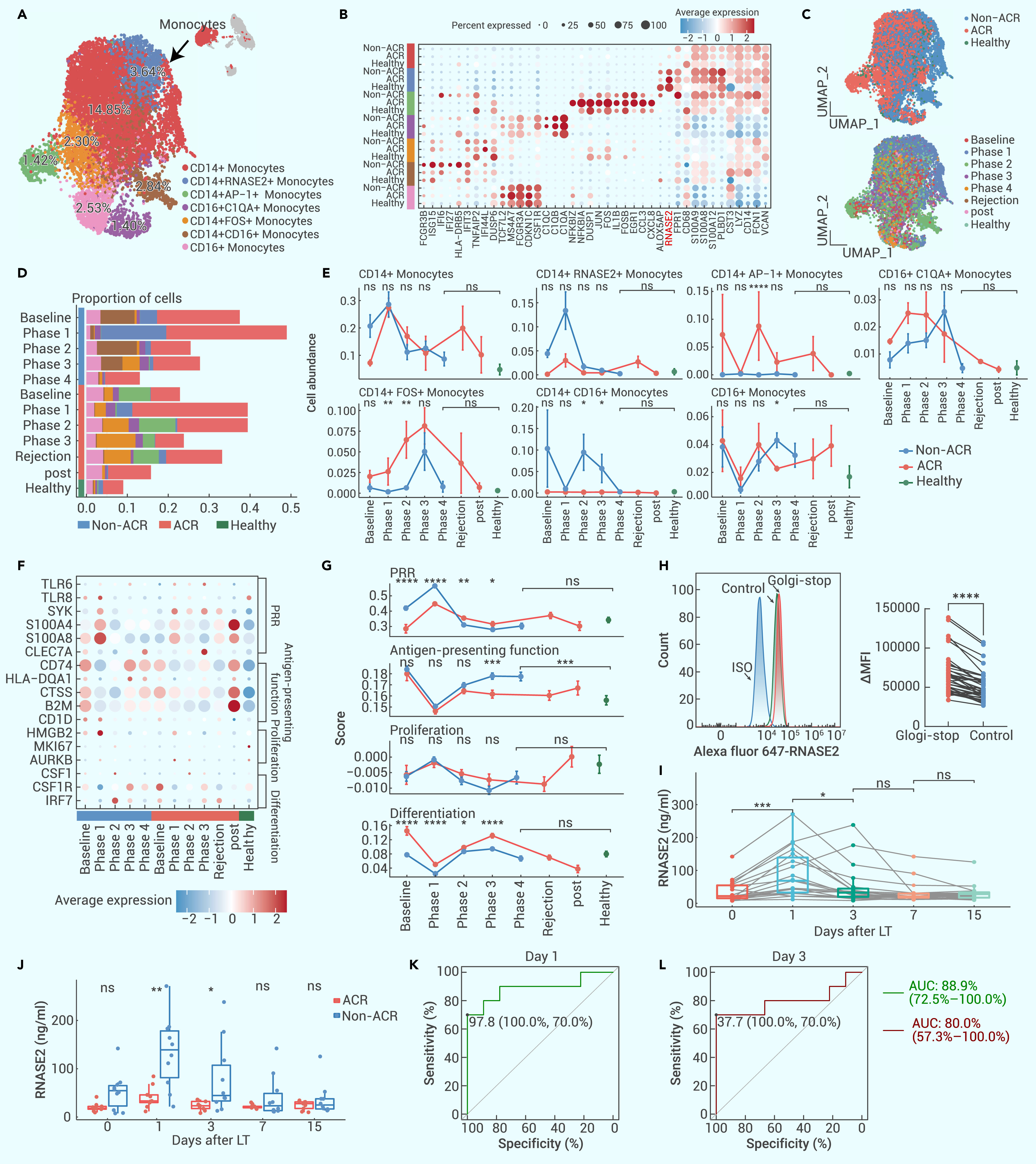
Figure 6.
Dynamics of monocytic subpopulations across LT
-
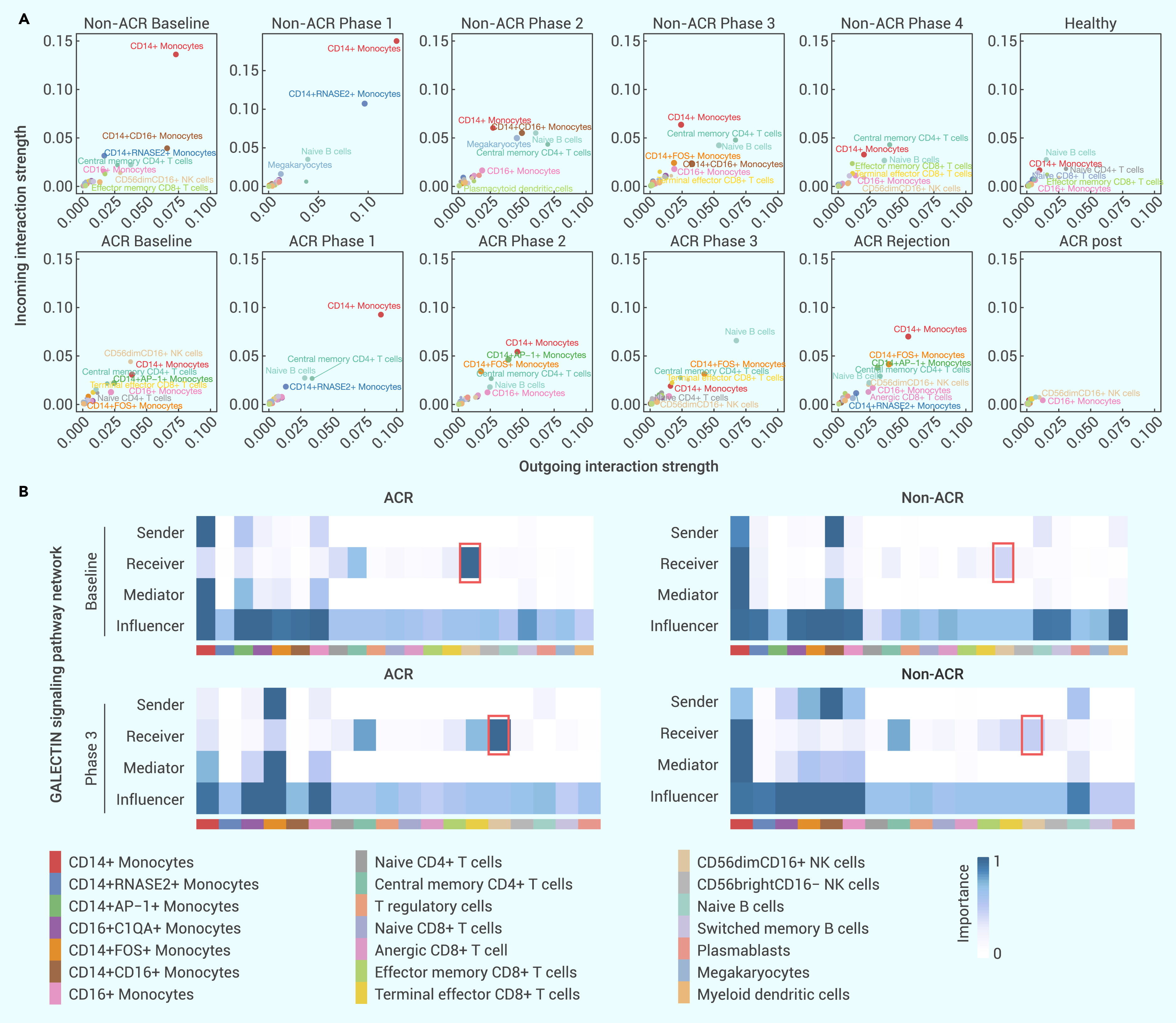
Figure 7.
Multicellular interaction analysis after LT
-
Figure 8.
A schematic illustration of a four-phase framework that aids in the clinical management of LT patients
