
Single-cell analysis reveals conserved regulons shaping muscle stem cell behavior during development and aging in mammals
-
GRAPHICAL ABSTRACT

 DownLoad:
Full size image
DownLoad:
Full size image
-
PUBLIC SUMMARY
-
A conserved developmental coordinate between human and mouse MuSCs sheds light on their shared biology.
Conserved regulons enhance the understanding of MuSC behavior during development and aging.
USF2 is a pivotal conserved regulon essential for muscle cell myogenesis and aging.
-
-
ABSTRACT
Muscle stem cells (MuSCs) play a pivotal role in skeletal muscle development, regeneration, and maintenance. Previous studies have extensively investigated the transcriptional changes in MuSCs during various developmental stages in mammals using single-cell RNA sequencing. However, a gap remains in cross-species integrative research examining the factors influencing MuSC activity during development and aging. To address this gap, we constructed a conserved single-cell landscape of MuSCs in mammals, encompassing their developmental and aging stages. Our findings unveil a conserved developmental coordinate between human and mouse MuSCs, shedding light on their shared biology. The single-cell coordinated gene association in pattern sets (scCoGAPS) algorithm and dynamic time warping algorithm were used to investigate the temporal dynamics of skeletal muscle regeneration related gene patterns. Additionally, we scrutinized the conservation of regulons, delineating groups of genes under the influence of common transcription factors, particularly emphasizing the identification of pivotal factors governing MuSC behavior during aging. Notably, we identified USF2, a conserved regulon, as a key regulator influencing muscle cell myogenesis and aging. This research provides critical insights into the conserved aspects that influence MuSC behavior and highlights the significance of USF2 in MuSC regulation. By unraveling the intricate mechanisms underlying MuSC development and aging, our study opens avenues for advancements in regenerative medicine and muscle-related therapeutics. -

-
REFERENCES
[1] Brack, A.S. and Rando, T.A. (2012). Tissue-specific stem cells: Lessons from the skeletal muscle satellite cell. Cell Stem Cell 10: 504−514. DOI: 10.1016/j.stem.2012.04.001. [2] Murphy, M. and Kardon, G. (2011). Origin of vertebrate limb muscle: The role of progenitor and myoblast populations. Curr. Top Dev. Biol. 96: 1−32. DOI: 10.1016/B978-0-12-385940-2.00001-2. [3] Hong, X.T., Campanario, S., Ramírez-Pardo, I., et al. (2022). Stem cell aging in the skeletal muscle: The importance of communication. Ageing Res. Rev. 73: 101528. DOI: 10.1016/j.arr.2021.101528. [4] Collins, C.A., Olsen, I., Zammit, P.S., et al. (2005). Stem cell function, self-renewal, and behavioral heterogeneity of cells from the adult muscle satellite cell niche. Cell 122: 289−301. DOI: 10.1016/j.cell.2005.05.010. [5] Tierney, M.T., and Sacco, A. (2016). Satellite cell heterogeneity in skeletal muscle homeostasis. Trends Cell Biol. 26: 434−444. DOI: 10.1016/j.tcb.2016.02.004. [6] Sacco, A., Doyonnas, R., Kraft, P., et al. (2008). Self-renewal and expansion of single transplanted muscle stem cells. Nature 456: 502−506. DOI: 10.1038/nature07384. [7] Tierney, M.T., Gromova, A., Sesillo, F.B., et al. (2016). Autonomous extracellular matrix remodeling controls a progressive adaptation in muscle stem cell regenerative capacity during development. Cell Rep. 14: 1940−1952. DOI: 10.1016/j.celrep.2016.01.072. [8] Cohen, S., Nathan, J.A., and Goldberg, A.L. (2015). Muscle wasting in disease: molecular mechanisms and promising therapies. Nat. Rev. Drug Discov. 14: 58−74. DOI: 10.1038/nrd4467. [9] Gopinath, S.D. and Rando, T.A. (2008). Stem cell review series: Aging of the skeletal muscle stem cell niche. Aging Cell. 7: 590−598. DOI: 10.1111/j.1474-9726.2008.00399.x. [10] De Micheli, A.J., Spector, J.A., Elemento, O., et al. (2020). A reference single-cell transcriptomic atlas of human skeletal muscle tissue reveals bifurcated muscle stem cell populations. Skelet. Muscle 10: 19. DOI: 10.1186/s13395-020-00236-3. [11] Rubenstein, A.B., Smith, G.R., Raue, U., et al. (2020). Single-cell transcriptional profiles in human skeletal muscle. Sci. Rep-UK 10: 229. DOI: 10.1038/s41598-019-57110-6. [12] Giordani, L., He, G.J., Negroni, E., et al. (2019). High-dimensional single-cell cartography reveals novel skeletal muscle-resident cell populations. Mol. Cell 74: 609−621. DOI: 10.1016/j.molcel.2019.02.026. [13] Xi, H.B., Langerman, J., Sabri, S., et al. (2020). A human skeletal muscle atlas identifies the trajectories of stem and progenitor cells across development and from human pluripotent stem cells. Cell Stem Cell 27: 181−185. DOI: 10.1016/j.stem.2020.06.006. [14] Li, H., Chen, Q., Li, C.Y., et al. (2019). Muscle-secreted granulocyte colony-stimulating factor functions as metabolic niche factor ameliorating loss of muscle stem cells in aged mice. Embo J. 38: e102154. DOI: 10.15252/embj.2019102154. [15] Xue, Z.G., Huang, K., Cai, C.C., et al. (2013). Genetic programs in human and mouse early embryos revealed by single-cell RNA sequencing. Nature 500: 593−597. DOI: 10.1038/nature12364. [16] Yan, L.Y., Yang, M.Y., Guo, H.S., et al. (2013). Single-cell RNA-Seq profiling of human preimplantation embryos and embryonic stem cells. Nat. Struct. Mol. Biol. 20: 1131−1139. DOI: 10.1038/nsmb.2660. [17] Nakamura, T., Okamoto, I., Sasaki, K., et al. (2016). A developmental coordinate of pluripotency among mice, monkeys and humans. Nature 537: 57−62. DOI: 10.1038/nature19096. [18] Gautam, P., Hamashima, K., Chen, Y., et al. (2021). Multi-species single-cell transcriptomic analysis of ocular compartment regulons. Nat. Commun. 12: 5675. DOI: 10.1038/s41467-021-25968-8. [19] Liu, T., Li, J., Yu, L., et al. (2021). Cross-species single-cell transcriptomic analysis reveals pre-gastrulation developmental differences among pigs, monkeys, and humans. Cell Discov. 7: 8. DOI: 10.1038/s41421-020-00238-x. [20] Stuart, T., Butler, A., Hoffman, P., et al. (2019). Comprehensive integration of single-cell data. Cell 177: 1888−1902. DOI: 10.1016/j.cell.2019.05.031. [21] Butler, A., Hoffman, P., Smibert, P., et al. (2018). Integrating single-cell transcriptomic data across different conditions, technologies, and species. Nat. Biotechnol. 36: 411−420. DOI: 10.1038/nbt.4096. [22] Tirosh, I., Izar, B., Prakadan, S.M., et al. (2016). Dissecting the multicellular ecosystem of metastatic melanoma by single-cell RNA-seq. Science 352: 189−196. DOI: 10.1126/science.aad0501. [23] Stein-O'Brien, G.L., Clark, B.S., Sherman, T., et al. (2019). Decomposing cell identity for transfer learning across cellular measurements, platforms, tissues, and species. Cell Syst. 8: 395−411. DOI: 10.1016/j.cels.2019.04.004. [24] Han, X.P., Zhou, Z.M., Fei, L.J., et al. (2020). Construction of a human cell landscape at single-cell level. Nature 581: 303−309. DOI: 10.1038/s41586-020-2157-4. [25] Bass, J.I.F., Diallo, A., Nelson, J., et al. (2013). Using networks to measure similarity between genes: Association index selection. Nat. Methods 10: 1169−1176. DOI: 10.1038/Nmeth.2728. [26] Wolf, F.A., Hamey, F.K., Plass, M., et al. (2019). PAGA: Graph abstraction reconciles clustering with trajectory inference through a topology preserving map of single cells. Genome Biol. 20: 59. DOI: 10.1186/s13059-019-1663-x. [27] Davis, M.P.A., van Dongen, S., Abreu-Goodger, C., et al. (2013). Kraken: A set of tools for quality control and analysis of high-throughput sequence data. Methods 63: 41−49. DOI: 10.1016/j.ymeth.2013.06.027. [28] Bolger, A.M., Lohse, M., and Usadel, B. (2014). Trimmomatic: A flexible trimmer for Illumina sequence data. Bioinformatics 30: 2114−2120. DOI: 10.1093/bioinformatics/btu170. [29] Kim, D., Landmead, B., and Salzberg, S.L. (2015). HISAT: A fast spliced aligner with low memory requirements. Nat. Methods 12: 357−360. DOI: 10.1038/Nmeth.3317. [30] Liao, Y., Smyth, G.K., and Shi, W. (2019). The R package is easier, faster, cheaper and better for alignment and quantification of RNA sequencing reads. Nucleic Acids Res. 47: e47. DOI: 10.1093/nar/gkz114. [31] Love, M.I., Huber, W., and Anders, S. (2014). Moderated estimation of fold change and dispersion for RNA-seq data with DESeq2. Genome Biol. 15: 550. DOI: 10.1186/s13059-014-0550-8. [32] Ge, Y.J., Sun, Y.T., and Chen, J. (2011). IGF-II is regulated by microRNA-125b in skeletal myogenesis. J. Cell Biol. 192: 69−81. DOI: 10.1083/jcb.201007165. [33] Zhang, J., Ying, Z.Z., Tang, Z.L., et al. (2012). MicroRNA-148a promotes myogenic differentiation by targeting the ROCK1 gene. J. Biol. Chem. 287: 21093−21101. DOI: 10.1074/jbc.M111.330381. [34] Cao, J., Spielmann, M., Qiu, X., et al. (2019). The single-cell transcriptional landscape of mammalian organogenesis. Nature 566: 496−502. DOI: 10.1038/s41586-019-0969-x. [35] Andersen, D.C., Laborda, J., Baladron, V., et al. (2013). Dual role of delta-like 1 homolog (DLK1) in skeletal muscle development and adult muscle regeneration. Development 140: 3743−3753. DOI: 10.1242/dev.095810. [36] Deák, F., Mátés, L., Korpos, E., et al. (2014). Extracellular deposition of matrilin-2 controls the timing of the myogenic program during muscle regeneration. J. Cell Sci. 127: 3240−3256. DOI: 10.1242/jcs.141556. [37] L'Honoré, A., Ouimette, J.F., Lavertu-Jolin, M., et al. (2010). Pitx2 defines alternate pathways acting through MyoD during limb and somitic myogenesis. Development 137: 3847−3856. DOI: 10.1242/dev.053421. [38] Jafarnejad, S.M., Ardekani, G.S., Ghaffari, M., et al. (2013). Pleiotropic function of SRY-related HMG box transcription factor 4 in regulation of tumorigenesis. Cell Mol. Life Sci. 70: 2677−2696. DOI: 10.1007/s00018-012-1187-y. [39] Chao, W., and D'Amore, P.A. (2008). IGF2: Epigenetic regulation and role in development and disease. Cytokine Growth Factor Rev. 19: 111−120. DOI: 10.1016/j.cytogfr.2008.01.005. [40] Karakaslar, E.O., Katiyar, N., Hasham, M., et al. (2023). Transcriptional activation of Jun and Fos members of the AP-1 complex is a conserved signature of immune aging that contributes to inflammaging. Aging Cell 22: e13792. DOI: 10.1111/acel.13792. [41] D'Angelo, M.A., Raices, M., Panowski, S.H., et al. (2009). Age-dependent deterioration of nuclear pore complexes causes a loss of nuclear integrity in postmitotic cells. Cell 136: 284−295. DOI: 10.1016/j.cell.2008.11.037. [42] Chen, X.N., He, L.Q., Zhao, Y., et al. (2017). Malat1 regulates myogenic differentiation and muscle regeneration through modulating MyoD transcriptional activity. Cell Discov. 17002. DOI: 10.1038/celldisc.2017.2. [43] Gerald, D., Berra, E., Frapart, Y.M., et al. (2004). JunD reduces tumor angiogenesis by protecting cells from oxidative stress. Cell 118: 781−794. DOI: 10.1016/j.cell.2004.08.025. [44] Gregorie, C.J., Wiesen, J.L., Magner, W.J., et al. (2009). Restoration of immune response gene induction in trophoblast tumor cells associated with cellular senescence. J. Reprod. Immunol. 81: 25−33. DOI: 10.1016/j.jri.2009.02.009. [45] Zhang, H., Zhang, Y., Zhou, X.Y., et al. (2020). Functional interrogation of HOXA9 regulome in MLLr leukemia via reporter-based CRISPR/Cas9 screen. eLife 9: e57858. DOI: 10.7554/eLife.57858. [46] De Micheli, A.J., Laurilliard, E.J., Heinke, C.L., et al. (2020). Single-cell analysis of the muscle stem cell hierarchy identifies heterotypic communication signals involved in skeletal muscle regeneration. Cell Rep. 30: 3583−3595. DOI: 10.1016/j.celrep.2020.02.067. [47] Dell'Orso, S., Juan, A.H., Ko, K.D., et al. (2019). Single cell analysis of adult mouse skeletal muscle stem cells in homeostatic and regenerative conditions. Development 146 : dev174177. DOI: 10.1242/dev.174177. [48] Schaum, N., Karkanias, J., Neff, N.F., et al. (2018). Single-cell transcriptomics of 20 mouse organs creates a Tabula Muris. Nature 562: 367−372. DOI: 10.1038/s41586-018-0590-4. [49] Rydell-Törmänen, K., and Johnson, J.R. (2019). The applicability of mouse models to the study of human disease. Methods Mol. Biol. 1940: 3−22. DOI: 10.1007/978-1-4939-9086-3_1. [50] Brehm, M.A., Shultz, L.D., and Greiner, D.L. (2010). Humanized mouse models to study human diseases. Curr. Opin. Endocrinol. 17: 120−125. DOI: 10.1097/MED.0b013e328337282f. [51] Ganeshan, K. and Chawla, A. (2017). Warming the mouse to model human diseases. Nat. Rev. Endocrinol. 13: 458−465. DOI: 10.1038/nrendo.2017.48. [52] Perlman, R.L. (2016). Mouse models of human disease: An evolutionary perspective. Evol. Med. Public Hlth. 2016: 170−176. DOI: 10.1093/emph/eow014. [53] Goldman, J.A. and Poss, K.D. (2020). Gene regulatory programmes of tissue regeneration. Nat. Rev. Genet. 21: 511−525. DOI: 10.1038/s41576-020-0239-7. [54] Malecova, B., Gatto, S., Etxaniz, U., et al. (2018). Dynamics of cellular states of fibro-adipogenic progenitors during myogenesis and muscular dystrophy. Nat. Commun. 9 : 3670. DOI: 10.1038/s41467-018-06068-6. [55] Wang, X., Allen, W.E., Wright, M.A., et al. (2018). Three-dimensional intact-tissue sequencing of single-cell transcriptional states. Science 361 : eaat5691. DOI: 10.1126/science.aat5691. [56] Luo, X., and Sawadogo, M. (1996). Antiproliferative properties of the USF family of helix-loop-helix transcription factors. Proc. Natl. Acad. Sci. USA 93: 1308−1313. DOI. DOI: 10.1073/pnas.93.3.1308. [57] Barruet, E., Garcia, S.M., Striedinger, K., et al. (2020). Functionally heterogeneous human satellite cells identified by single cell RNA sequencing. eLife 9: e51576. DOI: 5157610.7554/eLife.51576. DOI: 10.7554/eLife.51576. [58] Sato, A.Y.S., Antonioli, E., Tambellini, R., et al. (2011). ID1 inhibits USF2 and blocks TGF-β-induced apoptosis in mesangial cells. Am. J. Physiol-Renal. 301: F1260−F1269. DOI: 10.1152/ajprenal.00128.2011. [59] Chi, T.F., Khoder-Agha, F., Mennerich, D., et al. (2020). Loss of USF2 promotes proliferation, migration and mitophagy in a redox-dependent manner. Redox Biol. 37: 101750. DOI: 10.1016/j.redox.2020.101750. [60] Corre, S. and Galibert, M.D. (2005). Upstream stimulating factors: Highly versatile stress-responsive transcription factors. Pigm. Cell Res. 18: 337−348. DOI: 10.1111/j.1600-0749.2005.00262.x. -
ABOUT THIS ARTICLE
Cite this article:
Wang Z., Wang W., Li W., et al., (2024). Single-cell analysis reveals conserved regulons shaping muscle stem cell behavior during development and aging in mammals. The Innovation Life 2(2): 100075. https://doi.org/10.59717/j.xinn-life.2024.100075
Welcome!
To request copyright permission to republish or share portions of our works, please visit Copyright Clearance Center's (CCC) Marketplace website at marketplace.copyright.com.

Supplementary Information
-
download_supplementary
Table(S1-S6)
Article Metrics
Relative Articles
Cited by
Export File
Citation
Wang Z., Wang W., Li W., et al., (2024). Single-cell analysis reveals conserved regulons shaping muscle stem cell behavior during development and aging in mammals. The Innovation Life 2(2): 100075. https://doi.org/10.59717/j.xinn-life.2024.100075

Format
Content


-
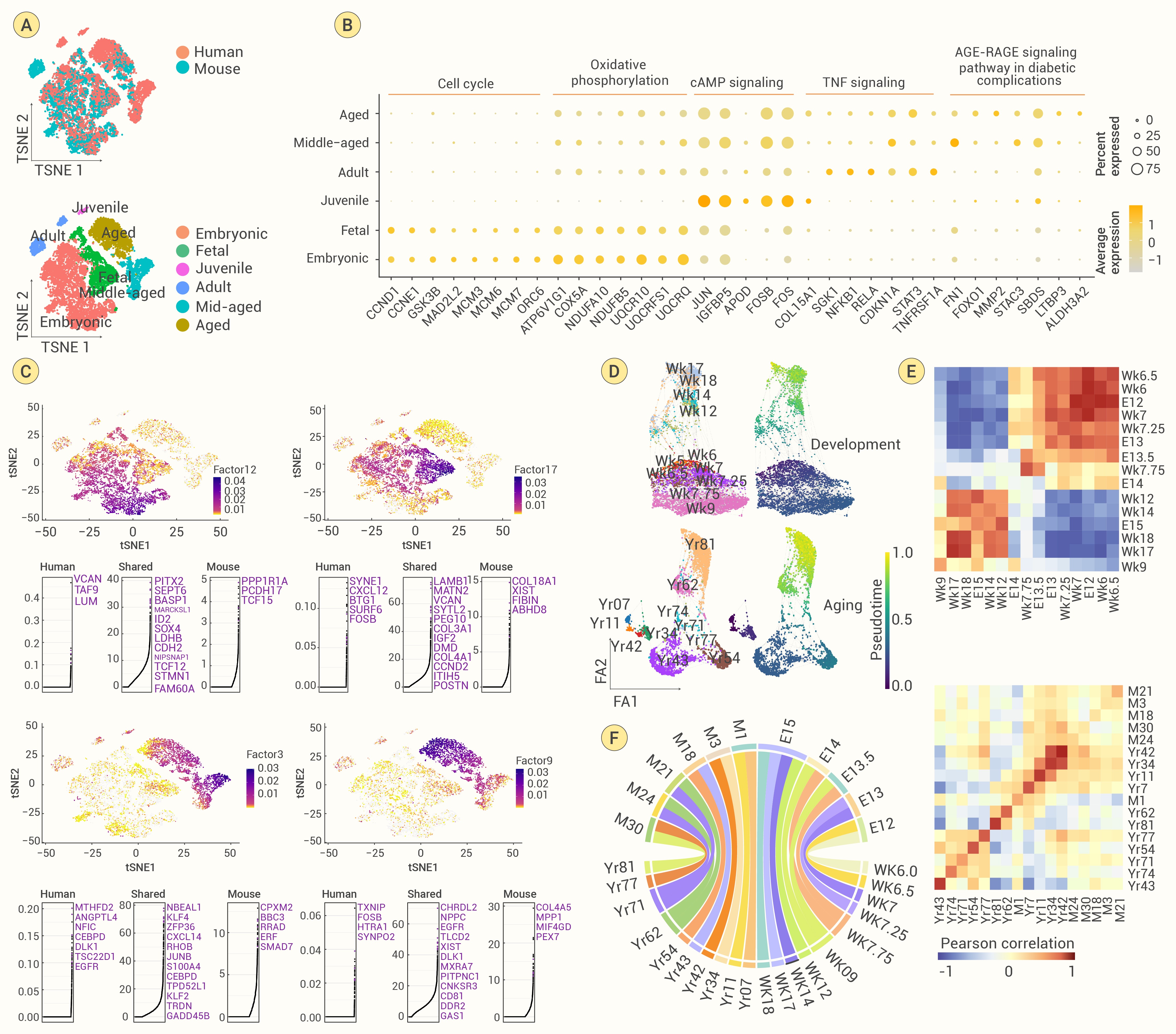
Figure 1.
Identification and comparative analysis of MuSCs in the human and murine skeletal muscle
-
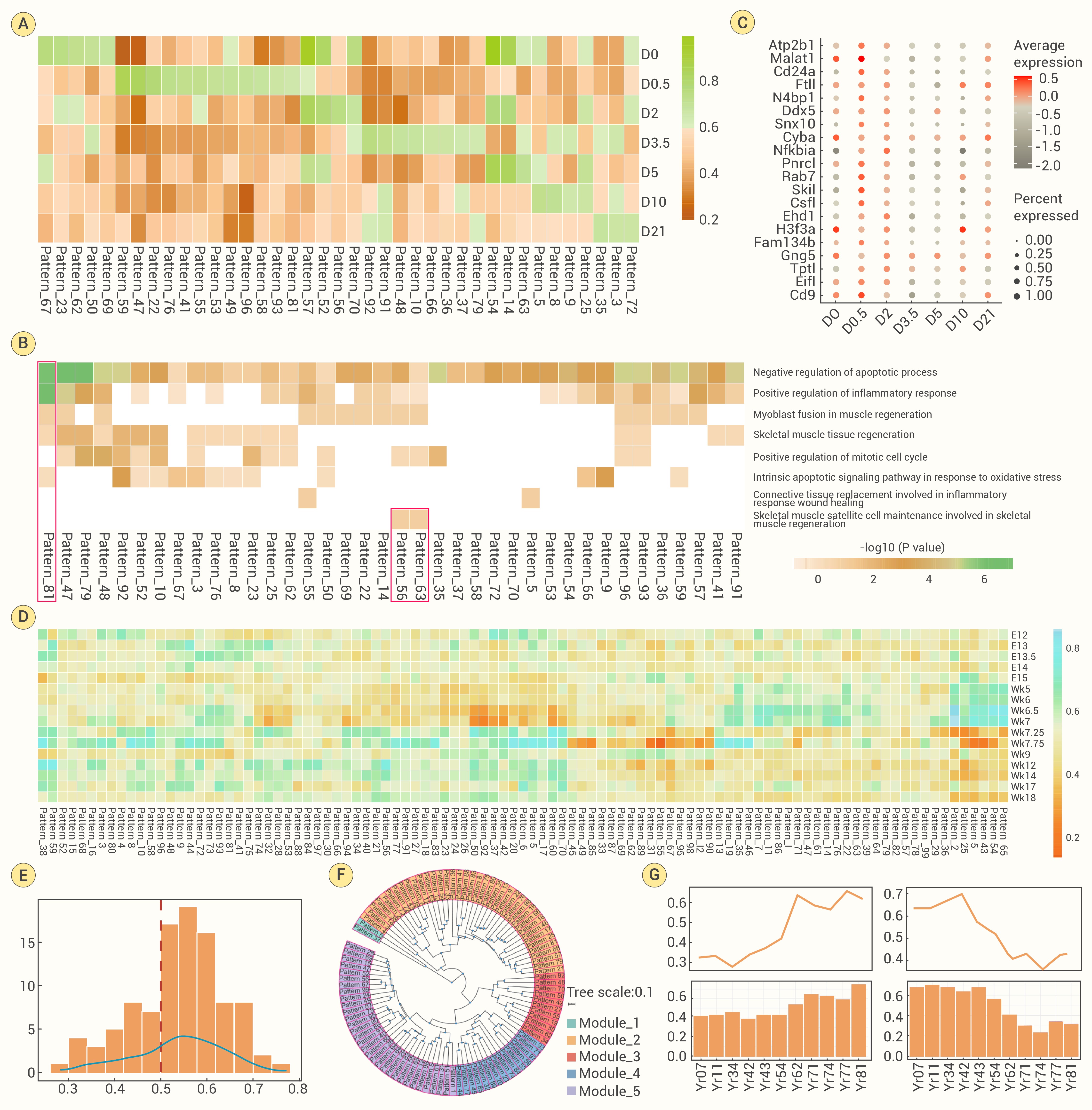
Figure 2.
Comparison analysis of MuSCs during development and aging in human and mouse
-
Figure 3.
Meta-analysis of MuSCs under regeneration in mammals
-
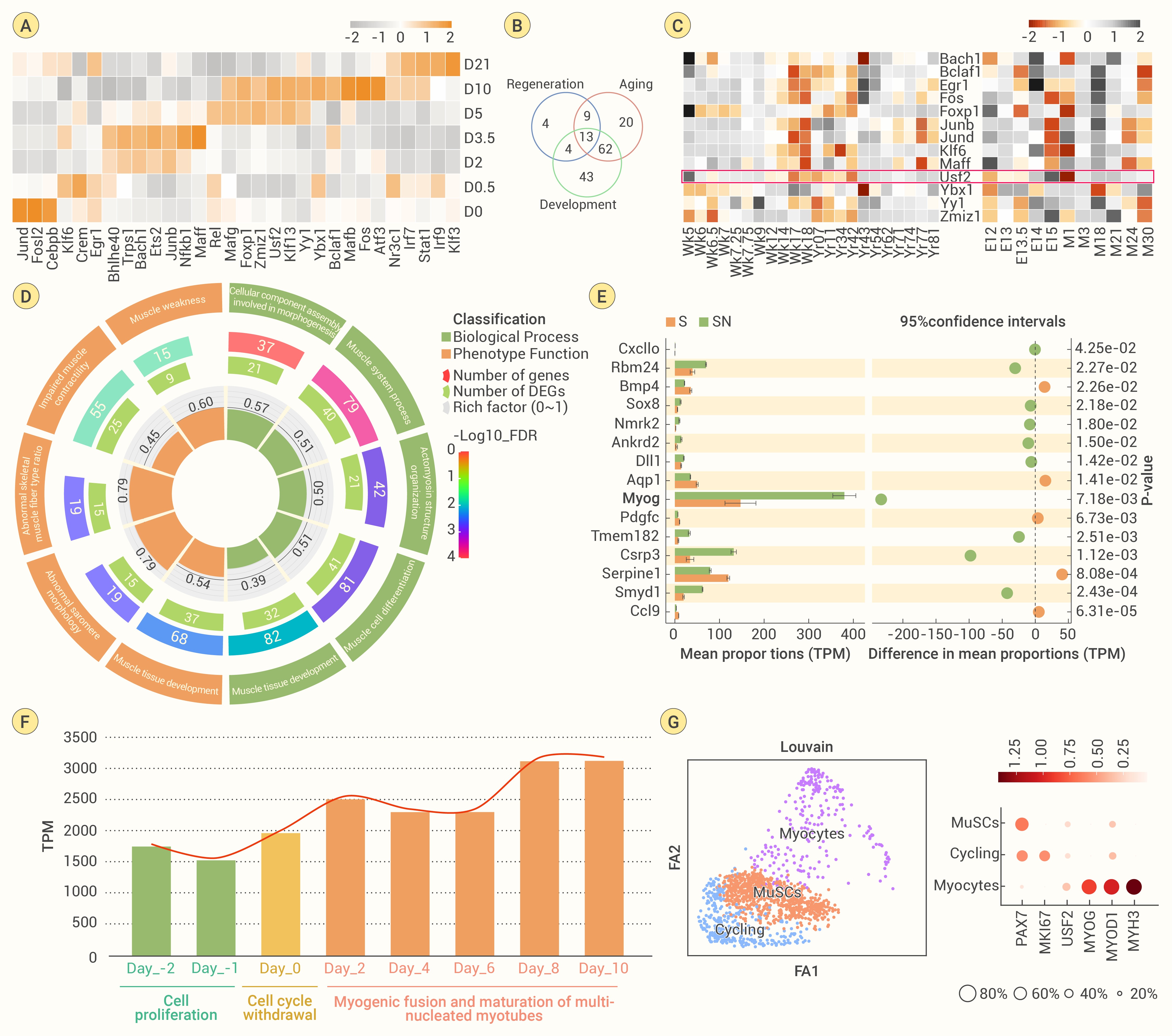
Figure 4.
Comparison of aging-related regulons in human and mouse
-
Figure 5.
USF2 participates in myogenesis in mammals
-
Figure 6.
USF2 enhances muscle regeneration and protects mouse myotubes from senescence by promoting myoblast differentiation
