
Electrodermal activity and its molecular mechanisms: Unraveling insights into skin diseases
-
GRAPHICAL ABSTRACT

 DownLoad:
Full size image
DownLoad:
Full size image
-
PUBLIC SUMMARY
-
Electrodermal activity (EDA) is increasingly vital due to non-invasive diagnostics, and real-time health monitoring.
Enhanced technology enables monitoring signals from inflammation, fibrosis, and sweat gland disorders.
Bioimpedance and machine learning integration significantly improve EDA reliability.
-
-
ABSTRACT
Electrodermal activity (EDA) refers to the changes in electrical potential recorded on the skin surface, which mainly reflect the electrical properties of the skin and sympathetic nerve activity reflected by sweat secretion. Various dermatoses impair the skin barrier and alter the function of innervated nerves, resulting in significant fluctuations in EDA. This review aims to provide a comprehensive overview of the molecular mechanisms underlying representative skin symptoms related to inflammation, fibrosis, and sweat gland disorders, and to explore the correlation of these mechanisms with EDA components. The physiological significance of EDA is discussed to provide a new perspective for the clinical application of EDA. -

-
REFERENCES
[1] Banganho, A., Santos, M., and da Silva, H.P. (2022). Electrodermal activity: Fundamental principles, measurement, and application. IEEE Potentials 41: 35−43. DOI: 10.1109/MPOT.2020.2983381. [2] Abe, Y. and Nishizawa, M. (2021). Electrical aspects of skin as a pathway to engineering skin devices. APL Bioeng 5: 041509. DOI: 10.1063/5.0064529. [3] Tronstad, C., Amini, M., Bach, D.R., et al. (2022). Current trends and opportunities in the methodology of electrodermal activity measurement. Physiol. Meas. 43: 02TR01. DOI: 10.1088/1361-6579/ac5007. [4] Edelberg, R. (1993). Electrodermal mechanisms: A critique of the two-effector hypothesis and a proposed replacement. Roy, J.-C., Boucsein, W., Fowles, D.C., et al. (ed). Progress in electrodermal research (Springer US), pp: 7-29. [5] Vetrugno, R., Liguori, R., Cortelli, P., et al. (2003). Sympathetic skin response: Basic mechanisms and clinical applications. Clin. Auton. Res. 13: 256−270. DOI: 10.1007/s10286-003-0107-5. [6] Tagami, H. (2014). Electrical measurement of the hydration state of the skin surface in vivo. Br. J. Dermatol. 171 Suppl 3 : 29-33. DOI: 10.1111/bjd.13245. [7] Calero, J.A.M., Páez-Montoro, A., López-Ongil, C., et al. (2023). Self-adjustable galvanic skin response sensor for physiological monitoring. IEEE Sens. J. 23: 3005−3019. DOI: 10.1109/JSEN.2022.3233439. [8] Posada-Quintero, H.F. and Chon, K.H. (2020). Innovations in electrodermal activity data collection and signal processing: A systematic review. Sensors 20: 479. DOI: 10.3390/s20020479. [9] Boucsein, W., Fowles, D.C., Grimnes, S., et al. (2012). Publication recommendations for electrodermal measurements. Psychophysiology 49: 1017−1034. DOI: 10.1111/j.1469-8986.2012.01384.x. [10] Pabst, O., Tronstad, C., Grimnes, S., et al. (2017). Comparison between the ac and dc measurement of electrodermal activity. Psychophysiology 54: 374−385. DOI: 10.1111/psyp.12803. [11] Sim, D.J.K., Kim, S.M., Kim, S.S., et al. (2019). Portable skin analyzers with simultaneous measurements of transepidermal water loss, skin conductance and skin hardness. Sensors 19: 3857. DOI: 10.3390/s19183857. [12] Jang, H., Sel, K., Kim, E., et al. (2022). Graphene e-tattoos for unobstructive ambulatory electrodermal activity sensing on the palm enabled by heterogeneous serpentine ribbons. Nat. Commun. 13: 6604. DOI: 10.1038/s41467-022-34406-2. [13] Nittala, A.S., Karrenbauer, A., Khan, A., et al. (2021). Computational design and optimization of electro-physiological sensors. Nat. Commun. 12: 6351. DOI: 10.1038/s41467-021-26442-1. [14] Kim, S., Lee, B., Reeder, J.T., et al. (2020). Soft, skin-interfaced microfluidic systems with integrated immunoassays, fluorometric sensors, and impedance measurement capabilities. Proc. Natl. Acad. Sci. USA. 117 : 27906-27915. DOI: 10.1073/pnas.2012700117. [15] Ray, P.P., Dash, D., and De, D. (2019). Analysis and monitoring of iot-assisted human physiological galvanic skin responsefactor for smart e-healthcare. Sensor Review 39: 525−541. DOI: 10.1108/SR-07-2018-0181. [16] Wruhs, M., Gleiß, A., Steiner, A., et al. (2017). Quantity and quality of sweating in atopic dermatitis. Arch. Dermatol. Res. 309: 787−793. DOI: 10.1007/s00403-017-1770-z. [17] Tronstad, C., Kalvøy, H., Grimnes, S., et al. (2013). Waveform difference between skin conductance and skin potential responses in relation to electrical and evaporative properties of skin. Psychophysiology 50 : 1070-1078. DOI: 10.1111/psyp.12092. [18] Kim, J., Ku, B., Bae, J.H., et al. (2018). Contrast in the circadian behaviors of an electrodermal activity and bioimpedance spectroscopy. Chronobiol. Int. 35: 1413−1422. DOI: 10.1080/07420528.2018.1486852. [19] Tekatas, A., Koca, S.S., Tekatas, D.D., et al. (2014). R-r interval variation and sympathetic skin response in systemic lupus erythematosus. Clin. Rheumatol. 33: 65−70. DOI: 10.1007/s10067-013-2391-3. [20] Badry, R., Gamal, R.M., Hassanien, M.M., et al. (2018). Sympathetic skin response in patients with systemic sclerosis and rheumatoid arthritis. Egypt. J. Neurol. Psychiat. Neurosurg. 54: 38. DOI: 10.1186/s41983-018-0044-9. [21] Emad, M.R., Farpour, H.R., Ahmed, F., et al. (2022). Is there any sympathetic skin response abnormality in raynaud phenomenon. Sultan Qaboos Univ. Med. J. 22: 274−279. DOI: 10.18295/squmj.4.2021.066. [22] Memis, Z., Çevik, N., Acar, H., et al. (2022). The first electrophysiological abnormality in new-onset DM: Autonomic tests. Noro. Psikiyatr. Ars. 59 : 197-200. DOI: 10.29399/npa.27952. [23] Margaritella, N., Mendozzi, L., Garegnani, M., et al. (2018). Sympathetic skin response in multiple sclerosis: A meta-analysis of case-control studies. Neurol. Sci. 39: 45−52. DOI: 10.1007/s10072-017-3111-6. [24] Halıgür, B.D., Cicek, D., Bulut, S., et al. (2012). The investigation of autonomic functions in patients with psoriasis. Int. J. Dermatol. 51: 557−563. DOI: 10.1111/j.1365-4632.2011.05111.x. [25] Ghandali, E., Hosseini, S.M., Moghimi, H.R., et al. (2020). Intra tester reliability of sympathetic skin responses in subjects with primary palmar hyperhidrosis. J. Bodyw. Mov. Ther. 24: 57−62. DOI: 10.1016/j.jbmt.2020.02.024. [26] Altunrende, B., Yildiz, S., Kandi, B., et al. (2013). Sympathetic skin responses from the scalp evoked by electrical stimulation in seborrheic dermatitis. J. Dermatol. 40: 458−462. DOI: 10.1111/1346-8138.12114. [27] Ulvi, H., Yoldaş, T., Yiğiter, R., et al. (2003). R-r interval variation and the sympathetic skin response in the assessment of the autonomic nervous system in leprosy patients. Acta Neurol. Scand 107: 42−49. DOI: 10.1034/j.1600-0404.2003.01307.x. [28] Cabalar, M., Yayla, V., Ulutas, S., et al. (2014). The clinical & neurophysiological study of leprosy. Pak J Med Sci 30: 501−506. DOI: 10.12669/pjms.303.5354. [29] Khan, A., Kamran, S., Ponirakis, G., et al. (2018). Peripheral neuropathy in patients with multiple sclerosis. PLoS One 13: e0193270. DOI: 10.1371/journal.pone.0193270. [30] Dolu, N., Ferahbaş, A., Özesmi, Ç., et al. (2005). Effect of puva therapy on electrodermal activity parameters in vitiligo patients. Auton. Neurosci. 118: 102−107. DOI: 10.1016/j.autneu.2004.12.005. [31] Eckert, R.L. and Rorke, E.A. (1989). Molecular biology of keratinocyte differentiation. Environ. Health Perspect. 80: 109−116. DOI: 10.1289/ehp.8980109. [32] Vičić, M., Kaštelan, M., Brajac, I., et al. (2021). Current concepts of psoriasis immunopathogenesis. Int. J. Mol. Sci. 22: 11574. DOI: 10.3390/ijms222111574. [33] Furue, M., Furue, K., Tsuji, G., et al. (2020). Interleukin-17a and keratinocytes in psoriasis. Int. J. Mol. Sci. 21: 1275. DOI: 10.3390/ijms21041275. [34] Zhou, X., Chen, Y., Cui, L., et al. (2022). Advances in the pathogenesis of psoriasis: From keratinocyte perspective. Cell Death Dis. 13: 81. DOI: 10.1038/s41419-022-04523-3. [35] Guo, Y., Luo, L., Zhu, J., et al. (2023). Multi-omics research strategies for psoriasis and atopic dermatitis. Int. J. Mol. Sci. 24: 8018. DOI: 10.3390/ijms24098018. [36] Chiricozzi, A., Romanelli, P., Volpe, E., et al. (2018). Scanning the immunopathogenesis of psoriasis. Int. J. Mol. Sci. 19: 179. DOI: 10.3390/ijms19010179. [37] Sabat, R., Wolk, K., Loyal, L., et al. (2019). T cell pathology in skin inflammation. Semin. Immunopathol. 41: 359−377. DOI: 10.1007/s00281-019-00742-7. [38] Kim, J., Kim, B.E., and Leung, D.Y.M. (2019). Pathophysiology of atopic dermatitis: Clinical implications. Allergy Asthma Proc. 40: 84−92. DOI: 10.2500/aap.2019.40.4202. [39] Sroka-Tomaszewska, J. and Trzeciak, M. (2021). Molecular mechanisms of atopic dermatitis pathogenesis. Int. J. Mol. Sci. 22: 4130. DOI: 10.3390/ijms22084130. [40] Koivisto, A.P., Belvisi, M.G., Gaudet, R., et al. (2022). Advances in TRP channel drug discovery: From target validation to clinical studies. Nat. Rev. Drug Discov. 21: 41−59. DOI: 10.1038/s41573-021-00268-4. [41] Zhang, M., Ma, Y., Ye, X., et al. (2023). TRP (transient receptor potential) ion channel family: Structures, biological functions and therapeutic interventions for diseases. Signal Transduct Target Ther. 8: 261. DOI: 10.1038/s41392-023-01464-x. [42] Slominski, A.T., Zmijewski, M.A., Zbytek, B., et al. (2013). Key role of crf in the skin stress response system. Endocr. Rev. 34: 827−884. DOI: 10.1210/er.2012-1092. [43] Rashighi, M. and Harris, J.E. (2017). Vitiligo pathogenesis and emerging treatments. Dermatol. Clin. 35: 257−265. DOI: 10.1016/j.det.2016.11.014. [44] McGeachy, M.J., Cua, D.J., and Gaffen, S.L. (2019). The IL-17 family of cytokines in health and disease. Immunity 50: 892−906. DOI: 10.1016/j.immuni.2019.03.021. [45] Li, X., Bechara, R., Zhao, J., et al. (2019). IL-17 receptor-based signaling and implications for disease. Nat. Immunol. 20: 1594−1602. DOI: 10.1038/s41590-019-0514-y. [46] Zenobia, C. and Hajishengallis, G. (2015). Basic biology and role of interleukin-17 in immunity and inflammation. Periodontol. 2000 69 : 142-159. DOI: 10.1111/prd.12083. [47] Zouboulis, C.C., Seltmann, H., Hiroi, N., et al. (2002). Corticotropin-releasing hormone: An autocrine hormone that promotes lipogenesis in human sebocytes. Proc. Natl. Acad. Sci. USA. 99: 7148−7153. DOI: 10.1073/pnas.102180999. [48] Zbytek, B., Pfeffer, L.M., and Slominski, A.T. (2006). CRH inhibits NF-kappa B signaling in human melanocytes. Peptides 27: 3276−3283. DOI: 10.1016/j.peptides.2006.07.017. [49] Iftinca, M., Defaye, M., and Altier, C. (2021). TRPV1-targeted drugs in development for human pain conditions. Drugs 81: 7−27. DOI: 10.1007/s40265-020-01429-2. [50] Kong, F., You, H., Zheng, K., et al. (2021). The crosstalk between pattern-recognition receptor signaling and calcium signaling. Int. J. Biol. Macromol. 192: 745−756. DOI: 10.1016/j.ijbiomac.2021.10.014. [51] Zhang, F., Yang, H., Wang, Z., et al. (2007). Transient receptor potential vanilloid 1 activation induces inflammatory cytokine release in corneal epithelium through mapk signaling. J. Cell. Physiol. 213: 730−739. DOI: 10.1002/jcp.21141. [52] Yuan, J., Liang, X., Zhou, W., et al. (2021). TRPA1 promotes cisplatin-induced nephrotoxicity through inflammation mediated by the mapk/nf-κb signaling pathway. Ann. Transl. Med. 9: 1578. DOI: 10.21037/atm-21-5125. [53] Kang, J., Ding, Y., Li, B., et al. (2017). TRPA1 mediated aggravation of allergic contact dermatitis induced by DINP and regulated by NF-κB activation. Sci. Rep. 7: 43586. DOI: 10.1038/srep43586. [54] Capobianchi, M.R., Uleri, E., Caglioti, C., et al. (2015). Type I IFN family members: Similarity, differences and interaction. Cytokine Growth Factor Rev. 26: 103−111. DOI: 10.1016/j.cytogfr.2014.10.011. [55] Conrad, C. and Gilliet, M. (2018). Psoriasis: From pathogenesis to targeted therapies. Clin. Rev. Allergy Immunol. 54: 102−113. DOI: 10.1007/s12016-018-8668-1. [56] Powell, M.D., Read, K.A., Sreekumar, B.K., et al. (2019). IL-12 signaling drives the differentiation and function of a TH1-derived TFH1-like cell population. Sci. Rep. 9: 13991. DOI: 10.1038/s41598-019-50614-1. [57] Johnson-Huang, L.M., Suárez-Fariñas, M., Pierson, K.C., et al. (2012). A single intradermal injection of IFN-γ induces an inflammatory state in both non-lesional psoriatic and healthy skin. J. Invest. Dermatol. 132: 1177−1187. DOI: 10.1038/jid.2011.458. [58] Mehta, N.N., Teague, H.L., Swindell, W.R., et al. (2017). IFN-γ and TNF-α synergism may provide a link between psoriasis and inflammatory atherogenesis. Sci. Rep. 7: 13831. DOI: 10.1038/s41598-017-14365-1. [59] Rashighi, M., Agarwal, P., Richmond, J.M., et al. (2014). CXCL10 is critical for the progression and maintenance of depigmentation in a mouse model of vitiligo. Sci. Transl. Med. 6: 223ra223. DOI: 10.1126/scitranslmed.3007811. [60] Rebane, A., Zimmermann, M., Aab, A., et al. (2012). Mechanisms of IFN-γ-induced apoptosis of human skin keratinocytes in patients with atopic dermatitis. J. Allergy Clin. Immunol. 129: 1297−1306. DOI: 10.1016/j.jaci.2012.02.020. [61] Shao, S., Tsoi, L.C., Sarkar, M.K., et al. (2019). Ifn-γ enhances cell-mediated cytotoxicity against keratinocytes via jak2/stat1 in lichen planus. Sci. Transl. Med. 11: eaav7561. DOI: 10.1126/scitranslmed.aav7561. [62] Okiyama, N. and Fujimoto, M. (2015). Clinical perspectives and murine models of lichenoid tissue reaction/interface dermatitis. J. Dermatol. Sci. 78: 167−172. DOI: 10.1016/j.jdermsci.2015.03.001. [63] Madonna, S., Scarponi, C., Pallotta, S., et al. (2012). Anti-apoptotic effects of suppressor of cytokine signaling 3 and 1 in psoriasis. Cell Death Dis. 3: e334. DOI: 10.1038/cddis.2012.69. [64] Wang, B., Han, D., Li, F., et al. (2020). Elevated IL-22 in psoriasis plays an anti-apoptotic role in keratinocytes through mediating Bcl-xl/Bax. Apoptosis 25: 663−673. DOI: 10.1007/s10495-020-01623-3. [65] Srivastava, A., Luo, L., Lohcharoenkal, W., et al. (2021). Cross-talk between IFN-γ and TWEAK through miR-149 amplifies skin inflammation in psoriasis. J. Allergy Clin. Immunol. 147: 2225−2235. DOI: 10.1016/j.jaci.2020.12.657. [66] Leone, G.M., Mangano, K., Petralia, M.C., et al. (2023). Past, present and (foreseeable) future of biological anti-TNF alpha therapy. J Clin Med 12: 1630. DOI: 10.3390/jcm12041630. [67] Monaco, C., Nanchahal, J., Taylor, P., et al. (2015). Anti-tnf therapy: Past, present and future. Int. Immunol. 27: 55−62. DOI: 10.1093/intimm/dxu102. [68] Wang, X., Cheng, D., Hu, G., et al. (2019). Tumor necrosis factor (TNF) receptor expression determines keratinocyte fate upon stimulation with TNF-like weak inducer of apoptosis. Mediators Inflamm. 2019: 2945083. DOI: 10.1155/2019/2945083. [69] Cheng, H., Xu, M., Liu, X., et al. (2016). TWEAK/Fn14 activation induces keratinocyte proliferation under psoriatic inflammation. Exp. Dermatol. 25: 32−37. DOI: 10.1111/exd.12820. [70] Peng, L., Li, Q., Wang, H., et al. (2018). Fn14 deficiency ameliorates psoriasis-like skin disease in a murine model. Cell Death Dis. 9: 801. DOI: 10.1038/s41419-018-0820-6. [71] Buechler, M.B., Pradhan, R.N., Krishnamurty, A.T., et al. (2021). Cross-tissue organization of the fibroblast lineage. Nature 593: 575−579. DOI: 10.1038/s41586-021-03549-5. [72] Lendahl, U., Muhl, L., and Betsholtz, C. (2022). Identification, discrimination and heterogeneity of fibroblasts. Nat. Commun. 13: 3409. DOI: 10.1038/s41467-022-30633-9. [73] Cai, X., Han, M., Lou, F., et al. (2023). Tenascin C+ papillary fibroblasts facilitate neuro-immune interaction in a mouse model of psoriasis. Nat. Commun. 14: 2004. DOI: 10.1038/s41467-023-37798-x. [74] Cohen, A.J., Nikbakht, N., and Uitto, J. (2023). Keloid disorder: Genetic basis, gene expression profiles, and immunological modulation of the fibrotic processes in the skin. Cold Spring Harb. Perspect. Biol. 15: a041245. DOI: 10.1101/cshperspect.a041245. [75] de Morais, J., Velosa, A.P.P., Andrade, P.C., et al. (2022). Collagen V α1 chain decrease in papillary dermis from early systemic sclerosis: A new proposal in cutaneous fibrosis molecular structure. Int. J. Mol. Sci. 23: 12654. DOI: 10.3390/ijms232012654. [76] Sawamura, S., Makino, K., Ide, M., et al. (2022). Elevated alpha 1(I) to alpha 2(I) collagen ratio in dermal fibroblasts possibly contributes to fibrosis in systemic sclerosis. Int. J. Mol. Sci. 23: 6811. DOI: 10.3390/ijms23126811. [77] Do, N.N. and Eming, S.A. (2016). Skin fibrosis: Models and mechanisms. Curr. Res. Transl. Med. 64: 185−193. DOI: 10.1016/j.retram.2016.06.003. [78] Driskell, R.R., Lichtenberger, B.M., Hoste, E., et al. (2013). Distinct fibroblast lineages determine dermal architecture in skin development and repair. Nature 504: 277−281. DOI: 10.1038/nature12783. [79] Lichtenberger, B.M., Mastrogiannaki, M., and Watt, F.M. (2016). Epidermal β-catenin activation remodels the dermis via paracrine signalling to distinct fibroblast lineages. Nat. Commun. 7: 10537. DOI: 10.1038/ncomms10537. [80] Li, B. and Wang, J.H. (2011). Fibroblasts and myofibroblasts in wound healing: Force generation and measurement. J. Tissue Viability 20: 108−120. DOI: 10.1016/j.jtv.2009.11.004. [81] Sacchetti, C., Bai, Y., Stanford, S.M., et al. (2017). PTP4A1 promotes TGFβ signaling and fibrosis in systemic sclerosis. Nat. Commun. 8: 1060. DOI: 10.1038/s41467-017-01168-1. [82] Varga, J. and Pasche, B. (2009). Transforming growth factor beta as a therapeutic target in systemic sclerosis. Nat. Rev. Rheumatol. 5: 200−206. DOI: 10.1038/nrrheum.2009.26. [83] Burgy, O. and Königshoff, M. (2018). The WNT signaling pathways in wound healing and fibrosis. Matrix Biol. 68-69: 67−80. DOI: 10.1016/j.matbio.2018.03.017. [84] Griffin, M.F., Huber, J., Evan, F.J., et al. (2022). The role of WNT signaling in skin fibrosis. Med. Res. Rev. 42: 615−628. DOI: 10.1002/med.21853. [85] Meng, X.M., Nikolic-Paterson, D.J., and Lan, H.Y. (2016). TGF-β: The master regulator of fibrosis. Nat. Rev. Nephrol. 12: 325−338. DOI: 10.1038/nrneph.2016.48. [86] Piersma, B., Bank, R.A., and Boersema, M. (2015). Signaling in fibrosis: TGF-β, WNT, and YAP/TAZ converge. Front. Med. 2: 59. DOI: 10.3389/fmed.2015.00059. [87] Cui, H.S., Lee, Y.R., Ro, Y.M., et al. (2023). Knockdown of CPEB1 and CPEB4 inhibits scar formation via modulation of TAK1 and SMAD signaling. Ann. Dermatol. 35: 293−302. DOI: 10.5021/ad.22.210. [88] Xu, Y.R. and Lei, C.Q. (2020). TAK1-TABs complex: A central signalosome in inflammatory responses. Front. Immunol. 11: 608976. DOI: 10.3389/fimmu.2020.608976. [89] Hara, A. and Tallquist, M.D. (2023). Fibroblast and immune cell cross-talk in cardiac fibrosis. Curr. Cardiol. Rep. 25: 485−493. DOI: 10.1007/s11886-023-01877-8. [90] Van Linthout, S., Miteva, K., and Tschöpe, C. (2014). Crosstalk between fibroblasts and inflammatory cells. Cardiovasc. Res. 102: 258−269. DOI: 10.1093/cvr/cvu062. [91] Johnson, B.Z., Stevenson, A.W., Prêle, C.M., et al. (2020). The role of IL-6 in skin fibrosis and cutaneous wound healing. Biomedicines 8: 101. DOI: 10.3390/biomedicines8050101. [92] Juhl, P., Bondesen, S., Hawkins, C.L., et al. (2020). Dermal fibroblasts have different extracellular matrix profiles induced by TGF-β, PDGF and IL-6 in a model for skin fibrosis. Sci. Rep. 10: 17300. DOI: 10.1038/s41598-020-74179-6. [93] Wang, W., Bhattacharyya, S., Marangoni, R.G., et al. (2020). The JAK/STAT pathway is activated in systemic sclerosis and is effectively targeted by tofacitinib. J. Scleroderma Relat. Disord. 5: 40−50. DOI: 10.1177/2397198319865367. [94] Li, Y., Zhao, J., Yin, Y., et al. (2022). The role of ILl-6 in fibrotic diseases: Molecular and cellular mechanisms. Int. J. Biol. Sci. 18: 5405−5414. DOI: 10.7150/ijbs.75876. [95] Andrews, J.P., Marttala, J., Macarak, E., et al. (2015). Keloid pathogenesis: Potential role of cellular fibronectin with the EDA domain. J. Invest. Dermatol. 135: 1921−1924. DOI: 10.1038/jid.2015.50. [96] Wang, K., Wen, D., Xu, X., et al. (2023). Extracellular matrix stiffness-the central cue for skin fibrosis. Front. Mol. Biosci. 10: 1132353. DOI: 10.3389/fmolb.2023.1132353. [97] Dey, A., Varelas, X., and Guan, K.L. (2020). Targeting the hippo pathway in cancer, fibrosis, wound healing and regenerative medicine. Nat. Rev. Drug Discov. 19: 480−494. DOI: 10.1038/s41573-020-0070-z. [98] Russo, B., Brembilla, N.C., and Chizzolini, C. (2020). Interplay between keratinocytes and fibroblasts: A systematic review providing a new angle for understanding skin fibrotic disorders. Front. Immunol. 11: 648. DOI: 10.3389/fimmu.2020.00648. [99] Amiri, N., Golin, A.P., Jalili, R.B., et al. (2022). Roles of cutaneous cell-cell communication in wound healing outcome: An emphasis on keratinocyte-fibroblast crosstalk. Exp. Dermatol. 31: 475−484. DOI: 10.1111/exd.14516. [100] Gomes, R.N., Manuel, F., and Nascimento, D.S. (2021). The bright side of fibroblasts: Molecular signature and regenerative cues in major organs. NPJ Regen. Med. 6: 43. DOI: 10.1038/s41536-021-00153-z. [101] Wiegand, C., Hipler, U.C., Elsner, P., et al. (2021). Keratinocyte and fibroblast wound healing in vitro is repressed by non-optimal conditions but the reparative potential can be improved by water-filtered infrared A. Biomedicines 9: 1802. DOI: 10.3390/biomedicines9121802. [102] Dufour, A.M., Borowczyk-Michalowska, J., Alvarez, M., et al. (2020). IL-17A dissociates inflammation from fibrogenesis in systemic sclerosis. J. Invest. Dermatol. 140: 103−112.e108. DOI: 10.1016/j.jid.2019.05.026. [103] Gruber, F., Kremslehner, C., Eckhart, L., et al. (2020). Cell aging and cellular senescence in skin aging - recent advances in fibroblast and keratinocyte biology. Exp. Gerontol. 130: 110780. DOI: 10.1016/j.exger.2019.110780. [104] Hu, Y., Converse, C., Lyons, M.C., et al. (2018). Neural control of sweat secretion: A review. Br. J. Dermatol. 178: 1246−1256. DOI: 10.1111/bjd.15808. [105] Murota, H., Matsui, S., Ono, E., et al. (2015). Sweat, the driving force behind normal skin: An emerging perspective on functional biology and regulatory mechanisms. J. Dermatol. Sci. 77: 3−10. DOI: 10.1016/j.jdermsci.2014.08.011. [106] Yamashita, M., Shibato, J., Rakwal, R., et al. (2023). Molecular and physiological functions of PACAP in sweat secretion. Int. J. Mol. Sci. 24: 4572. DOI: 10.3390/ijms24054572. [107] Takahashi, A., Tani, S., Murota, H., et al. (2016). Histamine modulates sweating and affects clinical manifestations of atopic dermatitis. Curr. Probl. Dermatol. 51: 50−56. DOI: 10.1159/000446758. [108] Yamashita, M., Takenoya, F., Hirabayashi, T., et al. (2021). Effect of PACAP on sweat secretion by immortalized human sweat gland cells. Peptides 146: 170647. DOI: 10.1016/j.peptides.2021.170647. [109] Lu, J., Piper, S.J., Zhao, P., et al. (2022). Targeting VIP and PACAP receptor signaling: New insights into designing drugs for the PACAP subfamily of receptors. Int. J. Mol. Sci. 23: 8069. DOI: 10.3390/ijms23158069. [110] Hanssens, L.S., Duchateau, J., and Casimir, G.J. (2021). CFTR protein: Not just a chloride channel. Cells 10: 2844. DOI: 10.3390/cells10112844. [111] Weidenfeld, S. and Kuebler, W.M. (2017). Cytokine-regulation of Na+-K+-Cl- cotransporter 1 and cystic fibrosis transmembrane conductance regulator-potential role in pulmonary inflammation and edema formation. Front. Immunol. 8: 393. DOI: 10.3389/fimmu.2017.00393. [112] Eisenhut, M. (2017). Commentary: Cytokine-regulation of Na+-K+-Cl- cotransporter 1 and cystic fibrosis transmembrane conductance regulator-potential role in pulmonary inflammation and edema formation. Front. Immunol. 8: 1490. DOI: 10.3389/fimmu.2017.01490. [113] Basantsova, N.Y., Starshinova, A.A., Dori, A., et al. (2019). Small-fiber neuropathy definition, diagnosis, and treatment. Neurol. Sci. 40: 1343−1350. DOI: 10.1007/s10072-019-03871-x. [114] Oaklander, A.L. and Nolano, M. (2019). Scientific advances in and clinical approaches to small-fiber polyneuropathy: A review. JAMA Neurol. 76: 1240−1251. DOI: 10.1001/jamaneurol.2019.2917. [115] Trbovich, M., Ford, A., Wu, Y., et al. (2021). Correlation of neurological level and sweating level of injury in persons with spinal cord injury. J. Spinal Cord Med. 44: 902−909. DOI: 10.1080/10790268.2020.1751489. [116] Glatte, P., Buchmann, S.J., Hijazi, M.M., et al. (2019). Architecture of the cutaneous autonomic nervous system. Front. Neurol. 10: 970. DOI: 10.3389/fneur.2019.00970. [117] Cheshire, W.P. (2020). Sudomotor dysfunction. Semin. Neurol. 40: 560−568. DOI: 10.1055/s-0040-1713847. [118] Akbar, M., Wandy, A., Soraya, G.V., et al. (2023). Sudomotor dysfunction in diabetic peripheral neuropathy (DPN) and its testing modalities: A literature review. Heliyon 9: e18184. DOI: 10.1016/j.heliyon.2023.e18184. [119] Xu, X., Wang, W., Wang, Z., et al. (2020). DW14006 as a direct AMPKα activator ameliorates diabetic peripheral neuropathy in mice. Diabetes 69: 1974−1988. DOI: 10.2337/db19-1084. [120] Yen, C., Lin, C.L., and Chiang, M.C. (2023). Exploring the frontiers of neuroimaging: A review of recent advances in understanding brain functioning and disorders. Life 13: 1472. DOI: 10.3390/life13071472. [121] Lanzone, J., Motolese, F., Ricci, L., et al. (2023). Quantitative measures of the resting eeg in stroke: A systematic review on clinical correlation and prognostic value. Neurol. Sci. 44: 4247−4261. DOI: 10.1007/s10072-023-06981-9. [122] Keller, S.M., Reyneke, C., Gschwandtner, U., et al. (2023). Information contained in eeg allows characterization of cognitive decline in neurodegenerative disorders. Clin. EEG Neurosci. 54: 391−398. DOI: 10.1177/15500594221120734. [123] Shellhaas, R.A. (2019). Seizure classification, etiology, and management. Handb. Clin. Neurol. 162: 347−361. DOI: 10.1016/b978-0-444-64029-1.00017-5. [124] Jackson, N., Cole, S.R., Voytek, B., et al. (2019). Characteristics of waveform shape in Parkinson's disease detected with scalp electroencephalography. eNeuro 6: ENEURO.0151−0119.2019. DOI: 10.1523/eneuro.0151-19.2019. [125] Chen, R., Berardelli, A., Bhattacharya, A., et al. (2022). Clinical neurophysiology of Parkinson's disease and parkinsonism. Clin. Neurophysiol. Pract. 7: 201−227. DOI: 10.1016/j.cnp.2022.06.002. [126] Chen, H. and Koubeissi, M.Z. (2019). Electroencephalography in epilepsy evaluation. Continuum 25: 431−453. DOI: 10.1212/con.0000000000000705. [127] Müller-Putz, G.R. (2020). Electroencephalography. Handb. Clin. Neurol. 168: 249−262. DOI: 10.1016/b978-0-444-63934-9.00018-4. [128] Ghita, M., Birs, I.R., Copot, D., et al. (2023). Bioelectrical impedance analysis of thermal-induced cutaneous nociception. Biomed. Signal Process. Control 83 : 104678. DOI: 10.1016/j.bspc.2023.104678. [129] Dean, D.A., Ramanathan, T., Machado, D., et al. (2008). Electrical impedance spectroscopy study of biological tissues. J. Electrostat. 66: 165−177. DOI: 10.1016/j.elstat.2007.11.005. [130] Ghita, M., Neckebroek, M., Juchem, J., et al. (2020). Bioimpedance sensor and methodology for acute pain monitoring. Sensors 20: 6765. DOI: 10.3390/s20236765. [131] Amin, R. and Faghih, R.T. (2022). Physiological characterization of electrodermal activity enables scalable near real-time autonomic nervous system activation inference. PLoS Comput. Biol. 18: e1010275. DOI: 10.1371/journal.pcbi.1010275. [132] McArthur, J.C., Stocks, E.A., Hauer, P., et al. (1998). Epidermal nerve fiber density: Normative reference range and diagnostic efficiency. Arch. Neurol. 55: 1513−1520. DOI: 10.1001/archneur.55.12.1513. [133] Provitera, V., Gibbons, C.H., Wendelschafer-Crabb, G., et al. (2016). A multi-center, multinational age- and gender-adjusted normative dataset for immunofluorescent intraepidermal nerve fiber density at the distal leg. Eur. J. Neurol. 23: 333−338. DOI: 10.1111/ene.12842. [134] Liu, Y., Fan, X., Wei, Y., et al. (2014). Intraepidermal nerve fiber density of healthy human. Neurol. Res. 36: 911−914. DOI: 10.1179/1743132814y.0000000377. [135] Verdugo, R.J., Matamala, J.M., Inui, K., et al. (2022). Review of techniques useful for the assessment of sensory small fiber neuropathies: Report from an IFCN expert group. Clin. Neurophysiol. 136: 13−38. DOI: 10.1016/j.clinph.2022.01.002. [136] Ackerley, R. and Watkins, R.H. (2018). Microneurography as a tool to study the function of individual C-fiber afferents in humans: Responses from nociceptors, thermoreceptors, and mechanoreceptors. J. Neurophysiol. 120: 2834−2846. DOI: 10.1152/jn.00109.2018. [137] Macefield, V.G. (2021). Recording and quantifying sympathetic outflow to muscle and skin in humans: Methods, caveats and challenges. Clin. Auton. Res. 31: 59−75. DOI: 10.1007/s10286-020-00700-6. [138] Kusayama, T., Wong, J., Liu, X., et al. (2020). Simultaneous noninvasive recording of electrocardiogram and skin sympathetic nerve activity (neuECG). Nat. Protoc. 15: 1853−1877. DOI: 10.1038/s41596-020-0316-6. -
ABOUT THIS ARTICLE
Cite this article:
Zhu X., Song J., Liu T., et al., (2024). Electrodermal activity and its molecular mechanisms: Unraveling insights into skin diseases. The Innovation Life 2(3): 100085. https://doi.org/10.59717/j.xinn-life.2024.100085
Welcome!
To request copyright permission to republish or share portions of our works, please visit Copyright Clearance Center's (CCC) Marketplace website at marketplace.copyright.com.

Export File
Citation
Zhu X., Song J., Liu T., et al., (2024). Electrodermal activity and its molecular mechanisms: Unraveling insights into skin diseases. The Innovation Life 2(3): 100085. https://doi.org/10.59717/j.xinn-life.2024.100085

Format
Content

-
Figure 1.
The formation of EDA
-
Figure 2.
Some typical design of EDA devices
-
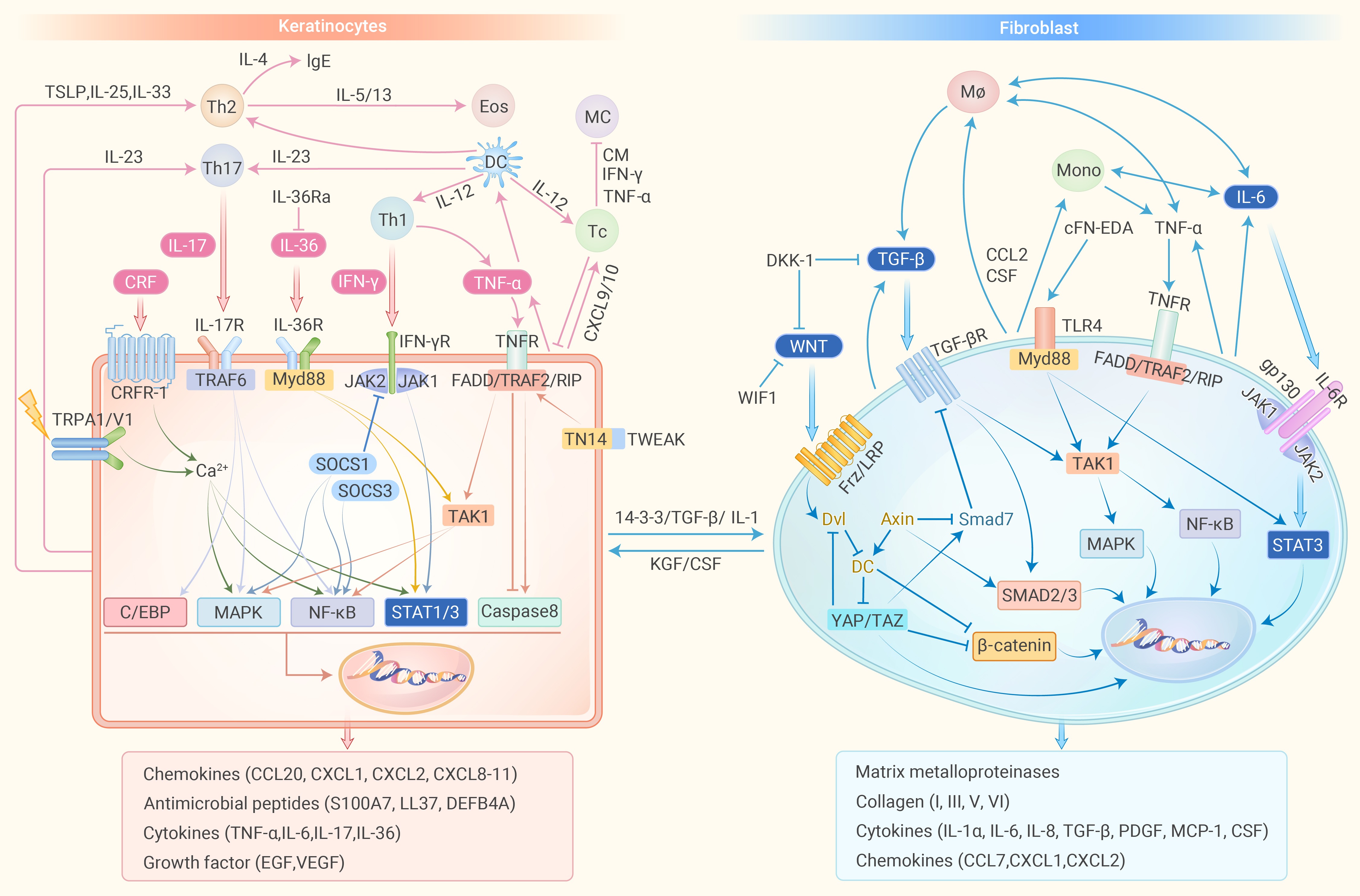
Figure 3.
Molecular mechanisms in inflammation and fibrosis.
