
Discovery of S1P/SPHK1 dysregulation in Parkinson’s disease and its role in pathological α-synuclein-induced neurodegeneration
-
GRAPHICAL ABSTRACT
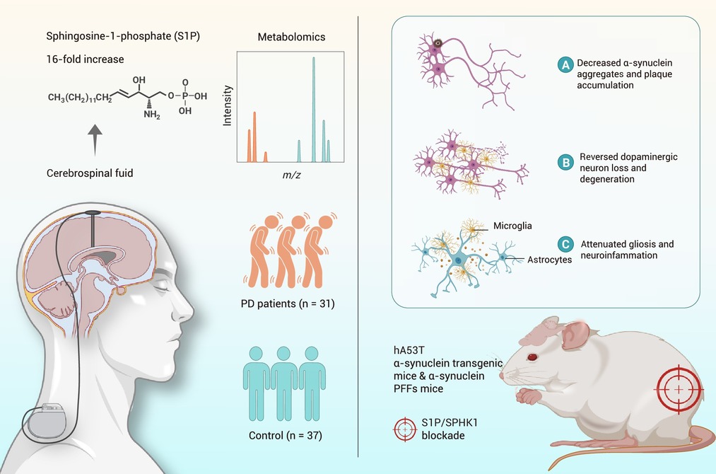
 DownLoad:
Full size image
DownLoad:
Full size image
-
PUBLIC SUMMARY
-
Sphingosine-1-phosphate is significantly elevated in the cerebrospinal fluid of Parkinson’s disease patients.
Pathologic α-synuclein selectively upregulates microglial SPHK1 over SPHK2.
S1P-mediated NF-κB signaling triggers pathologic α-synuclein-induced microglial activation.
Blockade of SPHK1 attenuates α-synuclein aggregation and neuroinflammation.
-
-
ABSTRACT
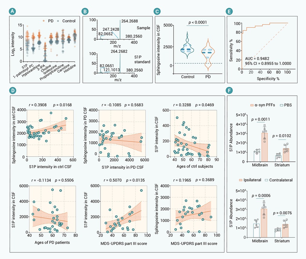
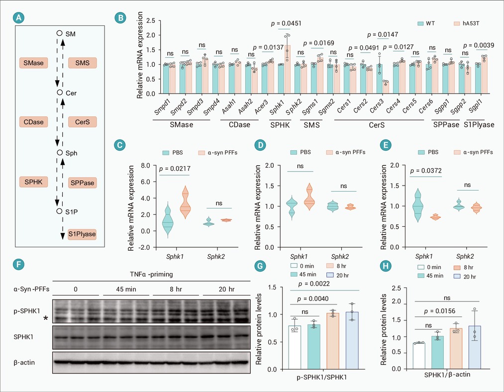
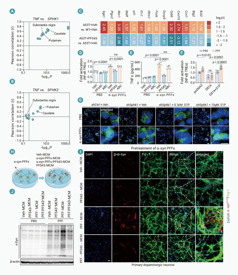
Parkinson's disease (PD) manifests a significant lipidopathy, marked by an enrichment of risk genes involved in sphingolipid metabolism. Sphingolipids are master regulators of cell fate, with sphingosine-1-phosphate (S1P) acting as a key signaling mediator in immunology and vascular biology. However, the fluctuation of S1P metabolism in PD and its role in synucleinopathy-induced neurodegeneration remain poorly understood. In this study, we discovered a 16-fold increase in cerebrospinal fluid (CSF) S1P levels synthesized by sphingosine kinase 1 (SPHK1) in PD patients compared to controls, accompanied by a reduction in sphingosine levels. Similarly, elevated S1P levels are observed in the striatum and midbrain of the α-synuclein preformed fibrils (α-syn PFFs)-induced mouse model of sporadic PD, with Sphk1 exhibiting the most pronounced upregulation among genes categorized under the “sphingosine metabolic process”, while Sphk2 expression remains unchanged. The increase of S1P levels is associated with the selective modulation of SPHK1 in microglia responding to pathological α-synuclein. Of note, α-syn PFFs treatment increases Sphk1 expression and activates SPHK1 in microglia. SPHK1 serves as a key contributor to pathologic α-syn-induced microglial activation via S1P-mediated NF-κB signaling. Knockdown of microglial Sphk1 alleviated neurodegenerative pathology in α-syn PFFs mice alleviates their neuropathological changes. Similarly, inhibition of SPHK1 with PF543 ameliorates PD-like neuropathological abnormalities in hA53T α-synuclein transgenic mice. These findings support a key role of S1P in the pathogenesis of PD linked to pathological α-synuclein.-
Keywords:
- Sphingolipids /
- metabolomics /
- neuroinflammation /
- α-synuclein aggregation /
- microgliosis
-

-
REFERENCES
[1] Bloem B.R., Okun M.S. and Klein C. (2021). Parkinson’s disease. The Lancet 397:2284−2303. DOI:10.1016/S0140-6736(21)00218-X [2] Dorsey E.R., Sherer T., Okun M.S., et al. (2018). The emerging evidence of the Parkinson pandemic. J. Parkinson’s Dis. 8:S3−S8. DOI:10.3233/JPD-181474 [3] Kalia L.V. and Lang A.E. (2015). Parkinson’s disease. The Lancet 386:896−912. DOI:10.1016/S0140-6736(14)61393-3 [4] Bartels T., De Schepper S. and Hong S. (2020). Microglia modulate neurodegeneration in Alzheimer’s and Parkinson’s diseases. Science 370:66−69. DOI:10.1126/science.abb8587 [5] Hickman S., Izzy S., Sen P., et al. (2018). Microglia in neurodegeneration. Nat. Neurosci. 21:1359−1369. DOI:10.1038/s41593-018-0242-x [6] Booms A., Pierce S.E., van der Schans E.J.C., et al. (2024). Parkinson’s disease risk enhancers in microglia. iScience 27:DOI:10.1016/j.isci.2024.108921 [7] Gosselin D., Skola D., Coufal N.G., et al. (2017). An environment-dependent transcriptional network specifies human microglia identity. Science 356:eaal3222. DOI:10.1126/science.aal3222 [8] Nott A., Holtman I.R., Coufal N.G., et al. (2019). Brain cell type–specific enhancer–promoter interactome maps and disease-risk association. Science 366:1134−1139. DOI:10.1126/science.aay0793 [9] Prinz M., Jung S. and Priller J. (2019). Microglia biology: One century of evolving concepts. Cell 179:292−311. DOI:10.1016/j.cell.2019.08.053 [10] Liddelow S.A., Guttenplan K.A., Clarke L.E., et al. (2017). Neurotoxic reactive astrocytes are induced by activated microglia. Nature 541:481−487. DOI:10.1038/nature21029 [11] Dickson D.W. (2012). Parkinson’s disease and parkinsonism: Neuropathology. Cold Spring Harb. Perspect. Med. 2:a009258. DOI:10.1101/cshperspect.a009258 [12] Tanriöver G., Bacioglu M., Schweighauser M., et al. (2020). Prominent microglial inclusions in transgenic mouse models of α-synucleinopathy that are distinct from neuronal lesions. Acta Neuropathol. Commun. 8:133. DOI:10.1186/s40478-020-00993-8 [13] Lee H.J., Bae E.J. and Lee S.J. (2014). Extracellular α-synuclein—A novel and crucial factor in Lewy body diseases. Nat. Rev. Neurol. 10:92−98. DOI:10.1038/nrneurol.2013.275 [14] Xiong M., Xia D., Yu H., et al. (2024). Microglia process α‐synuclein fibrils and enhance their pathogenicity in a TREM2‐dependent manner. Adv. Sci. 12:e2413451. DOI:10.1002/advs.202413451 [15] Panicker N., Sarkar S., Harischandra D.S., et al. (2019). Fyn kinase regulates misfolded α-synuclein uptake and NLRP3 inflammasome activation in microglia. J. Exp. Med. 216:1411−1430. DOI:10.1084/jem.20182191 [16] Long H., Zhang S., Zeng S., et al. (2022). Interaction of RAGE with α-synuclein fibrils mediates inflammatory response of microglia. Cell Rep. 40:111401. DOI:10.1016/j.celrep.2022.111401 [17] Mao X., Ou M.T., Karuppagounder S.S., et al. (2016). Pathological α-synuclein transmission initiated by binding lymphocyte-activation gene 3. Science 353:aah3374. DOI:10.1126/science.aah3374 [18] Zhang S., Liu Y.Q., Jia C., et al. (2021). Mechanistic basis for receptor-mediated pathological α-synuclein fibril cell-to-cell transmission in Parkinson’s disease. Proc. Natl. Acad. Sci. USA 118:e2011196118. DOI:10.1073/pnas.2011196118 [19] Saunders A., Macosko E.Z., Wysoker A., et al. (2018). Molecular diversity and specializations among the cells of the adult mouse brain. Cell 174:1015−1030.e16. DOI:10.1016/j.cell.2018.07.028 [20] Scheiblich H., Dansokho C., Mercan D., et al. (2021). Microglia jointly degrade fibrillar alpha-synuclein cargo by distribution through tunneling nanotubes. Cell 184:5089−5106.e21. DOI:10.1016/j.cell.2021.09.007 [21] Alaamery M., Albesher N., Aljawini N., et al. (2021). Role of sphingolipid metabolism in neurodegeneration. J. Neurochem. 158:25−35. DOI:10.1111/jnc.15044 [22] Várkonyi J., Rosenbaum H., Baumann N., et al. (2003). Gaucher disease associated with parkinsonism: Four further case reports. Am J. M. Genet. A 116A:348–351. DOI:10.1002/ajmg.a.10028 [23] Robak L.A., Jansen I.E., van Rooij J., et al. (2017). Excessive burden of lysosomal storage disorder gene variants in Parkinson’s disease. Brain 140:3191−3203. DOI:10.1093/brain/awx285 [24] Maceyka M. and Spiegel, S. (2014). Sphingolipid metabolites in inflammatory disease. Nature 510:58−67. DOI:10.1038/nature13475 [25] Cartier A. and Hla, T. (2019). Sphingosine 1-phosphate: Lipid signaling in pathology and therapy. Science 366:eaar5551. DOI:10.1126/science.aar5551 [26] Chao C.C., Gutiérrez-Vázquez C., Rothhammer V., et al. (2019). Metabolic control of astrocyte pathogenic activity via cPLA2-MAVS. Cell 179:1483−1498.e22. DOI:10.1016/j.cell.2019.11.016 [27] Mayo L., Trauger S.A., Blain M., et al. (2014). Regulation of astrocyte activation by glycolipids drives chronic CNS inflammation. Nat. Med. 20:1147−1156. DOI:10.1038/nm.3681 [28] Lee M.J., Thangada S., Claffey K.P., et al. (1999). Vascular endothelial cell adherens junction assembly and morphogenesis induced by sphingosine-1-phosphate. Cell 99:301−312. DOI:10.1016/S0092-8674(00)81661-X [29] Mizugishi K., Yamashita T., Olivera A., et al. (2005). Essential role for sphingosine kinases in neural and vascular development. Mol. Cell Biol. 25:11113−11121. DOI:10.1128/MCB.25.24.11113-11121.2005 [30] Mitroi D.N., Deutschmann A.U., Raucamp M., et al. (2016). Sphingosine 1-phosphate lyase ablation disrupts presynaptic architecture and function via an ubiquitin- proteasome mediated mechanism. Sci. Rep. 6:37064. DOI:10.1038/srep37064 [31] Atkinson D., Nikodinovic Glumac J., Asselbergh B., et al. (2017). Sphingosine 1-phosphate lyase deficiency causes Charcot-Marie-Tooth neuropathy. Neurology 88:533−542. DOI:10.1212/WNL.0000000000003595 [32] Schwedhelm E., Englisch C., Niemann L., et al. (2021). Sphingosine-1-phosphate, motor severity, and progression in Parkinson’s disease (MARK-PD). Mov. Disord. 36:2178−2182. DOI:10.1002/mds.28652 [33] Bonnaud S., Niaudet C., Legoux F., et al. (2010). Sphingosine-1-phosphate activates the AKT pathway to protect small intestines from radiation-induced endothelial apoptosis. Cancer Res. 70:9905−9915. DOI:10.1158/0008-5472.CAN-10-2043 [34] Morales-Ruiz M., Lee M.J., Zöllner S., et al. (2001). Sphingosine 1-Phosphate activates AKT, nitric oxide production, and chemotaxis through a Gi protein/phosphoinositide 3-kinase pathway in endothelial cells. J. Biol. Chem. 276:19672−19677. DOI:10.1074/jbc.M009993200 [35] Heras-Sandoval D., Pérez-Rojas J.M., Hernández-Damián J., et al. (2014). The role of PI3K/AKT/mTOR pathway in the modulation of autophagy and the clearance of protein aggregates in neurodegeneration. Cell. Signal. 26:2694−2701. DOI:10.1016/j.cellsig.2014.08.019 [36] Zheng Z., Zong Y., Ma Y., et al. (2024). Glucagon-like peptide-1 receptor: Mechanisms and advances in therapy. Sig. Transduct. Target. Ther. 9:234. DOI:10.1038/s41392-024-01931-z [37] Wang X., Wang B., Ji F., et al. (2024). Discovery of plasma biomarkers for Parkinson’s disease diagnoses based on metabolomics and lipidomics. Chin. Chem. Lett. 35:109653. DOI:10.1016/j.cclet.2024.109653 [38] Lee M.K., Stirling W., Xu Y., et al. (2002). Human α-synuclein-harboring familial Parkinson’s disease-linked Ala-53 → Thr mutation causes neurodegenerative disease with α-synuclein aggregation in transgenic mice. Proc. Natl. Acad. Sci. USA 99:8968−8973. DOI:10.1073/pnas.132197599 [39] Chung H., Ye Q., Park Y.J., et al. (2023). Very-long-chain fatty acids induce glial-derived sphingosine-1-phosphate synthesis, secretion, and neuroinflammation. Cell Metab. 35:855−874.e5. DOI:10.1016/j.cmet.2023.03.022 [40] Bennett M.L., Bennett F.C., Liddelow S.A., et al. (2016). New tools for studying microglia in the mouse and human CNS. Proc. Natl. Acad. Sci. USA 113:E1738−46. DOI:10.1073/pnas.1525528113 [41] Li Q., Cheng Z., Zhou L., et al. (2019). Developmental heterogeneity of microglia and brain myeloid cells revealed by deep single-cell rna sequencing. Neuron 101:207−223.e10. DOI:10.1016/j.neuron.2018.12.006 [42] Alvarez S.E., Harikumar K.B., Hait N.C., et al. (2010). Sphingosine-1-phosphate is a missing cofactor for the E3 ubiquitin ligase TRAF2. Nature 465:1084−1088. DOI:10.1038/nature09128 [43] Wang Z., Min X., Xiao S.H., et al. (2013). Molecular basis of sphingosine kinase 1 substrate recognition and catalysis. Structure 21:798−809. DOI:10.1016/j.str.2013.02.025 [44] Rothhammer V., Borucki D.M., Tjon E.C., et al. (2018). Microglial control of astrocytes in response to microbial metabolites. Nature 557:724−728. DOI:10.1038/s41586-018-0119-x [45] Schnute M.E., McReynolds M.D., Carroll J., et al. (2017). Discovery of a potent and selective sphingosine kinase 1 inhibitor through the molecular combination of chemotype-distinct screening hits. J. Med. Chem. 60:2562−2572. DOI:10.1021/acs.jmedchem.7b00070 [46] Yun S.P., Kam T.I., Panicker N., et al. (2018). Block of A1 astrocyte conversion by microglia is neuroprotective in models of Parkinson’s disease. Nat. Med. 24:931−938. DOI:10.1038/s41591-018-0051-5 [47] Luk K.C., Kehm V., Carroll J., et al. (2012). Pathological α-synuclein transmission initiates parkinson-like neurodegeneration in nontransgenic mice. Science 338:949−953. DOI:10.1126/science.1227157 [48] Liu J., Xu Y., Ji Y., et al. (2023). Sustained microglial activation and accelerated elimination of dendritic spines during acute sleep deprivation and restoration. The Innovation Life 1:100037. DOI:10.59717/j.xinn-life.2023.100037 [49] He X., Huang Y., Li B., et al. (2010). Deregulation of sphingolipid metabolism in Alzheimer’s disease. Neurobiol. Aging 31:398−408. DOI:10.1016/j.neurobiolaging.2008.05.010 [50] Mielke M.M., Maetzler W., Haughey N.J., et al. (2013). Plasma ceramide and glucosylceramide metabolism is altered in sporadic Parkinson’s disease and associated with cognitive impairment: A pilot study. PLoS One 8:e73094. DOI:10.1371/journal.pone.0073094 [51] Abbott S.K., Li H., Muñoz S.S., et al. (2014). Altered ceramide acyl chain length and ceramide synthase gene expression in Parkinson’s disease. Mov. Disord. 29:518−526. DOI:10.1002/mds.25729 [52] Filippatou A.G., Moniruzzaman M., Sotirchos E.S., et al. (2021). Serum ceramide levels are altered in multiple sclerosis. Mult. Scler. 27:1506−1519. DOI:10.1177/1352458520971816 [53] Vidaurre O.G., Haines J.D., Katz Sand I., et al. (2014). Cerebrospinal fluid ceramides from patients with multiple sclerosis impair neuronal bioenergetics. Brain 137:2271−2286. DOI:10.1093/brain/awu139 [54] Wang Z., Zheng Y., Wang F., et al. (2020). Mfsd2a and Spns2 are essential for sphingosine-1-phosphate transport in the formation and maintenance of the blood-brain barrier. Sci. Adv. 6:eaay8627. DOI:10.1126/sciadv.aay8627 [55] Jeya Paul J., Weigel C., Müller T., et al. (2020). Inflammatory conditions disrupt constitutive endothelial cell barrier stabilization by alleviating autonomous secretion of sphingosine 1-phosphate. Cells 9:928. DOI:10.3390/cells9040928 [56] Pisani V., Stefani A., Pierantozzi M., et al. (2012). Increased blood-cerebrospinal fluid transfer of albumin in advanced Parkinson’s disease. J. Neuroinflammation 9:670. DOI:10.1186/1742-2094-9-188 [57] Janelidze S., Hertze J., Nägga K., et al. (2017). Increased blood-brain barrier permeability is associated with dementia and diabetes but not amyloid pathology or APOE genotype. Neurobiol. Aging 51:104−112. DOI:10.1016/j.neurobiolaging.2016.11.017 [58] Männer A., Thomas D., Wagner M., et al. (2020). Sphingosine 1-phosphate levels in cerebrospinal fluid after subarachnoid hemorrhage. Neurol. Res. Pract. 2:49. DOI:10.1186/s42466-020-00093-x [59] Christoffersen C., Obinata H., Kumaraswamy S.B., et al. (2011). Endothelium-protective sphingosine-1-phosphate provided by HDL-associated apolipoprotein M. Proc. Natl. Acad. Sci. USA 108:9613–9618. 10.1073/pnas.1103187108 [60] Choe C., Petersen E., Lezius S., et al. (2021). Association of lipid levels with motor and cognitive function and decline in advanced Parkinson’s disease in the Mark-PD study. Parkinsonism Relat. Disord. 85:5−10. DOI:10.1016/j.parkreldis.2021.02.007 [61] Qiang J.K., Wong Y.C., Siderowf A., et al. (2013). Plasma apolipoprotein A1 as a biomarker for Parkinson disease. Ann. Neurol. 74:119−127. DOI:10.1002/ana.23872 [62] Liang J., Nagahashi M., Kim E.Y., et al. (2013). Sphingosine-1-phosphate links persistent STAT3 activation, chronic intestinal inflammation, and development of colitis-associated cancer. Cancer Cell 23:107−120. DOI:10.1016/j.ccr.2012.11.013 [63] Park K., Ikushiro H., Seo H.S., et al. (2016). ER stress stimulates production of the key antimicrobial peptide, cathelicidin, by forming a previously unidentified intracellular S1P signaling complex. Proc. Natl. Acad. Sci. USA 113:E1334−42. DOI:10.1073/pnas.1504555113 [64] Baker D.A., Barth J., Chang R., et al. (2010). Genetic sphingosine kinase 1 deficiency significantly decreases synovial inflammation and joint erosions in murine TNF-α–induced arthritis. J. Immunol. 185:2570−2579. DOI:10.4049/jimmunol.1000644 [65] Kunkel G.T., Maceyka M., Milstien S., et al. (2013). Targeting the sphingosine-1-phosphate axis in cancer, inflammation and beyond. Nat. Rev. Drug Discov. 12:688−702. DOI:10.1038/nrd4099 [66] Ji J., Wang J., Yang J., et al. (2019). The intra-nuclear SphK2-S1P axis facilitates M1-to-M2 shift of microglia via suppressing HDAC1-mediated KLF4 deacetylation. Front. Immunol. 10:1241. DOI:10.3389/fimmu.2019.01241 [67] Hait N.C., Allegood J., Maceyka M., et al. (2009). Regulation of histone acetylation in the nucleus by sphingosine-1-phosphate. Science 325:1254−1257. DOI:10.1126/science.1176709 [68] Weigert A., Von Knethen A., Thomas D., et al. (2019). Sphingosine kinase 2 is a negative regulator of inflammatory macrophage activation. Biochim. Biophys. Acta Mol. Cell Biol. Lipids 1864:1235−1246. DOI:10.1016/j.bbalip.2019.05.008 [69] Fitzner D., Bader J.M., Penkert H., et al. (2020). Cell-type- and brain-region-resolved mouse brain lipidome. Cell Rep. 32:108132. DOI:10.1016/j.celrep.2020.108132 [70] Meissner W.G., Remy P., Giordana C., et al. (2024). Trial of lixisenatide in early Parkinson’s disease. N. Engl. J. Med. 390:1176−1185. DOI:10.1056/NEJMoa2312323 [71] Vijiaratnam N., Girges C., Auld G., et al. (2025). Exenatide once a week versus placebo as a potential disease-modifying treatment for people with Parkinson’s disease in the UK: A phase 3, multicentre, double-blind, parallel-group, randomised, placebo-controlled trial. The Lancet 405:627−636. DOI:10.1016/S0140-6736(24)02808-3 [72] Athauda D., Maclagan K., Skene S.S., et al. (2017). Exenatide once weekly versus placebo in Parkinson’s disease: a randomised, double-blind, placebo-controlled trial. The Lancet 390:1664−1675. DOI:10.1016/S0140-6736(17)31585-4 [73] Christensen M., Sparre-Ulrich A.H., Hartmann B., et al. (2015). Transfer of liraglutide from blood to cerebrospinal fluid is minimal in patients with type 2 diabetes. Int. J. Obes. 39:1651−1654. DOI:10.1038/ijo.2015.136 [74] Zhang Y., Berka V., Song A., et al. (2014). Elevated sphingosine-1-phosphate promotes sickling and sickle cell disease progression. J. Clin. Invest. 124:2750−2761. DOI:10.1172/JCI74604 [75] Kim R., Kang N., Byun K., et al. (2023). Prognostic significance of peripheral neutrophils and lymphocytes in early untreated Parkinson’s disease: An 8-year follow-up study. J. Neurol. Neurosurg. Psychiatry. 94:1040−1046. DOI:10.1136/jnnp-2022-330394 -
ABOUT THIS ARTICLE
Cite this article:
Fang J., Wang X., Su X., et al. (2025). Discovery of S1P/SPHK1 dysregulation in Parkinson’s disease and its role in pathological α-synuclein-induced neurodegeneration. The Innovation Life 3:100164. https://doi.org/10.59717/j.xinn-life.2025.100164
Welcome!
To request copyright permission to republish or share portions of our works, please visit Copyright Clearance Center's (CCC) Marketplace website at marketplace.copyright.com.
Article Metrics
Relative Articles
Cited by
Export File
Citation
Fang J., Wang X., Su X., et al. (2025). Discovery of S1P/SPHK1 dysregulation in Parkinson’s disease and its role in pathological α-synuclein-induced neurodegeneration. The Innovation Life 3:100164. https://doi.org/10.59717/j.xinn-life.2025.100164

Format
Content
-
Figure 1.
Metabolomic profiling and S1P dysregulation in synucleinopathy-induced PD
-
Figure 2.
Selective upregulation of microglial SPHK1 in response to pathological α-synuclein
-
Figure 3.
Blockade of microglial SPHK1 alleviates pathological α-synuclein formation by suppressing S1P-mediated NF-κB activation
-
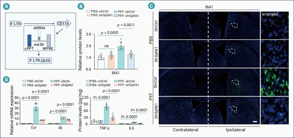
Figure 4.
Sphk1 knockdown alleviates α-syn PFFs-induced neuroinflammation
-
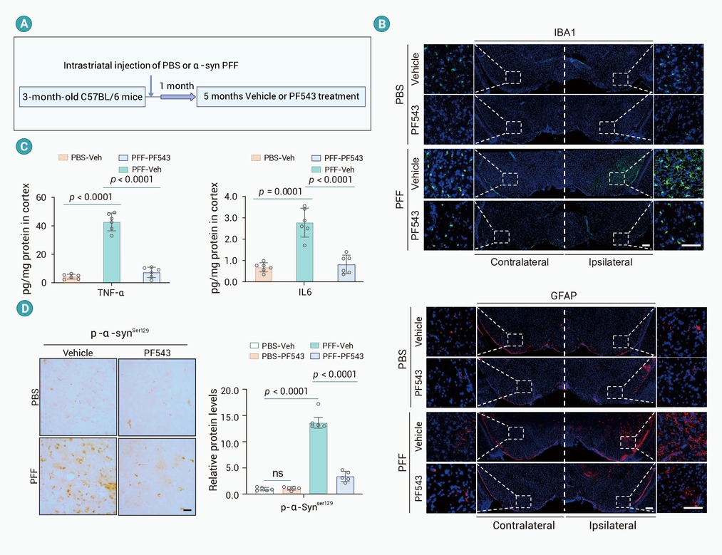
Figure 5.
SPHK1 inhibition diminishes α-syn PFFs-induced PD-like neuropathology
-
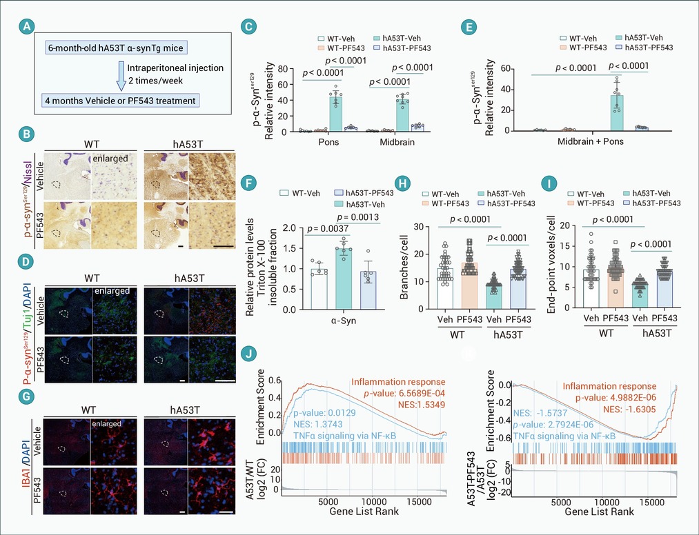
Figure 6.
SPHK1 inhibition reverses the neuropathology in hA53T α-synuclein transgenic mice
