
Maladaptive aging trajectory in late-onset Alzheimer's disease
-
GRAPHICAL ABSTRACT
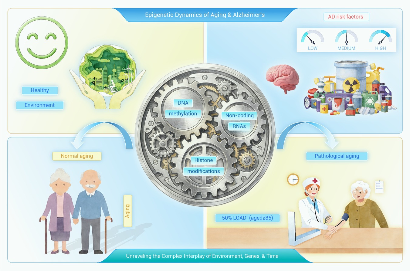
 DownLoad:
Full size image
DownLoad:
Full size image
-
PUBLIC SUMMARY
-
LOAD is fundamentally characterized as a pathological deviation from the trajectory of normal brain aging.
LOAD is underpinned by integrated genetic, epigenetic, and environmental influences.
Fostering healthy brain aging constitutes a central paradigm in the strategic shift toward preventing LOAD.
-
-
ABSTRACT
Dementia constitutes a significant public health crisis, accounting for one-third of deaths among the elderly and exceeding the combined mortality of breast and prostate cancers. Alzheimer's disease (AD), the predominant form of dementia (60–70%), primarily manifests as late-onset AD (LOAD), which constitutes over 95% of all AD cases. The risk of developing LOAD escalates dramatically with age, affecting approximately half of individuals over 85 years of age, thus positioning LOAD as a critical age-related medical challenge. The present study proposes a novel conceptual framework for understanding LOAD, positing it not merely as a discrete disease but rather as a maladaptive trajectory of brain aging, in which physiological aging processes progressively evolve into pathological states. This transition is driven by synergistic interactions between genetic susceptibility, environmental exposures, and aging-related mechanisms, with epigenetic alterations serving as a central dynamic mediator. This analysis demonstrates that genetic factors, aging, and environmental influences collectively regulate LOAD susceptibility through a multidimensional interplay: while genes directly drive pathological pathways, their expression is dynamically modulated by age and environmental context via epigenetic mechanisms. Deciphering these complex interactions provides crucial insights into LOAD pathogenesis. This reconceptualisation necessitates a paradigm shift in therapeutic strategy. Rather than focusing exclusively on amyloid-centric approaches, we advocate targeting core aging mechanisms to enhance brain resilience. The most promising interventions aim to counteract maladaptive cognitive decline by improving proteostasis, modulating epigenetic drivers, and suppressing chronic neuroinflammation. This would reposition healthy brain aging as the cornerstone of LOAD prevention and treatment.-
Keywords:
- Late-onset Alzheimer's disease /
- LOAD /
- Aging /
- Genes /
- Epigenetic.
-

-
REFERENCES
[1] Alzheimer’s Association. (2016). 2016 Alzheimer’s disease facts and figures. Alzheimer’s Dement. 12:459−509. DOI:10.1016/j.jalz.2016.03.001 [2] Alzheimer’s Association, Thies W. and Bleiler L. (2013). 2013 Alzheimer’s disease facts and figures. Alzheimer’s Dement. 9:208−245. DOI:10.1016/j.jalz.2013.02.003 [3] Cummings J.L. (2004). Alzheimer’s disease. N. Engl. J. Med. 351:56−67. DOI:10.1056/NEJMra040223 [4] Matthews F.E., Stephan B.C., Robinson L., et al. (2016). A two-decade dementia incidence comparison from the cognitive function and ageing studies I and II. Nat. Commun. 7:11398. DOI:10.1038/ncomms11398 [5] Reitz C. and Mayeux R. (2014). Alzheimer disease: Epidemiology, diagnostic criteria, risk factors and biomarkers. Biochem. Pharmacol. 88:640−651. DOI:10.1016/j.bcp.2014.01.017 [6] Eid A., Mhatre I. and Richardson J.R. (2019). Gene-environment interactions in Alzheimer’s disease: A potential path to precision medicine. Pharmacol. Ther. 199:173−187. DOI:10.1016/j.pharmthera.2019.03.005 [7] Rosenthal S.L. and Kamboh M.I. (2014). Late-onset Alzheimer’s disease genes and the potentially implicated pathways. Curr. Genet. Med. Rep. 2:85−101. DOI:10.1007/s40142-014-0034-x [8] Li Y., Macyczko J.R., Liu C.C., et al. (2022). ApoE4 reduction: An emerging and promising therapeutic strategy for Alzheimer’s disease. Neurobiol. Aging 115:20−28. DOI:10.1016/j.neurobiolaging.2022.03.011 [9] Alzheimer’s Association. (2010). 2010 Alzheimer’s disease facts and figures. Alzheimer’s Dement. 6:158−194. DOI:10.1016/j.jalz.2010.01.009 [10] Marfany A., Sierra C., Camafort M., et al. (2018). High blood pressure, Alzheimer disease and antihypertensive treatment. Panminerva Med. 60:8−16. DOI:10.23736/S0031-0808.17.03357-8 [11] James B.D., Leurgans S.E., Hebert L.E., et al. (2014). Contribution of Alzheimer disease to mortality in the United States. Neurology 82:1045−1050. DOI:10.1212/WNL.0000000000000240 [12] Alzheimer’s Association. (2023). 2023 Alzheimer’s disease facts and figures. Alzheimer’s Dement. 19:1598−1695. DOI:10.1002/alz.13016 [13] Evans D.A., Funkenstein H.H., Albert M.S., et al. (1989). Prevalence of Alzheimer’s disease in a community population of older persons: Higher than previously reported. JAMA 262:2551−2556. DOI:10.1001/jama.1989.03430180093036 [14] Abraham R., Moskvina V., Sims R., et al. (2008). A genome-wide association study for late-onset Alzheimer’s disease using DNA pooling. BMC Med. Genomics 1:44. DOI:10.1186/1755-8794-1-44 [15] Anand R., Gill K.D. and Mahdi A.A. (2014). Therapeutics of Alzheimer’s disease: Past, present and future. Neuropharmacology 76:27−50. DOI:10.1016/j.neuropharm.2013.07.004 [16] Beveridge J., Hachoumi L. and Sastre M. (2024). How promising are the latest monoclonal antibodies targeting amyloid-β for the treatment of early Alzheimer’s disease. Expert Opin. Emerg. Drugs 29:35−43. DOI:10.1080/14728214.2024.2304045 [17] Schiller E.R., Silverglate B.D. and Grossberg G.T. (2024). Profiling lecanemab as a treatment option for Alzheimer’s disease. Expert Rev. Neurother. 24:433−441. DOI:10.1080/14737175.2024.2344671 [18] Sims R., Hill M. and Williams J. (2020). The multiplex model of the genetics of Alzheimer’s disease. Nat. Neurosci. 23:311−322. DOI:10.1038/s41593-020-0599-5 [19] Pimenova A.A. and Goate A.M. (2020). Novel presenilin 1 and 2 double knock-out cell line for in vitro validation of PSEN1 and PSEN2 mutations. Neurobiol. Dis. 138:104785. DOI:10.1016/j.nbd.2020.104785 [20] Hang X. and Song W. (2013). The role of APP and BACE1 trafficking in APP processing and amyloid-β generation. Alzheimer’s Res. Ther. 5:46. DOI:10.1186/alzrt210 [21] Cervellati C., Valacchi G. and Zuliani G. (2021). BACE1: From biomarker to Alzheimer’s disease therapeutical target. Aging 13:12299−12318. DOI:10.18632/aging.203086 [22] McDade E., Voytyuk I., Aisen P., et al. (2021). The case for low-level BACE1 inhibition for the prevention of Alzheimer disease. Nat. Rev. Neurol. 17:703−714. DOI:10.1038/s41582-021-00545-1 [23] Bellenguez C., Küçükali F., Jansen I.E., et al. (2022). New insights into the genetic etiology of Alzheimer’s disease and related dementias. Nat. Genet. 54:412−436. DOI:10.1038/s41588-022-01024-z [24] Yamazaki Y., Zhao N., Caulfield T.R., et al. (2019). Apolipoprotein E and Alzheimer disease: Pathobiology and targeting strategies. Nat. Rev. Neurol. 15:501−518. DOI:10.1038/s41582-019-0228-7 [25] Farrer L.A., Cupples L.A., Haines J.L., et al. (1997). Effects of age, sex, and ethnicity on the association between apolipoprotein E genotype and Alzheimer disease. A meta-analysis. JAMA 278:1349−1356. DOI:10.1001/jama.1997.03550160069041 [26] Belloy M.E., Napolioni V. and Greicius M.D. (2019). A quarter century of APOE and Alzheimer’s disease: Progress to date and the path forward. Neuron 101:820−838. DOI:10.1016/j.neuron.2019.01.056 [27] Gao Y., Ren R.J., Zhong Z.L., et al. (2019). Mutation profile of APP, PSEN1, and PSEN2 in Chinese familial Alzheimer’s disease. Neurobiol. Aging 77:154−157. DOI:10.1016/j.neurobiolaging.2019.01.018 [28] Guela C., Wu C.K., Saroff D., et al. (1998). Aging renders the brain vulnerable to amyloid β-protein neurotoxicity. Nat. Med. 4:827−831. DOI:10.1038/nm0798-827 [29] Li X., Zhang J., Li D., et al. (2021). Astrocytic ApoE reprograms neuronal cholesterol metabolism and histone-acetylation-mediated memory. Neuron 109:957−970. DOI:10.1016/j.neuron.2021.01.005 [30] Wahlster L., Arimon M., Nasser-Ghodsi N., et al. (2013). Presenilin-1 adopts pathogenic conformation in normal aging and in sporadic Alzheimer’s disease. Acta Neuropathol. 125:187−199. DOI:10.1007/s00401-012-1062-9 [31] Kim J., Yoon H., Chung D., et al. (2016). miR-186 is decreased in aged brain and suppresses BACE1 expression. J. Neurochem. 137:436−445. DOI:10.1111/jnc.13507 [32] Halima S.B., Siegel G. and Rajendran L. (2016). miR-186 in Alzheimer’s disease: A big hope for a small RNA. J. Neurochem. 137:308−311. DOI:10.1111/jnc.13554 [33] Hou X., Zhang X., Zou H., et al. (2023). Differential and substrate-specific inhibition of γ-secretase by the C-terminal region of ApoE2, ApoE3, and ApoE4. Neuron 111:1898−1913. DOI:10.1016/j.neuron.2023.03.022 [34] Harman D. (2006). Alzheimer’s disease pathogenesis: Role of aging. Ann. N. Y. Acad. Sci. 1067:454−460. DOI:10.1196/annals.1354.065 [35] Zhu X.C., Tan L., Wang H.F., et al. (2015). Rate of early onset Alzheimer’s disease: A systematic review and meta-analysis. Ann. Transl. Med. 3:33. DOI:10.3978/j.issn.2305-5839.2015.01.19 [36] Reuter-Lorenz P.A. (2002). New visions of the aging mind and brain. Trends Cogn. Sci. 6:394−400. DOI:10.1016/S1364-6613(02)01957-5 [37] Godin S.K., Seo J. and Tsai L.H. (2018). Neurodegenerative diseases and the aging brain. Wolfe M.S. (ed). The molecular and cellular basis of neurodegenerative diseases (Academic Press), pp:509-526. DOI:10.1016/B978-0-12-811304-2.00017-1. [38] Sloane P.D., Zimmerman S., Suchindran C., et al. (2002). The public health impact of Alzheimer’s disease, 2000-2050: Potential implication of treatment advances. Annu. Rev. Public Health 23:213−231. DOI:10.1146/annurev.publhealth.23.100901.140525 [39] Kaplin A.I. and Williams M. (2007). How common are the “common” neurologic disorders. Neurology 69:410−411. DOI:10.1212/01.wnl.0000271137.67730.4b [40] Camandola S. and Mattson M.P. (2017). Brain metabolism in health, aging, and neurodegeneration. EMBO J. 36:1474−1492. DOI:10.15252/embj.201695810 [41] Jagust W. (2013). Vulnerable neural systems and the borderland of brain aging and neurodegeneration. Neuron 77:219−234. DOI:10.1016/j.neuron.2012.12.010 [42] Chiu M.J., Fan L.Y., Chen T.F., et al. (2017). Plasma tau levels in cognitively normal middle-aged and older adults. Front. Aging Neurosci. 9:51. DOI:10.3389/fnagi.2017.00051 [43] Cavedo E., Lista S., Houot M., et al. (2020). Plasma tau correlates with basal forebrain atrophy rates in people at risk for Alzheimer disease. Neurology 94:e30−e41. DOI:10.1212/WNL.0000000000008684 [44] Ono T., Uehara Y., Kurishita A., et al. (1993). Biological significance of DNA methylation in the ageing process. Age Ageing 22:S34−S43. DOI: 10.1093/ageing/22.suppl_1. S34. DOI:10.1093/ageing/22.suppl_1.S34 [45] Junnila R.K., List E.O., Berryman D.E., et al. (2013). The GH/IGF-1 axis in ageing and longevity. Nat. Rev. Endocrinol. 9:366−376. DOI:10.1038/nrendo.2013.67 [46] Liu X.M., Chan H.C., Ding G.L., et al. (2015). FSH regulates fat accumulation and redistribution in aging through the Gαi/Ca2+/CREB pathway. Aging Cell 14:409−420. DOI:10.1111/acel.12327 [47] Schafer M.J., Atkinson E.J., Vanderboom P.M., et al. (2016). Quantification of GDF11 and myostatin in human aging and cardiovascular disease. Cell Metab. 23:1207−1215. DOI:10.1016/j.cmet.2016.05.023 [48] Crunkhorn S. (2018). Aging: Promoting NAD+ production. Nat. Rev. Drug Discov. 17:864. DOI:10.1038/nrd.2018.188 [49] Trevisan K., Cristina-Pereira R., Silva-Amaral D., et al. (2019). Theories of aging and the prevalence of Alzheimer’s disease. Biomed. Res. Int. 2019:9171424. DOI:10.1155/2019/9171424 [50] Gefen T., Papastefan S.T., Rezvanian A., et al. (2018). Von economo neurons of the anterior cingulate across the lifespan and in Alzheimer’s disease. Cortex 99:69−77. DOI:10.1016/j.cortex.2017.10.015 [51] Hargis K.E. and Blalock E.M. (2017). Transcriptional signatures of brain aging and Alzheimer’s disease: What are our rodent models telling us. Behav. Brain Res. 322:311−328. DOI:10.1016/j.bbr.2016.05.007 [52] Jeong H., Shin J., Noh M., et al. (2019). Pan-HDAC inhibitors promote tau aggregation by increasing the level of acetylated tau. Int. J. Mol. Sci. 20:4283. DOI:10.3390/ijms20174283 [53] Smith C.D., Carney J.M., Starke-Reed P.E., et al. (1991). Excess brain protein oxidation and enzyme dysfunction in normal aging and in Alzheimer disease. Proc. Natl. Acad. Sci. USA 88:10540−10543. DOI:10.1073/pnas.88.23.10540 [54] Kirova A.M., Bays R.B. and Lagalwar S. (2015). Working memory and executive function decline across normal aging, mild cognitive impairment, and Alzheimer’s disease. Biomed. Res. Int. 2015:748212. DOI:10.1155/2015/748212 [55] Falco A., Cukierman D.S., Hauser-Davis R.A., et al. (2016). Doença de Alzheimer: Hipóteses etiológicas e perspectivas de tratamento. Quim. Nova 39:63−80. DOI:10.5935/0100-4042.20150163 [56] Cerami A. (1985). Hypothesis: Glucose as a mediator of aging. J. Am. Geriatr. Soc. 33:626−634. DOI:10.1111/j.1532-5415. 1985.tb01761.x. DOI:10.1111/j.1532-5415.1985.tb01761.x [57] Morrison N.A., Qi J.C., Tokita A., et al. (1994). Prediction of bone density from vitamin D receptor alleles. Nature 367:284−287. DOI:10.1038/367284a0 [58] Hayoz D., Ziegler T., Brunner H.R., et al. (1998). Diabetes mellitus and vascular lesions. Metabolism 47:16−19. DOI:10.1016/s0026-0495(98)90337-5 [59] Saez-Atienzar S. and Masliah E. (2020). Cellular senescence and Alzheimer disease: The egg and the chicken scenario. Nat. Rev. Neurosci. 21:433−444. DOI:10.1038/s41583-020-0325-1 [60] Blinkouskaya Y., Caçoilo A., Gollamudi T., et al. (2021). Brain aging mechanisms with mechanical manifestations. Mech. Ageing Dev. 200:111575. DOI:10.1016/j.mad.2021.111575 [61] Blinkouskaya Y. and Weickenmeier J. (2021). Brain shape changes associated with cerebral atrophy in healthy aging and Alzheimer’s disease. Front. Mech. Eng. 7:705653. DOI:10.3389/fmech.2021.705653 [62] Xue Q.L. (2011). The frailty syndrome: Definition and natural history. Clin. Geriatr. Med. 27:1−15. DOI:10.1016/j.cger.2010.08.009 [63] Grinin L., Grinin A. and Korotayev A. (2023). Global aging: An integral problem of the future. How to turn a problem into a development driver? Sadovnichy V., Akaev A., Ilyin I. et al. (eds) Reconsidering the limits to growth. World-systems evolution and global futures (Springer), pp:117-135. https://doi.org/10.1007/978-3-031-34999-7_7. [64] Fyfe I. (2018). Epigenetics links ageing with Alzheimer disease. Nat. Rev. Neurol. 14:254. DOI:10.1038/nrneurol.2018.32 [65] Bernstein A.I., Lin Y., Street R.C., et al. (2016). 5-Hydroxymethylation-associated epigenetic modifiers of Alzheimer’s disease modulate Tau-induced neurotoxicity. Hum. Mol. Genet. 25:2437−2450. DOI:10.1093/hmg/ddw110 [66] Rodríguez-Rodero S., Fernández-Morera J.L., Menéndez-Torre E., et al. (2010). Epigenetic regulation of aging. Discov. Med. 10:225–233. https://www.discoverymedicine.com/Sandra-Rodriguez-Rodero/2010/09/21/epigenetic-regulation-of-aging/#relatedArticles. [67] Jiang T., Yu J.T., Tian Y., et al. (2013). Epidemiology and etiology of Alzheimer’s disease: From genetic to non-genetic factors. Curr. Alzheimer Res. 10:852−867. DOI:10.2174/15672050113109990155 [68] Tecalco-Cruz A.C., Ramírez-Jarquín J.O., Alvarez-Sánchez M.E., et al. (2020). Epigenetic basis of Alzheimer disease. World J. Biol. Chem. 11:62−75. DOI:10.4331/wjbc. v11.i2.62. DOI:10.4331/wjbc.v11.i2.62 [69] Schueller E., Paiva I., Blanc F., et al. (2020). Dysregulation of histone acetylation pathways in hippocampus and frontal cortex of Alzheimer’s disease patients. Eur. Neuropsychopharmacol. 33:101−116. DOI:10.1016/j.euroneuro.2020.01.015 [70] Prasad H. and Rao R. (2018). Amyloid clearance defect in ApoE4 astrocytes is reversed by epigenetic correction of endosomal pH. Proc. Natl. Acad. Sci. USA 115:E6640−E6649. DOI:10.1073/pnas.1801612115 [71] Sen A., Nelson T.J. and Alkon D.L. (2015). ApoE4 and Aβ oligomers reduce BDNF expression via HDAC nuclear translocation. J. Neurosci. 35:7538−7551. DOI:10.1523/JNEUROSCI.1431-14.2015 [72] Sanchez-Mut J.V. and Gräff J. (2015). Epigenetic alterations in Alzheimer’s disease. Front. Behav. Neurosci. 9:347. DOI:10.3389/fnbeh.2015.00347 [73] Migliore L. and Coppedè F. (2022). Gene-environment interactions in Alzheimer disease: The emerging role of epigenetics. Nat. Rev. Neurol. 18:643−660. DOI:10.1038/s41582-022-00714-w [74] Jia J., Zhao T., Liu Z., et al. (2023). Association between healthy lifestyle and memory decline in older adults: 10-year, population based, prospective cohort study. BMJ 380:e072691. DOI:10.1136/bmj-2022-072691 [75] Gatz M., Pedersen N.L., Berg S., et al. (1997). Heritability for Alzheimer’s disease: The study of dementia in Swedish twins. J. Gerontol. A Biol. Sci. Med. Sci. 52:M117−M125. DOI:10.1093/gerona/52a.2.m117 [76] Marsit C.J. (2015). Influence of environmental exposure on human epigenetic regulation. J. Exp. Biol. 218:71−79. DOI:10.1242/jeb.106971 [77] Aguilera O., Fernández A.F., Muñoz A., et al. (2010). Epigenetics and environment: A complex relationship. J. Appl. Physiol. 109:243−251. DOI:10.1152/japplphysiol.00068.2010 [78] Pal S. and Tyler J.K. (2016). Epigenetics and aging. Sci. Adv. 2:e1600584. DOI:10.1126/sciadv.1600584 [79] Ridge P.G., Mukherjee S., Crane P.K., et al. (2013). Alzheimer’s disease: Analyzing the missing heritability. PLoS One 8:e79771. DOI:10.1371/journal.pone.0079771 [80] Mota M.P., Figueiredo P.A. and Duarte J.A. (2004). Teorias biológicas do envelhecimento. Rev. Port. Cienc. Desporto 4:81−110. DOI:10.5628/rpcd.04.01.81 [81] Campdelacreu J. (2014). Parkinsons disease and Alzheimer disease: Environmental risk factors. Neurología 29:541−549. DOI:10.1016/j.nrl.2013.04.001 [82] Lehallier B., Gate D., Schaum N., et al. (2019). Undulating changes in human plasma proteome profiles across the lifespan. Nat. Med. 25:1843−1850. DOI:10.1038/s41591-019-0673-2 [83] Federico A., Cardaioli E., Da Pozzo P., et al. (2012). Mitochondria, oxidative stress and neurodegeneration. J. Neurol. Sci. 322:254−262. DOI:10.1016/j.jns.2012.05.030 [84] Lionaki E., Markaki M. and Tavernarakis N. (2015). Mitochondria, autophagy and age-associated neurodegenerative diseases: New insights into a complex interplay. Biochim. Biophys. Acta 1847:1412−1423. DOI:10.1016/j.bbabio.2015.06.006 [85] Maruszak A. and Żekanowski C. (2011). Mitochondrial dysfunction and Alzheimer’s disease. Prog. Neuropsychopharmacol. Biol. Psychiatry 35:320−330. DOI:10.1016/j.pnpbp.2010.07.004 [86] Kim Y.S. and Joh T.H. (2006). Microglia, major player in the brain inflammation: Their roles in the pathogenesis of Parkinson’s disease. Exp. Mol. Med. 38:333−347. DOI:10.1038/emm.2006.40 [87] Goldgaber D., Harris H.W., Hla T., et al. (1989). Interleukin 1 regulates synthesis of amyloid beta-protein precursor mRNA in human endothelial cells. Proc. Natl. Acad. Sci. USA 86:7606−7610. DOI:10.1073/pnas.86.19.7606 [88] Plassman B.L., Havlik R.J., Steffens D.C., et al. (2000). Documented head injury in early adulthood and risk of Alzheimer’s disease and other dementias. Neurology 55:1158−1166. DOI:10.1212/WNL.55.8.1158 [89] Quintanilla R.A., Orellana D.I., González-Billault C., et al. (2004). Interleukin-6 induces Alzheimer-type phosphorylation of tau protein by deregulating the cdk5/p35 pathway. Exp. Cell Res. 295:245−257. DOI:10.1016/j.yexcr.2004.01.002 [90] Cuollo L., Antonangeli F., Santoni A., et al. (2020). The senescence-associated secretory phenotype (SASP) in the challenging future of cancer therapy and age-related diseases. Biology 9:485. DOI:10.3390/biology9120485 [91] Han X., Zhang T., Liu H., et al. (2020). Astrocyte senescence and Alzheimer’s disease: A review. Front. Aging Neurosci. 12:148. DOI:10.3389/fnagi.2020.00148 [92] Li Q., Liu Y. and Sun M. (2017). Autophagy and Alzheimer’s disease. Cell. Mol. Neurobiol. 37:377−388. DOI:10.1007/s10571-016-0386-8 [93] Li L., Zhang X. and Le W. (2010). Autophagy dysfunction in Alzheimer’s disease. Neurodegener. Dis. 7:265−271. DOI:10.1159/000276710 [94] Son J.H., Shim J.H., Kim K.H., et al. (2012). Neuronal autophagy and neurodegenerative diseases. Exp. Mol. Med. 44:89−98. DOI:10.3858/emm.2012.44.2.031 [95] Spaccavento S., Del Prete M., Craca A., et al. (2009). Influence of nutritional status on cognitive, functional and neuropsychiatric deficits in Alzheimer’s disease. Arch. Gerontol. Geriatr. 48:356−360. DOI:10.1016/j.archger.2008.03.004 [96] Aguzzi A. and O’Connor T. (2010). Protein aggregation diseases: Pathogenicity and therapeutic perspectives. Nat. Rev. Drug Discov. 9:237−248. DOI:10.1038/nrd3050 [97] Upadhya S.C. and Hegde A.N. (2007). Role of the ubiquitin proteasome system in Alzheimer’s disease. BMC Biochem. 8:S12. DOI:10.1186/1471-2091-8-S1-S12 [98] Vijg J. and Montagna C. (2017). Genome instability and aging: Cause or effect. Transl. Med. Aging 1:5−11. DOI:10.1016/j.tma.2017.09.003 [99] Cristofalo V.J., Gerhard G.S. and Pignolo R.J. (1994). Molecular biology of aging. Surg. Clin. North Am. 74:1−21. DOI:10.1016/s0039-6109(16)46226-2 [100] Soto C. and Pritzkow S. (2018). Protein misfolding, aggregation, and conformational strains in neurodegenerative diseases. Nat. Neurosci. 21:1332−1340. DOI:10.1038/s41593-018-0235-9 [101] Fukagawa N.K. (1999). Aging: Is oxidative stress a marker or is it causal. Proc. Soc. Exp. Biol. Med. 222:293−298. DOI:10.1046/j.1525-1373. 1999.d01-139.x. DOI:10.1046/j.1525-1373.1999.d01-139.x [102] Gella A. and Durany N. (2009). Oxidative stress in Alzheimer disease. Cell Adh. Migr. 3:88−93. DOI:10.4161/cam.3.1.7401 [103] Jiang H., Ju Z. and Rudolph K.L. (2007). Telomere shortening and ageing. Z. Gerontol. Geriatr. 40:314−324. DOI:10.1007/s00391-007-0480-0 [104] Cai Z. and Yan L.J. (2013). Telomere shortening and Alzheimer’s disease. Neuromolecular Med. 15:25−48. DOI:10.1007/s12017-012-8207-9 [105] Mastroeni D., Grover A., Delvaux E., et al. (2011). Epigenetic mechanisms in Alzheimer’s disease. Neurobiol. Aging 32:1161−1180. DOI:10.1016/j.neurobiolaging.2010.08.017 [106] Liu S., Gao J., Zhu M., et al. (2020). Gut microbiota and dysbiosis in Alzheimer’s disease: Implications for pathogenesis and treatment. Mol. Neurobiol. 57:5026−5043. DOI:10.1007/s12035-020-02073-3 [107] Bostancıklıoğlu M. (2018). Intestinal bacterial flora and Alzheimer’s disease. Neurophysiology 50:140−148. DOI:10.1007/s11062-018-9728-0 [108] Sarkar S.R., Banerjee S. and Das S. (2019). Gut microbiota in neurodegenerative disorders. J. Neuroimmunol. 328:98−104. DOI:10.1016/j.jneuroim.2018.12.004 [109] Lee C.Y., Chen B.C., Ho T.Y., et al. (2024). Characterizing dysregulations via cell-cell communications in Alzheimer’s brains using single-cell transcriptomes. BMC Neurosci. 25:24. DOI:10.1186/s12868-024-00862-3 [110] Garden G.A. and La Spada A.R. (2012). Intercellular (mis)communication in neurodegenerative disease. Neuron 73:886−901. DOI:10.1016/j.neuron.2012.02.017 [111] Puntambekar S.S., Moutinho M., Lin P.B.C., et al. (2022). CX3CR1 deficiency aggravates amyloid driven neuronal pathology and cognitive decline in Alzheimer’s disease. Mol. Neurodegener. 17:47. DOI:10.1186/s13024-022-00552-w [112] Sugaya K., Kwak Y.D., Ohmitsu O., et al. (2007). Practical issues in stem cell therapy for Alzheimer’s disease. Curr. Alzheimer Res. 4:370−377. DOI:10.2174/156720507781788945 [113] Vrotsos E.G. and Sugaya K. (2009). MCP-1-induced migration of NT2 neuroprogenitor cells involving APP signaling. Cell. Mol. Neurobiol. 29:373−381. DOI:10.1007/s10571-008-9330-x [114] Marutle A., Ohmitsu M., Nilbratt M., et al. (2007). Modulation of human neural stem cell differentiation in Alzheimer (APP23) transgenic mice by phenserine. Proc. Natl. Acad. Sci. USA 104:12506−12511. DOI:10.1073/pnas.0705346104 [115] Zhao L.R., Berra H.H., Duan W.M., et al. (2007). Beneficial effects of hematopoietic growth factor therapy in chronic ischemic stroke in rats. Stroke 38:2804−2811. DOI:10.1161/STROKEAHA.107.492132 [116] Wang J. and Chen G.J. (2016). Mitochondria as a therapeutic target in Alzheimer’s disease. Genes Dis. 3:220−227. DOI:10.1016/j.gendis.2016.05.001 [117] Moreira P.I., Cardoso S.M., Pereira C.M., et al. (2009). Mitochondria as a therapeutic target in Alzheimer’s disease and diabetes. CNS Neurol. Disord. Drug Targets 8:492−511. DOI:10.2174/187152709789824651 [118] Yuan S.H., Martin J., Elia J., et al. (2011). Cell-surface marker signatures for the isolation of neural stem cells, glia and neurons derived from human pluripotent stem cells. PLoS One 6:e17540. DOI:10.1371/journal.pone.0017540 [119] Karran E., Mercken M. and De Strooper B. (2011). The amyloid cascade hypothesis for Alzheimer’s disease: An appraisal for the development of therapeutics. Nat. Rev. Drug Discov. 10:698−712. DOI:10.1038/nrd3505 [120] Guo X., Yan L., Xu X., et al. (2024). Passive immunotherapy for Alzheimer’s disease. Ageing Res. Rev. 94:102192. DOI:10.1016/j.arr.2023.102192 [121] Hermann A. and Storch A. (2013). Induced neural stem cells (iNSCs) in neurodegenerative diseases. J. Neural Transm. 120:19−25. DOI:10.1007/s00702-012-0836-5 [122] Lee H.J., Kim K.S., Kim E.J., et al. (2007). Brain transplantation of immortalized human neural stem cells promotes functional recovery in mouse intracerebral hemorrhage stroke model. Stem Cells 25:1204−1212. DOI:10.1634/stemcells.2006-0405 [123] Yamasaki T.R., Blurton-Jones M., Morrissette D.A., et al. (2007). Neural stem cells improve memory in an inducible mouse model of neuronal loss. J. Neurosci. 27:11925−11933. DOI:10.1523/JNEUROSCI.1627-07.2007 [124] Zhang P., Kishimoto Y., Grammatikakis I., et al. (2019). Senolytic therapy alleviates Aβ-associated oligodendrocyte progenitor cell senescence and cognitive deficits in an Alzheimer’s disease model. Nat. Neurosci. 22:719−728. DOI:10.1038/s41593-019-0372-9 [125] Shapira R., Efrati S. and Ashery U. (2018). Hyperbaric oxygen therapy as a new treatment approach for Alzheimer’s disease. Neural Regen. Res. 13:817−818. DOI:10.4103/1673-5374.232475 [126] Yao Z., Yang W., Gao Z., et al. (2017). Nicotinamide mononucleotide inhibits JNK activation to reverse Alzheimer disease. Neurosci. Lett. 647:133−140. DOI:10.1016/j.neulet.2017.03.027 [127] Friesland M. and Orr M.E. (2023). Translating the biology of aging into new therapeutics for Alzheimer’s disease: Senolytics. J. Prev. Alzheimers Dis. 10:633−646. DOI:10.14283/jpad.2023.64 [128] Friedman L.G., Qureshi Y.H. and Yu W.H. (2015). Promoting autophagic clearance: Viable therapeutic targets in Alzheimer’s disease. Neurotherapeutics 12:94−108. DOI:10.1007/s13311-014-0320-z [129] Llanos-González E., Henares-Chavarino Á., Pedrero-Prieto C.M., et al. (2020). Interplay between mitochondrial oxidative disorders and proteostasis in Alzheimer’s disease. Front. Neurosci. 14:1444. DOI:10.3389/fnins.2020.571954 [130] Śliwińska S. and Jeziorek M. (2021). The role of nutrition in Alzheimer’s disease. Rocz. Panstw. Zakl. Hig. 72:15−23. DOI:10.32394/rpzh.2021.0156 [131] Cremonini A.L., Caffa I., Cea M., et al. (2019). Nutrients in the prevention of Alzheimer’s disease. Oxid. Med. Cell. Longev. 2019:9874159. DOI:10.1155/2019/9874159 [132] Yassine H.N. and Finch C.E. (2020). APOE alleles and diet in brain aging and Alzheimer’s disease. Front. Aging Neurosci. 12:150. DOI:10.3389/fnagi.2020.00150 [133] Banks W.A., Reed M.J., Logsdon A.F., et al. (2021). Healthy aging and the blood-brain barrier. Nat. Aging 1:243−254. DOI:10.1038/s43587-021-00043-5 [134] Jin L., Wang Q., Yang M., et al. (2022). Indirubin-3’-monoxime-loaded PLGA-PEG nanoparticles for potential Alzheimer’s disease treatment. Med. Nov. Technol. Devices 15:100150. DOI:10.1016/j.medntd.2022.100150 [135] Shri S.R., Sah S.S. and Subramaniam V. (2023). Role of GSK-3β inhibitors: New promises and opportunities for Alzheimer’s disease. Adv. Pharm. Bull. 13:688−700. DOI:10.34172/apb.2023.074 [136] Ren Q.G., Liao X.M., Wang Z.F., et al. (2007). Effects of tau phosphorylation on proteasome activity. FEBS Lett. 581:1521−1528. DOI:10.1016/j.febslet.2007.03.006 [137] Yu G. and Jia J. (2010). Is there an association of regulatory region polymorphism in the alpha-1-antichymotrypsin gene with sporadic Alzheimer’s disease in the northern Han-Chinese population. J. Clin. Neurosci. 17:766−769. DOI:10.1016/j.jocn.2009.10.009 [138] Hou Y., Dan X., Babbar M., et al. (2019). Ageing as a risk factor for neurodegenerative disease. Nat. Rev. Neurol. 15:565−581. DOI:10.1038/s41582-019-0244-7 [139] Thomas P., Wang Y.J., Zhong J.H., et al. (2009). Grape seed polyphenols and curcumin reduce genomic instability events in a transgenic mouse model for Alzheimer’s disease. Mutat. Res. 661:25−34. DOI:10.1016/j.mrfmmm.2008.10.012 [140] Min S.W., Cho S.H., Zhou Y., et al. (2015). Critical role of acetylation in tau-mediated neurodegeneration and cognitive deficits. Nat. Med. 21:1154−1162. DOI:10.1038/nm.3951 [141] Whittemore K., Derevyanko A., Martinez P., et al. (2019). Telomerase gene therapy ameliorates the effects of neurodegeneration associated to short telomeres in mice. Aging 11:2916−2948. DOI:10.18632/aging.101916 [142] Wang J., Yu J.T., Tan M.S., et al. (2013). Epigenetic mechanisms in Alzheimer’s disease: Implications for pathogenesis and therapy. Ageing Res. Rev. 12:1024−1041. DOI:10.1016/j.arr.2013.05.003 [143] Franco S., Blasco M.A., Siedlak S.L., et al. (2006). Telomeres and telomerase in Alzheimer’s disease: Epiphenomena or a new focus for therapeutic strategy. Alzheimer’s Dement. 2:164−168. DOI:10.1016/j.jalz.2006.03.001 [144] Zhu F., Li C., Chu F., et al. (2020). Target dysbiosis of gut microbes as a future therapeutic manipulation in Alzheimer’s disease. Front. Aging Neurosci. 12:544235. DOI:10.3389/fnagi.2020.544235 [145] Stafstrom C.E. and Rho J.M. (2012). The ketogenic diet as a treatment paradigm for diverse neurological disorders. Front. Pharmacol. 3:59. DOI:10.3389/fphar.2012.00059 [146] Nimgampalle M. and Kuna Y. (2017). Anti-Alzheimer properties of probiotic, Lactobacillus plantarum MTCC 1325 in Alzheimer’s disease induced albino rats. J. Clin. Diagn. Res. 11:KC01−KC05. DOI:10.7860/JCDR/2017/26106.10428 [147] Sanchez-Mut J.V., Aso E., Panayotis N., et al. (2014). Promoter hypermethylation of the phosphatase DUSP22 mediates PKA-dependent TAU phosphorylation and CREB activation in Alzheimer’s disease. Hippocampus 24:363−368. DOI:10.1002/hipo.22245 [148] Kandimalla R. and Reddy P.H. (2017). Therapeutics of neurotransmitters in Alzheimer’s disease. J. Alzheimer’s Dis. 57:1049−1069. DOI:10.3233/JAD-161118 [149] Mattsson P., Georgiopoulos C., Johansson A., et al. (2022). Decreased 5-HT1A binding in mild Alzheimer’s disease—A positron emission tomography study. Synapse 76:e22235. DOI:10.1002/syn.22235 [150] Nirogi R., Mohammed A.R., Shinde A.K., et al. (2023). Usmarapride (SUVN-D4010), a 5-HT4 receptor partial agonist for the potential treatment of Alzheimer’s disease: Behavioural, neurochemical and pharmacological profiling. Eur. J. Pharmacol. 947:175625. DOI:10.1016/j.ejphar.2023.175625 [151] Partyka A., Jastrzębska-Więsek M., Marciniec K., et al. (2023). Selective 5-HT6 receptor ligands (Agonist and Antagonist) show different effects on antipsychotic drug-induced metabolic dysfunctions in rats. Pharmaceuticals 16:154. DOI:10.3390/ph16020154 [152] Sharma K., Sharma N.K., Singh R., et al. (2021). Role of receptors in relation to plaques and tangles in Alzheimer’s disease pathology. Int. J. Mol. Sci. 22:12987. DOI:10.3390/ijms222312987 [153] de Paiva I.H.R., Duarte-Silva E.P., Silva-Fernandes A., et al. (2024). Semaglutide attenuates anxious and depressive-like behaviors and reverses the cognitive impairment in a type 2 diabetes mellitus mouse model via the microbiota-gut-brain axis. J. Neuroimmune Pharmacol. 19:1−26. DOI:10.1007/s11481-024-10111-3 [154] Mathew A., Sajan T.T., Paul J., et al. (2023). Current drug targets in Alzheimer’s associated memory impairment: A comprehensive review. CNS Neurol. Disord. Drug Targets 22:255−275. DOI:10.2174/1871527321666220401092149 [155] Flores-Clemente C., Rodríguez-Perdigón M., Tordera R.M., et al. (2021). Inhibition of astrocytic histamine N-methyltransferase as a possible target for the treatment of Alzheimer’s disease. Biomolecules 11:1408. DOI:10.3390/biom11101408 [156] Moreira N.C.S., Lima L.A., de Moraes W.B., et al. (2022). Neuroprotective effects of cholinesterase inhibitors: Current scenario in therapies for Alzheimer’s disease and future perspectives. J. Alzheimer’s Dis. Rep. 6:177−193. DOI:10.3233/ADR-210059 [157] Hayashi Y., Lin H.T., Lee C.C., et al. (2020). Effects of neural stem cell transplantation in Alzheimer’s disease models. J. Biomed. Sci. 27:29. DOI:10.1186/s12929-020-0623-9 [158] Lu M.H., Ji W.L., Chen D., et al. (2021). Intranasal transplantation of human neural stem cells ameliorates Alzheimer’s disease-like pathology in a mouse model. Front. Aging Neurosci. 13:650103. DOI:10.3389/fnagi.2021.650103 [159] Zhang H.A., Gao F., Xu H., et al. (2022). Neural stem cell transplantation alleviates functional cognitive deficits in a mouse model of tauopathy. Neural Regen. Res. 17:152−162. DOI:10.4103/1673-5374.314294 [160] Apodaca L.A., Baddour A.A.D., Garcia C., et al. (2021). Human neural stem cell-derived extracellular vesicles mitigate hallmarks of Alzheimer’s disease. Alzheimer’s Res. Ther. 13:57. DOI:10.1186/s13195-021-00791-x [161] Duncan T. and Valenzuela M. (2017). Alzheimer’s disease, dementia, and stem cell therapy. Stem Cell Res. Ther. 8:111. DOI:10.1186/s13287-017-0567-5 [162] Chang J., Zhao S., Zhang Y., et al. (2024). Neural stem cells promote neuroplasticity: A promising therapeutic strategy for the treatment of Alzheimer’s disease. Neural Regen. Res. 19:619−628. DOI:10.4103/1673-5374.380879 [163] Cummings J., Osse A.M.L., Cammann D., et al. (2024). Alzheimer’s disease drug development pipeline: 2024. Alzheimer’s Dement. 10:e12465. DOI:10.1002/trc2.12465 [164] Edwards M. and Corkill R. (2023). Disease-modifying treatments in Alzheimer’s disease. J. Neurol. 270:2342−2344. DOI:10.1007/s00415-023-11646-w [165] Pardo-Moreno T., González-Acedo A., Rivas-Domínguez A., et al. (2022). Therapeutic approach to Alzheimer’s disease: Current treatments and new perspectives. Pharmaceutics 14:1117. DOI:10.3390/pharmaceutics14061117 [166] Thoe E.S., Fauzi A., Tang Y.Q., et al. (2021). A review on advances of treatment modalities for Alzheimer’s disease. Life Sci. 276:119129. DOI:10.1016/j.lfs.2021.119129 [167] Kurihara M., Mano T., Saito Y., et al. (2019). Colocalization of BRCA1 with tau aggregates in human tauopathies. Brain Sci. 9:7. DOI:10.3390/brainsci9010007 [168] Heidebrink J.L. and Paulson H.L. (2024). Lessons learned from approval of aducanumab for Alzheimer’s disease. Annu. Rev. Med. 75:99−111. DOI:10.1146/annurev-med-052422-020540 [169] Zhang L., Sheng S. and Qin C. (2013). The role of HDAC6 in Alzheimer’s disease. J. Alzheimer’s Dis. 33:283−295. DOI:10.3233/JAD-2012-121636 [170] Miners J.S., Jones R. and Love S. (2014). Differential changes in Aβ42 and Aβ40 with age. J. Alzheimer’s Dis. 40:727−735. DOI:10.3233/JAD-132267 [171] Sjögren M., Vanderstichele H., Agren H., et al. (2001). Tau and Aβ42 in cerebrospinal fluid from healthy adults 21–93 years of age: Establishment of reference values. Clin. Chem. 47:1776−1781. DOI:10.1093/clinchem/47.10.1776 [172] Wang J., Dickson D.W., Trojanowski J.Q., et al. (1999). The levels of soluble versus insoluble brain Aβ distinguish Alzheimer’s disease from normal and pathologic aging. Exp. Neurol. 158:328−337. DOI:10.1006/exnr.1999.7085 [173] Piccini A., Russo C., Gliozzi A., et al. (2005). β-amyloid is different in normal aging and in Alzheimer disease. J. Biol. Chem. 280:34186−34192. DOI:10.1074/jbc.M505694200 [174] Querfurth H.W. and LaFerla F.M. (2010). Alzheimer’s disease. N. Engl. J. Med. 362:329−344. DOI:10.1056/NEJMra0909142 [175] Del Tredici K. and Braak H. (2008). Neurofibrillary changes of the Alzheimer type in very elderly individuals: Neither inevitable nor benign: Commentary on “No disease in the brain of a 115-year-old woman”. Neurobiol. Aging 29:1133−1136. DOI:10.1016/j.neurobiolaging.2007.10.018 [176] Drachman D.A. (2006). Aging of the brain, entropy, and Alzheimer disease. Neurology 67:1340−1352. DOI:10.1212/01.wnl.0000240127.89601.83 [177] Benayoun B.A., Pollina E.A. and Brunet A. (2015). Epigenetic regulation of ageing: Linking environmental inputs to genomic stability. Nat. Rev. Mol. Cell Biol. 16:593−610. DOI:10.1038/nrm4048 [178] Sidler C., Kovalchuk O. and Kovalchuk I. (2017). Epigenetic regulation of cellular senescence and aging. Front. Genet. 8:138. DOI:10.3389/fgene.2017.00138 [179] Dine J. and Deng C.X. (2013). Mouse models of BRCA1 and their application to breast cancer research. Cancer Metastasis Rev. 32:25−37. DOI:10.1007/s10555-012-9394-4 [180] Lardenoije R., Iatrou A., Kenis G., et al. (2015). The epigenetics of aging and neurodegeneration. Prog. Neurobiol. 131:21−64. DOI:10.1016/j.pneurobio.2015.05.002 [181] Pedersen N.L., Gatz M., Berg S., et al. (2004). How heritable is Alzheimer’s disease late in life. Findings from Swedish twins. Ann. Neurol. 55:180−185. DOI:10.1002/ana.10823 [182] Bacanu S.A., Devlin B., Chowdari K.V., et al. (2005). Heritability of psychosis in Alzheimer disease. Am. J. Geriatr. Psychiatry 13:624−627. DOI:10.1097/00019442-200507000-00012 [183] Ertekin-Taner N., Graff-Radford N., Younkin L.H., et al. (2001). Heritability of plasma amyloid β in typical late-onset Alzheimer’s disease pedigrees. Genet. Epidemiol. 21:19−30. DOI:10.1002/gepi.1018 [184] Zerbi V., Kleinnijenhuis M., Fang X., et al. (2013). Gray and white matter degeneration revealed by diffusion in an Alzheimer mouse model. Neurobiol. Aging 34:1440−1450. DOI:10.1016/j.neurobiolaging.2012.11.003 [185] Salat D.H., Chen J.J., van der Kouwe A.J., et al. (2011). Hippocampal degeneration is associated with temporal and limbic gray matter/white matter tissue contrast in Alzheimer’s disease. Neuroimage 54:1795−1802. DOI:10.1016/j.neuroimage.2010.10.045 [186] Guo J., Huang X., Dou L., et al. (2022). Aging and aging-related diseases: From molecular mechanisms to interventions and treatments. Signal Transduct. Target. Ther. 7:391. DOI:10.1038/s41392-022-01251-0 [187] Hunsberger H.C., Pinky P.D., Smith W., et al. (2019). The role of APOE4 in Alzheimer’s disease: Strategies for future therapeutic interventions. Neuronal Signal. 3:2. NS20180203. DOI:10.1042/NS20180203. [188] Aday J.S., Bloesch E.K. and Davoli C.C. (2020). Can psychedelic drugs attenuate age-related changes in cognition and affect. J. Cogn. Enhance. 4:219−227. DOI:10.1007/s41465-019-00143-6 [189] Liu C.C., Kanekiyo T., Xu H., et al. (2013). Apolipoprotein E and Alzheimer disease: Risk, mechanisms and therapy. Nat. Rev. Neurol. 9:106−118. DOI:10.1038/nrneurol.2012.263 [190] Apostolova L.G., Lane K.A., Logan P.E., et al. (2021). APOE4 is associated with earlier symptom onset in LOAD but later symptom onset in EOAD. Alzheimer’s Dement. 17:e056661. DOI:10.1002/alz.056661 [191] Zhao J., Fu Y., Yamazaki Y., et al. (2020). APOE4 exacerbates synapse loss and neurodegeneration in Alzheimer’s disease patient iPSC-derived cerebral organoids. Nat. Commun. 11:5540. DOI:10.1038/s41467-020-19264-0 [192] Han Y., Wang N., Kang X., et al. (2022). A class I HDAC inhibitor rescues synaptic damage and neuron loss in APP-transfected cells and APP/PS1 mice through the GRIP1/AMPA pathway. Molecules 27:4160. DOI:10.3390/molecules27134160 [193] Singh A., Kammala A.K., Benson M.K., et al. (2024). Aging and inflammation. Cold Spring Harb. Perspect. Med. 14:a041197. DOI:10.1101/cshperspect. a041197. DOI:10.1101/cshperspect.a041197 [194] Martínez-Morales P.L., Revilla A., Ocaña I., et al. (2013). Progress in stem cell therapy for major human neurological disorders. Stem Cell Rev. Rep. 9:685−699. DOI:10.1007/s12015-013-9443-6 [195] Xuan A.G., Luo M., Ji W.D., et al. (2009). Effects of engrafted neural stem cells in Alzheimer’s disease rats. Neurosci. Lett. 450:167−171. DOI:10.1016/j.neulet.2008.12.001 [196] Xuan A.G., Long D.H., Gu H.G., et al. (2008). BDNF improves the effects of neural stem cells on the rat model of Alzheimer’s disease with unilateral lesion of fimbria-fornix. Neurosci. Lett. 440:331−335. DOI:10.1016/j.neulet.2008.05.107 [197] Blurton-Jones M., Kitazawa M., Martinez-Coria H., et al. (2009). Neural stem cells improve cognition via BDNF in a transgenic model of Alzheimer disease. Proc. Natl. Acad. Sci. USA 106:13594−13599. DOI:10.1073/pnas.0901402106 [198] Kwak Y.D., Brannen C.L., Qu T., et al. (2006). Amyloid precursor protein regulates differentiation of human neural stem cells. Stem Cells Dev. 15:381−389. DOI:10.1089/scd.2006.15.381 [199] Lu P., Jones L.L. and Snyder E.Y. et al. (2003). Neural stem cells constitutively secrete neurotrophic factors and promote extensive host axonal growth after spinal cord injury. Exp. Neurol. 181:115−129. DOI:10.1016/S0014-4886(03)00037-2 [200] Zhao L.R., Singhal S., Duan W.M., et al. (2007). Brain repair by hematopoietic growth factors in a rat model of stroke. Stroke 38:2584−2591. DOI:10.1161/STROKEAHA.107.486712 [201] Zhu W., Mao Y., Zhao Y., et al. (2005). Transplantation of vascular endothelial growth factor-transfected neural stem cells into the rat brain provides neuroprotection after transient focal cerebral ischemia. Neurosurgery 57:325−333. DOI:10.1227/01.NEU.0000166685.26246.7E [202] Bang O.Y., Lee J.S., Lee P.H., et al. (2005). Autologous mesenchymal stem cell transplantation in stroke patients. Ann. Neurol. 57:874−882. DOI:10.1002/ana.20510 [203] Lee J.S., Hong J.M., Moon G.J., et al. (2010). A long-term follow-up study of intravenous autologous mesenchymal stem cell transplantation in patients with ischemic stroke. Stem Cells 28:1099−1106. DOI:10.1002/stem.430 [204] Zhu J., Huang Z., Yang F., et al. (2021). Cadmium disturbs epigenetic modification and induces DNA damage in mouse preimplantation embryos. Ecotoxicol. Environ. Saf. 219:112306. DOI:10.1016/j.ecoenv.2021.112306 [205] Min J. and Min K. (2016). Blood cadmium levels and Alzheimer’s disease mortality risk in older US adults. Environ. Health 15:69. DOI:10.1186/s12940-016-0155-7 [206] Wills N.K., Ramanujam V.M.S., Chang J., et al. (2008). Cadmium accumulation in the human retina: Effects of age, gender, and cellular toxicity. Exp. Eye Res. 86:41−51. DOI:10.1016/j.exer.2007.09.005 [207] Rahman M.A., Hannan M.A., Uddin M.J., et al. (2021). Exposure to environmental arsenic and emerging risk of Alzheimer’s disease: Perspective mechanisms, management strategy, and future directions. Toxics 9:188. DOI:10.3390/toxics9080188 [208] Bjørklund G., Aaseth J. and Chirumbolo S. (2018). Effects of arsenic toxicity beyond epigenetic modifications. Environ. Geochem. Health 40:955−965. DOI:10.1007/s10653-017-9965-y [209] Tyler C.R. and Allan A.M. (2014). The effects of arsenic exposure on neurological and cognitive dysfunction in human and rodent studies: A review. Curr. Environ. Health Rep. 1:132−147. DOI:10.1007/s40572-014-0012-1 [210] Kivipelto M., Ngandu T., Fratiglioni L., et al. (2005). Obesity and vascular risk factors at midlife and the risk of dementia and Alzheimer disease. Arch. Neurol. 62:1556−1560. DOI:10.1001/archneur.62.10.1556 [211] Lopomo A., Burgio E. and Migliore L. (2016). Epigenetics of obesity. Prog. Mol. Biol. Transl. Sci. 140:151−184. DOI:10.1016/bs.pmbts.2016.02.002 [212] Doherty G.H. (2011). Obesity and the ageing brain: Could leptin play a role in neurodegeneration. Curr. Gerontol. Geriatr. Res. 2011:708154. DOI:10.1155/2011/708154 [213] McKay J.A. and Mathers J.C. (2011). Diet induced epigenetic changes and their implications for health. Acta Physiol. 202:103−118. DOI:10.1111/j.1748-1716.2011. 02278.x. DOI:10.1111/j.1748-1716.2011.02278.x [214] Hullar M.A.J. and Fu B.C. (2014). Diet, the gut microbiome, and epigenetics. Cancer J. 20:170−175. DOI:10.1097/PPO.0000000000000046 [215] Shah R. (2013). The role of nutrition and diet in Alzheimer disease: A systematic review. J. Am. Med. Dir. Assoc. 14:398−402. DOI:10.1016/j.jamda.2013.01.014 [216] Stranahan A.M., Norman E.D., Lee K., et al. (2008). Diet-induced insulin resistance impairs hippocampal synaptic plasticity and cognition in middle-aged rats. Hippocampus 18:1085−1088. DOI:10.1002/hipo.20470 [217] Mattsson N., Andreasson U., Zetterberg H., et al. (2017). Association of plasma neurofilament light with neurodegeneration in patients with Alzheimer disease. JAMA Neurol. 74:557−566. DOI:10.1001/jamaneurol.2016.6117 [218] Somers E.C. and Richardson B.C. (2014). Environmental exposures, epigenetic changes and the risk of lupus. Lupus 23:568−576. DOI:10.1177/0961203313503911 [219] Kamenisch Y. and Berneburg M. (2009). Progeroid syndromes and UV-induced oxidative DNA damage. J. Invest. Dermatol. Symp. Proc. 14:8−14. DOI:10.1038/jidsymp.2009.6 [220] Caffo M., Caruso G., La Fata G., et al. (2014). Heavy metals and epigenetic alterations in brain tumors. Curr. Genomics 15:457−463. DOI:10.2174/1389202915666141031103234 [221] Lee H.J., Park M.K. and Seo Y.R. (2018). Pathogenic mechanisms of heavy metal induced-Alzheimer’s disease. Toxicol. Environ. Health Sci. 10:1−10. DOI:10.1007/s13530-018-0340-x [222] Ijomone O.M., Ifenatuoha C.W., Aluko O.M., et al. (2020). The aging brain: Impact of heavy metal neurotoxicity. Crit. Rev. Toxicol. 50:801−814. DOI:10.1080/10408444.2020.1845741 [223] Piekut T., Hurła M., Banaszek N., et al. (2022). Infectious agents and Alzheimer’s disease. J. Integr. Neurosci. 21:73. DOI:10.31083/j. jin2103073. DOI:10.31083/j.jin2103073 [224] Vedham V. and Verma M. (2015). Cancer-associated infectious agents and epigenetic regulation. Verma M.(ed). Cancer epigenetics: Methods in molecular biology (Humana Press), pp:333-354. DOI:10.1007/978-1-4939-1804-1_18. [225] De Chiara G., Marcocci M.E., Sgarbanti R., et al. (2012). Infectious agents and neurodegeneration. Mol. Neurobiol. 46:614−638. DOI:10.1007/s12035-012-8320-7 [226] Leso V., Macrini M.C., Russo F., et al. (2020). Formaldehyde exposure and epigenetic effects: A systematic review. Appl. Sci. 10:2319. DOI:10.3390/app10072319 [227] Li T., Wei Y., Qu M., et al. (2021). Formaldehyde and de/methylation in age-related cognitive impairment. Genes 12:913. DOI:10.3390/genes12060913 [228] Kou Y., Zhao H., Cui D., et al. (2022). Formaldehyde toxicity in age-related neurological dementia. Ageing Res. Rev. 73:101512. DOI:10.1016/j.arr.2021.101512 [229] Sawada Y. and Gallo R.L. (2021). Role of epigenetics in the regulation of immune functions of the skin. J. Invest. Dermatol. 141:1157−1166. DOI:10.1016/j.jid.2020.11.031 [230] Moro F., Pischiutta F., Portet A., et al. (2022). Ageing is associated with maladaptive immune response and worse outcome after traumatic brain injury. Brain Commun. 4:fcac036. DOI:10.1093/braincomms/fcac036 [231] Mitchelmore C. and Gede L. (2014). Brain derived neurotrophic factor: Epigenetic regulation in psychiatric disorders. Brain Res. 1586:162−172. DOI:10.1016/j.brainres.2014.06.037 [232] Wetherell J.L., Gatz M., Johansson B., et al. (1999). History of depression and other psychiatric illness as risk factors for Alzheimer disease in a twin sample. Alzheimer Dis. Assoc. Disord. 13:47−52. DOI:10.1097/00002093-199903000-00006 [233] Ivanovska M., Abdi Z., Murdjeva M., et al. (2020). CCL-11 or eotaxin-1: An immune marker for ageing and accelerated ageing in neuro-psychiatric disorders. Pharmaceuticals 13:230. DOI:10.3390/ph13090230 [234] Durazzo T.C., Mattsson N., Weiner M.W., et al. (2014). Smoking and increased Alzheimer’s disease risk: A review of potential mechanisms. Alzheimer’s Dement. 10:S122−S145. DOI:10.1016/j.jalz.2014.04.009 [235] Kaur G., Begum R., Thota S., et al. (2019). A systematic review of smoking-related epigenetic alterations. Arch. Toxicol. 93:2715−2740. DOI:10.1007/s00204-019-02562-y [236] Csiszar A., Podlutsky A., Wolin M.S., et al. (2009). Oxidative stress and accelerated vascular aging: Implications for cigarette smoking. Front. Biosci. 14:3128−3144. DOI:10.2741/3443 [237] Kaur R.P., Izumchenko E., Blakaj D.M., et al. (2021). The genomics and epigenetics of olfactory neuroblastoma: A systematic review. Laryngoscope Investig. Otolaryngol. 6:721−728. DOI:10.1002/lio2.630 [238] Devanand D.P., Lee S., Manly J., et al. (2015). Olfactory deficits predict cognitive decline and Alzheimer dementia in an urban community. Neurology 84:182−189. DOI:10.1212/WNL.0000000000001132 [239] Rawson N.E. (2006). Olfactory loss in aging. Sci. Aging Knowledge Environ. 2006:pe6−pe6. DOI:10.1126/sageke.2006.5.pe6 [240] Mortel K.F., Meyer J.S., Herod B., et al. (1995). Education and occupation as risk factors for dementias of the Alzheimer and ischemic vascular types. Dement. Geriatr. Cogn. Disord. 6:55−62. DOI:10.1159/000106923 [241] Liu Z., Chen B.H., Assimes T.L., et al. (2019). The role of epigenetic aging in education and racial/ethnic mortality disparities among older US women. Psychoneuroendocrinology 104:18−24. DOI:10.1016/j.psyneuen.2019.02.007 [242] Ardila A., Ostrosky-Solis F., Rosselli M., et al. (2000). Age-related cognitive decline during normal aging: The complex effect of education. Arch. Clin. Neuropsychol. 15:495−513. DOI:10.1093/arclin/15.6.495 [243] Campisano S., La Colla A., Echarte S.M., et al. (2019). Interplay between early-life malnutrition, epigenetic modulation of the immune function and liver diseases. Nutr. Res. Rev. 32:128−145. DOI:10.1017/S0954422418000237 [244] Navrátilová M., Jarkovský J., Cešková E., et al. (2007). Alzheimer disease: Malnutrition and nutritional support. Clin. Exp. Pharmacol. Physiol. 34:S11−S13. DOI:10.1111/j.1440-1681.2007. 04774.x. DOI:10.1111/j.1440-1681.2007.04774.x [245] Amarya S., Singh K. and Sabharwal M. (2015). Changes during aging and their association with malnutrition. J. Clin. Gerontol. Geriatr. 6:78−84. DOI:10.1016/j.jcgg.2015.05.003 [246] Itzhaki R.F., Lin W.R., Shang D., et al. (1997). Herpes simplex virus type 1 in brain and risk of Alzheimer’s disease. Lancet 349:241−244. DOI:10.1016/S0140-6736(96)10149-5 [247] Li H.P., Leu Y.W. and Chang Y.S. (2005). Epigenetic changes in virus-associated human cancers. Cell Res. 15:262−271. DOI:10.1038/sj.cr.7290296 [248] Fu Z.F. and Jackson A.C. (2005). Neuronal dysfunction and death in rabies virus infection. J. Neurovirol. 11:101−106. DOI:10.1080/13550280590900445 [249] Polli A., Ickmans K., Godderis L., et al. (2019). When environment meets genetics: A clinical review of the epigenetics of pain, psychological factors, and physical activity. Arch. Phys. Med. Rehabil. 100:1153−1161. DOI:10.1016/j.apmr.2018.09.118 [250] Harridge S.D.R. and Lazarus N.R. (2017). Physical activity, aging, and physiological function. Physiology 32:152−161. DOI:10.1152/physiol.00029.2016 [251] Zakhari S. (2013). Alcohol metabolism and epigenetics changes. Alcohol Res. 35:6−16. DOI:10.35946/arcr. v35.1.02. DOI:10.35946/arcr.v35.1.02 [252] White A.M., Orosz A., Powell P.A., et al. (2022). Alcohol and aging–An area of increasing concern. Alcohol 100:1−9. DOI:10.1016/j.alcohol.2021.10.001 [253] Collotta M., Bertazzi P.A. and Bollati V. (2013). Epigenetics and pesticides. Toxicology 307:35−41. DOI:10.1016/j.tox.2013.01.017 [254] Yan D., Zhang Y., Liu L., et al. (2016). Pesticide exposure and risk of Alzheimer’s disease: A systematic review and meta-analysis. Sci. Rep. 6:32222. DOI:10.1038/srep32222 [255] Sánchez-Santed F., Colomina M.T. and Hernández E.H. (2016). Organophosphate pesticide exposure and neurodegeneration. Cortex 74:417−426. DOI:10.1016/j.cortex.2015.10.003 [256] Kalaria R.N., Akinyemi R. and Ihara M. (2012). Does vascular pathology contribute to Alzheimer changes. J. Neurol. Sci. 322:141−147. DOI:10.1016/j.jns.2012.07.032 [257] Zhu M., Ding Q., Lin Z., et al. (2021). New insights of epigenetics in vascular and cellular senescence. J. Transl. Intern. Med. 9:239−248. DOI:10.2478/jtim-2021-0042 [258] Nagalakshmi B., Sagarkar S. and Sakharkar A.J. (2018). Epigenetic mechanisms of traumatic brain injuries. Prog. Mol. Biol. Transl. Sci. 157:263−298. DOI:10.1016/bs.pmbts.2018.01.002 [259] Lye T.C. and Shores E.A. (2000). Traumatic brain injury as a risk factor for Alzheimer’s disease: A review. Neuropsychol. Rev. 10:115−129. DOI:10.1023/A:1009068804787 [260] Wood R.L. (2017). Accelerated cognitive aging following severe traumatic brain injury: A review. Brain Inj. 31:1270−1278. DOI:10.1080/02699052.2017.1332387 [261] Purnell C., Gao S., Callahan C.M., et al. (2009). Cardiovascular risk factors and incident Alzheimer disease: A systematic review of the literature. Alzheimer Dis. Assoc. Disord. 23:1−10. DOI:10.1097/WAD.0b013e318187541c [262] Prasher D., Greenway S.C. and Singh R.B. (2020). The impact of epigenetics on cardiovascular disease. Biochem. Cell Biol. 98:12−22. DOI:10.1139/bcb-2019-0045 [263] Hayes S.M., Alosco M.L. and Forman D.E. (2014). The effects of aerobic exercise on cognitive and neural decline in aging and cardiovascular disease. Curr. Geriatr. Rep. 3:282−290. DOI:10.1007/s13670-014-0104-7 [264] Breton C.V. and Marutani A.N. (2014). Air pollution and epigenetics: Recent findings. Curr. Environ. Health Rep. 1:35−45. DOI:10.1007/s40572-013-0001-9 [265] Kilian J. and Kitazawa M. (2018). The emerging risk of exposure to air pollution on cognitive decline and Alzheimer’s disease–evidence from epidemiological and animal studies. Biomed. J. 41:141−162. DOI:10.1016/j.bj.2018.06.005 [266] Nicholson S., Baccarelli A. and Prada D. (2022). Role of brain extracellular vesicles in air pollution-related cognitive impairment and neurodegeneration. Environ. Res. 204:112316. DOI:10.1016/j.envres.2021.112316 [267] Huh I., Zeng J., Park T., et al. (2013). DNA methylation and transcriptional noise. Epigenetics Chromatin 6:9. DOI:10.1186/1756-8935-6-9 [268] Jafari Z., Kolb B.E. and Mohajerani M.H. (2020). Noise exposure accelerates the risk of cognitive impairment and Alzheimer’s disease: Adulthood, gestational, and prenatal mechanistic evidence from animal studies. Neurosci. Biobehav. Rev. 117:110−128. DOI:10.1016/j.neubiorev.2019.04.003 [269] Murphy D.R., Craik F.I., Li K.Z., et al. (2000). Comparing the effects of aging and background noise on short-term memory performance. Psychol. Aging 15:323−334. DOI:10.1037/0882-7974.15.2.323 [270] Sabat S.R. and Collins M. (1999). Intact social, cognitive ability, and selfhood: A case study of Alzheimer’s disease. Am. J. Alzheimer’s Dis. 14:11−19. DOI:10.1177/153331759901400102 [271] Barter J.D. and Foster T.C. (2018). Aging in the brain: New roles of epigenetics in cognitive decline. Neuroscientist 24:516−525. DOI:10.1177/1073858418780977 [272] Sharp E.S., Reynolds C.A., Pedersen N.L., et al. (2010). Cognitive engagement and cognitive aging: Is openness protective. Psychol. Aging 25:60−73. DOI:10.1037/a0018748 [273] Dhana K., Evans D.A., Rajan K.B., et al. (2020). Healthy lifestyle and the risk of Alzheimer dementia: Findings from 2 longitudinal studies. Neurology 95:e374−e383. DOI:10.1212/WNL.0000000000009816 [274] Alegría-Torres J.A., Baccarelli A. and Bollati V. (2011). Epigenetics and lifestyle. Epigenomics 3:267−277. DOI:10.2217/epi.11.22 [275] Peel N.M., McClure R.J. and Bartlett H.P. (2005). Behavioral determinants of healthy aging. Am. J. Prev. Med. 28:298−304. DOI:10.1016/j.amepre.2004.12.002 [276] Pilsner J.R., Lazarus A.L., Nam D.H., et al. (2010). Mercury-associated DNA hypomethylation in polar bear brains via the LUminometric Methylation Assay: A sensitive method to study epigenetics in wildlife. Mol. Ecol. 19:307−314. DOI:10.1111/j.1365-294X.2009. 04452.x. DOI:10.1111/j.1365-294X.2009.04452.x [277] Mutter J., Naumann J., Sadaghiani C., et al. (2004). Alzheimer disease: Mercury as pathogenetic factor and apolipoprotein E as a moderator. Neuro Endocrinol. Lett. 25:331–339. https://pubmed.ncbi.nlm.nih.gov/15580166/. [278] Cariccio V.L., Sama A., Bramanti P., et al. (2019). Mercury involvement in neuronal damage and in neurodegenerative diseases. Biol. Trace Elem. Res. 187:341−356. DOI:10.1007/s12011-018-1380-4 [279] Sarabi M.M., Babaeenezhad E., Amini M., et al. (2022). Bilirubin and epigenetic modifications in metabolic and immunometabolic disorders. Endocr. Metab. Immune Disord. Drug Targets 22:1178−1190. DOI:10.2174/1871530322666220324095255 [280] Ahmed A.I.A., Driessen S., van Schendel F.M.E., et al. (2014). Role of plasma bilirubin as a biomarker for Alzheimer’s disease: A retrospective cohort study. J. Am. Geriatr. Soc. 62:398−399. DOI:10.1111/jgs.12673 [281] Kim S.Y. and Park S.C. (2012). Physiological antioxidive network of the bilirubin system in aging and age-related diseases. Front. Pharmacol. 3:45. DOI:10.3389/fphar.2012.00045 [282] Huang X., Cuajungco M.P., Atwood C.S., et al. (1999). Cu (II) potentiation of Alzheimer Aβ neurotoxicity: Correlation with cell-free hydrogen peroxide production and metal reduction. J. Biol. Chem. 274:37111−37116. DOI:10.1074/jbc.274.52.37111 [283] Fragou D., Fragou A., Kouidou S., et al. (2011). Epigenetic mechanisms in metal toxicity. Toxicol. Mech. Methods 21:343−352. DOI:10.3109/15376516.2011.557881 [284] Zecca L., Youdim M.B., Riederer P., et al. (2004). Iron, brain ageing and neurodegenerative disorders. Nat. Rev. Neurosci. 5:863−873. DOI:10.1038/nrn1537 [285] Jevtic S., Sengar A.S., Salter M.W., et al. (2017). The role of the immune system in Alzheimer disease: Etiology and treatment. Ageing Res. Rev. 40:84−94. DOI:10.1016/j.arr.2017.08.005 [286] Onorati A., Sulli G., La Regina F., et al. (2022). Upregulation of PD-L1 in senescence and aging. Mol. Cell. Biol. 42:e0017121. DOI:10.1128/mcb.00171-21 [287] Giulietti A., Vignini A., Nanetti L., et al. (2016). Alzheimer’s disease risk and progression: The role of nutritional supplements and their effect on drug therapy outcome. Curr. Neuropharmacol. 14:177−190. DOI:10.2174/1570159X13666150528154240 [288] Cao X. and Südhof T.C. (2001). A transcriptionally active complex of APP with Fe65 and histone acetyltransferase Tip60. Science 293:115−120. DOI:10.1126/science.1058783 [289] Alzheimer’s Association. (2014). 2014 Alzheimer’s disease facts and figures. Alzheimer’s Dement. 10:e47−e92. DOI:10.1016/j.jalz.2014.02.001 [290] Scheltens P., De Strooper B., Kivipelto M., et al. (2021). Alzheimer’s disease. Lancet 397:1577−1590. DOI:10.1016/S0140-6736(20)32205-4 [291] Shariq I. and Khan H.A. (2015). Role of 5-hydroxymethylcytosine in neurodegeneration. Gene 570:17−24. DOI:10.1016/j.gene.2015.08.052 [292] Madrid A., Chopra P. and Alisch R.S. (2018). Species-specific 5mC and 5hmC genomic landscapes indicate epigenetic contribution to human brain evolution. Front. Mol. Neurosci. 11:39. DOI:10.3389/fnmol.2018.00039 [293] Hwang J.Y., Aromolaran K.A. and Zukin R.S. (2017). The emerging field of epigenetics in neurodegeneration and neuroprotection. Nat. Rev. Neurosci. 18:347−361. DOI:10.1038/nrn.2017.46 [294] Gao J., Zhang Z., Liu Y., et al. (2024). The role of histone H2B acetylation modification in aluminum-induced cognitive dysfunction. Biol. Trace Elem. Res. 202:3731−3739. DOI:10.1007/s12011-023-03944-1 [295] Wen K.X., Milic J., El-Khodor B., et al. (2016). The role of DNA methylation and histone modifications in neurodegenerative diseases: A systematic review. PLoS One 11:e0167201. DOI:10.1371/journal.pone.0167201 [296] Gräff J. and Tsai L.H. (2013). Histone acetylation: Molecular mnemonics on the chromatin. Nat. Rev. Neurosci. 14:97−111. DOI:10.1038/nrn3427 [297] Gaspar-Maia A., Qadeer Z.A., Hasson D., et al. (2013). MacroH2A histone variants act as a barrier upon reprogramming towards pluripotency. Nat. Commun. 4:1565. DOI:10.1038/ncomms2582 [298] Frost B., Hemberg M., Lewis J., et al. (2014). Tau promotes neurodegeneration through global chromatin relaxation. Nat. Neurosci. 17:357−366. DOI:10.1038/nn.3639 [299] Wang Y., Wang S., Xin Y., et al. (2022). Curcumin prevents Alzheimer’s disease progression by upregulating JMJD3. Am. J. Transl. Res. 14:5280–5294. https://pmc.ncbi.nlm.nih.gov/articles/PMC9452350/. [300] Lyu G., Guan Y., Zhang C., et al. (2018). TGF-β signaling alters H4K20me3 status via miR-29 and contributes to cellular senescence and cardiac aging. Nat. Commun. 9:2560. DOI:10.1038/s41467-018-04994-z [301] Rhodes C.T., Thompson J.J., Mitra A., et al. (2016). Cross-species analyses unravel the complexity of H3K27me3 and H4K20me3 in the context of neural stem progenitor cells. Neuroepigenetics 6:10−25. DOI:10.1016/j.nepig.2016.04.001 [302] Papadopoulou A.S., Dooley J., Linterman M.A., et al. (2015). Deficiency of the miR-29a/b-1 cluster leads to ataxic features and cerebellar alterations in mice. Neurobiol. Dis. 73:275−288. DOI:10.1016/j.nbd.2014.10.006 [303] Von Schimmelmann M., Feinberg P.A., Sullivan J.M., et al. (2016). Polycomb repressive complex 2 (PRC2) silences genes responsible for neurodegeneration. Nat. Neurosci. 19:1321−1330. DOI:10.1038/nn.4360 [304] Smith A.R., Smith R.G., Burrage J., et al. (2021). The histone modification H3K4me3 is altered at the ANK1 locus in Alzheimer’s disease brain. Future Sci. OA 7:FSO665. DOI:10.2144/fsoa-2021-0021 [305] Nativio R., Donahue G., Berson A., et al. (2018). Dysregulation of the epigenetic landscape of normal aging in Alzheimer’s disease. Nat. Neurosci. 21:497−505. DOI:10.1038/s41593-018-0101-9 [306] Plagg B., Ehrlich D., Kniewallner K.M., et al. (2015). Increased acetylation of histone H4 at lysine 12 (H4K12) in monocytes of transgenic Alzheimer’s mice and in human patients. Curr. Alzheimer Res. 12:752−760. DOI:10.2174/1567205012666150710111802 [307] Brochard T., Dugué B., Bourgeois M., et al. (2023). Repurposing nucleoside reverse transcriptase inhibitors (NRTIs) to slow aging. Ageing Res. Rev. 90:102132. DOI:10.1016/j.arr.2023.102132 [308] Marschallinger J., Iram T., Zardeneta M., et al. (2020). Lipid-droplet-accumulating microglia represent a dysfunctional and proinflammatory state in the aging brain. Nat. Neurosci. 23:194−208. DOI:10.1038/s41593-019-0566-1 [309] Yang A.N., Zhang H.P., Sun Y., et al. (2015). High-methionine diets accelerate atherosclerosis by HHcy-mediated FABP4 gene demethylation pathway via DNMT1 in ApoE-/- mice. FEBS Lett. 589:3998−4009. DOI:10.1016/j.febslet.2015.11.010 [310] Martínez-Iglesias O., Carrera I., Carril J.C., et al. (2020). DNA methylation in neurodegenerative and cerebrovascular disorders. Int. J. Mol. Sci. 21:2220. DOI:10.3390/ijms21062220 [311] Coppedè F., Zitarosa M.T., Migheli F., et al. (2012). DNMT3B promoter polymorphisms and risk of late onset Alzheimer’s disease. Curr. Alzheimer Res. 9:550−554. DOI:10.2174/156720512800618062 [312] de Bem C.M.B.E., Pezzi J.C., Borba E.M., et al. (2016). The synergistic risk effect of apolipoprotein ε4 and DNA (cytosine-5-)-methyltransferase 3 beta (DNMT3B) haplotype for Alzheimer’s disease. Mol. Biol. Rep. 43:653−658. DOI:10.1007/s11033-016-4015-x [313] Yin H., Ju Z., Zheng M., et al. (2023). Loss of the m6A methyltransferase METTL3 in monocyte-derived macrophages ameliorates Alzheimer’s disease pathology in mice. PLoS Biol. 21:e3002017. DOI:10.1371/journal.pbio.3002017 [314] Perkovic M.N., Strac D.S., Tudor L., et al. (2018). Catechol-O-methyltransferase, cognition and Alzheimer’s disease. Curr. Alzheimer Res. 15:408−419. DOI:10.2174/1567205014666171120141921 [315] Ma D., Fetahu I.S., Wang M., et al. (2020). The fusiform gyrus exhibits an epigenetic signature for Alzheimer’s disease. Clin. Epigenetics 12:129. DOI:10.1186/s13148-020-00920-7 [316] Kocinaj A., Chaudhury T., Uddin M.S., et al. (2021). High expression of nicotinamide N-methyltransferase in patients with sporadic Alzheimer’s disease. Mol. Neurobiol. 58:1769−1781. DOI:10.1007/s12035-020-02259-9 [317] Qu J., Li M., Zhong W., et al. (2021). MicroRNA-132-3p alleviates neuron apoptosis and impairments of learning and memory abilities in Alzheimer’s disease by downregulation of HNRNPU stabilized BACE1. Cell Cycle 20:2309−2320. DOI:10.1080/15384101.2021.1982507 [318] Foraker J., Millard S.P., Leong L., et al. (2015). The APOE gene is differentially methylated in Alzheimer’s disease. J. Alzheimer’s Dis. 48:745−755. DOI:10.3233/JAD-143060 [319] Guo X., Wu X., Ren L., et al. (2011). Epigenetic mechanisms of amyloid-β production in anisomycin-treated SH-SY5Y cells. Neuroscience 194:272−281. DOI:10.1016/j.neuroscience.2011.07.012 [320] Marambaud P., Wen P.H., Dutt A., et al. (2003). A CBP binding transcriptional repressor produced by the PS1/epsilon-cleavage of N-cadherin is inhibited by PS1 FAD mutations. Cell 114:635−645. DOI:10.1016/S0092-8674(03)00655-8 [321] Fuso A., Nicolia V., Cavallaro R.A., et al. (2008). B-vitamin deprivation induces hyperhomocysteinemia and brain S-adenosylhomocysteine, depletes brain S-adenosylmethionine, and enhances PS1 and BACE expression and amyloid-beta deposition in mice. Mol. Cell. Neurosci. 37:731−746. DOI:10.1016/j.mcn.2007.12.018 [322] Zong Y., Wang H., Dong W., et al. (2011). miR-29c regulates BACE1 protein expression. Brain Res. 1395:108−115. DOI:10.1016/j.brainres.2011.04.035 [323] Bao H. and Shen Y. (2022). Unmasking BACE1 in aging and age-related diseases. Trends Mol. Med. 28:705−718. DOI:10.1016/j.molmed.2022.05.006 [324] Shang J., Yao Y., Fan X., et al. (2016). miR-29c-3p promotes senescence of human mesenchymal stem cells by targeting CNOT6 through p53-p21 and p16-pRB pathways. Biochim. Biophys. Acta 1863:520−532. DOI:10.1016/j.bbamcr.2015.12.004 [325] Kim J., Yoon H., Ramírez C.M., et al. (2012). MiR-106b impairs cholesterol efflux and increases Aβ levels by repressing ABCA1 expression. Exp. Neurol. 235:476−483. DOI:10.1016/j.expneurol.2012.03.010 [326] Dong X., Hu X., Chen J., et al. (2018). BRD4 regulates cellular senescence in gastric cancer cells via E2F/miR-106b/p21 axis. Cell Death Dis. 9:203. DOI:10.1038/s41419-017-0181-6 [327] An F., Gong G., Wang Y., et al. (2017). MiR-124 acts as a target for Alzheimer’s disease by regulating BACE1. Oncotarget 8:114065−114071. DOI:10.18632/oncotarget.23119 [328] Angelopoulou E., Paudel Y.N. and Piperi C. (2019). miR-124 and Parkinson’s disease: A biomarker with therapeutic potential. Pharmacol. Res. 150:104515. DOI:10.1016/j.phrs.2019.104515 [329] Long J.M. and Lahiri D.K. (2011). MicroRNA-101 downregulates Alzheimer’s amyloid-β precursor protein levels in human cell cultures and is differentially expressed. Biochem. Biophys. Res. Commun. 404:889−895. DOI:10.1016/j.bbrc.2010.12.053 -
ABOUT THIS ARTICLE
Cite this article:
Lv T., Sun H., Xia T., et al. (2026). Maladaptive aging trajectory in late-onset Alzheimer's disease. The Innovation Life 4:100206. https://doi.org/10.59717/j.xinn-life.2026.100206
Welcome!
To request copyright permission to republish or share portions of our works, please visit Copyright Clearance Center's (CCC) Marketplace website at marketplace.copyright.com.
Export File
Citation
Lv T., Sun H., Xia T., et al. (2026). Maladaptive aging trajectory in late-onset Alzheimer's disease. The Innovation Life 4:100206. https://doi.org/10.59717/j.xinn-life.2026.100206

Format
Content
-
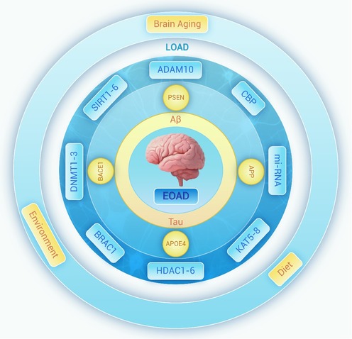
Figure 1.
Direct and indirect links between AD and genes
-
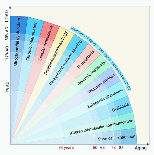
Figure 2.
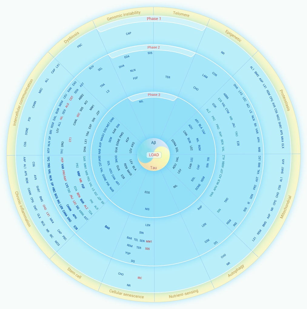
The prevalence of LOAD rises with hallmarks of aging
-
Figure 3.
Epigenetics links environment with aging or LOAD
-
Figure 4.
The effect of the target of clinical LOAD drugs on aging
-
Figure 5.
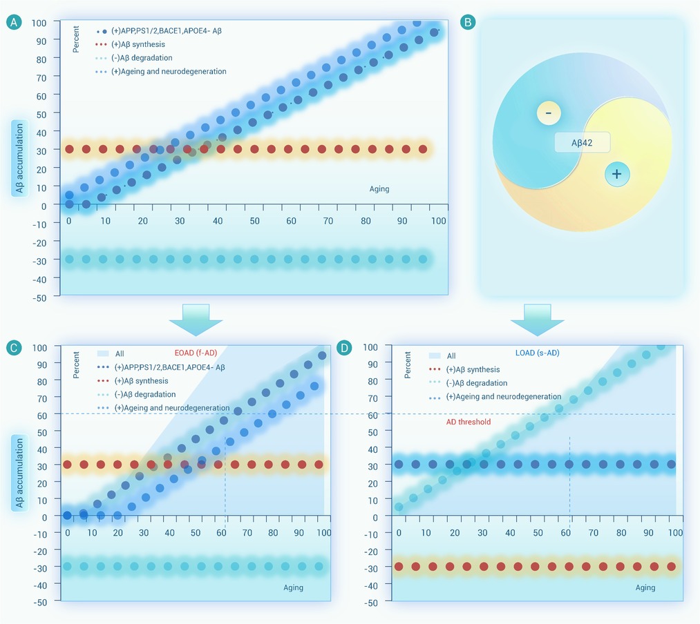
A dynamic equilibrium model for EOAD and LOAD
-
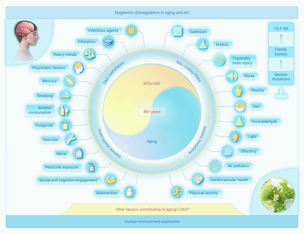
Figure 6.
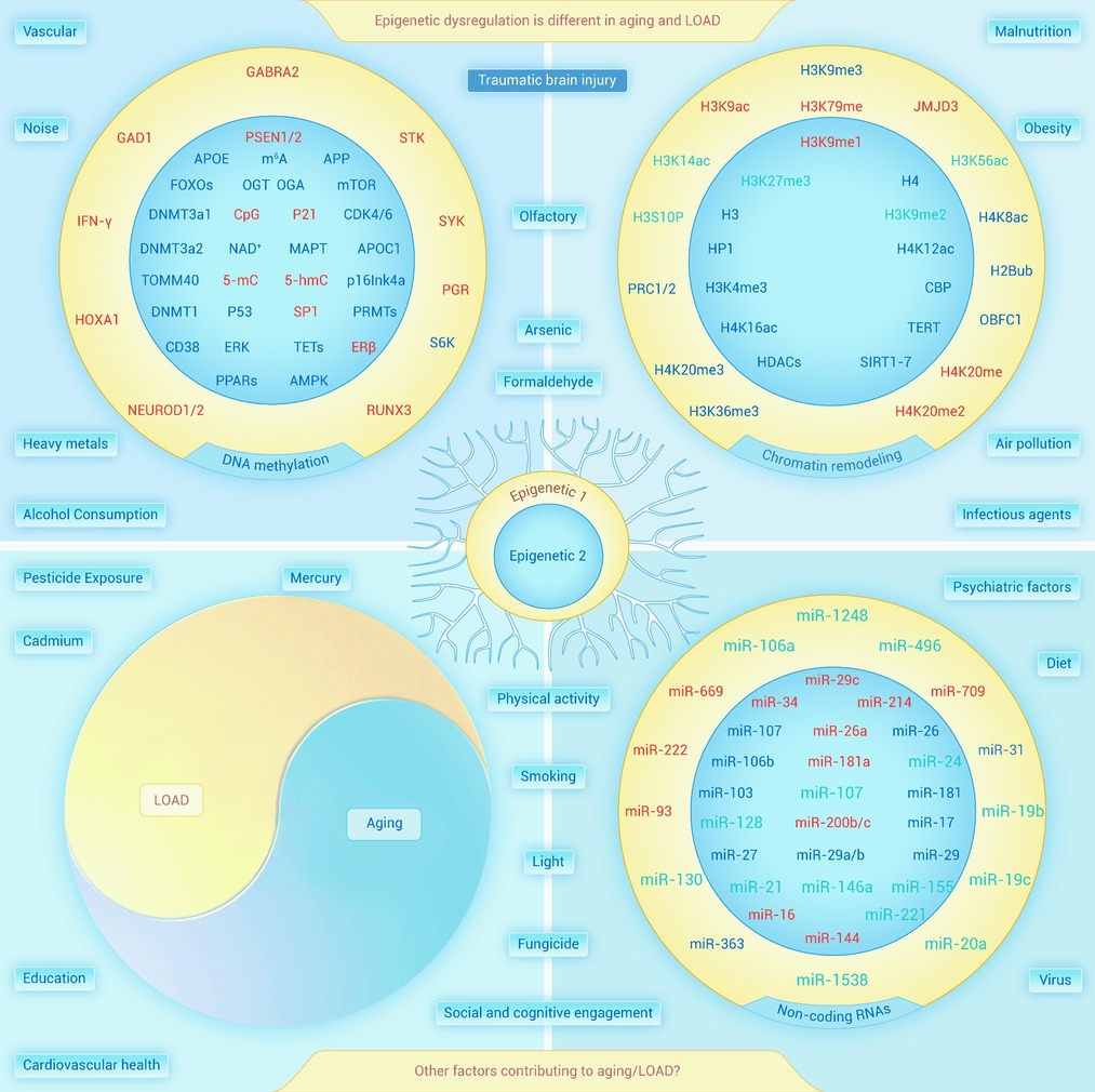
Epigenetic dysregulation in LOAD
